
Abstract
Background:
In-office bleaching is one of the most commonly used procedures for discolored tooth treatment. Although the efficacy of tooth bleaching has been investigated, depending on the applied technique and the used materials, bleaching procedures might irritate the tooth pulp and induce surface changes in enamel. The aim of this study was to evaluate the effect of four different bleaching techniques on the penetration of hydrogen peroxide (HP) into the pulp chamber.
Materials and methods:
Seventy-two single-rooted sound human teeth were used. The samples were prepared and evaluated in four groups. Group 1: 35% HP gel alone (HP Gel); group 2: Nd:YAG laser (0.25 W and 10 Hz with a fiber tip size of 200 μm) irradiation was added (HP Gel+laser); group 3: HP gel mixed with graphite particles (10th of millimeter in size) derived from crashed pencil lead in association with Nd:YAG laser (HP Gel+laser+graphite); and group 4: HP gel in association with light-emitting diode (LED) (litex 686, 50 Hz, 450–490 nm) (HP Gel+LED). The amount of HP penetrating into the pulp chamber was evaluated using acetate buffer and standard graphs. Data were analyzed with one-way ANOVA test, using SPSS 17. Post hoc Tukey test was used for between-group comparisons (α = 0.05).
Results:
Statistical analyses showed that the HP Gel+laser+graphite group had significantly higher level of HP penetration than other groups (p < 0.001). Moreover, pulp chamber penetration of HP in the HP Gel+laser group was greater than the LED and control groups (p < 0.001). The difference between control and LED groups was not significant (p = 0.99).
Conclusions:
Laser bleaching associated with HP Gel and graphite particles resulted in increased penetration of HP into the pulp chamber compared with the LED and control groups.
Introduction
The chromatic alterations affecting the tooth structure can be of extrinsic or intrinsic etiology. Bleaching procedure has been common in aesthetic rehabilitation planning. 1 Recently, there have been many advances in bleaching techniques due to increasing demands for healthy teeth and aesthetic smile. Vital tooth bleaching is a safe and nondestructive treatment modality for tooth discoloration. 2 Teeth are bleached using in-office bleaching and night guard vital bleaching. 3
Nowadays the available bleaching systems rely on hydrogen peroxide (HP) as the principal agent or on one of its products, especially carbamide peroxide, and are used in association with activating agents such as light or heat. 4 Various radiation sources have been used to improve the efficacy of tooth bleaching gels, including curing lights, light-emitting diode (LED) lamps, and diode lasers. The first lasers used in bleaching procedures were argon laser (480 nm) and CO2 laser (1060 nm); however, currently mostly diode lasers (810‒980 nm), Nd:YAG (1064 nm), potassium-titanyl-phosphate (KTP) (532 nm), and Er:YAG (2940 nm) are used. 2 Vanderstricht indicated that combination of a light source with bleaching agents can result in faster changes in color. 5
During dental bleaching, the oxidation of organic structures of the dental tissue occurs through the action of reactive oxygen species released from HP, 6 which leads to changes in the hard tissue and in the pulp tissue. 1 Complications have been reported with the use of these agents, including external root resorption and pulpal irritation. 7 Penetration of HP through enamel and dentin into the pulp chamber is a major concern in tooth bleaching. 8 –13 Animal studies have reported the increase in HP concentration and the prolonged activation of CD5-positive cells and a significant increase in the levels of pain-related neuropeptides in pulp. 14
Benetti et al. showed that interleukin (IL)-6 and IL-17 caused inflammation in the pulp tissue of rats after tooth bleaching, particularly at 2 days. 15 Ferreira et al. demonstrated that tooth bleaching increased IL-6 and tumor necrosis factor-α (TNF-α) in the pulp tissue. 16
Berger et al. demonstrated that three concentrations (10%, 35%, and 50%) of HP penetrated into the pulp chamber, with no significant differences between their penetration capacity. 17 Further, LED light and Nd:YAG laser bleaching increase 35% HP penetration into the pulp chamber. 18
Recently it has been shown that mixing some coloring agents to HP gel can significantly increase the efficacy of bleaching by enhancing the absorption of light. In KTP-laser bleaching with the Smart Bleach gel, a photocatalytic effect is induced that is the activation of peroxide gel by means of light, also referred to as a photochemical reaction. 5
The Nd:YAG laser is frequently used for laser-activated bleaching, Q-Switch dye wavelength corresponds with Nd:YAG laser and so is used in association with peroxide gel for power bleaching. However, it is an expensive and toxic material that confounds its application. 19
Moreover, the Nd:YAG laser wavelength corresponds with graphite. Recently some studies have evaluated the efficiency of the Nd:YAG laser in association with graphite for the treatment of dentinal hypersensitivity. Maamary et al. revealed significant pain reduction with respect of aesthetics and the natural aspect of the treated teeth. 20 Maleki-Pour et al. showed that Nd:YAG laser used at 0.25 and 0.5 W with application of graphite smear was able to reduce the number and diameter of dentinal tubules. 21
Pencil lead graphite particles addition to HP gel, a very cheap and accessible material, as coloring agent may enhance the efficiency of Nd:YAG laser bleaching by means of light absorption that is also referred to as a photochemical reaction. 5 However, the higher absorption of light with bleaching gel may increase the penetration of HP to pulp chamber. Therefore, the ideal treatment would be a technique able to chromatically change the substrate without side effects related to peroxide penetration to pulp. 1
The aim of this study was to evaluate the penetration of HP into the pulp chamber of Nd:YAG laser bleaching, Nd:YAG laser bleaching associated with graphite particle–gel mixture, and LED light bleaching in comparison with the conventional gel bleaching procedure. The null hypothesis was that different power bleaching methods and graphite particle addition to bleaching gel do not increase the HP penetration into the pulp chamber.
Materials and Methods
This research was approved by dental and periodontal research center with the ethical code of
The thickness of dentin was measured using Vernier caliper (Asimeto, China) to make sure of its equality in all the teeth. Human teeth extracted due to orthodontic and periodontal problems were used in this study.
The Randlist software program was used to randomly assign the teeth to four groups and different bleaching techniques were used in each group to bleach the samples. The teeth were stored in normal saline solution and the roots were cut away 2 mm below the cemento enamel junction (CEJ) in a buccolingual direction perpendicular to the long axis of each tooth using a diamond disk (JOTA AG, Swiss). The pulp chambers of the teeth were enlarged using a #6 Gates-Glidden drill. During the preparation, pulp chambers of all the teeth were irrigated with 110 mL of 1% NaCl solution. Then the samples were immersed in the same solution for 24 h to remove pulp tissue remnants, rinsed under running water for 1 h, and dried at room temperature for 6 h.
The four study groups were subjected to the following bleaching protocols: Group 1: The samples were exposed to 35% HP gel (Opalescence PF; Ultradent) for 40 min (4 × 10) (HP Gel:control). Group 2: In addition to 35% HP gel, Nd:YAG laser (LAMBDA Scientifica, S.r.l., Vicenza, Italy) with the following parameters was irradiated on the tooth: 0.25 W and 10 Hz with a fiber tip size of 200 μm (Fig. 1A). HP gel was applied to the tooth surface and activated for 20 sec with laser beams. The gel was left on the tooth surface for 10 min and then removed.
22
This was repeated for four times in accordance with the effective application period specified in the company instructions (HP Gel+laser). Group 3: Graphite particles (10th of millimeter in size) derived from crashed pencil lead were mixed with 35% HP gel (equal weight percent). The same Nd:YAG laser bleaching protocol (group 2) was applied (Gel+laser+graphite). Group 4: Bleaching with 35% HP bleaching gel and activation with LED (Litex LED 686; Dent America) 50 Hz, 220 V, 450–490 nm, for 40 min according to the instruction of clinical use of the Litex 686 unit (Gel+LED)
23
(Fig. 1B).
The whole surface was subjected to a uniform sweeping motion to guarantee deposition of equal radiation energy values over the tooth surface in all groups.

The quality of the laser and LED was measured and controlled by power meter of each company.
A similar amount of HP was used in all the groups, utilizing a graduated plate. To determine the amount of HP penetrating into the pulp chamber, the pulp chamber was irrigated with 30 μL of distilled water twice and dried with paper cones. Then 30 μL of acetate buffer at a concentration of 2 M and pH of 4.5 was placed within the pulp chamber. After the bleaching procedure, the acetate buffer was collected and poured into 10 mL flasks.
Then the pulp chamber was rinsed with 30 μL of distilled water twice and added to the flask; 1 mL of leucocrystal violet horseradish peroxidase (Sigma Aldrich) (0.5 mg/mL) and 0.5 mL of horseradish peroxidase (1 mg/mL; Sigma Aldrich) were added to the flask.
After adding 4 mL of the acetate buffer, the volume of the mixture increased to 10 mL by adding distilled water (Fig. 2). Then the color intensity was determined using a ultraviolet/visible spectrophotometer (Sicily, Italy) at a wavelength of 596 nm, and the amount of HP penetrating into the pulp chamber was determined in micrograms using a standard and calibrated graph (Fig. 3). 24 The person assessing HP concentration was blinded to study groups.
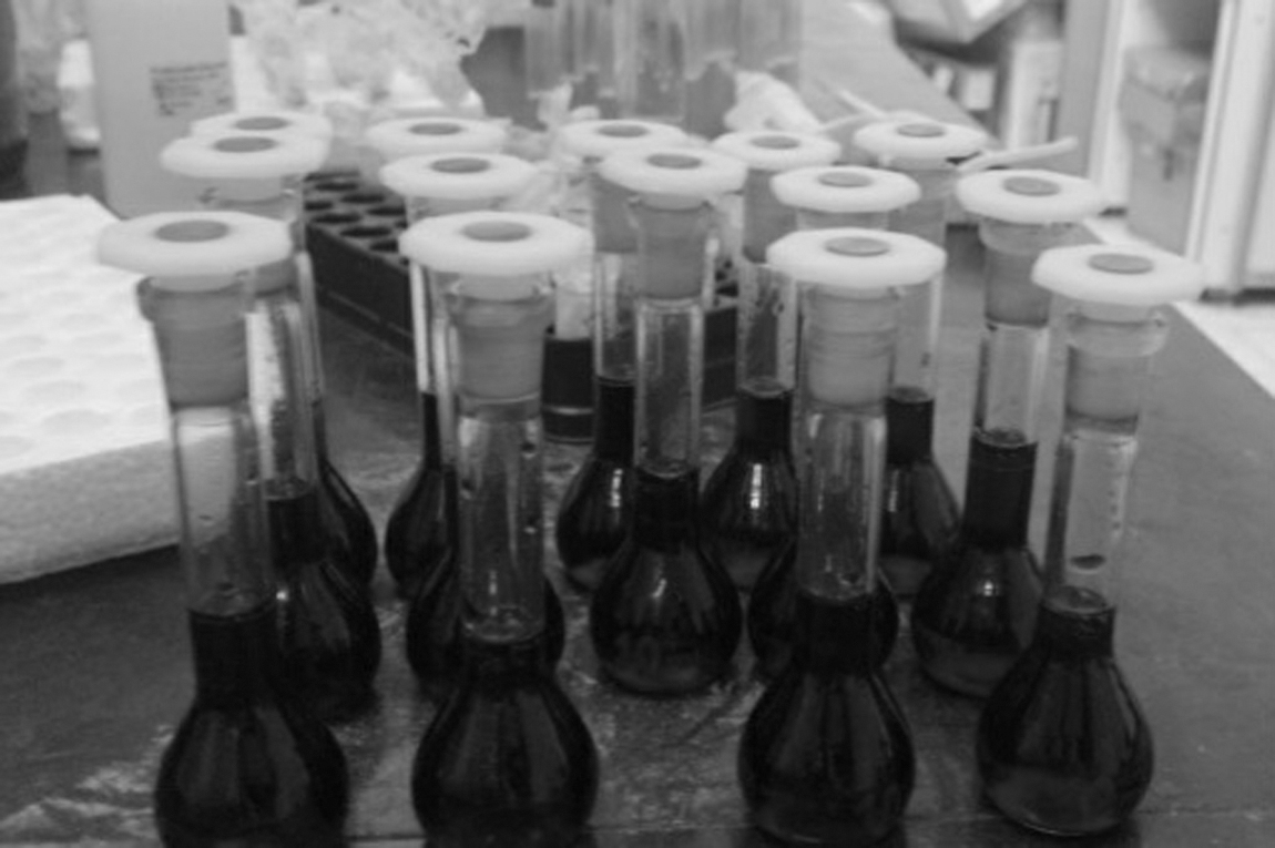
The flask containing the fluid collected from the samples after application of different bleaching techniques, which had been prepared for the measurement of penetration of hydrogen peroxide.
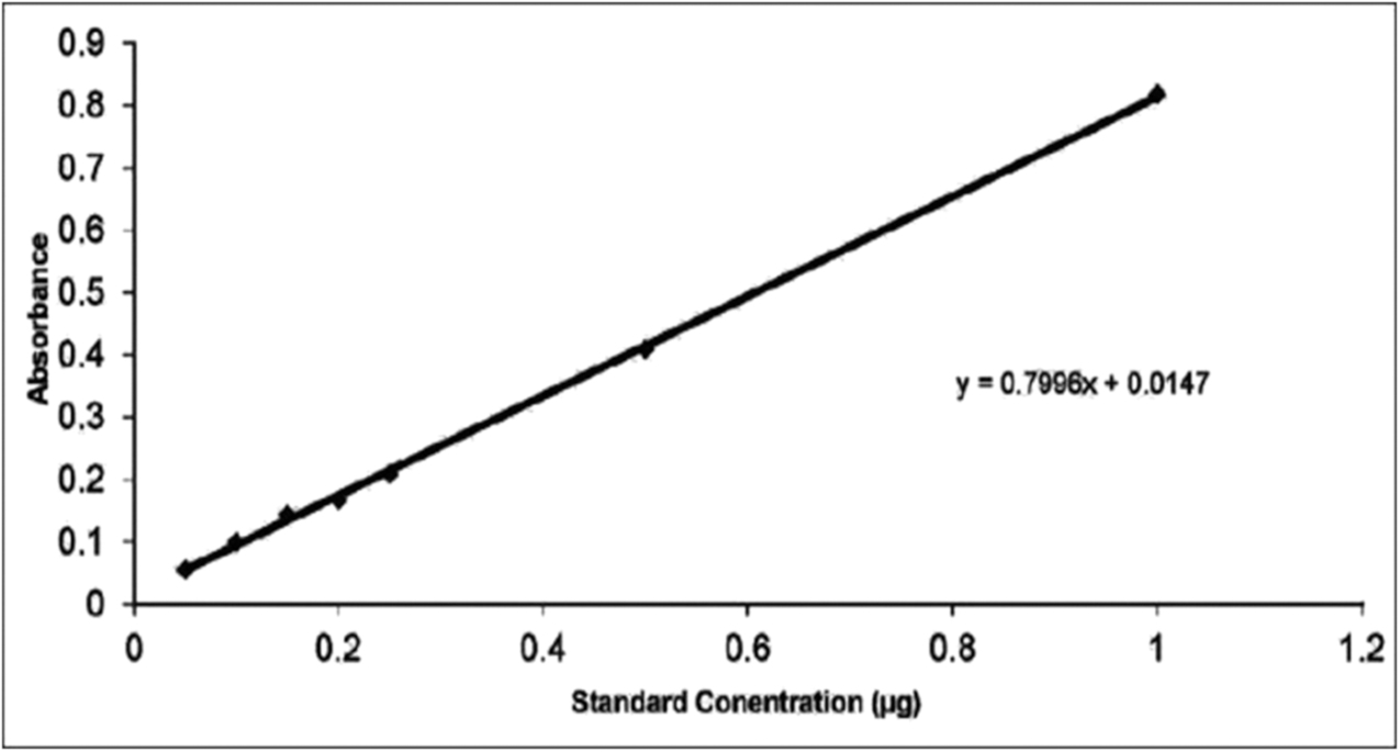
The light absorption‒standard concentration of hydrogen peroxide.
Statistical analysis
Data are presented as means ± SD. To compare the HP penetration between groups, one-way ANOVA and Tukey post hoc test were used. Kolmogorov–Smirnov test was used to assess the normality of penetration values. All statistical analyses were conducted using SPSS for Windows 25.0 (SPSS). Statistical significance was preset at p = 0.05.
Results
Mean ± SD values of HP concentrations of study groups are presented in Table 1, with the highest concentration in the Gel+graphite+laser group.
Descriptive Statistics of the Concentration of Hydrogen Peroxide within the Pulp Chamber During Different Bleaching Protocols
LED, light-emitting diode; SD, standard deviation.
One-way ANOVA test showed significant differences in the concentration of HP between study groups (p < 0.001). Post hoc Tukey tests revealed that HP concentration in the HP Gel+laser group was greater than that in the LED and control groups (p < 0.001). However, the difference between the control and LED groups was not significant (p = 0.099) (Table 2 and Fig. 4).
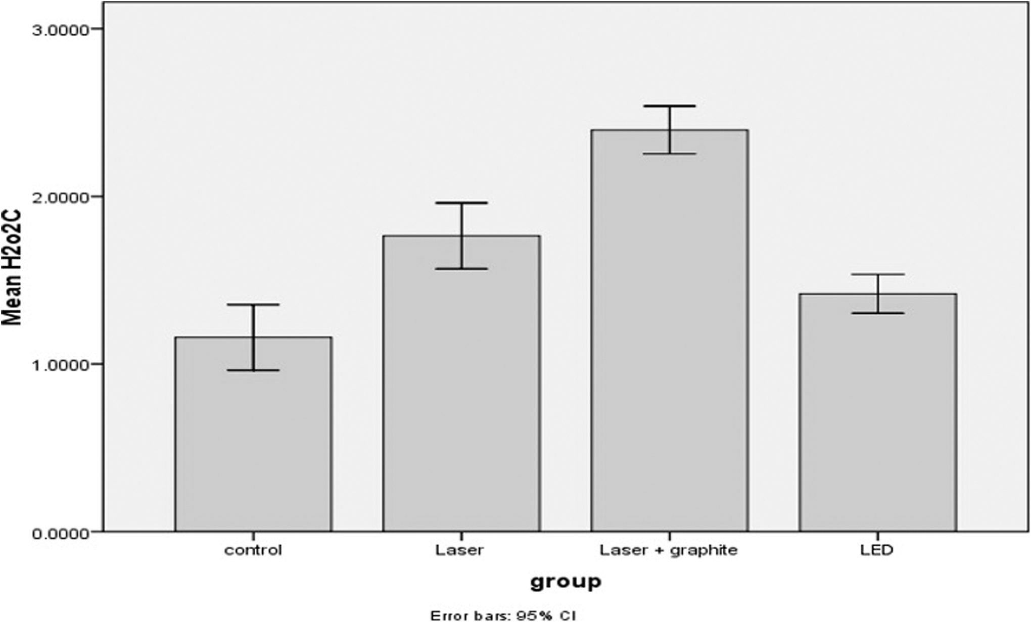
Changes in the concentration of hydrogen peroxide within the pulp chamber in different bleaching protocols.
Evaluation of Differences in the Concentration of Hydrogen Peroxide Between Different Study Groups (n = 18) with Post Hoc Tukey Tests
HP, hydrogen peroxide.
Discussion
Tooth discoloration is one of the problems that makes patients increasingly visit dental offices in search of aesthetic treatments. Tooth bleaching techniques have been used to achieve lighter smile. 25
Various studies aimed at using different light sources in bleaching procedure to accelerate the decomposition of HP and releasing free radicals. 26 Recently, for accelerating the light absorption by gel, coloring agents have been added commercially. The Nd:YAG laser is frequently used for laser-assisted bleaching, which emits at 1064 nm and corresponds to yellow, brown, and black colors. 27 Therefore, pencil lead graphite particles (black color), as a very cheap and accessible material, were used in this study in association with laser.
The advantages of bleaching techniques are well known; however, some complications have been reported, including changes in tooth structure and pulp irritation. 7 One of the concerns related to tooth bleaching procedures is the penetration of HP through enamel and dentin into the pulp chamber; 8 –13 however, if a proper bleaching technique is used, there will be minimal pulpal inflammation. 13,28
Penetration of HP into the pulp chamber is accomplished due to its low molecular weight, its ability to denature proteins, and to decrease calcium and phosphate content of enamel and dentin. Nathanson et al. showed that although vital bleaching with 35% HP can lead to pulp sensitivity, pulp retains its health, and the sensitivity is completely reversible. 26 HP penetrates easily into the pulp chamber and affects pulpal enzymes.
When the concentration of HP increases sixfold, the penetration increases only fourfold, indicating that the concentration of HP is not proportional to its diffusion into the pulp chamber space. 29 Torres et al. showed that the higher the concentration of HP, the higher is its rate of penetration into the pulp chamber. 25
Use of HP solution along with heat or light cannot necrotize the pulp except when the tooth is overheated or traumatized. 30
Abbasi et al. investigated the effect of diode laser-assisted bleaching with three different wavelengths on HP penetration to pulp chamber. Their results showed that the 980 nm wavelength is absorbed readily by water causing its evaporation and increasing the temperature of gel and accelerating the chemical reaction. 31
The results of this study revealed that penetration of HP into the pulp chamber in the Gel+laser and Gel+laser+graphite groups was significantly higher than that in the control group, so the null hypothesis of study was rejected.
Light activation with Nd:YAG laser causes an increase in the temperature of the gel. Use of a color that has a wavelength similar to that of Nd:YAG laser increases the efficacy of laser absorption by the surfaces. One way to apply this technology in bleaching of teeth is to mix colors with wavelengths similar to Nd:YAG laser with the HP gel. Each laser beam is absorbed better in specific tissues of the body depending on its wavelength. Based on previous studies, Nd:YAG laser exhibits the highest absorption in melanin-containing tissues 27 and this is the cause for selecting graphite particles for incorporation into HP gel.
Since coloring agents are incorporated into gels to absorb more energy from the light sources and to achieve maximum activation, it appears this higher absorption results in an increase in molecular movements; the whole system requires a high level of kinetic energy. Based on the molecular collision theory, during the collision, some of the kinetic energy is used, making bonding cleavage and accelerating the diffusion speed leading to increased penetration into the pulp. 25
Also De Moor et al. showed that when warming the gel (photothermal effect), colorants can also trigger other photochemical reactions such as photodynamic reactions in which photosensitive dyes are excited by light. In their triplet excited state, they can donate highly energetic electrons and produce O2 and H2O2 forming H2O2, HO2 •, and HO•. Further, during intersystem crossing (from singlet to triplet excitement), the dyes will form singlet oxygen ( 1 O2) by spin-orbit coupling with triplet oxygen ( 3 O2), the common form of molecular oxygen. Altogether, photodynamic bleaching enhancement enables the formation of the two most potent bleaching agents, that is, hydroxyl radical and singlet oxygen, and is thus a very powerful enhancement. 32
Bleaching gels that have been activated by light exhibit the complications of penetration, including direct destruction of odontoblasts and a decrease in their metabolic activity. 33 Torres et al. reported that photoactivation decreased the penetration time of 20% HP, but not 35% HP. They showed that diffusion time of HP was dependent on activation and concentration of HP. 25 Kwon showed that 40% HP penetration was not affected by light activation with a xenon power arc lamp, contrary to the results of this study. It can be speculated that different HP concentrations and activation source may have contributed to the contradictory results. 34
The results of this study showed that penetration of HP into the pulp chamber was higher when light activated with Nd:YAG laser compared with the LED light, which might be attributed to the lower increase in temperature in LED light activation. 35 –37 The light-assisted methods used in this study were according to clinical protocols instructed by the manufacturer in order to compare them properly.
A study by Ladalardo showed that use of LED light resulted in a 2°C increase in the temperature of the bleaching agent without increasing the temperature of tooth structure, which may favor the molecular mobility and radicals release. 35
Clemente et al. recently suggested an alternative to the conventional bleaching treatment by using a source of violet LED and offered an immediate bleaching effect on the dentin tissue, without using oxidizing agent. The mechanism of action of this new technology is based on the emission of light at a wavelength of 405–410 nm, which coincides with the absorption peak of chromophore molecules, in which instability and breakdown could occur, thus resulting in a photolytic effect. 1
In this way, using a whitening protocol without the side effects of HP offers safer treatments. Moreover Gallinari et al. observed that the isolated use of the violet LED caused a chromatic alteration and significant bleaching effect. 38 However, using violet LED in pigmentations with blood and red wine is less effective. Molecules with iron atoms are grouped so that they have a smaller distance between them, being less permeable to the passage of photons like those of violet light. 1
de Oliveira Gallinari et al. demonstrated the use of light potentiated chromatic alteration. They showed that violet LED light had no effect on self-reported pain sensation; however, its use sensitized the front teeth in low-temperature situations. Although it is yet to be confirmed, this could be the beginning of an evolution in bleaching therapy, if significant aesthetic results can be achieved with a lower oxidative stress. 39
In this study, bleaching gel was applied four times each for 10 min and refreshed similarly in all groups. Balladares et al. showed that for acidic pH gels, longer application results in more HP penetration. 40 Another study showed that an increase in the duration of contact between the enamel and HP increases its penetrability in relation to both the amount and depth. 14 However, a study by Camps et al. showed that after a certain duration of contact of the gel with tooth structure, the penetrability decreases. 41
The pulpal fluid decreases the toxicity of HP. Therefore, a longer duration for reaching of HP to the pulp chamber decreases its toxicity because pulpal fluid will have more time to dilute peroxide and prevent it from entering the pulp. In addition, pulpal mechanisms protect the pulp against free radicals. 42 It should be kept in mind that under in vitro conditions, different results might be achieved as there is no pulpal pressure that might serve as a barrier against the penetration of HP.
As mentioned, different studies have been developed to evaluate the effectiveness of different bleaching techniques as well as their possible side effects. Laser bleaching along with different concentrations of gel and graphite addition could be an efficient method, however, based on the limitations of this study, we suggest future research to evaluate the effect of graphite addition on pulp chamber temperature and bleaching efficacy. Moreover, different concentrations of bleaching gels should be investigated.
Conclusions
In conclusion, adding graphite to HP gel along with Nd:YAG laser enhanced the penetration significantly. Further, in the laser-assisted bleaching process, greater penetration was observed comparing with the control group due to chemical activation. However, graphite particle addition and light-activated bleaching may be an efficient method that needs color-evaluating studies in future.
Footnotes
Authors' Contributions
Concept or design of the study was carried out by F.P.-A. Acquisition of data was contributed by M.M. Analysis or interpretation of data was carried out by M.M. and S.K. Drafting of the article was by F.P.-A. Critical revision for important intellectual content was by S.K. All authors had full access to the data, contributed to the study, approved the final version for publication, and take responsibility for its accuracy. All authors are from the Tabriz University of Medical Sciences, where education and research are the primary functions.
Author Disclosure Statement
No competing financial interests exist.
Funding Information
No funding was received for this article.