
Abstract
Objective:
The green synthesis of Tin(IV) oxide (SnO2): Gold (Au) nanoparticles (NPs) using Teucrium polium medicinal plant extract was investigated, and the NPs were characterized and tested as photosensitizers to produce reactive oxygen species (ROS).
Methods:
The cytotoxic effect on C26 cells was investigated using the 3-(4,5-dimethylthiazol-2-yl)-2,5-diphenyltetrazolium bromide (MTT) technique. The results showed their toxicity in a dose-dependent manner. The green synthesis of SnO2:Au NPs was achieved for the first time using an extract of T. polium medicinal plant as a reducing and stabilizing agent. The produced NPs were examined for their application in photodynamic therapy (PDT) for cancer.
Results:
Methylene blue and anthracene were used to confirm that the photosensitizer could produce ROS when excited with UVA radiation. The anticancer activity of SnO2:Au was investigated in vitro using the C26 cell line and an MTT assay, showing that PDT with SnO2:Au NPs could inhibit cancer cell proliferation.
Conclusions:
The significant afterglow of the SnO2:Au NPs could cause the generation of ROS to continue several minutes after switching off the light source.
Introduction
Colorectal cancer poses a significant risk to human health, ranking fourth in terms of its prevalence worldwide, and is the fifth cause of death for both men and women. The incidence of colon cancer has risen in recent times, particularly among young adults and teenagers. As the world economy progresses leading to lifestyle and dietary changes, it is anticipated that the incidence of colon cancer will rise. Cancer treatment is a global challenge that affects the whole of society. 1,2 At present, various treatment methods are used such as surgery, chemotherapy, radiotherapy, and immunotherapy. However, these treatments have severe side effects and can damage healthy tissues. 3 –5 Therefore, a method that has the least damage to healthy cells and the most effect on cancer cells should be preferred.
Photodynamic therapy (PDT) is a novel approach that does not require any invasive procedures. It involves the use of photosensitizers to produce cytotoxic reactive oxygen species (ROS) in response to light exposure, selectively targeting and destroying cancer cells. Clinical trials over the past few decades have demonstrated that PDT is a viable and beneficial treatment choice for several types of cancer. 6 –11 The action of PDT depends on three basic factors as follows: photosensitizer, light, and oxygen. Each factor is harmless on its own but their simultaneous presence within cancerous tissues produces toxic ROS leading to cell death by apoptosis and/or necrosis. When photosensitizers are activated by a suitable light source, PDT has the most effect on cancer cells and the least effect on healthy cells since the drug accumulation is targeted to the tumor and the light radiation is spatially confined. 12 –17 The photosensitizers used in PDT are often composed of organic molecules such as porphyrins, phthalocyanines, and other tetrapyrroles, but these compounds have limitations such as hydrophobicity and poor stability in the tumor environment that limit their clinical usage. High radiation doses must be used for some photosensitizers to be effective in PDT, which in turn may cause long-term damage. PDT is also used to treat superficial and skin cancers requiring a low light penetration depth. 6,13,18,19 NPs can be used to overcome some of these limitations. For instance, optical nanoparticle sensors can improve PDT with a high quantum yield. The development of nanotechnology could provide new biomedical applications for PDT and play a significant role in improving cancer treatment. 16,20,21 The so-called “afterglow NPs” can emit light from several minutes to a few hours after the light source has been turned off. Nanoparticles that exhibit strong afterglow characteristics or persistent luminescence are capable of generating ROS, such as hydroxyl radicals, superoxide radicals, as well as singlet oxygen, which are all useful in PDT. Although other types of oxygen radicals can also be effective, singlet oxygen is particularly important in PDT. 22,23 Due to their small size, NPs have been used in smart and purposeful approaches to treat cancer. 24,25
Semiconductor NPs are capable of producing ROS due to the large electronic band gap and can be used as photosensitizers. 8,26,27 Nanoparticles utilized in PDT tend to accumulate more in tumors compared with normal tissues, mainly because of the enhanced permeability and retention effect. This enables passive uptake by cancerous tissues with leaky blood vessels and poor lymphatic drainage. ZnO quantum dots with a size of approximately 6–11 nm when exposed to visible light in the range of 400–500 nm could produce ROS such as hydroxyl radicals (•OH) and superoxide radicals (O2•−). 28 SnO2 acts as a significant n-type semiconductor material with various biomedical applications. SnO2 NPs doped with metals and conjugated with biomolecules can show antibacterial, anticancer, antitumor, biosensors, and antioxidant activity. Hang’s research group reported the use of gold nanorods in PDT without the need for any organic photosensitizer. The gold nanorods could generate singlet oxygen that led to the destruction of melanoma B16FO tumors in mice. 29 Titanium dioxide nanoparticles have been used in PDT to produce ROS using UV light, which caused apoptotic cell death in HeLa, T-24, and U937 cancer cell lines. 30,31 Nanomaterials have been extensively investigated for their unique optical properties in PDT, with Cadmium selenide (CdSe) being one of the materials studied. CdSe has an active surface and can produce ROS when exposed to UV radiation. 32
Although chemically synthesized NPs are effective and suitable for PDT, they are expensive, toxic, and can be polluting to the environment. As an alternative to conventional physical and chemical synthetic processes, which may be detrimental to both humanity and the environment, there has been an increased interest in recent years in biosynthetic methods using plants, microbes, or algae. Green synthesis is an effective, inexpensive, and nontoxic method with minimal side effects and needs only a simple process. 33 –35 This improved performance is attributable to the many biomolecules that are present in the natural source of biological material. These biomolecules, which include plant metabolites, flavonoids, carbohydrates, proteins, enzymes, pigments, and vitamins, work as reducing agents to generate nanoparticles from metal salts without creating any toxic by-products. 36 As a result, the manufacture of green nanoparticles could be a possible answer to the rising need for the development of environmentally friendly approaches to battle microbial resistance and control diseases. Therefore, green nanotechnology allows for the manufacture of nanomaterials without endangering either human or environmental health. 37
In the present investigation, the plant Teucrium polium was used as a reducing and stabilization agent in the production of SnO2:Au NPs. Other reports have described the use of T. polium as a reducing and stabilizing agent for the synthesis of nanoparticles. 38 –40 T. polium is a medicinal plant that has been used for more than two thousand years. It is a member of the Lamiaceae family of plants. In the Middle East, people have been using this plant for treatment of diabetes, as well as liver problems, renal disorders, stomach discomfort, and indigestion for a very long time. Moreover, it has been shown to have anti-inflammatory, anticancer, antifungal, and antibacterial properties. 40 –43
In this work, the green synthesis of SnO2:Au NPs using T. polium medicinal plant extract was investigated, and the NPs were characterized and tested as photosensitizers to produce ROS. The cytotoxic effect on C26 cells was investigated using the MTT technique. The results showed their toxicity in a dose-dependent manner.
Materials and Methods
Preparation of Teucrium polium extract
In a light-restricted environment, the plant material underwent a cleaning and drying process. Subsequently, the desiccated plant material was delicately ground into a fine powder using a mortar and pestle. An aqueous extract was then created using 10 g of T. polium powder, followed by filtration and centrifugation to remove smaller plant particles. The final extract was stored in the dark at 4°C until it was needed for the preparation of NPs.
SnO2:Au NP synthesis
SnO2:Au NPs were synthesized using a green method employing T. polium plant extract. Initially, the precursors of SnCl2.2H2O and HAuCl4.3H2O and the high-purity herbal extract of T. polium were prepared. First, 1.75 g of tin (II) chloride (SnCl2) was dissolved in 50 mL of double-distilled water. After 30 min, 0.2 g HAuCl4.3H2O was added to the solution, which was stirred on a magnetic stirrer for 30 min, followed by the addition of 30 mL of T. polium extract. After 5 min, a brown precipitate was obtained, which was separated by centrifugation and washed with double-distilled water. The solution was heated at 80°C for 24 h in an electric oven to facilitate solvent evaporation and leave a powder. Subsequently, in the calcination phase, the powder was subjected to a 2-h heating process at 600°C in an electric furnace to destroy the remnants of the T. polium extract.
Modification of SnO2:Au NPs with PEG
PEG is the best-known polymer for increasing biodegradability, used for surface modification of NPs, as well as increasing solubility and stability in an aqueous environment. Surface modification by Polyethylene glycol (PEG) allows NPs to circulate in the blood for a longer time and to penetrate more easily into tumor tissues and is effective for targeted PDT treatment. 25 To modify the surface of SnO2:Au NPs with PEG, 50 mL of a suspension of SnO2:Au NPs at a concentration of 0.5 g/L was prepared. Then 0.2 g of PEG was added to the solution. The optimum surface modification was achieved after magnetic stirring for 24 h, and the product was used for cellular experiments.
SnO2:Au NP characterization
Structural investigation was carried out by X-ray diffraction (XRD) using a Philips Instrument with CuKα radiation filtered by Nickel. A scanning electron microscope (SEM; model: TESCAN Mira 3-XMU) and a transmission electron microscope (TEM; model: Zeiss EM900) were used to analyze the NP morphology, composition, and particle size. To confirm the presence of functional groups, a Fourier transform infrared spectrometer (FTIR; model: Magna-IR550) was used. At room temperature, the optical properties were investigated using an ultraviolet-visible spectrometer (UV-Vis; UVS-2500, Phystech) scanning from 200–800 nm, and the photoluminescence spectrum was recorded using a PL; Perkin-Elmer spectrometer model LS55 equipped with a photomultiplier tube and xenon lamp.
ROS measurement
Methylene blue (MB) is a dye that is decomposed to CO2 and H2O in the presence of hydroxyl radicals, so it was used as a free radical detector. First, 0.001 g of MB was dissolved in 50 mL of DI water, 2 mL of this solution was combined with photosensitizer NPs, and the absorption spectrum was recorded using a UV-Vis spectrometer. In the next step, the MB solution containing light-sensitive nanoparticles was irradiated with UV light for 1 h, and the absorption spectrum was recorded. The production of hydroxyl radicals was determined by comparing the reduction of adsorption intensity before and after irradiation. Anthracene was used as a probe to measure single oxygen production. Thus, 0.001 g anthracene was dissolved in 50 cc ethanol, then photosensitizer NPs were added to 0.04 mL of anthracene solution, and the absorption spectrum was recorded before illumination. The solution was then exposed to UVA light, and the singlet oxygen produced was determined by recording the reduction in adsorption intensity after illumination.
Cell culture
The C26 cell line, which was derived from human colon cancer cells, was obtained from the Pasteur Institute in Tehran, Iran. The C26 cells were cultured in RPMI 1640 medium supplemented with 10% fetal bovine serum and penicillin–streptomycin (Gibco, USA). The cells were maintained in a humidified incubator with 5% CO2 at 37°C.
UV-LED irradiation protocol and optimization for cell viability
The cells were seeded on suitable plates and then incubated with SnO2:Au NPs for 24 h at 37°C and 5% CO2. Subsequently, the cells were irradiated with a UV-LED (manufactured by Shenzhen Hatter Photoelectricity Co, Ltd. from Shenzhen, China) at a power density of 0.21 mW/cm2 for 15 min delivering a fluence of 0.189 J/cm2. The UV-LED used in this experiment had a wavelength of 365 nm. The intensity of the light was initially measured in Lux and then converted to a power density measurement in mW cm−2. Varying durations of 5, 10, 15, 20, 25, and 30 min of irradiation with the UV-LED at a distance of 5 cm from the cell culture plate were used to determine the appropriate UV irradiation dose. Fifteen min of irradiation was determined to be the optimum time, which did not significantly affect the cell viability on its own (Fig. 10).
Assessment of cell toxicity
The cytotoxicity of PDT mediated by SnO2:Au NPs was evaluated using the MTT assay. The C26 colon cancer cell line was used for this experiment. About 10,000 cells per well were seeded in two 96-well plates. A concentration of 1000 µg/mL of SnO2:Au NPs was suspended in Dulbecco's Modified Eagle Medium. The stock solution of nanoparticles in the culture medium was sonicated for 20 min at 80 W at room temperature in an ultrasonic water bath sonicator before being added to the cells to disaggregate the NPs and ensure a uniform suspension. It was observed that the NP solution tended to form clusters over time, a phenomenon influenced by environmental factors such as storage temperature, concentration, and pH. Sonication helped to mitigate this issue by breaking down the aggregates and maintaining the NPs in a dispersed state. The suspension was further diluted to different concentrations (0, 5, 10, 25, 50, 100, 200, 400, and 800 µg/mL). The serial dilutions of SnO2:Au NPs were added to the wells in both plates in triplicate. To find the appropriate irradiation time, after culturing the cells, irradiation was performed for 5, 10, 15, 20, 25, and 30 min. Then one of the plates was exposed to light for 15 min, whereas the other was kept in the dark. After 24 h of incubation, the culture medium was discarded, and new medium containing 0.5 mg/mL MTT was added and incubated for 2 h. Then the medium was removed, and the purple sediment was dissolved in Dimethyl sulfoxide. The optical density (OD) was measured at 570 nm using a microplate reader (Epoch; USA). By comparing the mean OD of the treated wells with the control wells without treatment, the percentage of cell viability was calculated.
Statistical analysis
Statistical comparisons between the groups were conducted using the nonparametric Kruskal–Wallis test for non-normally distributed data. A p value of <0.05 was considered significant. In addition, an independent sample t test was used for further statistical analysis. All analyses were performed using SPSS software version 19 (SPSS Inc., Chicago, IL, USA).
Results and Discussion
Evaluating the characteristics of SnO2:Au NPs
X-ray diffraction was used to identify the phases and estimate the size of SnO2:Au crystals generated by T. polium medicinal plant extract. Figure 1 shows the single phase and tetragonal structure for NPs according to the JCPDS card number 01-077-0450. The XRD of the prepared NPs had additional peaks, which disappeared after optimal annealing at 600οC for 2 h.
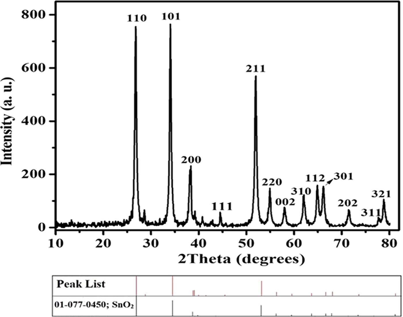
XRD pattern of the SnO2:Au NPs. XRD, X-ray diffraction; NPs, nanoparticles.
The Scherrer equation was used to estimate the size of the crystallites.
In this equation, Dhkl is the approximate size of the crystallites, k is the shape factor (k = 0.9), λ is the wavelength of the X-ray beam (CuKα), β is the full width at half maximum of the primary peak diffracted from the (101) plane, and θ is the diffraction angle. Based on the aforementioned equation, the estimated size of the synthesized SnO2:Au NPs was approximately 22 nm.
The morphology and shape of the SnO2:Au NPs were visualized using SEM, as shown in Figure 2. The SnO2:Au NPs were homogeneous and uniform with a median size between 30–40 nm.
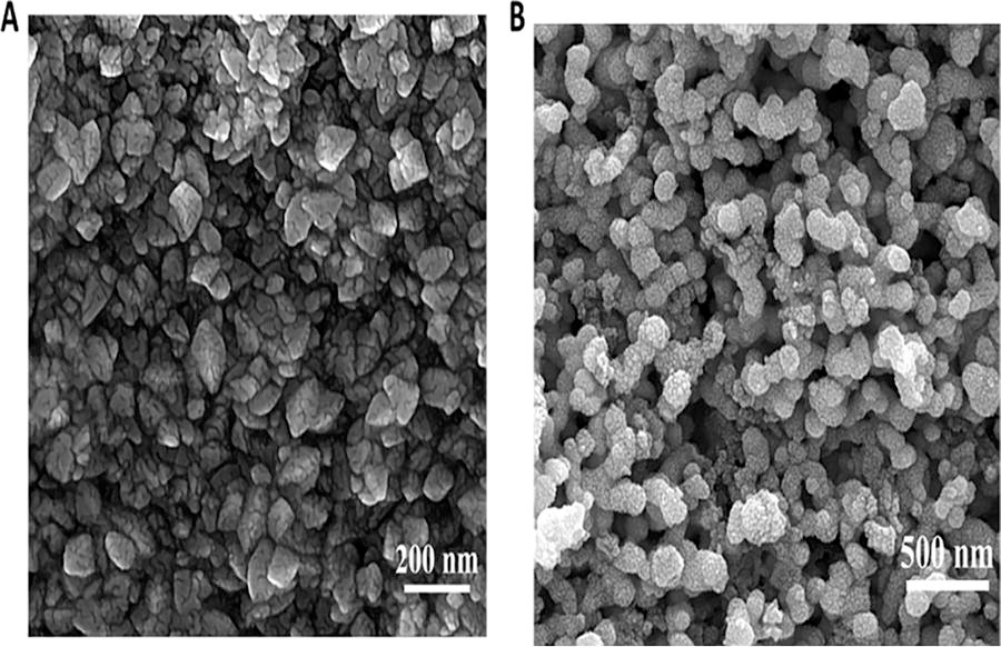
SEM images of SnO2:Au NPs.
Figure 3 shows the Energy-dispersive X-ray spectroscopy (EDS spectrum of the SnO2:Au NPs, which confirmed the presence of Au, O, and Sn elements within the specimen.
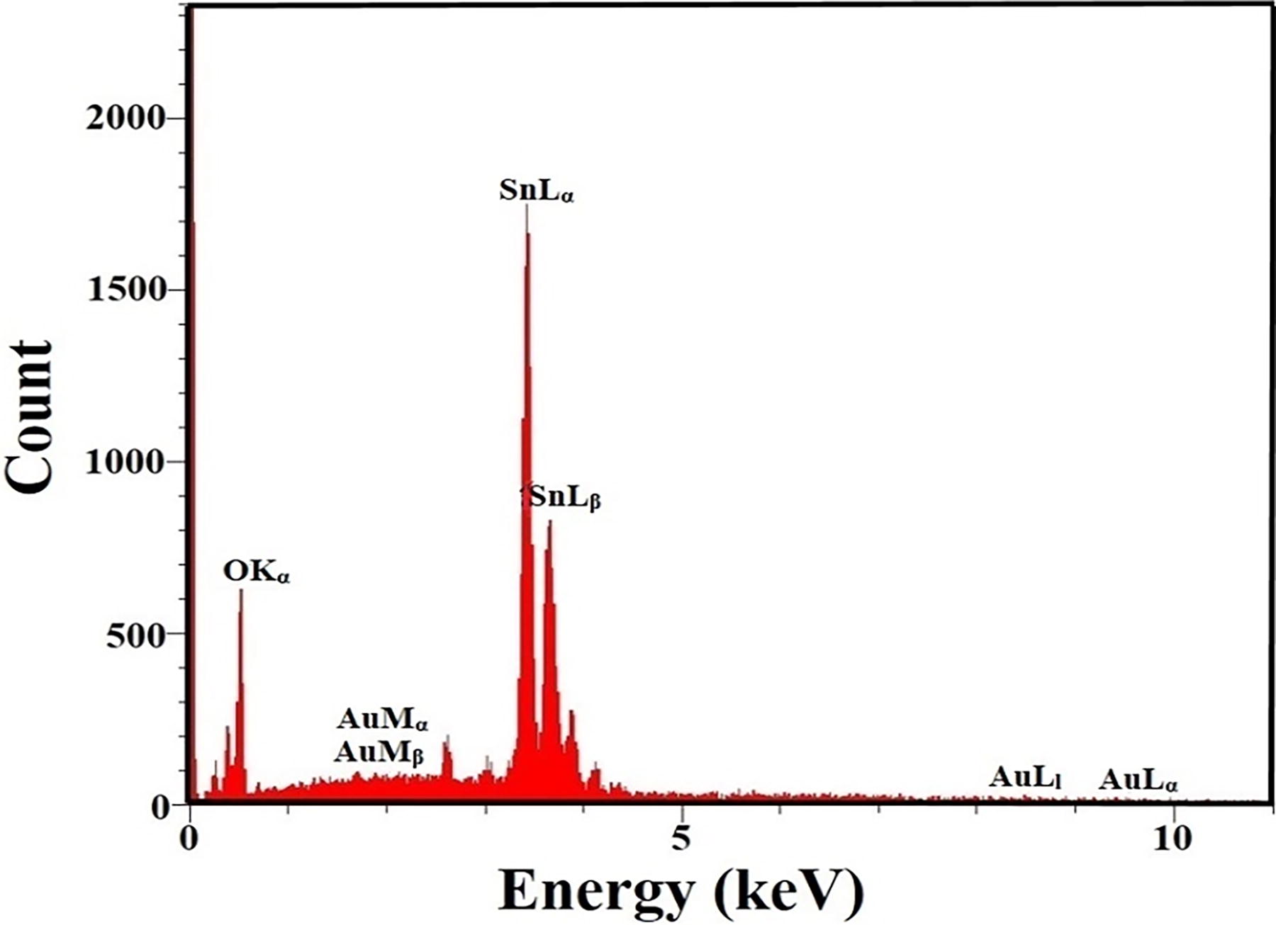
Energy dispersive X-ray (EDX) spectrum of SnO2:Au NPs.
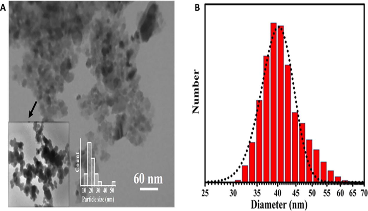
To further verify the shape, size, morphology, and uniformity of the synthesized SnO2:Au NPs, TEM images were captured. As shown in Figure 4A, the particles were predominantly spherical in shape, with diameters ranging between 20 nm and 30 nm. In some regions, they appeared to be piled up or scattered. The formation of NPs was further confirmed by dynamic light scattering analysis as shown in Figure 4B. The histogram of size distribution showed that the average particle size was about 20–40 nm with a peak at 35 nm and a polydispersity index of 0.5. Accordingly, there was no significant NP aggregation in the suspension.
The surface modification of SnO2:Au NPs was carried out by coating with polyethylene glycol to increase the biocompatibility of NPs and lengthen their circulation in the bloodstream. The coated NPs could eventually be used as a photosensitizer in vivo. The FTIR spectrum shows that hydrogen bonds were formed between the SnO2:Au NPs and the PEG polymer. The FTIR spectrum of modified SnO2:Au NPs is shown in Figure 6. The SnO2:Au NP spectrum contains the vibrations of the Sn–O–Sn bond of SnO2 from 670 to 570 cm−1, and the characteristic Sn–O–Sn stretching band is seen at 635 cm−1. The signals observed in the range of 1720–1250 cm−1 are attributed to vibrations of the –OH, CO, and CH bonds of the organic molecules. The absorption band at about 2872 cm−1 is associated with the stretching band C-H and at 3400 cm−1 correspond mainly to the stretching vibration of the hydroxyl group (–OH). The presence of PEG bands in the SnO2:Au NP spectrum confirms the coating of this polymer onto the NPs (Fig. 5). The wide band between 670 and 570 cm−1, attributed to the Sn–O–Sn bond, confirms the presence of SnO2 in the crystalline phase. 44
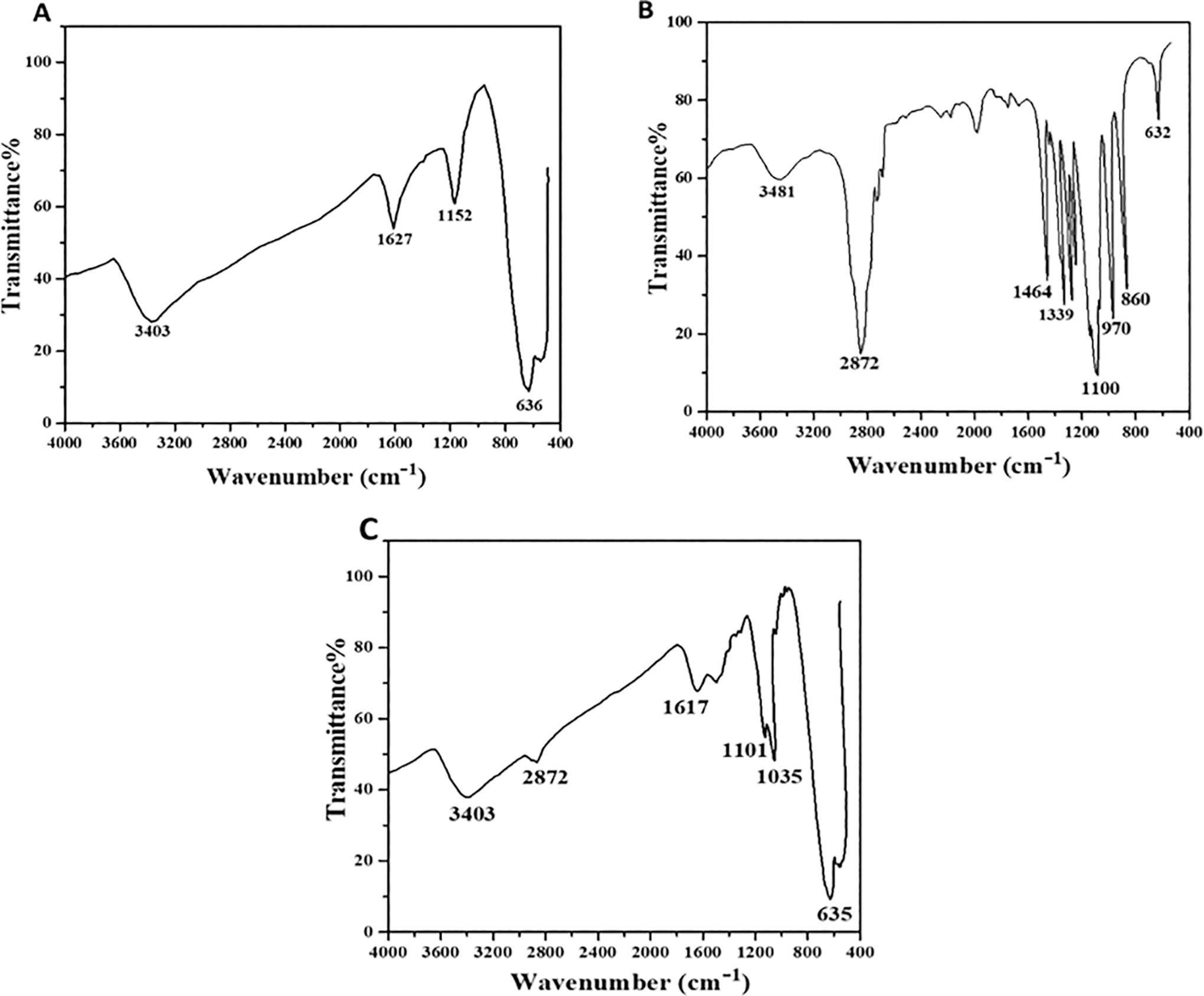
FTIR spectra
Enhanced evaluation of SnO2:Au NPs for PDT in cancer therapy
Semiconductor NPs such as SnO2:Au have unique photoluminescence (PL) and electrical properties, making them potential candidates for PDT in cancer treatment. Thus, this study explored their ability to generate ROS and serve as effective photosensitizers.
Photoluminescence properties
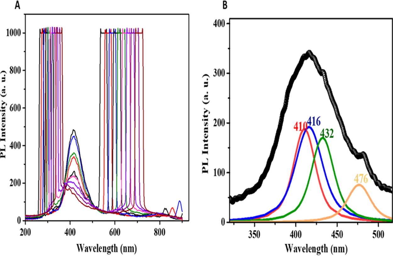
The PL spectrum of SnO2:Au NPs was measured with excitations at wavelengths ranging from 250 nm to 340 nm in 10 nm increments (Fig. 6). The highest emission was observed at 270 nm (Fig. 6A). The emission spectrum after excitation at 270 nm was deconvolved into components at 410, 416, 432, and 476 nm (Fig. 6B). In nanocrystalline oxides, oxygen sites are known as crystalline defects that significantly influence the emission spectrum. Oxygen vacancies (VO 0, VO +, and VO ++) play a crucial role, with VO++ groups contributing to emissions in the 400–500 nm range. Thus, the observed emission in SnO2:Au NPs is primarily due to oxygen deficiencies and defects within NPs. 45,46
Phosphorescence and afterglow
To evaluate the phosphorescence emission, SnO2:Au NPs were irradiated with UV light for 15 min, and the afterglow was recorded at room temperature, as shown in Figure 7. Afterglow refers to the delayed emission of light by the material after the excitation light source is turned off. The prolonged re-emission time is due to forbidden transitions from excited energy states to the ground state. To utilize SnO2:Au NPs for cancer treatment through PDT and address the limitation of light penetration, the afterglow spectrum of the NPs was examined. As shown in Figure 7, the afterglow of SnO2:Au NPs initially decreased over 200 sec, but then remained nearly constant up to 400 sec. This suggests the possibility of exciting the photosensitizer NPs outside the body and then rapidly injecting the material into tumors deep within the body. The long-lasting afterglow of the NPs could enable the continuous generation of ROS without requiring further excitation, potentially leading to the sustained death of deep-seated cancer cells.
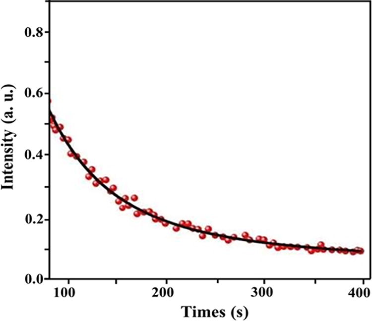
Afterglow spectrum of SnO2:Au NPs.
Generation of ROS
Singlet Oxygen Detection Using Anthracene
Anthracene (C14H10), a blue-purple fluorescent molecule, serves as an indicator of singlet oxygen, a predictor of cellular toxicity. When oxidized in the liquid or gas phase, anthracene is converted to nonfluorescent anthraquinone. In the presence of ROS, more anthracene molecules are converted to anthraquinone, leading to a decrease in absorption intensity. In this study, anthracene was initially exposed to UV light for 60 min, and then its absorption spectrum was recorded (Fig. 8). As shown in Figure 8, the absorption spectra of anthracene before and after irradiation were almost identical in the absence of NPs. However, after adding NPs to the anthracene solution and illuminating it with UV light for 60 min, the absorption intensity of anthracene decreased by 25%, indicating singlet oxygen production by SnO2:Au NPs.
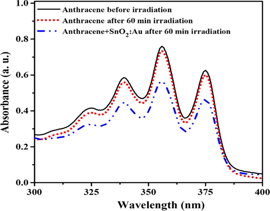
Detection of singlet oxygen produced by SnO2:Au NPs irradiated with UV.
Hydroxyl radical generation using MB
The generation of hydroxyl radicals by the afterglow NPs was examined indirectly using MB as a probe. MB, with the molecular formula C16H18N3SCl, is highly soluble in water. When MB reacts with hydroxyl radicals generated by the afterglow NPs, it degrades and is eventually converted to water and carbon dioxide (Fig. 9). This degradation causes a decrease in the absorption intensity of MB, resulting in visible discoloration. The MB solution was irradiated alone and in the presence of NPs for 60 min. It was observed that MB alone was not degraded. As shown in Figure 9, there was a decrease of about 55% in MB absorbance in the presence of SnO2:Au NPs, suggesting that the NPs are capable of producing hydroxyl radicals.
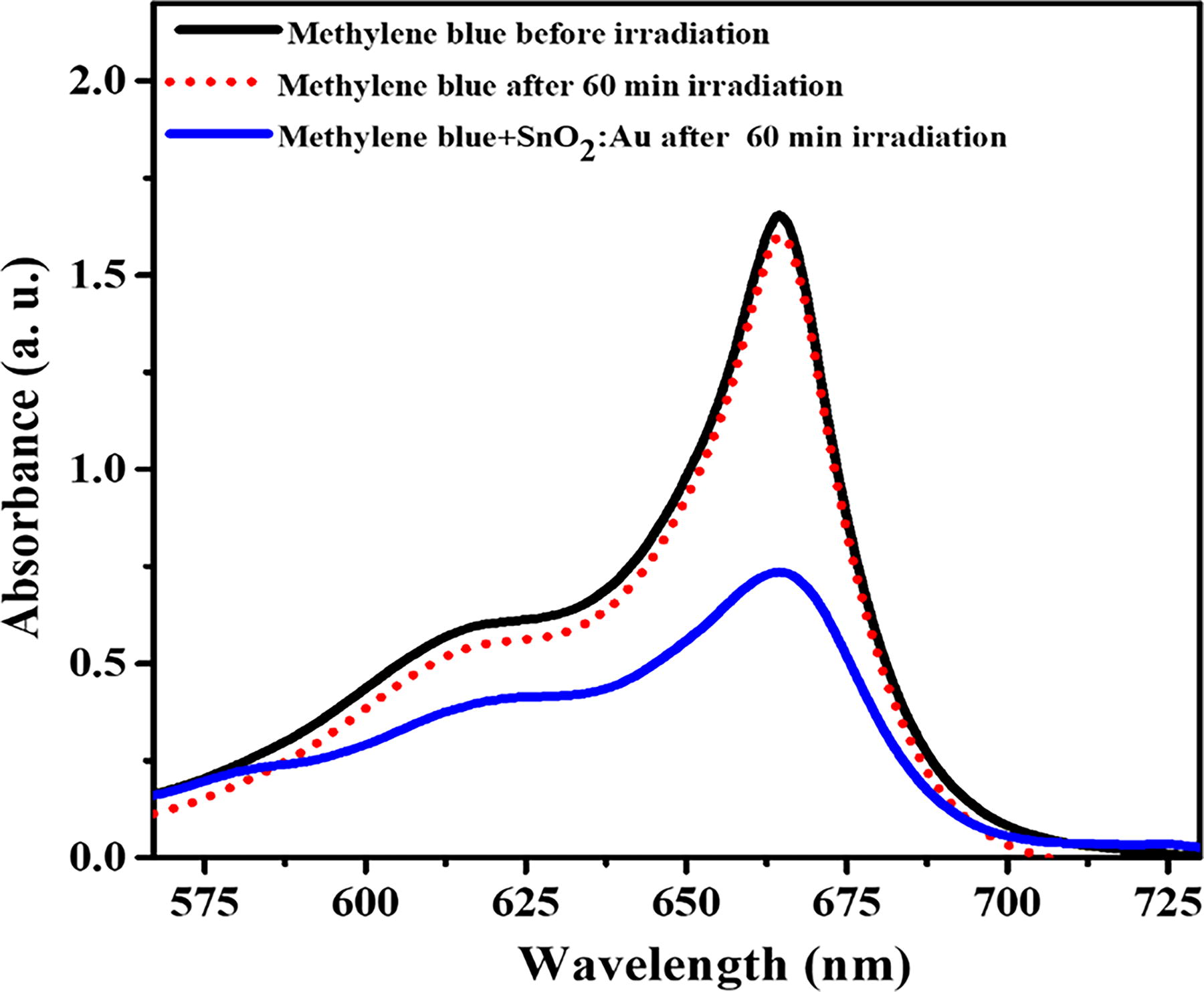
Hydroxyl radical detection of SnO2:Au NPs by bleaching of MB. MB, methylene blue.
The ability of SnO2:Au NPs to generate ROS, including singlet oxygen and hydroxyl radicals, is a crucial factor in their effectiveness as photosensitizers in PDT for cancer therapy. When SnO2:Au NPs are exposed to UV light, the photoexcitation process results in the transfer of energy to molecular oxygen, forming singlet oxygen. This singlet oxygen is highly reactive and can induce oxidative stress by damaging cellular components such as lipids, proteins, and DNA. 47,48 In addition, the photoexcitation of SnO2:Au NPs leads to the generation of electron-hole pairs. The photo-generated electrons can interact with oxygen molecules to form superoxide anions, which further dismutate to hydrogen peroxide and subsequently form hydroxyl radicals through the Fenton reaction. These hydroxyl radicals are among the most reactive ROS and can cause extensive cellular damage, leading to cell death. 49,50 Thus, SnO2:Au NPs demonstrate significant potential as photosensitizers for PDT in cancer therapy. Their ability to produce ROS, including singlet oxygen and hydroxyl radicals, combined with their prolonged afterglow emission, makes them promising candidates for effective and sustained cancer cell eradication. The enhanced generation of ROS upon UV activation underscores the therapeutic potential of SnO2:Au NPs in targeting and destroying cancer cells through oxidative stress mechanisms.
Cytotoxic effects of SnO2:Au NPs in PDT for cancer treatment
To investigate the potential of SnO2:Au NPs in PDT for cancer treatment, this study focused specifically on their ability to generate ROS and serve as effective photosensitizers. The phototoxicity of SnO2:Au NPs against C26 cells was assessed using the MTT assay. First, the effect of UV LED exposure alone on the cells was tested by irradiating them for durations ranging from 0 to 30 min, as shown in Figure 10A. An irradiation time of 15 min was selected for subsequent experiments as it minimally affected cell viability while being sufficient to activate the NPs. Exposures longer than 15 min resulted in significant phototoxicity.
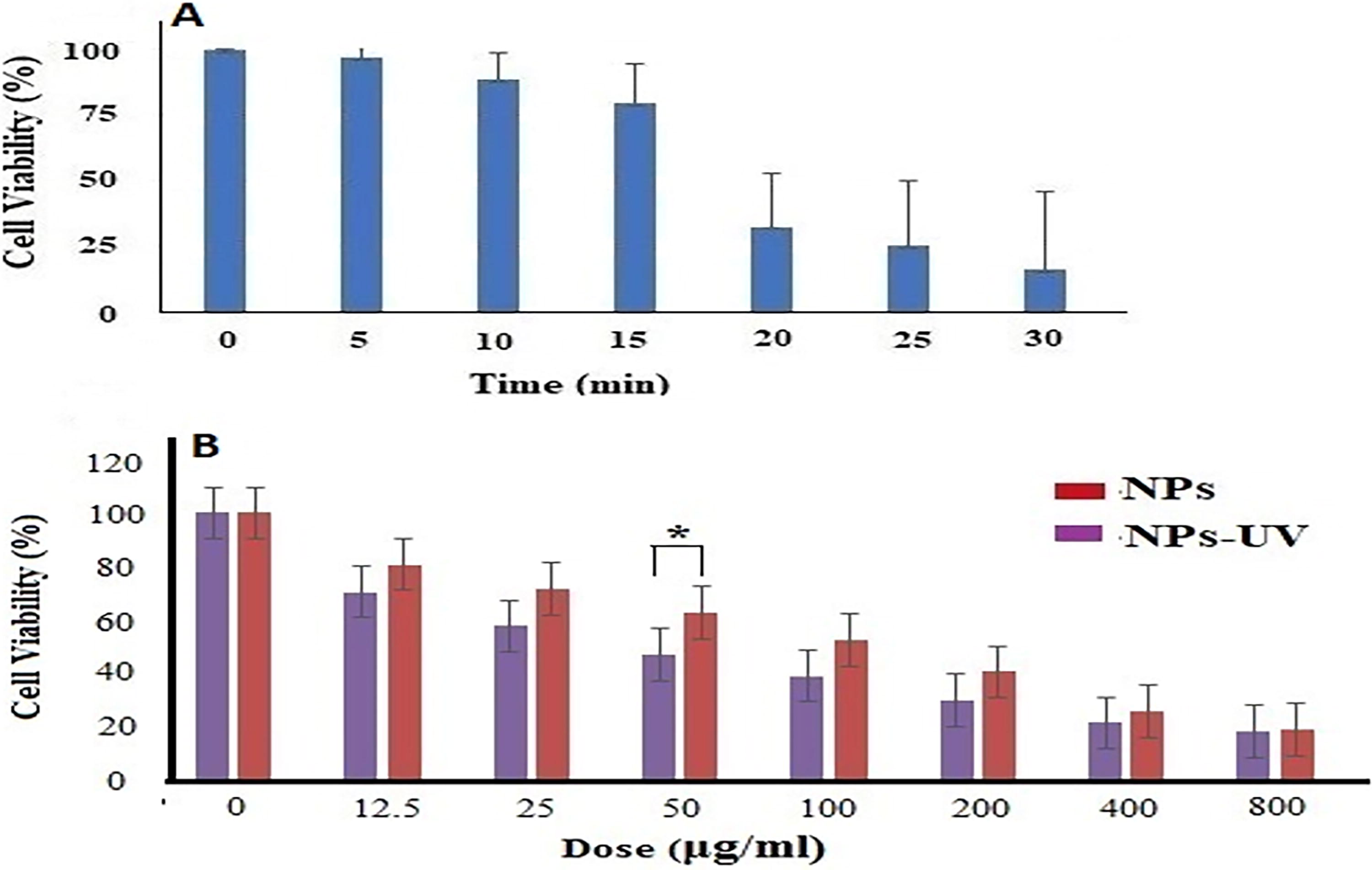
Cell viability by MTT assay.
Next, the cells were exposed to various concentrations of SnO2:Au NPs for 24 h and either kept in the dark or exposed to 15 min of UV light. The results are shown in Figure 10B. Increasing the concentration of SnO2:Au NPs resulted in higher cell death rates both in the dark and after UV light exposure. The IC50 value (concentration that inhibits cell growth by 50%) of SnO2:Au on C26 cells after 24 h of treatment was 118 μg/mL. However, the IC50 decreased to 42 μg/mL (p = 0.021) after 15 min of illumination, indicating enhanced efficacy upon light activation. UV radiation alone (15 min) without NPs did not significantly affect cell viability (p = 0.086). These findings demonstrate that SnO2:Au NPs are effective photosensitizers capable of generating ROS, which is critical for inducing cell death in PDT. Statistical tests confirmed the significance of these findings (Fig. 10B). The decrease in cell viability with UV-activated NPs was statistically significant compared with both the control and NPs without UV exposure (p < 0.05).
One of the primary mechanisms by which SnO2:Au NPs exert anticancer effects is through ROS generation upon UV activation. ROS, such as singlet oxygen, hydroxyl radicals, and superoxide anions, cause oxidative stress and damage to cellular components, leading to cell death. 51 The enhanced ROS generation with UV-activated SnO2:Au NPs, as shown by the significant reduction in anthracene fluorescence and MB absorbance, corroborates this mechanism. Specifically, upon UV activation, SnO2:Au NPs undergo a photoexcitation process where electrons in the NPs are excited to higher energy states. These excited electrons interact with molecular oxygen, generating ROS. The generated ROS can induce oxidative damage to vital cellular structures, including lipids, proteins, and DNA, thereby triggering cell death pathways. 52 Thus, SnO2:Au NPs are effective photosensitizers capable of inducing cell death through ROS generation, making them promising candidates for PDT. The ability to activate these NPs with UV light and achieve sustained ROS production offers a potential strategy for treating deep-seated tumors. Further, the prolonged afterglow of the NPs could enable the continuous generation of ROS even after the initial UV exposure, enhancing the therapeutic efficacy against cancer cells located in less accessible areas.
In related research, Ag@SiO2 core-shell NPs were prepared and structurally verified using techniques such as UV-vis, XRD, FTIR, TEM, and EDX. The findings showed that Ag@SiO2 core-shell NPs could eliminate cancer cells when exposed to light. This suggests that using SiO2 as a shell can not only prevent clumping but also mediate the light activation of Ag NPs. 53 In addition, another study exposed human liver adenocarcinoma cells (HepG2) to various types of zinc oxide NPs (ZnO-NPs), including those doped with different metals such as Iron (Fe), Silver (Ag), Lead (Pb), and Cobalt (Co), as well as silica-coated ZnO-NPs. They also tested titanium dioxide nanoparticles (TiO2-NPs), titanium dioxide nanotubes (TiO2-NTs), and a combination of ZnO-NPs and TiO2-NTs excited by UV light. The findings showed that ZnO-NPs, Fe-ZnO-NPs, Ag-ZnO-NPs, Pb-ZnO-NPs, and Co-ZnO-NPs all displayed toxic effects on HepG2 cells, with IC50 concentrations of 42.6, 37.2, 45.1, 77.2, and 56.5 μg/mL, respectively, compared with doxorubicin (IC50: 20.1 μg/mL). When the cancer cells were treated with these NPs, there was a significant increase in the activity of superoxide dismutase, as well as levels of hydrogen peroxide and nitric oxide, along with a significant decrease in catalase and glutathione peroxidase (GSH-Px) activity. In addition, decreased amounts of reduced GSH, total protein, and nucleic acids were observed. They concluded that metal-doped ZnO-NPs combined with UV irradiation could provide an antiproliferative effect on HepG2 cells due to the generation of ROS. However, further in vivo studies using photodynamic therapy are necessary before these NPs can be considered for use in modern clinical trials. 54
Conclusions
The green synthesis of gold-doped tin dioxide NPs was performed by a coprecipitation method mediated by T. polium medicinal plant extract. The green NPs were characterized by TEM, SEM, XRD, PL, UV-Vis, and FTIR analysis. The highest intensity PL emission corresponded to excitation at 270 nm and contained four emission bands at 410, 416, 432, and 476 nm. The FTIR spectra confirmed the formation of Sn and O bonds and the surface modification of the NPs with PEG. A 25% decrease in anthracene and a 55% reduction in MB in the presence of SnO2:Au NPs plus UV demonstrated the ability of the NPs to generate ROS. Using an MTT test, the cytotoxicity of SnO2:Au NPs with or without UV was quantified in a time and dose-dependent manner. According to the findings, the use of SnO2:Au NPs alone could inhibit the proliferation of cancer cells, and exposure to UV radiation could significantly increase the anticancer effects. Analysis of the cellular uptake of NPs would be important to assess the effects of NPs on different cells. Unfortunately, due to the high costs, we were not able to do this. Hence, this is an important limitation of the study. Another limitation is that the PDT effect was relatively modest compared with the PDT effects of most traditional photosensitizers. In conclusion, the synthesized NPs have remarkable afterglow and the ability to produce ROS upon UV excitation and could be suitable for cancer treatment, but this needs further investigation.
Footnotes
Authors’ Contributions
M.K.: Investigation (lead); methodology (equal); and writing—original draft (equal). M.N., H.M., and E.S.: Data curation (supporting); formal analysis (supporting); investigation (supporting), critical revised. E.S., H.M., and M.N.: Data curation (supporting); investigation (supporting); methodology (supporting), critical revised. M.K.: Data curation (supporting); formal analysis (equal); investigation (supporting); methodology (supporting); critical revised; and writing—review and editing (equal). M.R.H. and M.Z.: Critical revised and writing—review and editing (equal). M.Z.: Conceptualization (equal); funding acquisition (lead); investigation (lead); resources (equal); supervision (equal); and writing—review and editing (equal). All authors confirmed the final version of article.
Author Disclosure Statement
No competing financial interests exist.
Funding Information
No funding was received for this article.