
Abstract
Objective:
To investigate the effects of laser-treated enamel surface roughness on the adhesion of Streptococcus mutans.
Methods:
A total of 176 premolars extracted for subtractive orthodontic treatment were collected. Samples were randomly divided into four groups: control group, 35% phosphoric acid, Er:YAG laser, Er,Cr:YSGG laser. Surface roughness was detected using three-dimensional white light interferometric surface topography (n = 8/group), and surface morphology was observed by scanning electron microscope [(SEM), n = 8/group]. Samples were cultured in S. mutans solution for 12 h, 1 day, and 2 days. The colony forming units on the enamel surface were calculated (n = 8 per time point). The biofilm and activity of S. mutans were observed by SEM (n = 2/group) and confocal laser scanning microscope (n = 2/group). Correlation analysis was carried out between bacterial adhesion on the enamel surface at different time points and the surface roughness of the enamel treated by different methods.
Results:
After Er:YAG and Er,Cr:YSGG laser etching, the surface roughness of enamel increased (p < 0.05), but there was no difference in S. mutans adhesion and biofilm morphology after 1 day compared with traditional phosphoric acid etching (p > 0.05). S. mutans adhesion was positively correlated with the enamel roughness in the early stage.
Conclusions:
Er:YAG and Er,Cr:YSGG laser etching may be an alternative to traditional phosphoric acid etching.
Introduction
Currently, the phosphoric acid etching method routinely used before orthodontic bracket bonding has drawbacks, such as demineralization of the tooth’s hard tissue surface, increased caries rates around the bracket, and a high bonding failure rate after the bracket falls off. 1 Laser etching technology was introduced in orthodontic bonding in the 1990s. 2 With the continuous development of laser technology and deeper research, Er family lasers have gained focus because their wavelengths are easily absorbed by hydroxyapatite and water, allowing them to effectively interact with dental hard tissues. 3 –5
Colucci et al. demonstrated that Er:YAG laser ablation of enamel and dentin in water-cooled mode does not cause any thermal damage to the dental pulp nor does it produce any cracking or carbonization on the enamel surface. 6 However, studies have shown that when the Er:YAG laser is used, the water molecules in the enamel’s surface structure absorb energy to produce micro-blasts, cutting the enamel or causing surface roughening. 7,8 Lee also found that Er:YAG laser (300 mJ/Pulse, 10 sec) etched enamel can produce rougher and more irregular ablation patterns than phosphoric acid etched surfaces. 9 In 2018, Teutle-Coyotecatl found that the roughness of Er:YAG laser-etched enamel in different energy density groups (12.7, 25.5, and 38.2 J/cm2) increased significantly. Increased roughness is the basis for increased bond strength of enamel, but it may also favor the adhesion of cariogenic bacteria, such as Streptococcus mutans. 10
Our previous research compared different working parameters and confirmed that after Er:YAG laser etching at 250 mJ, 30 Hz, and 6/8 water cooling, the surface morphology and bonding performance of enamel were similar to those after 35% (mass fraction) phosphoric acid etching. 11 Moreover, Er:YAG laser re-etching of enamel under this parameter produced a uniform honeycomb structure and similar shear strength to 35% phosphoric acid-mediated re-etching. 12 Further in-depth research by our group found that Er:YAG laser etching under this parameter enhanced the acid resistance of the enamel surface. 13 However, the effect of the resulting increase in roughness on bacterial adhesion is still unknown.
A previous study has shown that laser conditioning of enamel using Er,Cr:YSGG with 4.5 W could be a promising alternative to acid conditioning, as it produces less surface roughness, reduced plaque accumulation, and lower bacterial adhesion. 14 However, other studies have indicated that surface roughness was significantly increased with the Er,Cr:YSGG laser. Scanning electron microscope (SEM) analysis showed that the irradiated surface produced a rough texture that completely lacked a smear layer, and there was also no cracking of enamel or dentin. 15 This difference may be due to varying parameters selected, so further research is needed to investigate the effects of Er,Cr:YSGG laser irradiation on enamel surface roughness and bacterial adhesion. We hypothesize that Er:YAG and Er,Cr:YSGG laser treatments will significantly increase the roughness of the enamel surface; however, these treatments will have little effect on the bacteria adhesion. Therefore, we compare and discuss the surface roughness of enamel after Er:YAG and Er,Cr:YSGG laser treatment, as well as the effects of variation on the adhesion of S. mutans to surfaces.
Materials and Methods
Tooth selection and sample preparation
One hundred and seventy-six premolars extracted for orthodontic treatment were collected. The crown enamel was required to be intact, without cracks, caries, fillings, incomplete mineralization, wear, or root canal treatment. The extracted teeth were immediately cleaned with saline and then stored in distilled water at 37°C (replaced every 7 days) for no more than 6 months. The protocol for this study was approved by the Human Subjects Ethics Board of Xuanwu Hospital, Capital Medical University (Ethical Approval: LYWS[2024]002-002) and conducted in accordance with the Helsinki Declaration of 1975.
The teeth were sectioned with a diamond bur and embedded in a 2 × 2 × 1 cm mold with epoxy resin. A double-disc grinder (UltrMet6, Beijing Zhongxing Bairui) was used to grind the buccal enamel under running water (rotation speed: 200 r/min, silicon carbide sandpaper: 240#, 400#, 1000#, 2000#) to form a facet (parallel to the horizontal plane) of approximately 9 mm2. Each plane was marked left and right along the long axis of the clinical crown, with the left side serving as the blank control side and the right side treated according to the grouping.
Surface treatment
Two hundred and sixty samples were divided into four groups (n = 65/group): control group, where no surface treatment was done; 35% phosphoric acid group, using a 35% mass fraction of phosphoric acid etching agent (Gruma, Heraeus Gusa, Germany) that was acid-etched for 60 sec and then rinsed with distilled water; Er:YAG group, where the Er:YAG laser (LiteTouch™, Syneron, Israel) with a wavelength of 2.94 µm was used to etch for 15 sec, with the following working parameters: 250 mJ, 30 Hz, 6/8 water, pulse width 150 sec; fiber working tip diameter 1.3 mm, length 19 mm; working distance 2 mm, working angle 60°; and Er,Cr:YSGG laser group, where the Er,Cr:YSGG laser (Waterlase MD, BioLase Technology Inc., Irvine, CA, USA.) with wavelength of 2.78 µm was also used to etch for 15 sec, with working parameters of 1.5 W, fiber working tip diameter 0.8 mm, working distance 2 mm, and working angle 60°. 16
Surface roughness analysis
The surface roughness of each sample was evaluated using a three-dimensional (3D) white light interferometric surface topography instrument (NexView, Zhai Kelamda, USA) to obtain their 3D images. Three nonoverlapping 0.8 × 0.8 mm areas were randomly selected from both the blank control side and the treatment side of each sample to measure the roughness. The Gaussian filter was used to calculate the 3D contour arithmetic mean deviation Sa (µm) according to the international standard ISO 4288, which was recorded as Sa0 (before treatment) and Sa1 (after treatment).
Bacterial adhesion test
S. mutans ATCC 25715 (UA159, Baina Biology) was grown on a brain heart infusion [(BHI), OXOID, CM1135, USA] plate at 37°C under humidified air containing 5% CO2. Overnight cultures of S. mutans were then diluted to an optical density at 660 nm of 0.2, corresponding to 1 × 10⁹ CFU/mL.
Prior to the bacterial adhesion test, waterproof nail polish was applied to the samples, creating a window of 3 × 3 mm in the center of the buccal side, and allowed to dry naturally. All four groups of samples were placed into 24-well cell culture plates, with one sample in each well and the buccal side window facing upward. One milliliter of S. mutans solution was added to each culture well, and then cultured under anaerobic conditions at 37°C with 5% CO2 for 12 h, 1 day, and 2 days (the medium was replaced once a day). After incubation, each sample was washed three times with phosphate-buffered saline [(PBS), BL302A, Biosharp, China] for 10 min each to remove nonadherent bacteria. Subsequently, S. mutans within the window was scraped off using a metal blade and placed in an EP tube containing 1 mL of PBS. The samples were then sonicated in three 15-sec cycles, and after serial dilution, they were streaked onto BHI agar plates. After 48 h of anaerobic incubation, colony counting (CFU/mL) was performed.
SEM
At each time point, samples were gently washed with 5 mL of PBS three times, with each wash lasting 10 min. They were then fixed with 2.5% glutaraldehyde (Tianjin Damao) at 4°C for more than 4 h, followed by three washes with PBS. Finally, 50%, 70%, 80%, 90%, and 100% alcohol (Beijing Tongguang Fine Chemicals) were used for gradient dehydration. SEM was then used to observe the S. mutans biofilm on the enamel surface, with three areas per sample being randomly selected.
Confocal laser scanning microscopy
At each time point, two samples of each subgroup were gently washed with 5 mL 85% normal saline three times for 10 min each time and then stained with BacLight biofilm (LIVE/DEAD BacLight™ Bacterial Viability Kits, L7012, Thermo, U.S). After staining, rinse with 5 mL of 85% normal saline three times, and finally place the sample upside down on the coverslip (FCGF50, Biyuntian, Shanghai) that has been dripped with fixed oil. The confocal laser scanning microscope (CLSM) (FV1000, OLYMPUS, Japan) equipped with a UPLSAPO 60× oil immersion objective and 10× eyepiece was used to observe biofilm activity (three regions/sample were randomly selected).
Statistical analysis
Statistical analyses of the results were conducted using SPSS version 25.0. Measurement data for surface roughness were expressed as mean ± standard deviation (mean ± SD). The comparison of measurements before and after treatment was assessed using a paired t-test, while differences between groups were evaluated using one-way analysis of variance (ANOVA). Nonparametric tests were employed for measurement data that did not conform to a normal distribution or exhibited nonhomogeneous variance. The adherence of S. mutans (measured in CFU/mL) was also expressed as mean ± SD after logarithmic transformation. In addition, repeated measures ANOVA was utilized for intra-group and inter-group comparisons. A p value of less than 0.05 was considered indicative of statistical significance.
Results
Alterations in the surface roughness of enamel after Er laser treatment
Observations utilizing a 3D white light interferometric surface topography instrument indicated that the enamel surface in the control group remained relatively flat. In contrast, the acid group exhibited a uniform needle-like appearance; while the surfaces treated with Er:YAG and Er,Cr:YSGG lasers were characterized by rough and uneven textures, displaying prominent grooves and ridges. SEM analysis revealed no significant morphological changes in the enamel surfaces of the control group. The acid group displayed a uniform morphology with a consistent squamous appearance. Notably, the centers of the enamel rods in both the Er:YAG and the Er,Cr:YSGG treatment groups presented a sunken appearance (Fig. 1). In the roughness assessment, enamel treated with acid, Er:YAG, and Er,Cr:YSGG, exhibited an increase in surface roughness compared with levels measured before treatment (p < 0.05). Further, the enamel roughness after treatment with Er:YAG and Er,Cr:YSGG was significantly greater than that observed after acid treatment (p < 0.05) (Table 1).
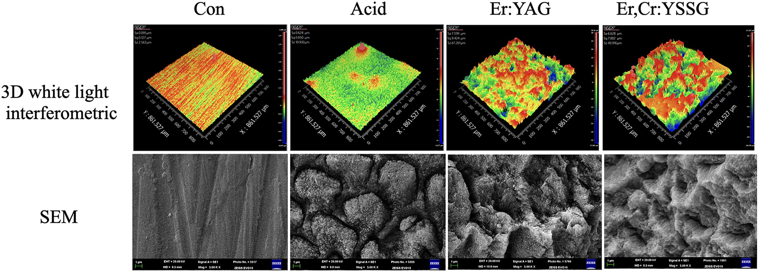
3D white light interference topography and SEM image (5000×) of enamel surface treated by different methods. 3D, three-dimensional; SEM, scanning electron microscope.
Comparison of Surface Roughness (µm) of Enamel with Different Treatment
Represents significant difference compared with pretreatment.
Represents significant differences compared with the control group.
Represents a significant difference compared with the acid group.
S. mutans adhesion on tooth enamel surface after laser treatment
The adhesion of S. mutans to the enamel surface was analyzed across all treatment groups. Results indicated that bacterial adhesion increased at each time point examined (Table 2). A comparison of different treatment methods revealed that, at 12 h post-treatment, bacterial adhesion on the enamel surface in all treatment groups was significantly higher than that observed in the control group. Further, the number of S. mutans on the enamel surface after Er:YAG and Er,Cr:YSGG laser treatments was greater than that in the acid treatment group (p < 0.05) (Table 2). As time progressed, at both 1 day and 2 days post-treatment, no significant difference in the number of adhered S. mutans was observed among all treatment groups compared with the control group, nor was there any significant difference between the various treatment groups (p > 0.05) (Table 2). In addition, the amount of S. mutans adhesion demonstrated a positive correlation with enamel surface roughness at 12 h (r = 0.97, p = 0.034), although the correlation coefficient decreased by 1 and 2 days (Table 3).
Comparison of S. mutans Lg (CFU/mL) on the Enamel Surface at Different Time Points
Represents significant difference compared with the control group at 12 h.
Represents significant difference compared with the acid group at 12 h.
CFU, colony forming units; S. mutans, Streptococcus mutans.
Correlation Coefficient Matrix of S. mutans Adhesion Amount and Roughness at Different Time Points
r: Pearson correlation coefficient.
Represents p < 0.05.
S. mutans, Streptococcus mutans.
SEM observation of S. mutans adhesion on tooth enamel surface
After 12 h, S. mutans appeared scattered and clustered locally within the control group. There were significantly more S. mutans present in the acid, Er:YAG, and Er,Cr:YSGG groups, where the bacteria formed clusters, forming a relatively loose biofilm structure with large gaps between substrates (Fig. 2A–D). At 1 day, bacterial colonies in the control group increased and connected to form a bridge, resulting in a biofilm adherent to the enamel surface. In the acid, Er:YAG, and Er,Cr:YSGG groups, the S. mutans colonies on the enamel surface further expanded, with the biofilm structure becoming progressively denser and the inter-matrix gaps decreasing (Fig. 2E–H). By 2 days, the enamel surface in each group was nearly covered by bacterial colonies, forming a dense network-like structure. The biofilm had matured further, with increased thickness. At this stage, there was no significant difference in the morphology of the biofilm among the different treatment groups (Fig. 2I–L).
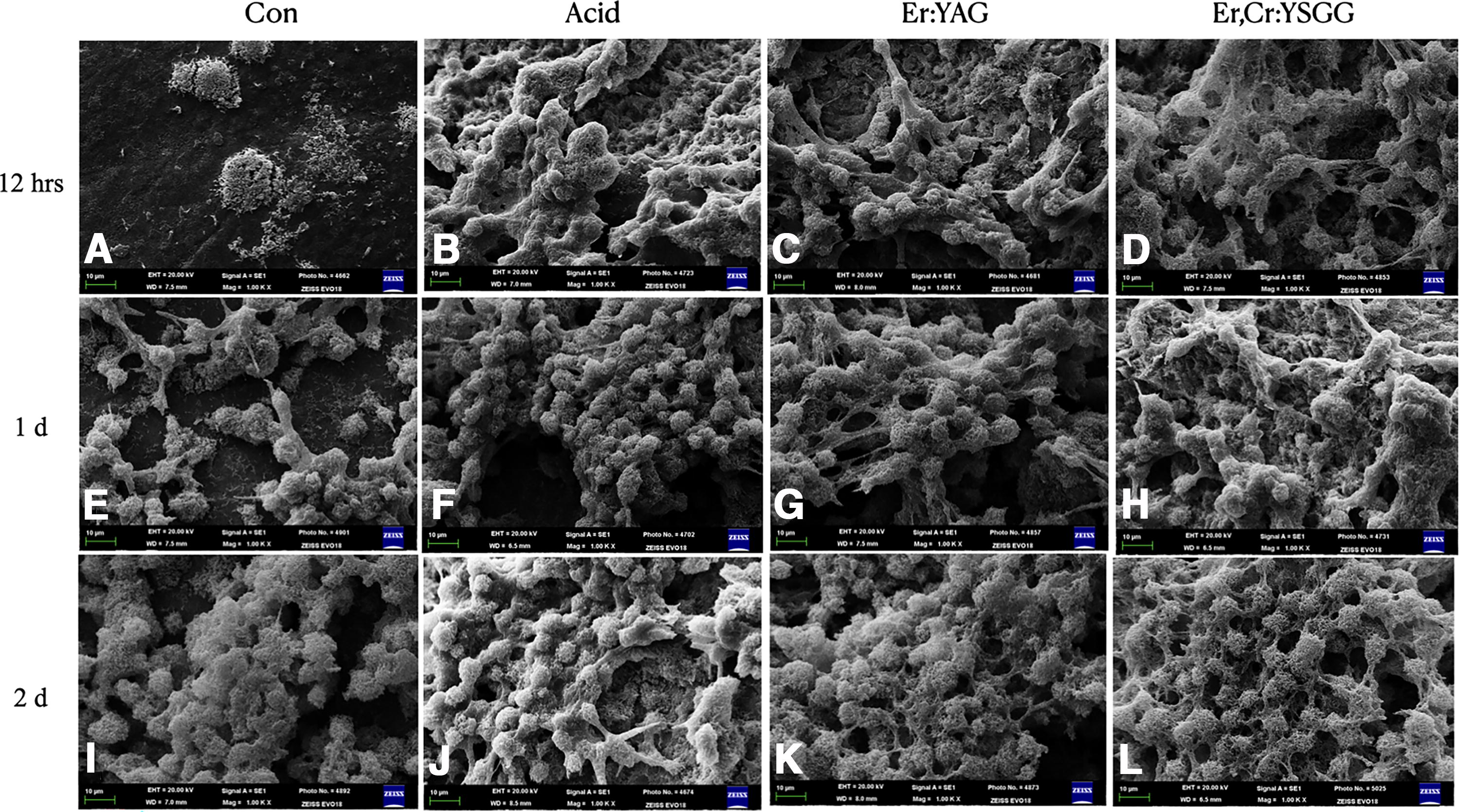
SEM images of S. mutans adhered to the enamel surface with different methods:
CLSM observation of S. mutans activity on tooth enamel surface
After fluorescent staining with SYTO9 and PI, green fluorescence indicated live bacteria, while red fluorescence indicated dead bacteria. The results showed that the percentage of viable bacteria in nearly all groups gradually increased from 12 h to 1 day, reaching a peak of approximately 97% viability at 1 day, before gradually decreasing (Fig. 3). Statistical analysis showed that at 12 h, the percentages of viable bacteria within the biofilms of acid, Er:YAG, and Er,Cr:YSGG groups were significantly higher than those in the control group at 12 h (p < 0.05). By the second day, the percentage of viable bacteria in each group was significantly lower than that on the first day (p < 0.05) (Table 4).
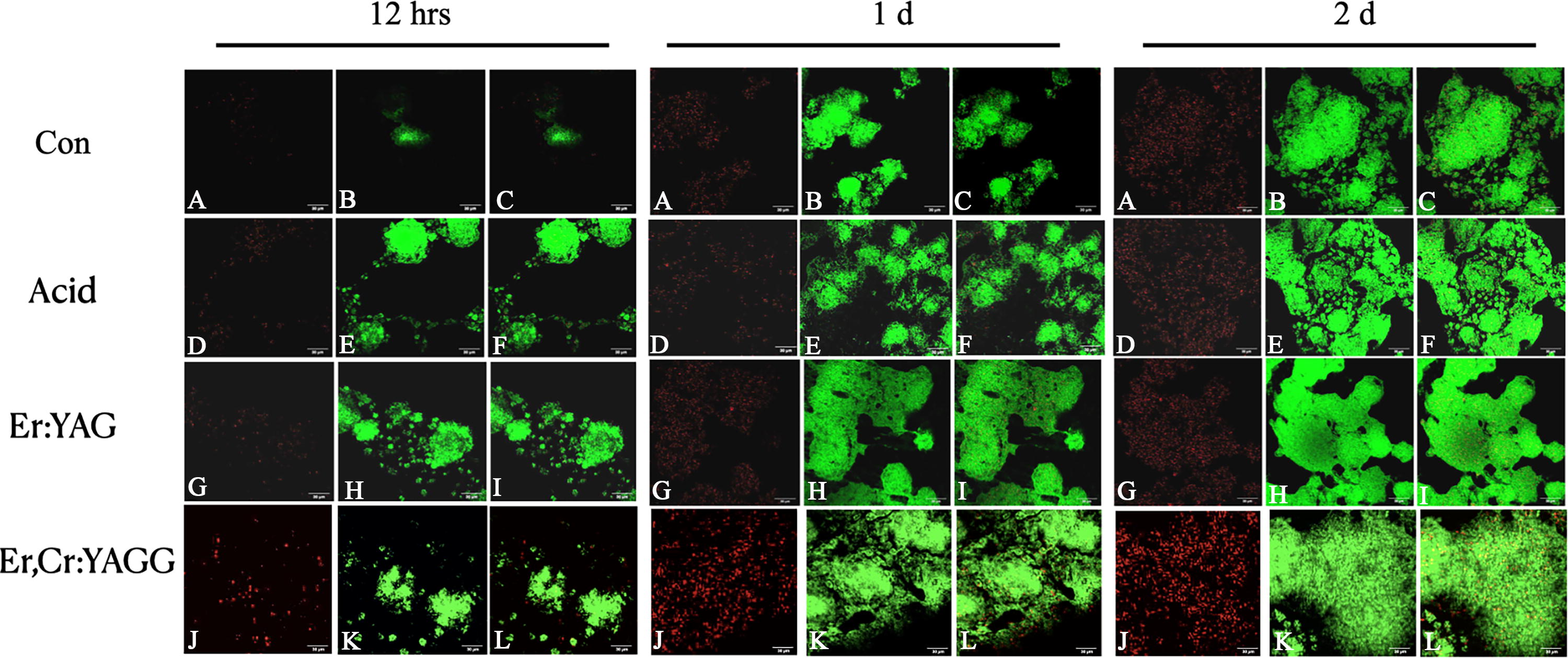
CLSM images of S. mutans adhered to the enamel surface with different methods:
Comparison of the Percentage of Viable Biofilm Bacteria on the Surface of Tooth Enamel at Different Time Points
Represents a significant difference compared with the control group, p < 0.05.
Represents a significant difference compared with 12 h, p < 0.05.
Represents a significant difference compared with 1 day, p < 0.05.
Con, control.
Discussion
The surface structure of natural enamel exhibits micro-roughness within the range of 0.59–0.66 μm. 17 Surface etching involves the risk of damaging the enamel surface and changing its original morphology. The roughened enamel surface can hinder proper cleaning, subsequently leading to plaque deposition, bacterial retention, and stain formation. 18 In recent years, laser etching techniques have developed to address these issues using various types of lasers. 19 Laser-etched enamel surfaces display a porous surface similar to that produced by acid etching, suggesting that acid etching could serve as an alternative to traditional acid etching. 20
Previous studies using profilometers to assess surface roughness have limitations, as they cannot form a comprehensive image of the surface. 21 Although SEM can describe surface roughness, it does not allow quantitative analysis of this parameter. Although atomic force microscopy is capable of performing quantitative assessments of surface roughness, its measuring range is limited to 2.5–20 µm, which restricts its precision. 22 In the present study, the 3D white light interferometric surface topography instrument was used to assess the enamel surface roughness after acid etching or laser irradiation. This instrument not only ensures high-precision measurement of 0.1 nm across a full range of 0–150 mm but is also noncontact, noninvasive, and nondestructive. Our findings indicate that the surface roughness of the enamel in the Er:YAG and Er,Cr:YSGG laser etching group was greater than that of the phosphoric acid etching group, consistent with previous studies. 15,23 This supports our first hypothesis that Er laser treatment increases enamel roughness compared to acid etching. The mechanism behind this increased roughness may be related to the ablation process of the laser. The emission wavelengths of the Er:YAG and Er,Cr:YAGG lasers are 2.94 and 2.78 μm, respectively, which coincide with the absorption peak of water and are well absorbed by the OH− groups in hydroxyapatite. 24 When the laser is applied to the tooth surface, the water in the enamel absorbs the laser energy and boils, resulting in the formation of high-pressure steam. This vaporization transforms the smooth enamel into irregular shapes and micro-crack structures. 7 In addition to creating a roughened enamel surface, laser irradiation may also enhance acid resistance. This effect could be related to the ability of the laser-irradiated area to capture free ions necessary for remineralization. 25 These findings suggest that laser etching could serve as a viable alternative to traditional acid etching techniques, providing improved surface characteristics beneficial for orthodontic bonding.
Biofilms in the oral cavity comprises a variety of complex microorganisms, with S. mutans recognized as one of the most important pathogenic bacteria associated with dental caries. S. mutans plays a crucial role in the occurrence and progression of dental caries. 26,27 Studies have demonstrated that S. mutans are initial colonizers of dental plaque and exhibit a strong affinity for enamel hydroxyapatite. 28,29 Therefore, this study selected S. mutans to compare the differences in bacterial adhesion on the enamel surface after various treatments.
In the present study, Er:YAG and Er,Cr:YSGG lasers exhibited significantly greater S. mutans adhesion compared with traditional phosphoric acid etching at 12 h, which can be attributed to the increased surface roughness. By 2 days, there were no statistical differences in the levels of bacterial adhesion among the groups. Quantification of the CLSM images revealed that the percentage of viable bacteria in the biofilm on the enamel surface gradually increased from 12 h to 1 day, reaching a maximum of approximately 97%, followed by a gradual decline. Analysis indicated that extended culture time resulted in diminished nutrients within the bacterial solution and an accumulation of metabolites, thereby impacting the further increase in the percentage of viable bacteria in the biofilm. Further, the percentage of viable bacteria in the acid, Er:YAG, and Er,Cr:YSGG groups increased more slowly from 12 h to 1 day. This phenomenon may be attributed to the fact that both phosphoric acid etching and laser etching treatments enhance the surface roughness of the enamel. In the early stages of the experiment, this roughness serves as the dominant factor for bacterial adhesion, allowing viable bacteria to adhere rapidly and form biofilms on the surface of the tooth enamel. From 1 day to 2 days, due to the substantial initial adhesion of S. mutans, the Er:YAG and the Er,Cr:YSGG groups experienced greater limitations in nutritional availability and bacterial growth space, leading to a rapid decrease in the percentage of viable bacteria during this period. In addition, although laser etching altered the surface morphology of the enamel, showing honeycomb and melting features, no significant differences were observed in the morphology of S. mutans biofilm on the enamel surface among the various processing methods in CLSM images, which is consistent with Zancope’s observations. 30
The adhesion amount of S. mutans was correlated with surface roughness at 12 h (r = 0.97, p = 0.034), consistent with the findings of Nogueira et al. 31 As time progressed, the correlation coefficient decreased to a point of no correlation. These results further support our hypothesis that Er laser treatment significantly influences bacterial adhesion in the early stages, but this effect diminishes over time. This study suggests an interaction between the bacterial adhesion duration and the different treatment methods, indicating that the efficacy of various treatments on S. mutans adhesion may change over time. This phenomenon may arise because, in addition to roughness, other surface properties such as charge, hydrophilicity, hydrophobicity, and morphology, as well as the bacterial response to these surface characteristics, all impact bacterial adhesion. 32,33
The limitations of this study include the examination of bacterial adhesion using a single species, S. mutans, which may not fully capture the complexities of bacterial interactions within diverse microbial communities. In addition, the influence of environmental factors, such as pH and temperature, was not considered, potentially limiting the generalizability of the findings. In the early stages, bacterial growth was higher in the treated groups but later became similar across all groups due to limitations in nutrition and available space—conditions that may not accurately reflect the oral environment, where variations in diet can affect bacterial behavior. Further, the long-term effects of the treatment methods on bacterial adhesion were not explored, which could provide valuable insights into the stability and efficacy of these approaches over time. Future research aims to develop a more realistic model of the oral cavity to better understand bacterial interactions and adhesion after Er laser treatment.
Conclusions
Compared with traditional phosphoric acid etching, Er:YAG and Er,Cr:YSGG laser etching significantly increased the surface roughness of enamel; however, there was no difference in the amount of S. mutans adhesion and biofilm morphology after 1 day. The adhesion of S. mutans on the enamel surface was positively correlated with roughness during the initial stage. In summary, Er:YAG and Er,Cr:YSGG laser etching technologies may serve as viable alternatives to traditional phosphoric acid etching.
Footnotes
Authors’ Contributions
L.T. and X.T. were principal investigators and major contributors to this article. L.T. made contributions to the bacterial experimental design, article writing, submission, and revision. X.T. completed most of the experiments and data analysis. N.Y. is responsible for the study of changes in enamel surface morphology. M.L. completed the experiment on the roughness of the enamel surface. Y.Z. was the organizer of the entire project, including experimental design, guidance, and supervision. All authors have read and approved the article.
Author Disclosure Statement
The funders had no role in the design of the study; in the collection, analysis, or interpretation of data; in the writing of the article; or in the decision to publish the results.
Funding Information
This study was supported by Capital’s Funds for Health Improvement and Research (2022-2-2012), Beijing, China, and the R&D Program of Beijing Municipal Education Commission (KZ202210025032).