
Abstract
Walnuts kernels (Juglans regia L.) have rich antioxidants content and have been used in both cosmetic and pharmaceutical industry. This study dealt with the protective role of walnut kernels extracts (WK) on isoproterenol (ISO)-induced myocardial infarction (MI) in rats. Rats were pretreated with WK extracts (300 mg/kg) daily for 35 days. Then, ISO (100 mg/kg) was injected subcutaneously into rats to induce MI. Cardiac diagnostic markers (LDH and CPK), cardiac troponin, heart lipid peroxidation (TBARS and hydroperoxide), antioxidant system (CAT, SOD, GPx, GST, GSH, and GSSG) and the levels of lipid profile were evaluated in rats, and the results revealed WK significantly prevented myocardial injury induced by ISO (p < 0.05). WK significantly alleviated the oxidative damage and dyslipidemia in ISO-induced MI rats (p < 0.05). The effect produced by WK was compared with α-tocopherol. The mechanisms for the protective effects of WK could be attributed to its antilipid peroxidative, antioxidant, and antilipidemic properties. In conclusion, we demonstrated that WK has a significant protective effect against ISO-induced MI.
Introduction
Myocardial infarction (MI) occurs when coronary blood flow is inadequate, hence the oxygen/nutrients supply is insufficient for the demand of the myocardium, resulting in irreversible damage to the heart. 1 From 1990 to now, cardiovascular diseases remains the burden of many regions of the world. 2 Isoproterenol (ISO), a synthetic catecholamine, causes severe pathological changes in the myocardium, resembling MI in humans. 3 To study the cardioprotective effect of drugs, induction of MI by ISO is considered as a well standardized model. 4 It is widely accepted that an increase in oxidative stress is one of the most causative factor. Additionally, dyslipidemia, an independent risk factor for cardiovascular diseases, is also caused by ISO. 5
Walnut (Juglans regia L.) is a valuable crop largely consumed. Not only dry fruits (nuts) are used but also green walnuts, shells, kernels, and leaves have been used in both cosmetic and pharmaceutical industry. 6 Walnuts kernels (WK) are rich in components containing abundant phospholipids (PL), proteins, tocopherols, and unsaturated fatty acids. 7 In terms of antioxidant content, walnuts ranked second when 1,113 different food items were tested. 8 Of note, ellagic acid, a major component of walnuts, has been proposed to exert antiatherogenic and antioxidative properties. 9,10 Recent investigations reported that a walnut diet improved arteriosclerosis, 11 hypercholesterolemia, 12 and diabetes mellitus. 13
However, no research has been carried out to investigate the effect of WK in myocardial infarcted rats. This study has been designed to evaluate the cardioprotective activity of WK in ISO-induced cardiac damage in rats and attempts to explore the mechanism of its therapeutic effect with reference to the roles of antioxidation and regulation of lipid metabolisms.
Materials and Methods
Experimental animals
Male Sprague-Dawley rats, weighing 190–230 g, maintained in a clean room at a temperature of 21–25°C, with a 12: 12 hours light/dark cycle and 50% relative humidity. Filtered tap water and a standard animal diet were available ad libitum.
ISO was obtained from Sigma Chemic al Company (St. Louis, MO). All other chemicals used were of analytic al grade. ISO (100 mg/kg) was dissolved in saline and injected subcutaneously into rats at an interval of 24 hours for 2 days to induce MI. 14,15 The ISO-induced MI was confirmed by elevated activity/level of serum creatine kinase-MB (CK-MB) in rats. The study was approved by the Ethics Committee of The First Affiliated Hospital of Zhengzhou University (Zhengzhou, China).
Preparation of WK extracts
Extracts of WK was prepared as described previously and extracted by methanol. 16 Dried kernel pellicles (10 kg) of walnuts cultivated in China were powdered and extracted at 80°C for 2 hours with 50 L of 50% (v/v) ethanol. The solvent was subsequently evaporated. The contents of principal polyphenols were determined by HPLC equipped with a 250 × 4.6 mm i.d. column Diamonsil-C18 (Dikma, Beijing, China). The methanolic extracts showed efficacy and was suspended in water to prepare the required dilution at the time of dosing.
Experimental design
To determine the dose-dependent effects of WK in ISO-induced myocardial infarcted rats, a pilot study was conducted with three different doses of WK dissolved in normal saline. The dose (100, 200 and 300 mg/kg) was chosen based on previous study. 17 We found that it was clear that 300 mg/kg of WK exhibited the highest significant (p < 0.05) effect and we have chosen 300 mg/kg for our further study (Fig. 1).
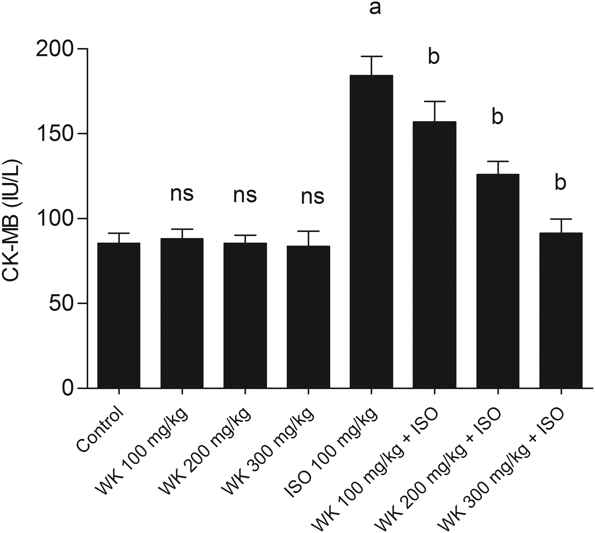
Effect of WK on serum CK-MB activity (dose-dependent study). Values are expressed as mean ± SD (n = 8) ns: not significant compared to normal control. a p < 0.05 significantly different from normal control. b p < 0.05 significantly different from ISO control. ISO, isoproterenol; WK, walnut kernels
The experimental animals were divided into five groups of 10 rats each. Group I: Control rats. Group II: Normal animals were administered ISO (100 mg/kg b.w., subcutaneously twice at an interval of 24 hours) in saline. Group III: Animals were orally treated with WK extracts (300 mg/kg/day, for a period of 35 days). Group IV: Animals were orally treated with WK extracts (300 mg/kg/day, for a period of 35 days and ISO (100 mg/kg) was administered subcutaneously once a day for 2 days. Group V: Animals were orally treated with α-tocopherol (60 mg/kg) orally for a period of 35 days and ISO (100 mg/kg) was administered subcutaneously once a day for 2 days.
After the experimental period, all groups of rats were anesthetized with 30 mg/kg pentobarbital. Blood was collected, and plasma were separated by centrifugation. Heart tissue was excised immediately and rinsed in ice-chilled normal saline. A known weight of the heart tissue was homogenized in 5.0 mL of 0.1 M Tris-HCl buffer (pH 7.4) solution. The homogenate was centrifuged and the supernatant was used for the estimation of various biochemical parameters.
Tissue weights and histopathological examination
The hearts were removed and weighed. The wet heart weight to body weight ratio was calculated to assess the degree of myocardial weight gain. Hearts samples were fixed in 10% buffered formalin before being embedded in paraffin wax and sectioned at 5 μm thickness. Sections were stained with hematoxylin and eosin, and analyzed using optical microscopy (Olympus Corporation, Tokyo, Japan).
Estimation of cardiac diagnostic markers
Biochemical markers of myocardial ischemia injury such as, CK-MB, CK, and lactate dehydrogenase (LDH) were assayed using commercial kits purchased (Nanjing Jiancheng Bioengineering Institute, China) according to the manufacturers' instructions.
Estimation of cardiac troponin, lipid peroxidation products, and antioxidants
The level of tissue thiobarbituric acid reactive substances (TBARs) was estimated by the method of Yagi. 18 Tissue lipid hydroperoxides was estimated by the method of Jiang et al. 19 Malondialdehyde (MDA) levels and superoxide dismutase (SOD) activity were assayed using commercial kits purchased (Nanjing Jiancheng Bioengineering Institute, China) according to the manufacturers' instructions. 20
Estimation of reduced glutathione (GSH) in the heart tissue was performed by the method of Ellman. 21 This method is based on the development of yellow color, when dithionitrobenzoic acid is added to compounds containing sulfhydryl groups. The color developed was read at 412 nm. Glutathione peroxidase (GPx) activity in the heart tissue was assayed by the method of Rotruck et al. 22 A known amount of enzyme preparation was allowed to react with hydrogen peroxide and GSH for a specified time period. The GSH content remaining after the reaction was measured by Ellman's reaction. The activity of glutathione-S-transferase (GST) was assayed in the cardiac tissue following the increase in the absorbance at 340 nm using 1-chloro-2, 4-dinitrobenzene as substrate by the method of Habig and Jakoby. 23 Oxidized glutathione was measured according to the method described by Asen et al. 24
Estimation of lipids
Lipids were extracted from the heart tissue by the method of Folch et al. using chloroform methanol mixture (2:1 v/v). 25 The level of total cholesterol (TC) was estimated by the method of Roeschlau et al. 26 The level of triglyceride (TG) was estimated by the method of McKenzie et al. 27 Free fatty acids (FFA) levels in heart tissue were estimated by the method of Falholt et al. 28 PL was estimated by the method of Zilversmit and Davis. 29
Statistical analysis
Statistical analysis was performed by one-way analysis of variance (ANOVA) followed by Duncan's Multiple Range Test using PASW Statistics 19.0. Results were expressed as mean ± SD for each group. p values <0.05 were considered significant.
Result
WK exert protective effects against MI
To determine the dose-dependent effects of WK in ISO-induced myocardial infarcted rats, a pilot study was conducted with three different doses of WK (100, 200, and 300 mg/kg body weight). From this result (Table 1), it was clear that 300 mg/kg body weight of WK exhibited the highest significant (p < 0.05) effect and near normalized the activity of serum CK-MB with respect to the other two doses (100 and 200 mg/kg body weight) (Fig. 1). Hence, we have chosen 300 mg/kg of WK for our further study.
Effect of Walnut Kernels on Heart Weight, Body Weight, and Heart Weight to Body Weight Ratio in Control and Experimental Groups of Animals
Values are expressed as mean ± SD (n = 8).
ns: not significant compared to normal control.
p < 0.05 significantly different from normal control.
p < 0. 05 significantly different from ISO control.
ISO, isoproterenol.
ISO induction caused significant increase in the activities of serum and heart myocardial injury marker enzymes (CK and LDH) when compared with control rats. WK and α-tocopherol almost restored all the ISO-induced alterations of serum and heart diagnostic maker enzymes to normal levels (Table 2). HE staining showed that the control group exhibited normal space between the myocardial nuclei and regular shape of the nuclei along the heart muscle (Fig. 2). ISO treatment resulted in marked myofbrillar degeneration, necrosis, edema, and infiltration with neutrophil granulocytes in rats. The WK group showed a reduction compared with the ISO group in the shape of the nuclei, and myocardial interstitial fibrous.
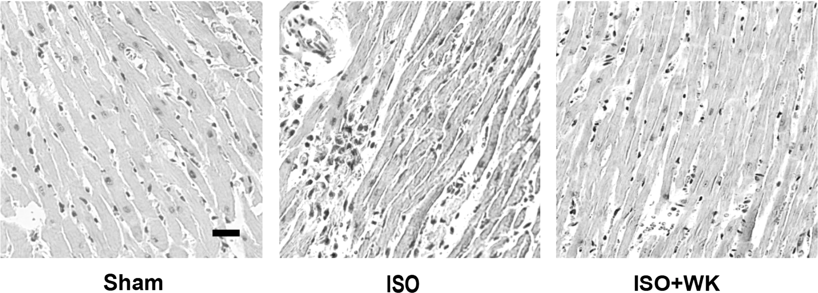
Myocardial biopsy of rat myocardial tissues stained with hematoxylin and eosin. control group; ISO group; WK+ISO group (bar = 40 μm).
Effect of Walnut Kernels on Creatine Kinase and Lactate Dehydrogenase Activities in Control and Experimental Groups of Animals
Values are expressed as mean ± SD (n = 8).
ns: not significant compared to normal control.
p < 0.05 significantly different from normal control.
p < 0. 05 significantly different from ISO control.
Values are expressed as mean ± SD for six animals in each group. The level of CK and LDH in serum are expressed as μkat/L. Tissue AST, ALT and LDH—mmol of pyruvate liberated/min/mg protein.
CK, creatine kinase; LDH, lactate dehydrogenase.
Effect of WK on the troponin T level in myocardial ischemic rats
Table 3 depicts the levels of TBARS and hydroperoxide in heart of normal and experimental rats. Rats induced with ISO, showed a significant (p < 0.05) increase in the levels of TBARS and hydroperoxide in the heart when compared with normal control rats. Oral treatment with WK and α-tocopherol significantly (p < 0.05) decreased the levels of TBARS and HP in the heart when compared with ISO control rats.
Effect of Walnut Kernels on Levels of TBARS and Hydroperoxides in Heart of Control and Experimental Groups of Animals
Values are expressed as mean ± SD (n = 8).
ns: not significant compared to normal control.
p < 0.05 significantly different from normal control.
p < 0. 05 significantly different from ISO control.
TBARS, thiobarbituric acid reactive substances.
Effect of WK on oxidative stress parameters in cardiac tissue
The activities of enzymic antioxidants such as SOD and CAT in the heart of control and experimental groups of rats are shown in Figure 3. The antioxidant enzyme activities were decreased significantly (p < 0.05) in ISO-treated rats when compared with those of control rats. Oral treatment with WK and α-tocopherol daily for a period of 35 days to ISO-induced rats significantly increased the activities of these enzymes compared with ISO alone induced rats.
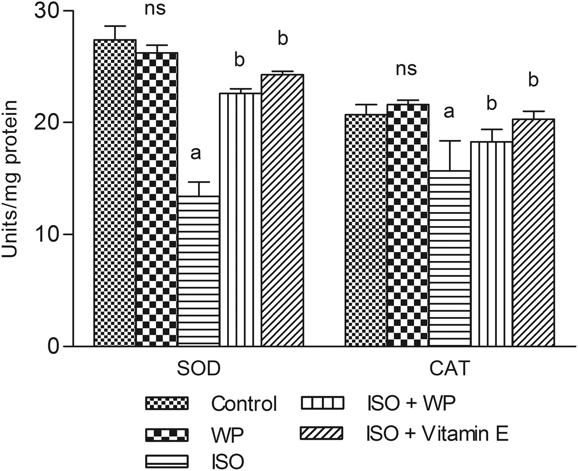
Effect of WK on Activities of SOD and CAT in heart of control and ISO-induced rats. Values are expressed as mean ± SD (n = 8). a p < 0.05 significantly different from normal control. b p < 0.05 significantly different from ISO control. CAT, catalase; NS, not significant; SOD, superoxide dismutase.
Table 4 has shown the levels of GPx, GST, GSH, and GSSG in hearts of control and experimental groups of rats. There was a significant (p < 0.05) decrease in the level of GPx, GST, GSH, and concomitant increased in the level of GSSG in ISO-treated rats compared with the control rats. Oral administration of WK and α-tocopherol to ISO-treated rats significantly increased the levels of GPx, GST, and GSH and decreased the level of GSSG when compared with ISO control rats.
Effect of Walnut Kernels on the Levels/Activities of Reduced Glutathione, Glutathione Peroxidase, and Glutathione-S-Transferase in the Heart of Control and ISO, Isoproterenol-Induced Rats
Values are the mean ± SD; n = 6.
ns: not significant compared to normal control.
p < 0.05 significantly different from normal control.
p < 0.05 significantly different from ISO control.
CDNB, 1-chloro-2, 4-dinitrobenzene; GPx, glutathione peroxidase; GSH, reduced glutathione; GSSG, oxidized glutathione; GST, glutathione S-transferase.
Effect of WK on lipids parameters in cardiac tissue
Figure 4 represents the levels of myocardial tissue lipids in control and ISO-induced rats. ISO-induced rats showed significant increase in the levels of TC, TG, and FFA with a significant decrease in PL. WK administration brought the levels of myocardial tissue lipids to near normality.
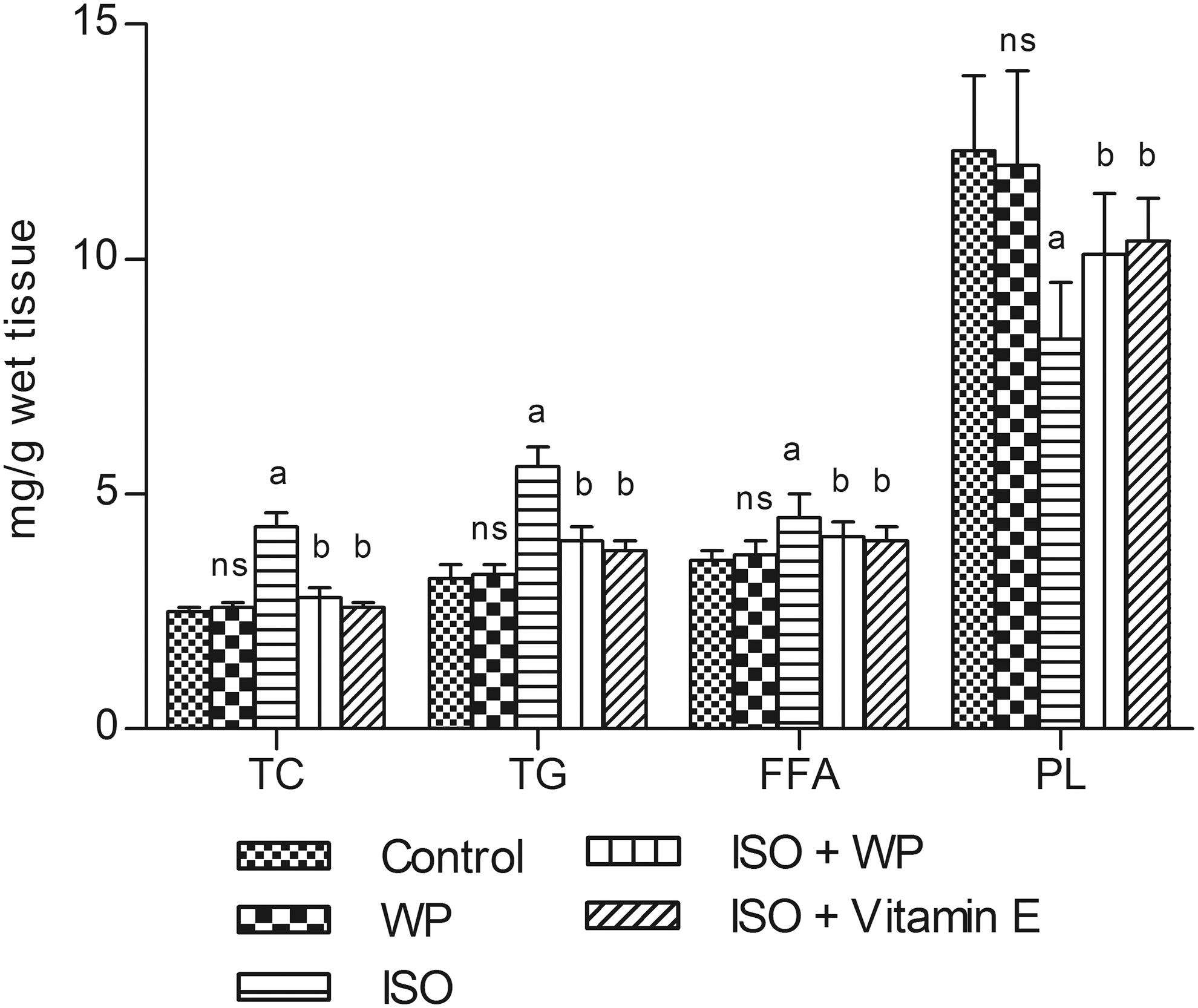
Effect of WK on the levels of TC, TG, FFA, and PL in heart of control and ISO-induced rats. Values are expressed as mean ± SD (n = 8). a p < 0.05 significantly different from normal control. b p < 0. 05 significantly different from ISO control. TC, total cholesterol; TG, triglyceride; FFA, free fatty acids; NS, not significant; PL, phospholipids.
Discussion
A dose-dependent study was performed with three different doses of WK on serum CK-MB activity in the ISO-induced myocardial infarcted rats. ISO causes an increase in the activity of diagnostic marker enzyme, CK-MB. The increased activity of CK-MB in serum might be due to ISO-induced myocardial necrosis. Pretreatment with WK (100, 200, and 300 mg/kg) daily for 35 days dose dependently decreased the elevated activity of serum CK-MB in the ISO-induced rats. But 300 mg/kg of WK elicited the highest significant (p < 0.05) effect on reducing serum CK-MB activity; this dose was used in the following experiments in this study.
An increase in the weight of heart observed in ISO-induced myocardial infarcted rats indicates cardiac hypertrophy. WK decreased the heart weight and prevented cardiac hypertrophy in ISO-induced myocardial infarcted rats. HE staining showed that the control group exhibited normal space between the myocardial nuclei and regular shape of the nuclei along the heart muscle (Fig. 1). The ISO group had increased myocardial interstitial components and widened nuclear space. Fibrosis shapes were disordered. The WK group showed a reduction in the shape of the nuclei compared with the ISO group, and myocardial interstitial fibrous and interstitial fiber arrangements were disordered.
Cardiac troponin, low molecular weight proteins, and the components of the myofibrillary contractile apparatus of cardiac muscle is the markers of myocardial cell injury. 30 In our study, we observed increased levels of cardiac troponin in serum of ISO-induced rats. Although serum cardiac troponin generally is thought to originate from cardiomyocytes breakdown and circulates as a complex with the other cardiac troponins, it is possible that some may originate from leakage of the cytosolic pool and circulate as free cTnI. 31 WK decreased the levels of serum cardiac troponins in ISO-induced myocardial infarcted rats. This could be due to the protective effect of WK on myocardium, preventing the cardiac damage thereby restricting the leakage of troponins from the myocardium into the blood stream.
Therapeutic intervention showing antioxidant or free radical scavenging activity should exert beneficial effects against oxidative stress associated with various cardiovascular diseases, including ischemic heart disease. 32 The use of antioxidants is the most preferred way to inhibit lipid oxidation. Dietary with WK has a beneficial effect on lipids and may reduce hepatic steatosis for nonalcoholic fatty liver disease. 33 Similarly, another recent study demonstrated the effect of walnut leaf in reduction of oxidative stress due to paracetamol in liver of rat. 34
When myocardial cells are injured, many enzymes can be released from the myocardial cells to the extracellular fluid. Hence, in ISO myocardial infarcted rats, there was a decrease in activities of the marker enzymes LDH and CPK in the heart homogenate. 35 In this study, we observed a decrease in the activities of LDH and CPK in ISO-induced rats, which is in consonance with previously reported studies. 36 Pretreatment with WK significantly lowered the ISO-induced elevation of serum levels of these diagnostic marker enzymes. It demonstrated that WK could maintain membrane integrity thereby restricting the leakage of these enzymes.
The generation of reactive oxygen species occurs by the leakage of electrons into oxygen from various systems. Endogenous antioxidant enzymatic defense is a very important source to neutralize the oxygen free radical-mediated tissue injury. 37 –39 In this study, a significantly lower activity of the enzymes SOD and CAT was observed in heart of ISO exposed rats when compared with control rats. Studies found decreased SOD and CAT activities in ISO-treated rats. 40,41 The observed decrease in the activities of these enzymes might be due to their increased utilization for scavenging ROS and their inactivation by excessive ISO oxidants. Treatment with WK improved the activities of CAT and SOD by scavenging superoxide and hydrogen peroxides produced by ISO.
GSH and GSSG levels are commonly used markers for oxidative stress. 42 The observed decrease in reduced glutathione levels might be due to increased utilization in protecting thiol containing proteins from lipid peroxides and from other reactive oxygen species that causes the reduction in the activities of GPx, GRx, and GST. 43 WK increased the levels of reduced glutathione and also increased the activities of GPx and GST in heart of ISO-induced cardiotoxic rats. This shows the antioxidant property of WK.
Lipid peroxidation is a well-established mechanism of cellular injury and has been used as an indicator of oxidative stress that leads to pathogenesis of MI. 44 In this study, significant increase in the levels of lipid peroxidation was observed in ISO-treated rats. Oral treatment with WK decreased the levels of lipid peroxidation products in ISO-induced rats. Thus, WK scavenges the lipid peroxidation products produced excessively by ISO, protected the cardiac tissue because of its antioxidant effect. This could be due to the inhibitory effect of WK on lipid peroxidation by virtue of its antilipid peroxidation property. ISO also causes an increase in the levels of circulatory and myocardial lipids, indicating its hyperlipidemic effect. 45 High levels of circulating cholesterol and its accumulation in heart affects the hormone-sensitive lipase, 46 resulting in marked hyperlipidemia. WK administration significantly restored these alterations thereby maintaining the normal fluidity and function of the myocardial membrane.
This study focused on the protective effect of WK against ISO-induced MI and did not explore in detail signaling pathways involved. Another limitation was that just merely a single dose of WK was used in most parts of this study. Similarly, previous study has shown that WK exerted dose-dependent in vitro and ex vivo potent antiplatelet and anticoagulant effects.
Conclusion
α-Tocopherol had been reported to protect the heart against ISO-induced cardiotoxicity due to their abilities in scavenging free radicals, improving antioxidants, and maintaining lipids levels. The observed cardioprotective nature of WK may be due to the same effects. 47 In conclusion, WK exhibited protective effect against ISO-induced MI in rats by modulating antioxidant defense system, the production of lipid peroxidation and the levels of lipids and thereby maintaining the levels of cardiac marker enzymes.
Footnotes
Acknowledgment
This work was supported by the Foundation of Beijing Medical and Health (YWJKJJHKYJJ-B16239).
Author Disclosure Statement
The authors declare that they have no conflicts of interest.