
Abstract
Abstract
Background:
In 2015 a new device for the collection of mediastinal fluid from patients with deep sternal wound infection (DSWI) in the presence of negative-pressure wound therapy (NPWT) became available. The present study was designed to evaluate whether changing sample collection devices increased micro-organism detection in patients undergoing NPWT.
Methods:
During 2013–2014, 207 samples were collected and cultured from NPWT patients (n = 23) to demonstrate the presence of DSWI using reticulated polyurethane sponge culture, a swab, and blood culture. In 2015, a new collection device was introduced for specimen collection. A total of 357 samples (n = 17) were collected using the ESwab™ (Copan, Murrieta, CA) for deep and superficial wound sample collection. In addition, blood culture devices were used for collecting mediastinal fluid aspirated directly from the wound and biologic fluid obtained from the NPWT device. Fisher exact test was performed to test the rate of independence rate of micro-organism identification using the NPWT sponge device and taking blood culture results as a reference for micro-organism identification.
Results:
After the introduction of the new collection device in our hospital, an overall increase in the detection of micro-organisms (46.7%) was reported. During 2013–2014 our traditional microbiologic collection method did not detect a pathogen in 30.4% of patients. During 2015, the new sample collection approach, direct from the NPWT device, improved micro-organism detection by 10.4% and reduced DSWIs with undetected pathogens to 17.6% (p < 0.01).
Conclusions:
As a result of proficiency gained in the last year, the most representative specimen in wound infection was represented by mediastinal fluid collected directly from the wound and the NPWT device. Given the correlation between the blood culture of micro-organisms detected using the ESwab device from the wound, mediastinal drainage, and drainage from the NPWT device, we can assume that the NPWT device may replace the other biologic sampling devices.
D
Multiple techniques to treat DSWIs have been described. The most common conventional treatments for DSWIs involve surgical revision, open dressing, closed mediastinal irrigation, debridement, and removal of all infected sternal tissue including eventual complete sternotomy or reconstruction by plastic surgery techniques.
According to El Oakley and Wright [4], the management of mediastinitis is usually based on available procedures, the choice of surgical strategies according to the time of presentation, and other clinical risk factors. Moreover, soft tissue dehiscence not involving the sternum and not requiring rewiring of bone edges but requiring surgical debridement and reconstruction of soft tissues involves a complex multi-factorial process [5]. In the last two decades, the surgical approach has changed toward more conservative techniques because the introduction of negative-pressure wound therapy (NPWT) in the modalities used for DSWI treatment, which has been used successfully in cardiac surgery for the treatment of these wounds. Appropriate timing of wound closure is important to avoid infection recidivism, and collection of the most representative microbiologic samples from the infected wound is necessary to identify the micro-organism responsible for the infection.
Several risk factors have been identified in the pathogenesis of DSWI, which are both patient- and procedure-dependent [5], such as the presence of micro-organisms that clinically follow a more insidious course [3]. According to the U.S. Centers for Disease Control and Protection (CDC) guidelines [6], the diagnosis of DSWI requires an organism isolated from purulent discharge from the mediastinum or an organism isolated from mediastinal drainage and blood culture.
In routine practice, wound swabs are the most frequently used method for microbiologic specimen collection, however, the micro-organism detected is often not representative of the site of infection because of sample contamination by normal skin microflora during swab extraction from the wound. On the other hand, according to the manufacturer, the polyurethane sponge of NPWT devices is not designed to hold back bacteria but to reduce bacterial colonization [3,6].
This study was designed to demonstrate that changing the method of collecting biologic material in patients undergoing NPWT results in increased sensitivity when using new sample collection methods/devices from the wound and the NPWT device. The performance of this method was investigated in order to obtain greater microbiologic detection/sensitivity from biologic material representative of the infection.
Patients and Methods
The Cardiovascular Division of the University Hospital Luigi Sacco in Milan, Italy, has 24 licensed beds and performs 700 operations per year. The demographic, clinical, and microbiologic data of 40 patients who underwent cardiac surgical procedures between January 2013 and September 2015 were entered into an electronic database.
Pre-operative risk factors (gender, age, and indication for surgery), peri-operative, and post-operative risk factors (pre-operative stay, surgery duration, hospitalization, micro-organism detection, and clinical recovery) were investigated. A multiple correspondence analysis (MCA) was performed to outline the associations between risk factors and the occurrence of a pathogen either alone or as a co-infection. For this purpose, risk factors were treated as active variables and pathogen occurrence as supplementary variables. The analysis was performed using Statistica, version 8.0 (Dell Software, Round Rock, TX).
A two-tailed Fisher exact test was performed to test the independence of the rate of micro-organism identification in the NPWT device with respect to the sponge, taking identification by blood culture as a reference. The criterion for patient enrollment was the presence of NPWT (V.A.C, KCI, and San Antonio TX) as a treatment for DSWI. All enrolled patients were first assessed for wound exudation and a microbiologic investigation was performed before NPWT placement; the samples were always collected at that time (n = 40). Wound NPWT, in combination with antimicrobial therapy and surgical debridement, were performed immediately. An empirical broad-spectrum antibiotic treatment including bancomycin and pipercillin tazobactam was administered against the suspected infection. After obtaining the appropriate micro-organism identification, target therapy was administered according to the susceptibility tests. As recommended by the NPWT device manufacturer, the tubing system and device were changed every three to five days and samples were collected during NPWT.
Before 2015 (2013–2014), in order to diagnose a DSWI, a total of 207 samples were collected and cultured from patients who underwent NPWT (n = 23) as follows: Reticulated polyurethane sponge culture from the NPWT device, swab (Steward agar gel medium, Copan Italia), and blood culture (Biomerieux, Marcy l'Etoile, France). In 2015, a total of 357 samples were collected from 17 patients. According to recent publications [7,8], a new collection device has been introduced for routine specimen collection. The ESwab™ device (Copan, Murrieta, CA), which is filled with liquid Aimes transport medium, and a specimen collection swab that has a tip flocked with soft nylon fiber (FLOQSwab™, Copan Italia, Brescia, Italy) able to sustain the viability of a variety of organisms including clinically important aerobes, anaerobes, and fastidious bacteria was used for the collection of samples from deep and superficial wounds. The Eswab was also investigated for difficult and anaerobic bacteria.
Blood culture devices were used for the collection of both mediastinal fluid aspirated directly from the wound and biologic fluid obtained from the NPWT device. After removal from the patient, the NPWT canister was opened under sterile conditions (Fig. 1). Using a syringe with a large needle, the gel was aspirated from the back of the container, where a small window is located. To facilitate gel extraction and microbial collection, an enrichment broth was used (BBL™, Brain Heart Infusion, BD, Franklin Lakes, NJ) and introduced into blood culture devices. Micro-organism identification and antimicrobial susceptibility were determined using the automated analyzer Vitek.2 System (BioMérieux, Marcy l'Etoile, France).
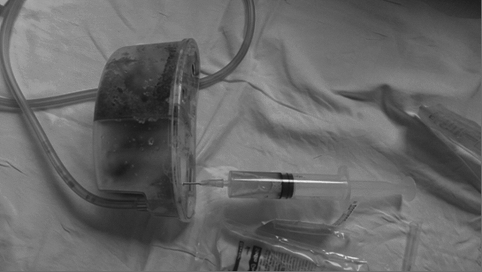
Aspiration of the gel from the back of the negative-pressure wound therapy (NPWT) canister.
Results
Since 2005, the Cardiovascular Division of Hospital Luigi Sacco has used the NPWT device as the first treatment option for superficial or deep sternal wound infections with purulent discharge and for accelerating the healing process by bringing the wound margins closer together. During a three-year period (January 2013 to September 2015), 2,100 patients underwent cardiac surgery at our institution with an incidence of DSWI of 1.9% (40/2100). In all patients, NPWT in combination with antimicrobial therapy and surgical debridement were performed promptly.
The mean age of the patients enrolled in our study (32 males and 8 females) was 69 y with an average hospitalization of 43 d (12 days in the intensive care unit) and a mean preoperative stay of 4.5 d. An NPWT device was positioned in 32 of 40 patients with wound dehiscence and in 8 of 40 patients with mediastinitis; 65% (26/40) of patients had a mono-microbial infection and 10% (4/40) had co-infections. In 25% (10/40) of cases, microbiologic investigation did not detect the pathogen responsible for the infection.
A hospital mortality rate of 22.5% (9/40) was reported: 7.5% (3/40) resulting from wound dehiscence and 15% (6/40) resulting from mediastinitis (Table 1). To determine the patient cohort with DSWI better, an MCA was performed. A comparison of patient demographics, intra-operative, and post-operative variables, and the micro-organism detected was performed. The analysis did not find strong relevant associations between pathogen presence and risk factors, probably because of the occasional occurrence of many species. No correlation was noted between most of the pathogens and these variables. However, spurious associations were observed between Enterococcus faecalis and Klebsiella pneumoniae.
MED = mediastinitis; DST = dehiscence of soft tissue.
Gram-positive (n = 15) and gram-negative (n = 16) bacteria were equally represented. The most commonly isolated gram-positive organism in patients with DSWI was Staphylococcus aureus (25%, 10/40), followed by S. epidermidis (12.5%, 5/40). The most frequently identified gram-negative organism was K. pneumoniae (12.5%, 5/40; Table 1).
During 2013 and 2014 (n = 23), 69.6% (16/23) of DSWIs were caused by micro-organisms and in 30.4% (7/23) of wound infections micro-organisms were not detected. From January 2015 (n = 17), mediastinal fluid aspirated directly from the wound and biologic fluid obtained from the aspirate through the membrane at the back of the NPWT device demonstrated an overall increase of 46.7% in the detection of micro-organisms in comparison with traditional methods (80% [12/17] versus 33.3% [5/17]). The detection of pathogens was documented in all four collection methods (mediastinal fluid drained from the NPWT device into blood culture, ESwab samples collected directly from the deep wound and mediastinal fluid and from mediastinal exudate).
Statistical evaluation was performed to test the independence of the rate of micro-organism detection using the NPWT device with respect to the sponge (using blood culture as a reference), which showed high significance (p < 0.001) between the rate of correct identification by the two techniques. The results of micro-organism detection using different sample collection devices/methods are reported in Table 2. A correlation between drainage from NPWT and blood culture was reported in 16 of 17 cases (94%) who underwent NPWT and who were enrolled in 2015. Moreover, in our study using anaerobic bacteria, no growth in thiol broth was observed. Finally, during 2015 the percentage of undetected pathogens was 17.6% (3/17) compared with 30.4% (7/23) observed in 2013–2014.
NPWT = negative-pressure wound therapy; DST = dehiscence of soft tissue; MED = mediastinitis; NP = not performed.
Discussion
Negative-pressure wound therapy is a safe and cost-effective method for treating complex sternal and thoracic wounds in patients undergoing cardiothoracic surgery and has been used in the Cardiovascular Division of Hospital Luigi Sacco for 10 y. The present study (2013–2015) reported an incidence of DSWI of 1.9%, which is consistent with data reported in other European and Australian studies [9–18].
The rapid and appropriate identification of the micro-organisms responsible for mediastinal infections is critical for specific antibiotic therapy and timely wound closure. Appropriate antimicrobial therapy and clinical recovery depends on the most representative microbiologic samples collected before and during NPWT positioning. During 2013 and 2014, microbiologic collection devices were unable to detect a pathogen in 30.4% of cases (sponge versus blood culture). The high percentage of negative cultures, in agreement with other similar investigations in Italy, led us to investigate these sample collection methods. During 2015, a new approach to sample collection was introduced that allowed direct sample collection from the NPWT device. This approach improved micro-organism detection by 10.4% (80% versus 69.6%), and reduced the number of DSWIs with undetected pathogens by 17.6% (3/17 cases). Standardization of the new collection devices within our hospital for the collection of mediastinal fluid direct from the NPWT device led to an overall increase of 46.7% in the sensitivity of microbiologic identification.
The microbiologic influence of DSWI in Hospital Luigi Sacco was comparable to reported rates in the literature for S. aureus and S. epidermidis cultured [19] among gram-positive bacteria. However, microbiologic identification showed an equal prevalence of gram-positive (n = 16) and gram-negative (n = 15) bacteria.
According to our experience, gained during 2015, micro-organism detection achieved using different sample collection devices/methods showed that the most representative specimen in wound infections was mediastinal fluid collected directly from the wound and the NPWT device (p < 0.001). A good rate of micro-organism detection was also reported for ESwab sampling (Table 2).
Pre-operative contamination of the sternal wound with gram-negative bacteria appears unlikely considering the known bacterial flora in the chest region, and the isolation of gram-negative bacteria from air in the operating room or from the wound after a long operation is rare [20]. However, gram-negative infection has been shown to be associated with other gram-negative bacteria and Candida spp. in co-infections.
In addition, no difference in mortality was observed between sternal infections caused by gram-positive compared with gram-negative bacteria [21]. In our population, of the patients who died from mediastinitis, only three of nine pathogens detected were gram positive. However, a correlation between the prognosis and the micro-organism identified was not documented: K. pneumoniae resistant to carbapenems (KPC) found in sternal wounds did not lead to patient death. However, methicillin-resistant S. aureus (MRSA) did lead to patient death. Despite the average hospitalization of 43 days, no hospital-acquired infections were reported.
The multiple devices/methods (ESwab, mediastinal drainage, blood cultures with drainage from NPWT) used in our experiments lead us to assume that the NPWT devices may be used for other biologic samples. In particular, the collection of mediastinal fluid from a device detached from the patient could be useful during the process of wound healing, when direct sampling might be too invasive.
Given the correlation between the blood culture of micro-organisms detected using ESwab from the wound, mediastinal drainage, and drainage during NPWT, this sampling method can be used in clinical practice for patients with DSWIs treated with NPWT and could replace or reduce the amount of biologic exudates collected in order to facilitate prompt and appropriate antimicrobial therapy.
Footnotes
Author Disclosure Statement
No competing financial interests exist.