
Abstract
The small leucine-rich proteoglycans (SLRPs), prevalent in collagenous tissues, regulate collagen fibrillogenesis and provide a host of biochemical cues critical to tissue function and homeostasis. Incorporating SLRPs may enhance tissue engineering designs that mimic the native extracellular matrix, although SLRPs purified from animal sources bear low yields and lack design control. Consequently, we have designed synthetic peptidoglycans, inspired by the native SLRP decorin, that contain a collagen-binding peptide attached to a glycosaminoglycan (GAG) chain. These peptidoglycans modulate collagen fibrillogenesis and decrease fibril diameter in vitro, similarly to decorin, while maintaining the characteristic D-banded fibrils. Application for tissue engineering is demonstrated as these peptidoglycans are incorporated into collagen gels seeded with smooth muscle cells. Gels formed with peptidoglycans and decorin show a faster rate of gel compaction, and one peptidoglycan uniquely increases elastin production. The peptidoglycan design can be tailored with respect to the peptide sequence and GAG identity and is expected to have versatile application in tissue engineering.
Introduction
The small leucine-rich proteoglycans (SLRPs), prevalent in collagenous tissues, have gained interest for their role in regulating collagen fibrillogenesis and providing a host of biochemical cues critical to tissue function and homeostasis.9–18 Several SLRPs have been identified and are known to contain a protein core with an attached glycosaminoglycan (GAG) chain.19,20 Decorin is a well-studied SLRP and is known to delay fibrillogenesis in vitro and modulate fibril diameter in vitro and in vivo.17,21–23 The core protein of decorin (decoron) 24 contains many functional domains, including two regions known to bind collagen.25,26 Decorin binding to collagen inhibits lateral aggregation of collagen during fibrillogenesis, thus modulating the fibril diameter.17,27
Simplifying decorin to its collagen-binding function with attached GAG, we have previously designed a peptidoglycan, which contains a collagen-binding peptide and a covalently attached dermatan sulfate (DS) GAG. 28 This peptidoglycan modulates collagen fibrillogenesis while incorporating a biochemically active GAG chain. It has also been shown to increase the stiffness of collagen gels, similarly to decorin. For collagen-based tissue engineering, peptidoglycans may provide unique application; they mimic in part, native SLRPs, by binding collagen and incorporating a GAG chain, but unlike SLRPs purified from animal tissues, peptidoglycans have the benefit of design control and ease of synthesis.
Collagen-based tissue-engineered scaffolds have been used for vascular tissue engineering with promising results. 29 One drawback to collagen-based scaffolds is that smooth muscle cells (SMCs), which maintain the vessel ECM, only minimally degrade collagen and synthesize little new matrix.2,30 Incorporating GAGs, SLRPs, or peptidoglycans may improve the design of collagen-based scaffolds by modulating collagen fibril structure and incorporating the biochemically active ingredients present in the native tissue.2,31,32
The present work broadens the scope of our previously described peptidoglycan, by demonstrating tailorability of the peptidoglycan molecule, analyzing the morphological characteristics of collagen fibrils with incorporated peptidoglycans, and demonstrating tissue engineering application. We hypothesized that these molecules would behave similarly to native SLRPs in modulating collagen fibrillogenesis and would influence cellular remodeling of collagen matrices. The designed peptidoglycans offer a tailorable biomimetic approach to influence collagen fibril morphology while incorporating biochemical cues that influence cell function; as such, these peptidoglycans are expected to have versatile application in tissue engineering.
Materials and Methods
Materials
Pepsin-treated Nutragen Type I Collagen purified from bovine hide was purchased from Inamed Biomaterials (Freemont, CA) at a stock concentration of 6.4 mg/mL in 10 mM of hydrochloric acid (HCl). DS (41 kDa, 6.85% sulfur) and its oxidized form, containing an average of 1.1 aldehydes per polymer chain, were purchased from Celsus Laboratories (Cincinnati, OH). Decorin from bovine tendon (100 kDa, 50 kDa of which is DS) was purchased from Sigma-Aldrich (St. Louis, MO). Amino acids were purchased from Anaspec (San Jose, CA), Knorr resin was purchased from SynBioSci Corporation (Livermore, CA), and all other supplies were purchased from VWR or Sigma-Aldrich, West Chester, PA unless otherwise noted.
Synthesis/characterization DS-Dc13
The peptide sequences RRANAALKAGELYKSILYGC (SILY), SYIRIADTNITGC (Dc13), and ZSYIRIADTNITGC (ZDc13), where Z designates dansyl glycine, were synthesized on a solid-phase Symphony Peptide Synthesizer (Protein Technologies, Tucson, AZ) with Knorr resin. Peptides were cleaved from the resin using 92.5% trifluoroacetic acid, 2.5% MilliQ water, 2.5% triisopropylsilane, and 2.5% ethane dithiol. Peptides were purified using reverse-phase chromatography on an ÄKTA Explorer FPLC (GE Healthcare, Piscataway, NY) using a C-18 column (Grace-Vydac, Deerfield, IL) with acetonitrile/water gradient, and purity was confirmed using mass spectroscopy (Applied Biosystems 4700, Foster City, CA).
Conjugation of peptides to dermatan sulfate
Peptides were conjugated to oxidized DS as described previously using the heterobifunctional cross-linker 3-(2-pyridyldithio)propionyl hydrazide (PDPH; Pierce, Rockford, IL), reactive to aldehyde and thiol groups. 28 Briefly, PDPH was reacted in coupling buffer (0.1 M of sodium phosphate, 0.25 M of sodium chloride, pH 7.2) at 10-fold molar excess with oxidized DS containing 1.1 aldehyde groups per DS polymer chain. Excess PDPH was removed using gel filtration by a column packed with Sephadex G-25 medium (GE Healthcare). Purified DS-PDPH was then reacted in coupling buffer with 5-fold molar excess of peptide. The reaction was monitored according to the production of pyridine-2-thione as described. Excess peptide was removed using gel filtration, and pure DS-peptide was lyophilized and stored at −20°C until further testing.
Binding of DS-Dc13 to collagen
Collagen and bovine serum albumin was immobilized on a high-bind 384-well black/clear-bottom plate by incubating each at 2 mg/mL for 1 h at 37°C in 10 mM of HCl and 1 × phosphate buffered saline (PBS), respectively. Wells were then washed three times with 1 × PBS. Untreated wells were used as a positive control. A mix of 60% DS-PDPH capped with L-cysteine and 40% fluorescently labeled DS-ZDc13 was dissolved in 1 × PBS at varying concentrations from 100 μM to 1 nM and incubated in the treated or control wells for 30 min at 37°C. Unbound DS-ZDc13 was removed by washing three times with 1 × PBS, and fluorescence (ex: 335 nm em: 490 nm) was measured on a SpectraMax M-5 spectrophotometer (Molecular Devices, Sunnyvale, CA). All measurements were performed in triplicate.
Collagen gel preparation and fibrillogenesis assays
Samples compared in this study include collagen alone and collagen mixed with DS, DS-SILY, DS-Dc13, intact decorin, or free peptides SILY or Dc13. Unless noted, treatment concentrations were added at a 10:1 molar ratio of collagen:additive. Molar ratios were calculated by an estimate of molecular weights: collagen, 285 kDa, DS 41, kDa; DS-SILY, 43.2 kDa; and decorin, 100 kDa. Gels were formed by mixing stock collagen solution with 10 × PBS, 1 M of sodium hydroxide, and 1 × PBS on ice to a final concentration of 4 mg/mL, pH 7.4, and ionic strength of 164 mM. Treatments were added as a part of the final 1 × PBS addition and incubated on ice for 30 min before initializing fibrillogenesis by warming to 37°C. Fibrillogenesis was monitored turbidimetrically by absorbance at 313 nm as described previously.8,20,22 Samples of 50 μL were added at 4°C to each well of a 384-well plate, and measurements were taken at 1-min intervals for up to 6 h in a SpectraMax M-5 spectrophotometer set at 37°C.
Cryo-scanning electron microscopy and fibril diameters
Gels for cryo-scanning electron microscopy (SEM) were formed directly on the scanning electron microscope stage at 37°C overnight as described previously. 33 The stages were then secured in a cryo-holder and plunged into liquid nitrogen slush. Samples were then transferred to a Gatan Alto, Pleasanton, CA, 2500 pre-chamber cooled to −170°C under vacuum. A free-break surface was created using a cooled scalpel, and the sample was sublimated for 20 min, followed by platinum sputter coating for 120 s. Samples were transferred to the cryo-stage at −130°C, where regions with similar orientation were imaged for comparison across treatments. Fibril diameters were measured using ImageJ software (National Institutes of Health (NIH), Bethesda, MD), in which regions orthogonal to the fibril were measured. Three independent observers blinded to the treatment recorded at least 45 measurements for each treatment and were instructed to measure diameters only of fibrils that could be resolved to individual fibrils, rather than those appearing fused, overlapping, or obscured by other artifacts.
Atomic force microscopy
Gels were formed on cut glass cover slides and incubated overnight at 37°C. Samples were dehydrated and washed with MilliQ water, and the glass slides were fixed to the atomic force microscope (AFM) metal specimen disc (#16208) purchased from Ted Pella (Redding, CA). Tapping mode AFM was performed using multimode scanning probe microscopy from Veeco Instruments (Santa Barbara, CA). A single-beam silicon tip (#OTESPA) was purchased from Veeco Probes (Camarillo, CA) and had spring constant of 42 N/m. AFM imaging was performed at room temperature under ambient humidity. Images were collected at a 1 Hz scan rate.
Cell culture and gel compaction
Human coronary artery (HCA) SMCs (Cascade Biologics, Portland, OR) were cultured in growth medium (Medium 231 supplemented with smooth muscle growth factor). Cells from passage 3 were used for all experiments. Differentiation medium (Medium 231 supplemented with 1% fetal bovine serum and 1 × penicillin/streptomycin) was used for all experiments unless otherwise noted. This medium differs from manufacturer protocol in that it does not contain heparin.
Collagen gels were prepared with each additive, as described, with the exception that the 1 × PBS addition was omitted to accommodate the addition of cells in media. Collagen pregel was incubated on ice for 30 min, followed by addition of HCA SMCs in differentiation medium to a final concentration of 1 × 10⁶ cells/mL. Gels were formed in quadruplicate in 48-well non-tissue culture treated plates (Costar, Corning, NY) for 6 h before adding 500 μL per well of differentiation medium. Gels were freed from the well edges after 24 h to allow free gel compaction. After 24 h, growth medium was collected and then replaced every 2 to 3 days; images for compaction were taken at the same time points using a Gel Doc System (Bio-Rad, Hercules, CA). The cross-sectional area of circular gels correlating to degree of compaction was determined using ImageJ software (NIH). Gels containing no cells were used as a negative control, and cells in collagen gels absent additive were used as a positive control.
Collagen degradation
Collagen degradation was measured by determining the hydroxyproline content released from gels into the cell medium, following a protocol adopted from Reddy and Enwemeka, assuming that 12% of the weight of collagen is hydroxyproline.28,34 Medium (200 μL) was collected every 2 to 3 days and was hydrolyzed in 6 M of HCl at 110°C overnight. After HCl removal by evaporation, Chloramine T reagent was freshly made, and 450 μL was added to each sample and allowed to incubate at room temperature for 25 min. Ehrlich's aldehyde reagent was freshly made and 500 μL mixed with each sample and then incubated at 65°C for 20 min. Absorbance was read at 550 nm, and hydroxyproline concentration was determined from a collagen standard curve.
Elastin production
Collagen gels seeded with HCA SMCs were prepared as described. Differentiation medium was changed every three days and gels were cultured for 10 days. Collagen gels containing no cells were used as a control. At day 10, gels were rinsed in 1 × PBS overnight, and gels were tested for elastin content using the Biocolor, United Kingdom, Fastin elastin assay, per the manufacturer's protocol (Biocolor, County Atrim, United Kingdom). Briefly, gels were solubilized in 0.25 M of oxalic acid by incubating at 100°C for 1 h. Elastin was precipitated using Fastin Precipitation Solution, and samples were then centrifuged at 11,000 × g for 10 min. The solubilized collagen supernatant was removed, and the elastin pellet was stained using Fastin Dye Reagent for 90 min at room temperature. Samples were centrifuged at 11,000 × g for 10 min, and unbound dye in the supernatant was removed. Dye from the elastin pellets was released by the Fastin Dye Dissociation Reagent, and 100-μL samples were transferred to a 96-well plate (Costar). Absorbance was measured at 513 nm, and elastin content was calculated from an α-elastin standard curve.
Cytotoxicity
HCA SMCs were seeded at 4.8 × 10⁴ cells/mL in growth medium onto a 96-well tissue-culture black/clear-bottom plate (Costar) and allowed to adhere for 4 h. Growth medium was aspirated, and 600 μL of differentiation medium containing each additive at a concentration equivalent to the concentration within collagen gels (1.4 × 10⁻⁶ M) was added to each well. Cells were incubated for 48 h and then tested for cytotoxicity using Live-Dead and CyQuant (Invitrogen, Carlsbad, CA) assays, per the manufacturer's protocol. Cells in differentiation medium containing no additive were used as control.
Statistics
All experiments were performed in at least triplicate and are presented as averages ± standard errors. Significance was determined using Design Expert (StatEase, Minneapolis, MN) software, with α = 0.05.
Results and Discussion
We have previously described the design of a synthetic peptidoglycan based on the SLRPs such as decorin in which the GAG DS is conjugated to a collagen-binding peptide. Here we have expanded the design, demonstrating tailoribility using two different collagen-binding peptide sequences, one derived from decorin SYIRIADTNITGC (Dc13) and one from the platelet receptor to type I collagen RRANAALKAGELYKSILYGC (SILY).35,36 Although the exact binding location along the collagen fibril was not determined in this study, it is assumed that these sequences, derived from different sources and with different primary structure, bind collagen in different locations, and they were thus studied for comparison. In addition, each sequence not containing an internal cysteine could be modified using a terminal glycine (spacer) and cysteine to employ specific sulfhydryl chemistry. We have compared the effects of these peptidoglycans on collagen fibrillogenesis and morphology of collagen gels, showing a level of control over the gel biophysical properties, and demonstrated tissue engineering application by culturing HCA SMCs in collagen gels with incorporated peptidoglycans.
Synthesis and characterization of DS-Dc13 and DS-SILY
As shown in Figure 1, approximately 1 Dc13 peptide was coupled per DS molecule, resulting in a peptidoglycan that is similar to DS-SILY and is designed to mimic in part native SLRPs such as decorin, which contains one collagen-binding decoron per GAG chain.28,37 To ensure that conjugation of DS to Dc13 did not inhibit the ability of Dc13 to bind to collagen, fluorescently labeled DS-ZDc13, where Z denotes dansyl glycine, was conjugated to DS and in a microplate assay, bound specifically to collagen in a dose-dependent manner shown in Fig. 2. Because saturation binding was not achieved at the maximum concentration of 100 μM, the binding affinity could not be calculated. DS is known to interact electrostatically with collagen and can self-associate; thus increasing the concentration to reach saturation binding of the peptidoglycan was not possible, although specific binding to collagen was demonstrated, making DS-Dc13 a suitable collagen-binding peptidoglycan. 38
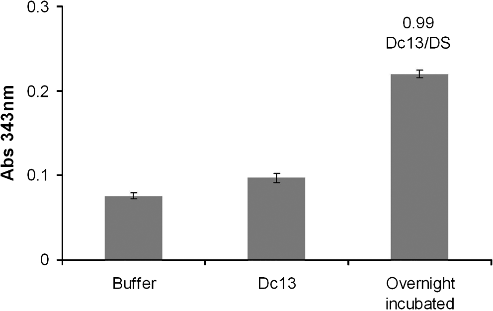
Conjugation of Dc13 to dermatan sulfate (DS). Production of pyridine-2-thione, measured according to an increase in absorbance at 343 nm, indicates 0.99 Dc13 peptides per DS polymer chain (101 × 65 mm) (300 × 300 DPI).
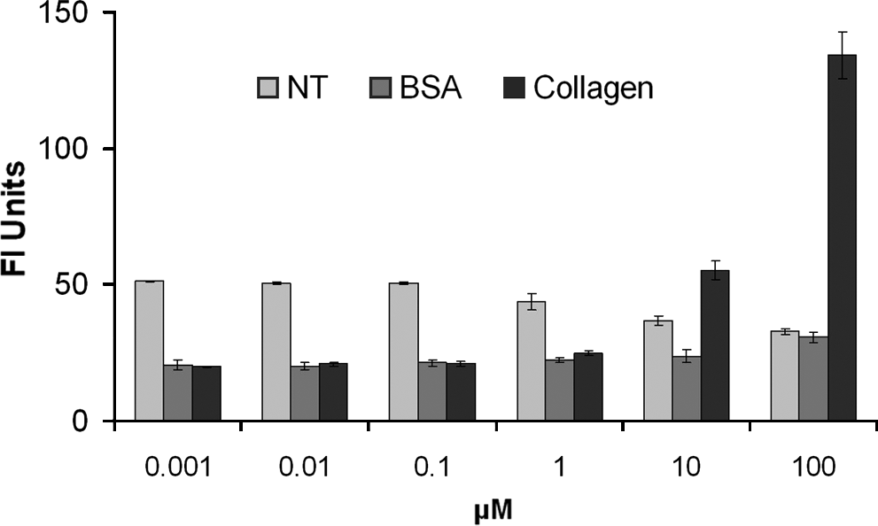
Microplate fluorescence binding of dermatan sulfate (DS)-ZDc13 to collagen. DS-ZDc13 bound specifically to the collagen surface in a dose-dependent manner, although saturation was not achieved at the maximum concentration. (101 × 62 mm) (300 × 300 DPI). NT, no treatment; BSA, bovine serum albumin.
Collagen fibrillogenesis and morphology
The effects on fibrillogenesis and fibril morphology were assessed according to turbidity. DS-SILY and DS-Dc13 delay fibrillogenesis in a dose-dependent manner, as shown in Figure 3, in which, at high concentrations (1:1 molar ratio), marked inhibition is observed, following the same trends as observed for native SLRPs.22,39 Free peptide controls did not have an effect on fibrillogenesis even at the high concentrations shown in Figure 3 and previously. 28
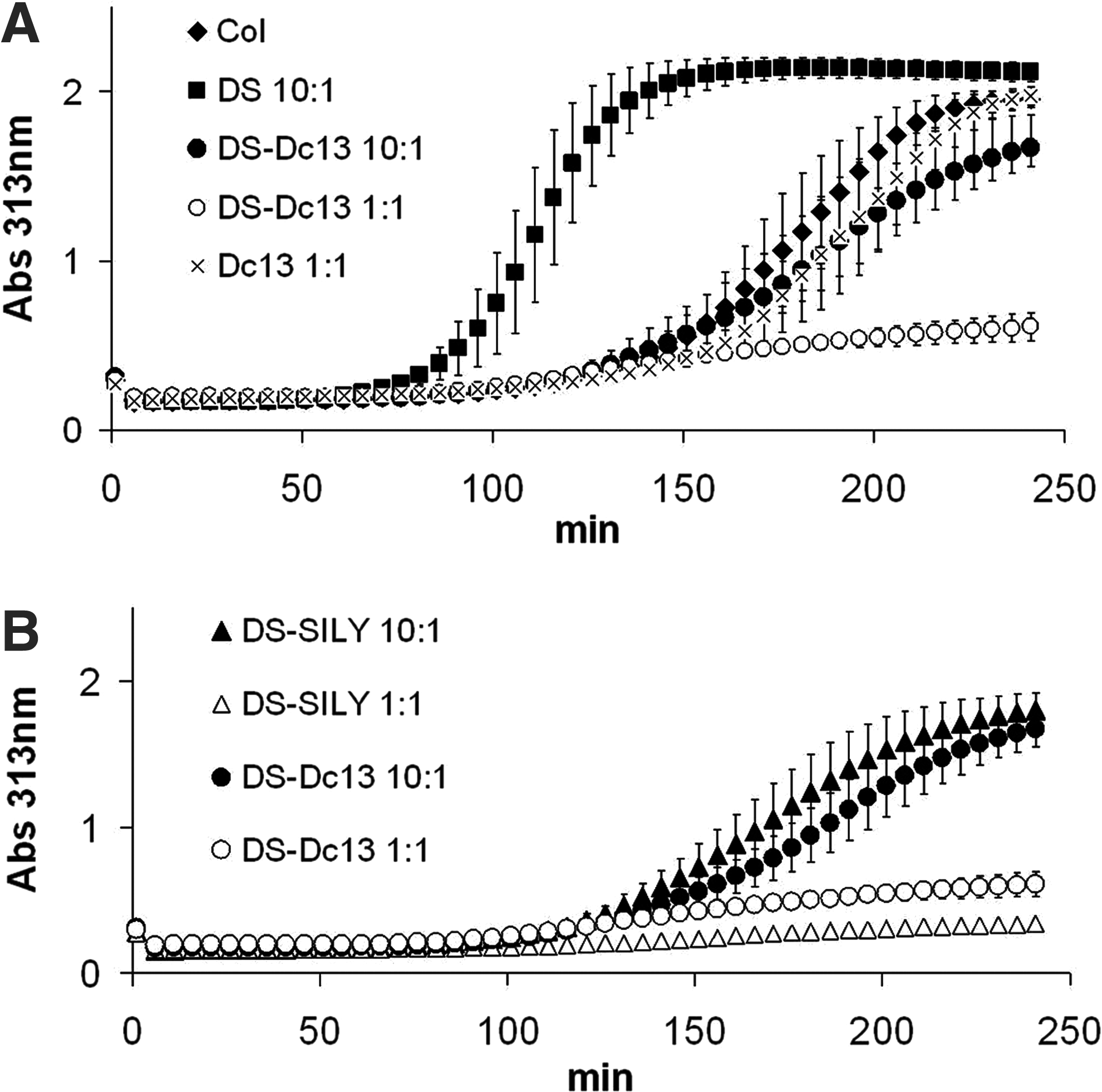
Collagen fibrillogenesis according to turbidity measurements. (
A proposed mechanism for the interaction of collagen-binding peptidoglycans, depicted in Figure 4, is the same as that proposed for SLRPs, in which the binding of these molecules to collagen monomer or onto forming fibrils inhibits lateral fibril growth during fibrillogenesis.15,17,22,37,40,41 In vitro, collagen self-associates into aggregates in the form of fibrils, which are specifically arranged, quarter-staggered collagen molecules. 42 Delayed fibrillogenesis is dose dependent, as illustrated at the 1:1 molar ratio of collagen:peptidoglycan, indicating that, as more peptidoglycans are bound to collagen, they inhibit the required quarter-stagger arrangement of collagen, thus preventing or delaying fibril formation. Unlike peptidoglycans, DS alone increases the rate of fibrillogenesis. Because DS interacts electrostatically with collagen, it is likely that collagen fibrils are brought together, nucleating fibrils more quickly and speeding fibrillogenesis. Because the interaction between collagen and DS is weaker than that between collagen and peptide, DS does not remain bound where it would inhibit lateral aggregation of collagen. 43
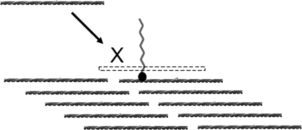
Peptidoglycan–collagen interaction mechanism. Proposed inhibition of lateral aggregation by binding of peptidoglycan or small leucine-rich proteoglycans (SLRPs) along a collagen molecule. The required quarter-stagger arrangement of collagen in the lateral direction is prevented by bound peptidoglycan or SLRPs, limiting growth to the axial direction. The exact binding location of peptidoglycan is not proposed here but a schematic of the mechanism of lateral inhibition (101 × 47 mm) (300 × 300 DPI).
Fibril morphology was assessed using cryo-SEM techniques and is depicted in Figure 5. Cryo-SEM does not require dehydration steps, which can collapse the fibrillar structure, making fibril diameter measurements less accurate, and is thus a preferred method for assessing collagen gel structure. 33 Inhibited lateral aggregation would result in smaller diameter fibrils, which is indicated by decreased optical density observed from turbidity studies and confirmed by direct measurement of fibril diameters (Fig. 4). Histograms illustrating fibril size distribution show non-normal distribution for some treatments, so the Kolmogorov-Smirnov test, which does not assume normal distribution, was used for statistical analysis. Gels formed in the presence of decorin, DS-Dc13, and DS-SILY resulted in significantly smaller average fibril diameter (10%, 15%, and 27%, respectively) than collagen. The ability to modulate collagen fibril diameter under physiological conditions is significant for collagen-based tissue engineering applications because fibril size plays an important role in cellular response, as well as the mechanical properties of collagen scaffolds. Native tissues contain fibrils ranging in diameter from 10s to 100s of nanometers, depending on the tissue, which contributes to the unique mechanical and cellular environments. 44 Synthetic peptidoglycans also incorporate the biochemically active DS GAG chain, so fibril diameter cannot be isolated as a distinct variable in cellular response or mechanical properties.
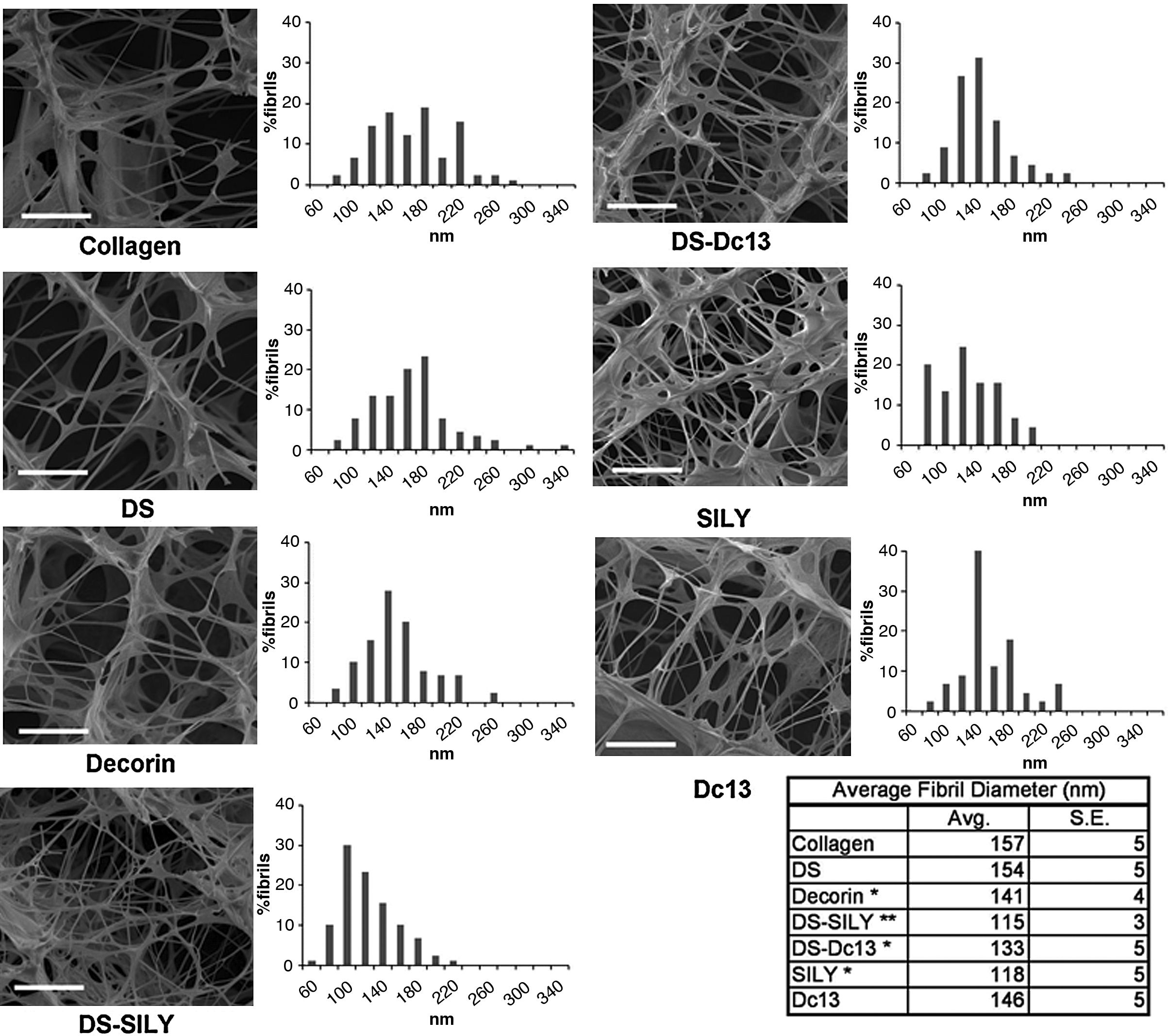
Cryo-canning electron microscopy images and fibril diameter distribution. The addition of decorin, dermatan sulfate (DS)-SILY, and DS-Dc13 resulted in significantly smaller fibril diameter than collagen alone or collagen with DS. Free peptide SILY also reduced fibril diameter, whereas Dc13 had no effect. *Significance vs collagen or collagen + DS, **DS-SILY significant vs decorin (254 × 190 mm) (300 × 300 DPI).
The free peptide SILY also decreased fibril diameter, whereas Dc13 did not. The size of these peptide sequences, in which SILY containing 20 amino acid residues could sterically hinder collagen–collagen interactions, whereas Dc13 containing only 13 amino acid residues may not may in part explain this. Likewise, when the 40-kDa DS molecule is attached, both peptidoglycans are large enough to inhibit lateral aggregation of collagen. Because free SILY peptide does not incorporate DS, its may be a useful peptide for studying the effect of fibril diameter independently.
In addition to fibril diameter, cryo-SEM images also provided information on the branching structure of the fibrillar network of collagen gels, revealing a highly branched network, especially in the presence of DS-SILY. The specific binding location of the peptidoglycan on growing collagen fibrils may influence fibril branching, which is currently being studied.
AFM images, depicted in Figure 6 were used to confirm that properly arranged D-banded fibrils form in the presence of peptidoglycans. The quarter-staggered orientation of collagen, which gives rise to characteristic D-banding, is critical to the mechanical and biochemical function of collagen. This particular organization presents a host of active binding sites for cellular activity, and a collagen-based engineered ECM that does not preserve the native D-banded arrangement may negate the benefits of choosing collagen instead of its denatured form gelatin.45,46 Both peptidoglycans preserve D-banded fibril structure, indicating that these molecules do not interrupt the quarter-stagger orientation of collagen monomer as depicted in Figure 4. Fibril diameters were not measured using this method because sample dehydration was required and can result in collapse of fibrils.
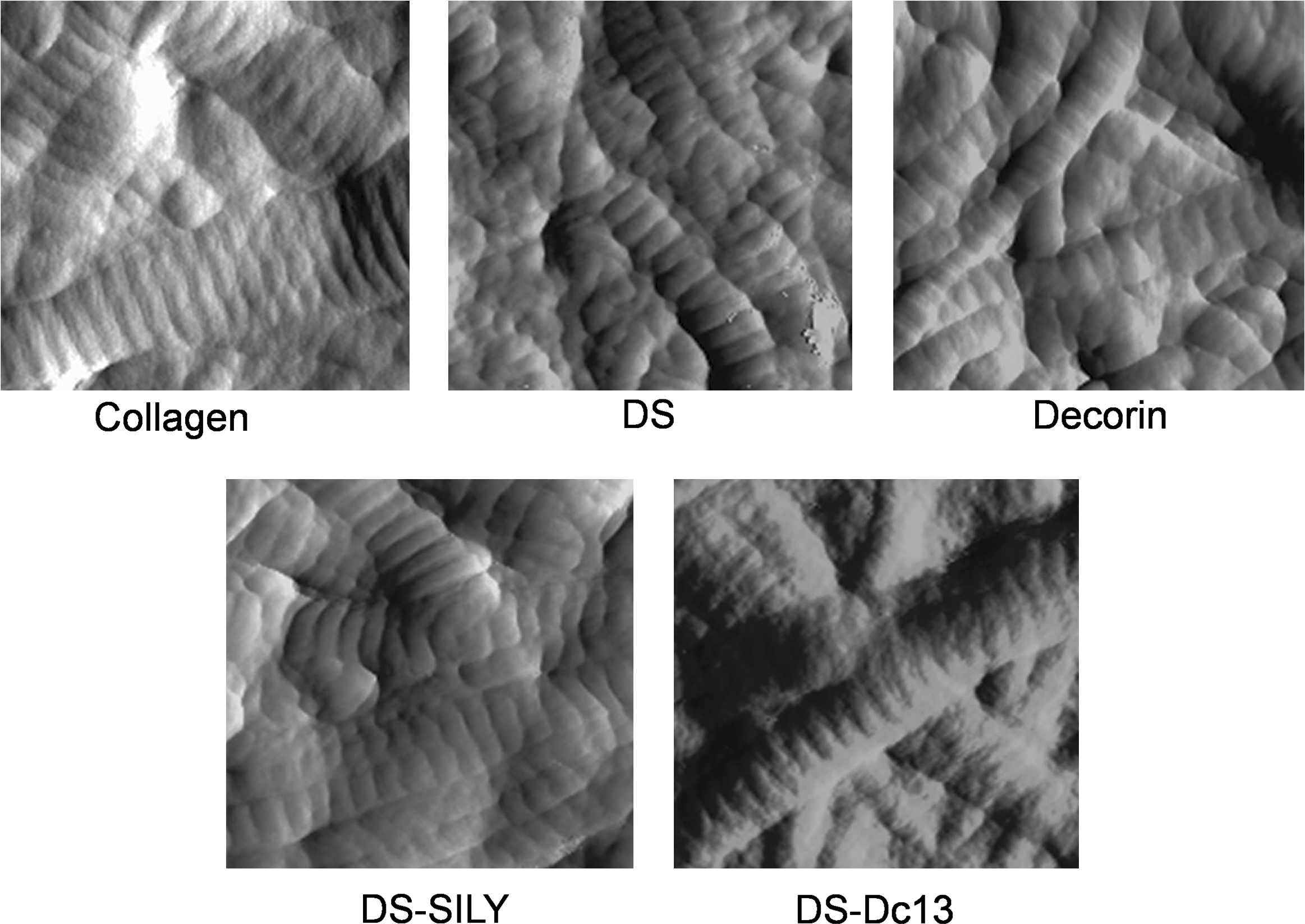
Atomic force microscopy images of collagen gels. D-banding was observed in all treatments, demonstrating that peptidoglycans do not prevent quarter-stagger collagen arrangement. Images are 1 μm by 1 μm (203 × 149 mm) (300 ×300 DPI).
Cell Studies
To demonstrate tissue engineering application of peptidoglycans, HCA SMCs were cultured in the presence of peptidoglycans in two-dimensional (2D) and 3D culture. In 2D culture, neither peptidoglycan showed an effect on cytotoxicity, which was tested according to cell permeability (Live-Dead) and confirmed according to total DNA content (CyQuant). Because the 3D cell-seeded collagen gel contracts to a dense diffusion-limited structure, 2D culture was assumed sufficient to assess cell viability.
HCA SMCs seeded into 3D collagen gels with incorporated peptidoglycans characteristically contracted the gel to varying degrees, which correlated to fibril diameter at early time points, as shown in Figure 7. Gels with incorporated peptidoglycans or decorin, containing fibrils of smaller diameters, contracted faster, which a greater surface area on which cells can exert traction could explain. By day 10, the compaction differences were largely lost, because the gels all had substantially compacted from their original size, and subtle differences in compaction were undetectable. Free peptide controls were compared with collagen alone and showed that, at day 5, SILY resulted in greater compaction than collagen, but by day 10, both peptides resulted in less-compact gels than collagen alone (data not shown). The implications of a greater rate of compaction indicate a faster rate of remodeling, which is necessary for restoring the function of tissue-engineered constructs. 3
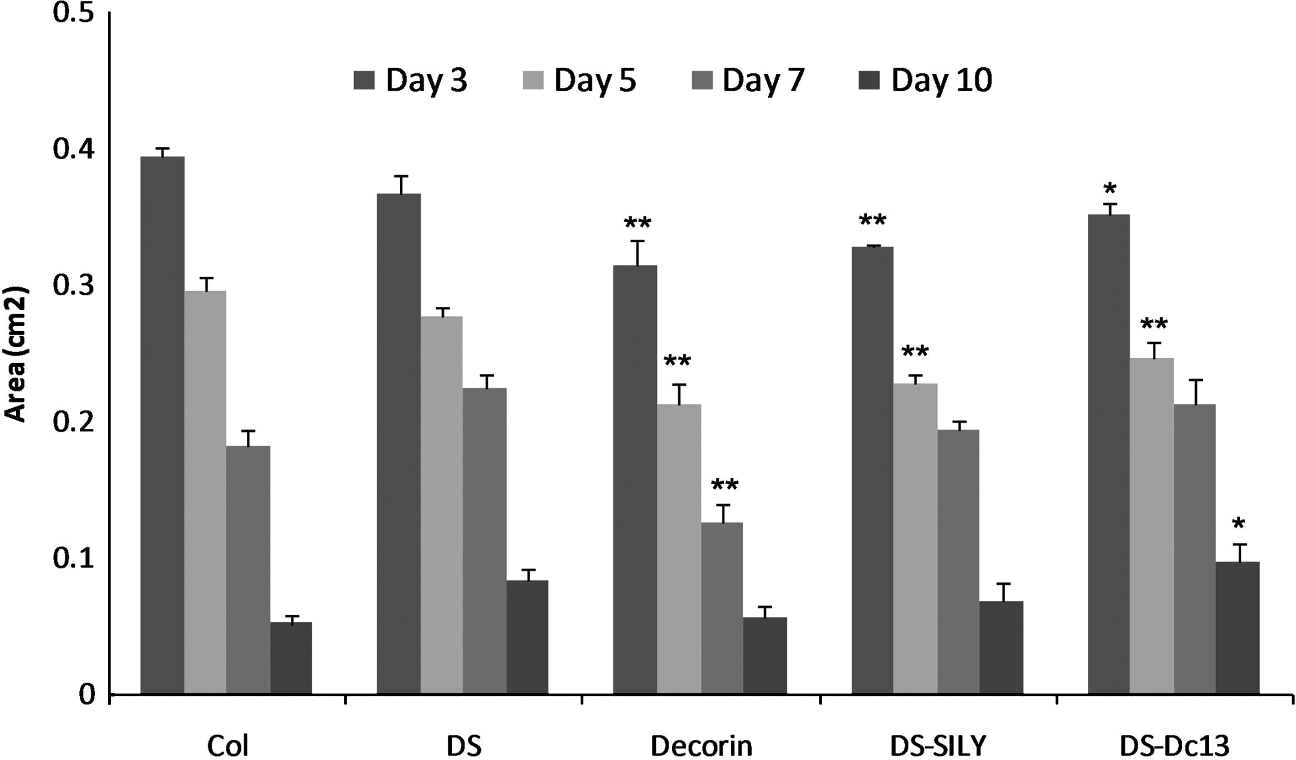
Compaction of collagen gels seeded with smooth muscle cells. The addition of peptidoglycans and decorin increases the rate of gel compaction, indicating faster scaffold remodeling. *Significance vs collagen control. **Significance vs collagen and dermatan sulfate (190 × 113 mm) (300 × 300 DPI).
Degradation of collagen matrices by incorporation of peptidoglycans or decorin did not increase over the 2-week culture time, indicating a lack of remodeling by breaking down of the existing collagen matrix (data not shown). No variations in trends were found at each time point, so hydroxyproline totals for the 2-week culture time were used for analysis.
For tissue-engineered vessels, the synthesis of elastin, which is responsible for the elasticity of arteries, is critical. Incorporation of DS-SILY into HCA SMC–seeded collagen gels increased elastin production more than all other treatments, as shown in Figure 8, indicating great promise for this particular peptidoglycan in collagen-based vascular constructs. Although the increase over collagen with no additive was significant, but modest, DS itself resulted in a decrease in elastin, and DS-SILY–incorporated gels resulted in a more substantial increase than DS-incorporated gels. This suggests that not only chemical identity of the GAG, but also presentation of the GAG in the form of a peptidoglycan is important not only for biophysical function, but also for biochemical function.
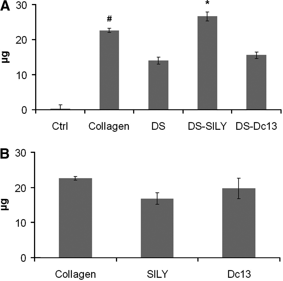
Elastin production according to Fastin assay. (
Conclusions
We have described here the synthesis of collagen-binding peptidoglycans that can be tailored with respect to the peptide sequence as well as the attached GAG chain. These peptidoglycans modulate collagen fibrillogenesis and decrease fibril diameter in collagen gels similarly to the SLRP decorin. Characteristic D-banding is observed in collagen fibrils formed with and without peptidoglycans or decorin, and turbidity results together with smaller fibril diameters suggest inhibited lateral aggregation of collagen molecules by the binding of peptidoglycans. We are further investigating this mechanism and the possible role of binding location along the collagen molecule on morphology of collagen fibrils.
The potential use of peptidoglycans for tissue engineering applications was demonstrated by culturing HCA SMCs within collagen gel matrices formed with added peptidoglycans. Free peptides and peptidoglycans did not exhibit cytotoxicity in 2D culture studies. Cell-seeded collagen gels formed with added peptidoglycans resulted in a faster rate of compaction, and incorporation of DS-SILY increased elastin synthesis. Further studies for vascular tissue engineering, as well as other applications for collagen-based tissue-engineered constructs with incorporated peptidoglycans, are being studied. Additionally, variant forms of peptidoglycans designed with different GAG identities and peptide sequences are being explored as a biomimetic method of modulating collagen-based constructs.
Footnotes
Acknowledgments
This work was funded in part by the NIH (K25HL074968) and the National Science Foundation (CBET-0651643). The authors thank Debby Sherman at Purdue Life Sciences Microscopy Facility for help with cryo-SEM images.
Disclosure Statement
No competing financial interests exist.