
Abstract
In this study, poly (ɛ-caprolactone) [PCL] and its collagen composite blend (PCL/Col) were fabricated to scaffolds using electrospinning method. Incorporated collagen was present on the surface of the fibers, and it modulated the attachment and proliferation of pig bone marrow mesenchymal cells (pBMMCs). Osteogenic differentiation markers were more pronounced when these cells were cultured on PCL/Col fibrous meshes, as determined by immunohistochemistry for collagen type I, osteopontin, and osteocalcin. Matrix mineralization was observed only on osteogenically induced PCL/Col constructs. Long bone analogs were created by wrapping osteogenic cell sheets around the PCL/Col meshes to form hollow cylindrical cell-scaffold constructs. Culturing these constructs under dynamic conditions enhanced bone-like tissue formation and mechanical strength. We conclude that electrospun PCL/Col mesh is a promising material for bone engineering applications. Its combination with osteogenic cell sheets offers a novel and promising strategy for engineering of tubular bone analogs.
Introduction
The inclusion of collagen has been postulated as a method of improving the functionality of tissue engineering scaffolds. Collagen coating on electrospun scaffolds by physical adsorption results in enhanced attachment, spreading, and viability of endothelial cells. 5 Similar results were observed with fibroblasts when collagen was co-electrospun as a blend with PCL. 6 The immobilization of collagen onto poly (lactic-co-glycolic acid) [PLGA] modulates attachment, proliferation, and functioning of cartilage and bone cells.7,8 Further, grafting of short peptide sequences of type I collagen onto hydroxyapatite scaffolds supports osteogenic differentiation of bone marrow derived progenitor cells. 9
We hypothesized that tubular bone tissue substitutes can be engineered using electrospun biodegradable fibrous scaffolds in combination with osteogenically induced bone marrow derived cell sheets. PCL and its collagen-blend meshes (PCL/Col) were fabricated using electrospinning technique and characterized for their mechanical and cellular properties. Although the inclusion of collagen into the structure compromised the resulting mechanical strength, PCL/Col fibrous meshes were superior in terms of their ability to support the attachment, proliferation, and osteogenic differentiation of bone marrow derived mesenchymal cells. Tubular bone grafts were made using these porous meshes in combination with osteogenic cell sheets. Cell sheets are an alternative method of cell delivery with the major advantage of keeping cell- and matrix-adhesion molecules intact together with the extracellular matrix.10,11 These constructs showed extensive bone-like tissue formation after 4 and 8 weeks of dynamic culturing. This method presents a novel strategy for engineering tubular bone analogs.
Materials and Methods
Materials and reagents
All materials and reagents were purchased from Sigma Aldrich (St. Louis, MO) unless otherwise stated. Porcine collagen particulates were obtained from Nippon Meat Packers (Osaka, Japan). Chloroform used as electrospinning solvent was from EM Science (Gibstown, NJ). Cell culture media and reagents were purchased from Gibco (Grand Island, NY). Trypsin solution (0.25%) was purchased from Hyclone (Logan, UT). Mouse monoclonal antibody against osteocalcin and rabbit polyclonal antibody against osteopontin were purchased from QED Bioscience (San Diego, CA) and Abcam (Cambridge, UK), respectively. FITC-labeled secondary antibody against mouse IgG and developmental kit for HRP immunohistochemistry were purchased from DAKO Cytomation (Glostrup, Denmark).
Scaffold fabrication and characterization
Electrospinning
Electrospun PCL meshes were made by dissolving PCL pellets in a solvent mixture of HFIP:chloroform at 4:1 volume ratio to make a 12.5% w/v concentration. For PCL/Col meshes, 2.5% w/v collagen was added, replacing the same weight of PCL to maintain concentration. The polymer solutions were loaded into a 5 mL syringe mounted on a syringe pump. Polymer was fed to a metal capillary (21-G needle) at a constant rate of 0.75 mL/hour. The metal capillary was charged to 10 kV to initiate electrospinning. Grounded aluminum foil was placed 15 cm below the tip to collect the fibers. The nonwoven fibers were then dried in a dessicator.
Scanning electron microscopy (SEM)
Dried fibrous meshes were cut into 1 cm × 1 cm squares for SEM viewing. Samples were mounted on SEM stubs and sputter coated with gold using BALTEC glow discharge sputter coater (BAL-TEC AG, Balzers, Liechtenstein). Viewing was done with JEOL 5600 electron microscope system under 10 kV accelerating voltage.
Immunofluorescence
Dried fibrous meshes were cut into 1 cm × 1 cm squares and incubated with mouse monoclonal antibody against porcine collagen type I at 4°C for 16 hours. Following washing with phosphate-buffered saline (PBS), the samples were incubated with FITC-labeled antibody against mouse IgG at ambient temperature for 30 min. Viewing was done using Olympus FV500 laser scanning confocal microscope (LCSM) system.
Tensile test
Tensile tests were done on the meshes previously cut into 3.5 cm × 1 cm strips using an Instron tensile tester with 100 N load cell. All tests were done in PBS immersion at 37°C with a crosshead speed of 10 mm/min until scaffold failure. Stress was calculated using the nominal cross-sectional area of the individual mesh samples. Surface wettability measurements were done using static sessile water contact angle method on a VCA Optima system.
Cell culture and in vitro osteogenic differentiation
PBMMC culture
Pig bone marrow mesenchymal cells (pBMMCs) were isolated from the iliac crest of Duroc-Yorkshire pigs weighing 45–50 kg and aged 16–18 weeks. Briefly, a 20 mL syringe containing 1.5 mL of 5000 IU/mL heparin was used to extract 10–15 mL of bone marrow aspirate. The aspirate was then gently mixed and placed in tissue culture flasks. Approximately 3 mL of the aspirate was plated on a T150 culture flask containing 25 mL expansion culture media (DMEM supplemented with 10% fetal bovine serum and 1% antibiotics). The cells were then cultured in a heat sterilizable humidified incubator at 5% CO2 (Binder GmbH, Tuttlingen, Germany) and 37°C. Subculturing of cells was performed by 0.25% trypsin enzymatic digestion once cells reached 70–80% confluency. Passages 2–3 cells were used in these studies.
Scaffold seeding and culture
pBMMCs (20,000 cells/cm2) were seeded on electrospun meshes cut into 1.5 cm ×1.5 cm squares. The scaffolds were previously ethanol sterilized, UV irradiated for 15 min, and conditioned overnight in culture media. PCL and PCL/Col meshes used in this study were divided into two subgroups: induced and uninduced. A total of four study groups were present. Induced groups were cultured in media supplemented with osteogenic differentiation factors (10 nM dexamethasone, 50 μM ascorbic acid 2-phosphate, and 10 mM β-glycerophosphate), while uninduced groups were maintained in basal media. Experiments were conducted up to 4 weeks with weekly time points for various assays.
Cell proliferation assay
At predetermined time points, cell-scaffold constructs were collected from four study groups. Samples were then incubated with an enzymatic solution of 0.1% w/v trypsin and collagenase for 1 hour at 37°C. Total DNA in the resulting suspensions was measured using PicoGreenTM dsDNA quantitation assay in accordance with the manufacturer's protocol. Fluorescence measurements were done at 485 nm (excitation) and 535 nm (emission) using a TECAN spectrophotometer (Salzburg, Austria).
Cell morphology and attachment assay
At predetermined time points, samples were collected from all four groups and fixed with 2.5% glutaraldehyde. The samples were then serially dehydrated using ethanol gradients followed by drying. Prior to SEM observations, the cell-scaffold constructs were gold sputter coated. Viewing was performed using a JEOL electron microscope system under 10 kV accelerating voltage.
Histology and immunohistochemistry
Samples were collected and sectioned using a cryo-microtome (10 μm thickness of each section). General tissue formation was studied by hematoxylin and eosin (H&E) staining of the constructs. Immunohistochemical methods were used to study specific matrix protein deposition (i.e., collagen type I, osteopontin, and osteocalcin). All immunohistochemical staining were developed using a HRP-DAB development kit. Matrix mineralization of the constructs was studied using alizarin red S staining.
Engineering of tubular cell-scaffold constructs
Creation of tubular constructs
Tubular cell-scaffold constructs were prepared by wrapping osteogenic cell sheets onto PCL/Col meshes preseeded with pBMMCs. Osteogenic cell sheets were obtained by culturing pBMMCs in osteogenic media for 2–3 weeks. The cell sheets were detached from the culture substrates mechanically using cell scrapers. PCL/Col electrospun scaffolds of 2 cm × 2 cm size were rolled into cylindrical shapes of diameter 6 mm. The scaffolds were seeded with pBMMCs on both sides (approximate density 20,000 cells/sq cm) and then further wrapped with the cell sheet to form the tubular constructs. Plastic sterile serological pipettes were used as substrate in the rolling-up process to maintain the tubular shape of the constructs. The constructs were then cultured in static condition for 1 week in a humidified incubator, at 5% CO2 atmosphere under osteogenic induction condition (10 nM dexamethasone, 50 μM ascorbic acid 2-phosphate, and 10 mM β-glycerophosphate). The constructs were transferred into spinner flasks for dynamic culturing under osteogenic induction condition for 1–2 months. The tubular constructs were suspended using a thin wire to the spinner arms such that the long axis of the constructs was parallel to the ground. The spinner flasks were filled with 100 mL of media and set to 30 rpm. In this manner, culture media was allowed to flow through the long axis, exposing both the inner and outer surfaces of the constructs to fluid shear stresses. PCL/Col tubes without cell-sheet wrapping were cultured as well as controls. At 1- and 2-month time points, samples were collected for histological and mechanical analyses.
Histology and immunohistochemistry
After 4 and 8 weeks, constructs were collected and fixed in 3.7% formaldehyde. They were then dehydrated and embedded in paraffin after decalcification in 30% formic acid. Sections of 10 μm thickness were obtained using Leica microtome. H&E staining and immunohistochemical methods were used to study the tissue and bone specific matrix proteins (collagen type I, collagen type II, and osteocalcin). HRP-DAB development kit from DAKO was used to view the staining.
Compression test
Samples collected were mounted on a Bose material testing system (Bose, Eden Prairie, MN) fitted with a 225 N load cell. Specimens were kept wet by dropping PBS prior to test. A crosshead speed of 2 mm/min was used, and a compression load of up to 20 N was applied. Testing in the axial direction was done using an indenter. The stiffness value was calculated from the linear region of the load-elongation curve. The modulus was computed from the stress-strain curve.
Results
Scaffold fabrication and characterization
Electrospun PCL fibrous meshes and its collagen blend were fabricated and their morphology, collagen localization, and mechanical properties assayed (Fig. 1). Irrespective of composition, the fibers exhibited smooth and uniform morphology (Fig. 1A). Average fiber diameters were 564 ± 267 nm and 513 ± 83 nm for PCL and PCL/Col, respectively. A second, much finer fiber distribution was present in PCL/Col. Immunofluorescence analysis revealed surface localization of collagen on PCL/Col. Addition of collagen however caused a 62.8% and 53.4% reduction in tensile modulus and strength-at-break, respectively, while improving the elongation-at-break by 27.2% compared to pure PCL (Fig. 1B). Collagen significantly improved surface wettability based on the water contact angle data.
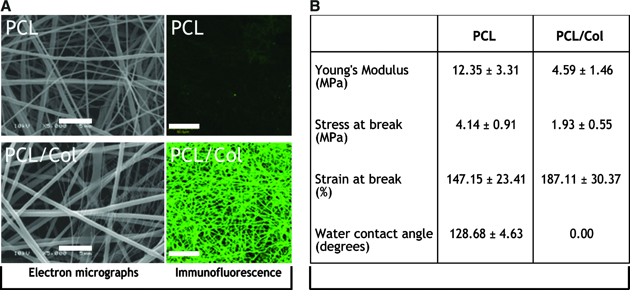
(
Cell culture study
To examine pBMMCs morphology and proliferation on the electrospun scaffolds, SEM and total DNA measurements were done. pBMMCs attached and proliferated in all four study groups (Fig. 2). Spreading of the cells however was more pronounced on PCL/Col scaffolds, and more rounded cell morphology was found on PCL (Fig. 2A). After 1 week of culture, qualitative differences in proliferation were observed. Induction groups showed the highest cellular density, followed by uninduced PCL/Col and lastly uninduced PCL (Fig. 2B). On PCL meshes, pBMMCs existed in clusters due to the hydrophobic nature of the surface. Complete surface coverage was achieved after 4 weeks in all but uninduced PCL group.
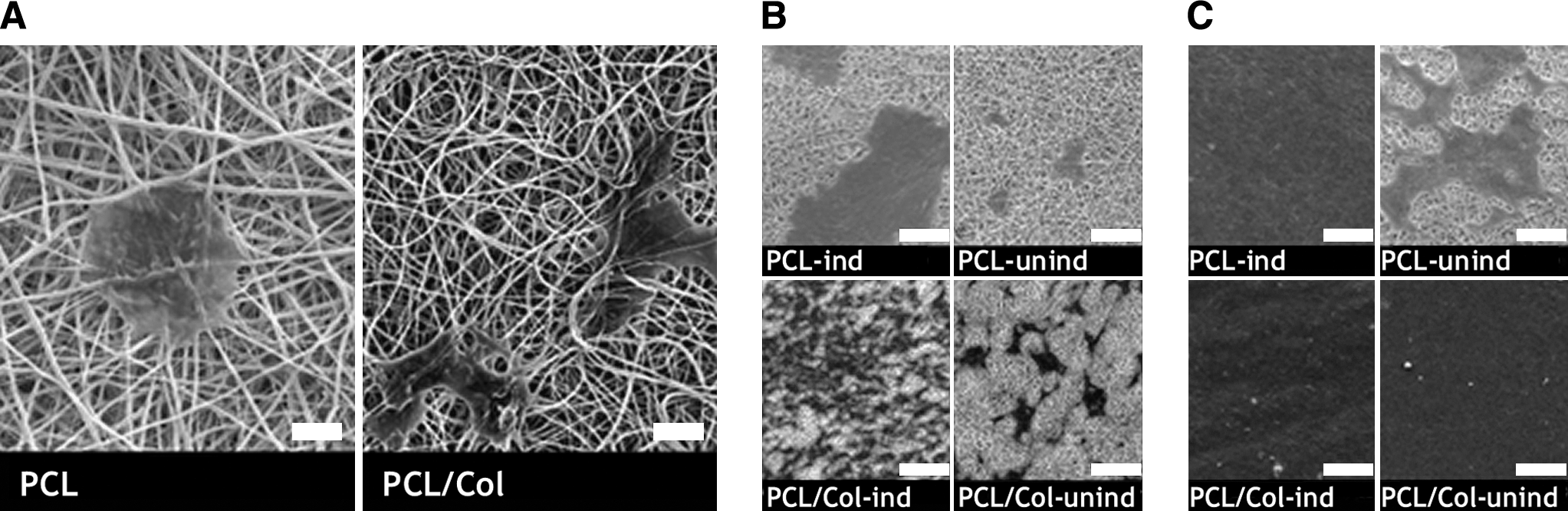
Morphological differences were observed when pig bone marrow mesenchymal cells (pBMMCs) were cultured on electrospun PCL and PCL/Col meshes. (
Quantitative total DNA analysis revealed that pBMMCs proliferated in all four groups albeit at different rates (Fig. 3A). Overall, PCL/Col groups (with and without osteogenic supplements) were able to support more pBMMCs proliferation compared to PCL. Cell number reached a plateau at much lower levels in PCL groups. Without osteogenic supplements, PCL group was unable to support significant cellular proliferation. A significant reduction in cell number occurred in PCL/Col induced group after 2 weeks of culture. Maturation of the bone-like tissue was responsible for this as indicated by the presence of calcium deposits, which was analyzed by SEM/EDX (Fig. 3B).
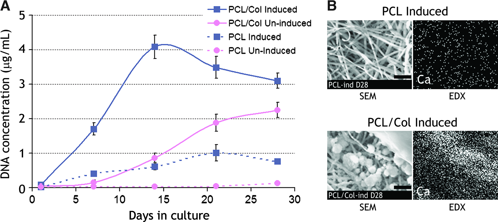
Electrospun PCL and PCL/Col meshes exhibited differences in their ability to support proliferation of pBMMCs over the course of 28 days. (
To examine the tissue morphology and bone-specific matrix deposition on the electrospun scaffolds, histology and immunohistochemistry staining were conducted (Fig. 4). Osteogenically induced pBMMCs showed thick tissue and multilayered tissue formation on PCL/Col meshes. Its PCL counterpart remained largely monolayered throughout. No cellular penetration was apparent in any of the fibrous scaffolds' structure. The new tissue formed on PCL/Col showed intense expression of collagen type I and osteopontin, while osteocalcin was marginally expressed. Osteonectin was also found to be expressed in the PCL/Col group (data not shown). Calcium deposition was observed only after 4 weeks of osteogenic culturing. Osteogenically cultured pBMMCs on PCL meshes showed weaker expression of collagen type I and osteopontin. However, osteocalcin, osteonectin, and calcium depositions were not observed.
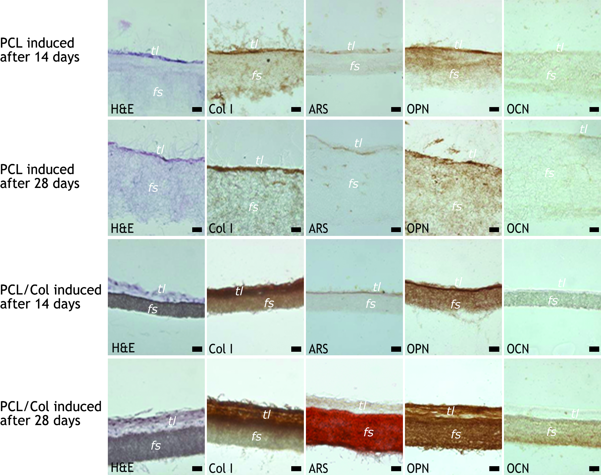
Histology and immunohistochemical staining of pBMMCs on PCL and PCL/Col electrospun fibrous scaffolds (fs) after 14 and 28 days culture in osteogenic-supplemented culture media. Osteopontin (OPN) signal was found in all groups, and osteocalcin (OCN) was found only in PCL/Col induced group after 4 weeks (induced cultures). Formation of tissue layer (tl) was more prominent on PCL/Col scaffolds with dense cell layers and collagen deposition. After 4 weeks of culture, the PCL/Col cell-scaffold construct was stained positive for calcium (ARS). Bar in all pictures represents 20 μm. Color images available online at www.liebertonline.com/ten.
Engineering of tubular cell-scaffold constructs
The ability of PCL/Col electrospun meshes to support the formation of bone tissue in a design mimicking the long bone was tested by combining them with cell sheet technology. Construction of such tubular cell-scaffold constructs is shown in Figure 5. pBMMCs proliferated and formed cell sheets within 2 weeks of osteogenic culture. These cell sheets were detached and wrapped around PCL/Col tubes. The cells were viable after assembly and grew over time to form tubular constructs of 1–2 mm wall thickness and 6 mm diameter. Mechanical analyses revealed that the constructs were stronger in the radial direction compared to the axial direction. In the radial direction, construct stiffness was 27 N/mm, 200 times more than that of tubes without cells (control, 0.13 N/mm). Modulus of the construct was 21 MPa, 20 times higher than the control value of 1 MPa. In the axial direction, construct stiffness and modulus were 0.86 N/mm and 2.4 MPa, respectively. These values were 4 to 7 times higher than for the controls (0.11 N/mm and 0.6 MPa).
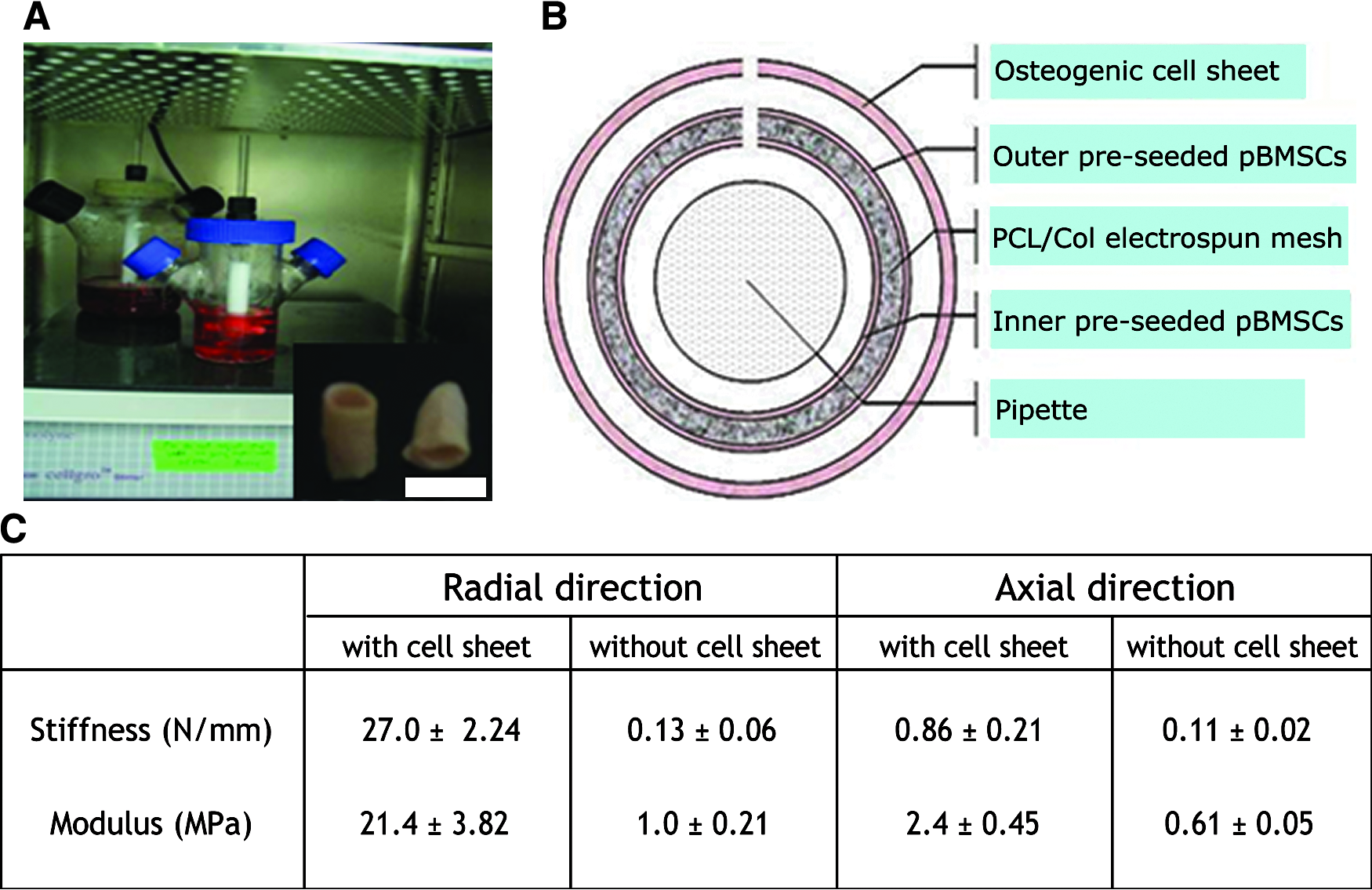
Tubular bone constructs were obtained by wrapping preseeded PCL/Col electrospun meshes around a glass pipette. Osteogenic cell sheets were then wrapped around the structure, and the entire constructs were cultured in spinner flasks (
Morphology and identity of the newly formed tissue were analyzed by histology and immunohistochemical assays. Distinct layers of tissues formed after 1 and 2 months of dynamic culturing (Fig. 6A, B). A denser layer formed at the interior of the constructs and was more visible after 2 months. This dense tissue formation showed intense staining of safranin-O, indicating cartilage-like matrix (Fig. 6C). Certain regions of this layer contained chondrocyte-like cells with typical vacuoles. Further analysis also showed that only this dense middle layer expressed bone specific matrix deposition, that is, collagen (Fig. 6D), calcium (Fig. 6E), and osteocalcin (Fig. 6F). Tissues flanking this layer showed weak collagen I expression and negative safranin-O, osteocalcin, and calcium signals.
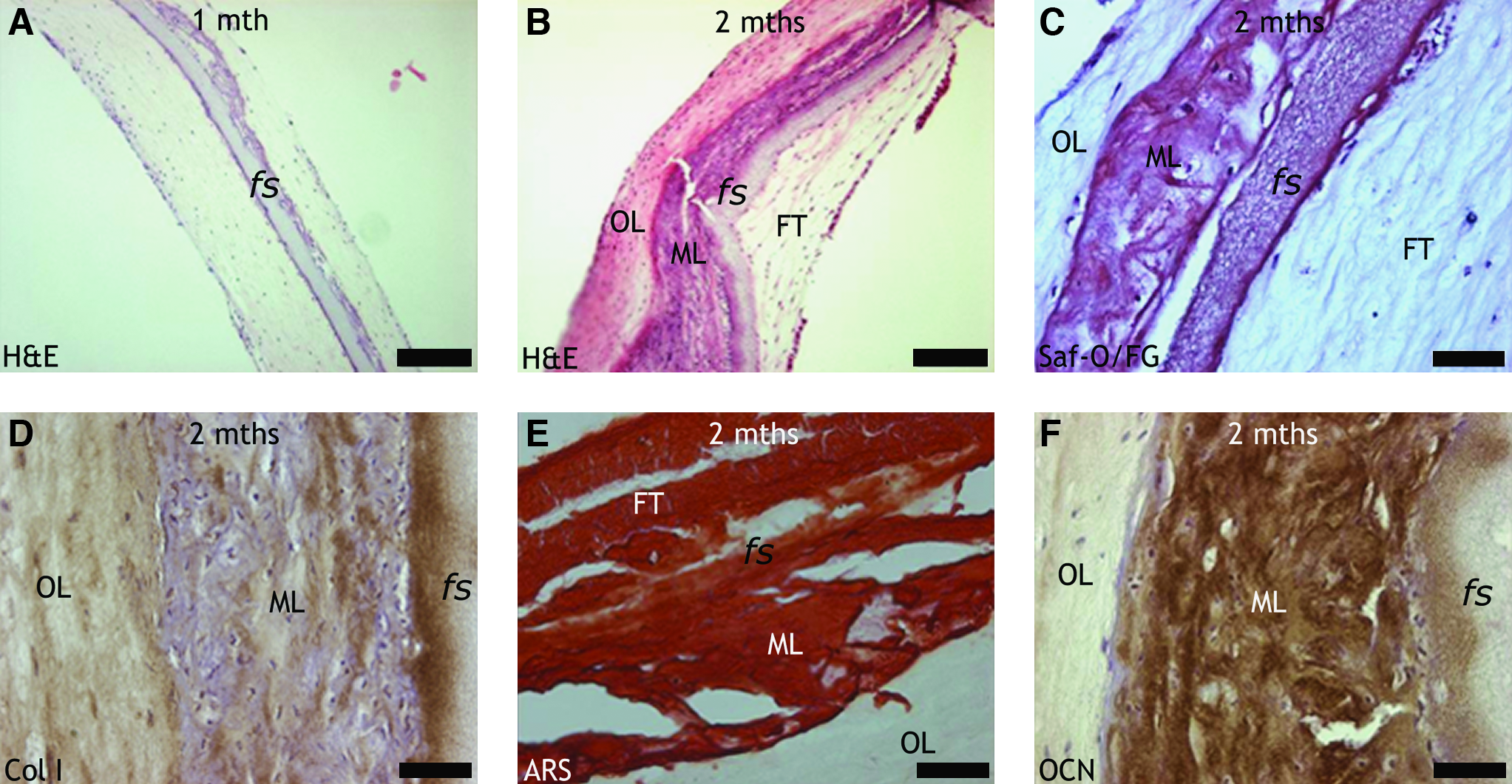
One month of in vitro dynamic culturing resulted in thick tissue formation on both sides of the constructs with tissue adjacent to the electrospun fibrous scaffold (fs) exhibiting strong eosin staining (
Discussion
Synthetic-natural hybrid materials offer desirable mechanical and manufacturing properties while retaining their bioactive properties and are hence suitable for tissue engineering applications.12,13 Modifications of material surfaces by collagen or Arg-Gly-Asp (RGD)-containing proteins augment cell attachment, proliferation, and differentiation.9,14–16 In the present study, a composite blend of electrospun PCL and collagen (PCL/Col) material is investigated as scaffold for bone engineering.
A micro- to nanoscale topography and environment elicit diverse cell behaviors such as attachment, orientation, proliferation, and differentiation. Incorporation of natural polymers is a promising avenue for developing more biomimetic scaffolding materials.17,18 Interestingly, in this study, the co-electrospun collagen was found covering the surface of the fibers. While the mechanism of preferential surface segregation of the collagen remains to be studied, it is thought that the electric field applied to the polymer jet attracted the more charged natural polymer to the surface of the jet. This fact renders the fabrication of natural polymer–coated PCL fibers less complicated. However, we have shown in another study that the combined action of the solvent (fluoroalcohols) and electrical field leads to extensive denaturation of the collagen. 19 In the case of our polymer blend, the collagen molecules may not have been completely denatured, as monoclonal antibody recognizing native helical structure could still detect the presence of collagen on the fibers' surface.
Previous studies have shown that prolonged passage and better differentiation potential of mesenchymal cells on denatured collagen (DC) could modulate cell phenotypes in two-dimensional (2D) monolayer cultures.20,21 Studies performed in 2D cultures have demonstrated that heat denaturation of type I collagen results in the dissociation of its triple helical structure, exposing the internal RGD cell adhesion sequences that are inaccessible in the native type I collagen conformation. Several studies of cell adhesion to type I collagen have shown that cells primarily adhere to the native structure through RGD-independent integrin interactions, whereas RGD-dependent binding of cells can be observed when DC is used.22,23
Most notable effects of collagen inclusion into the PCL matrix were on the fibers' apparent DC coat and mechanical properties. We hypothesized that during electrospinning the applied electric field caused preferential segregation of DC on the fibers' surface, forming a coaxial structure with PCL as the core. This may prove beneficial as direct contact of DC with cells and tissues is now possible. The inclusion of DC however resulted in a decrease in stiffness and ultimate stress. It is believed that the DC may not have been perfectly miscible with PCL, thus forming collagen-rich phases or domains in the fiber. These DC-rich phases would have caused discontinuity in an otherwise uninterrupted PCL matrix. The resulting interface between the phases coupled with a reduction in overall material crystallinity resulted in mechanically compromised fibers. Parallel observations were reported by Kwon et al. in electrospun blends of PLCL and collagen, where increasing the amount of collagen resulted in progressively much weaker fibers. 24 Similar findings were observed as well in PLGA polymer when gelatin/elastin or chitosan was incorporated into the electrospinning solution blend.25,26 Thus, depending on the nature of application, a balance should be met between mechanical strength and biological activity of the scaffolds.
Biological activity of the scaffolds was then investigated using bone marrow mesenchymal cells. Bone marrow contains a population of progenitor cells capable of differentiating into mesenchymal lineages.27–30 The spreading and proliferation of these cells were modulated by the surface-bound DC. In fact, the scaffolds with DC were able to support more proliferating cells on their surface. Binding of cells to collagen, presumably via integrins, has been reported to trigger pathways that lead to actin-mediated cellular spreading 31 and possibly the activation of Cbfa1 transcription factor, initiating osteoblastic phenotype development.32–34 Supplementation of the media with osteogenic inductive chemicals accelerated the differentiation process based on the steeper DNA amount profile. This proliferative phase is considered as the earliest principal period in osteoblast developmental sequence before matrix development and mineralization.35,36
Within the time frame of the experiment, bone-like tissue could be formed only with PCL/Col electrospun scaffolds under osteogenic conditions. The new tissue formed exhibited protein markers often found in bone, such as collagen type I, osteopontin, and osteocalcin. Collagen type I and osteopontin were observed at time points preceding matrix mineralization corresponding to typical osteoblastic phenotype development. 35 While collagen I acts as the main structural fiber and framework for calcium deposition, osteopontin is postulated to play a crucial role in regulating hydroxyapatite-specific calcification.37,38 Mature bone-like tissue was observed only in PCL/Col induced group at the late time point as indicated by calcium deposition and expression of osteocalcin in line with terminal osteoblastic differentiation. 36 Osteocalcin, a bone-specific matrix protein, is found in fully developed mineralized matrix of bone and plays a crucial role in bone remodeling signaling. 39 Although the PCL counterpart expressed collagen I and osteopontin to a certain degree, further maturation of tissue did not occur.
Having established the superior biological properties of PCL/Col meshes, it was taken further to construct tubular cell-scaffold constructs mimicking the long bone. Osteogenic cell sheets were used for cell delivery due to their ability to maintain cell-to-cell junctions and extracellular matrix.40,41 To our knowledge, this is the first study combining electrospun scaffolds with cell sheet technology. Such combination together with dynamic culturing conditions resulted in cylindrical bone-like tissue constructs. The use of bioreactors is aimed to provide a controlled dynamic in vitro system for the culture of three-dimensional (3D) cell-scaffold constructs. 42 The spinner flask itself exposed the constructs to media flow, minimizing diffusion gradients and providing mechanical stimuli in the form of shear stress. It has been established that fluid flow modulates the osteogenic differentiation of bone marrow mesenchymal cells and matrix mineralization.43–45
Interestingly, distinct layers of tissue formed in the constructs and bone-like tissue markers were almost exclusively found in the middle layer. While the exact cause of this phenomenon is a subject of further investigations, it is thought that endochondral ossification occurred in the middle tissue layer. As the middle layer was flanked on both sides by thick tissues, low oxygen tension may have played a crucial role in directing chondrogenic differentiation of the cells. 46 Further, the application of fluid flow on cultures of mesenchymal cells has been reported to upregulate expression of SOX9 gene, which was critical for endochondral ossification.47–49 Later on, these chondrocytes became hypertrophic and initiated matrix mineralization. Formation of the mainly fibrous inner and outer tissues may have been caused by supra-physiological shear stress levels. 50 Calcification of the structure in turn explains the observed increase in strength and stiffness of the constructs.
Conclusions
In terms of supporting the attachment, spreading, and proliferation of pBMMCs, compositing PCL with collagen was proven to be advantageous. These natural-synthetic fibers also modulated the osteogenic differentiation of pBMMCs. These findings led to the construction of tubular cell-scaffold constructs by combining electrospun PCL/Col meshes with osteogenic cell sheets. The approach proved promising in the engineering of a long bone tissue substitute. Further improvements will involve devising more intricate 3D electrospun fibrous structures that better mimic the long bone anatomy.
Footnotes
Acknowledgment
The authors acknowledge the support from A-Star under the grant “A composite material platform for bone engineering.”
Disclosure Statement
The authors declare that no competing financial interests exist in relation to the studies conducted or the writing of this manuscript.