
Abstract
Microspheres (MSs) can function as multifunctional scaffolds in different approaches of tissue repair (TR), as a filler, a slow-release depot for growth factors, or a delivery vehicle for cells. Natural cell adhesion–supporting extracellular matrix components like gelatin are good materials for these purposes. Recombinant production of gelatin allows for on-demand design of gelatins, which is why we aim at developing recombinant gelatin (RG) MSs for TR. Two types of MSs (50 < Ø < 100 μm) were prepared by crosslinking two RGs, Syn-RG, and the arginine-glycine-aspartate-containing Hu-RG. The MSs were characterized, and their tissue reaction and degradation in rats was examined. Histological analysis of the explants after 14 and 28 days in vivo also showed that Syn-RG was degraded slower than Hu-RG, which correlated with the in vitro degradation assay. Hu-RG explants displayed more cellular ingrowth (60% vs. 15% for Syn-RG at day 14), which was associated with extracellular matrix deposition and vascularization. The infiltrating cells consisted of mainly macrophages, part of which fused to giant cells locally, and fibroblasts. No differences were found in matrix metalloproteinase mRNA levels, whereas gelatinase activity was clearly higher in Hu-RG explants. In conclusion, the in vitro and in vivo results of these novel formulations pave the way for cell- and/or factor-driven TR by these RG MSs.
Introduction
Scaffolds are prepared from a broad range of natural and synthetic biomaterials. These biomaterials should ideally be nontoxic, be biocompatible, and promote cellular interactions to orchestrate TR. Furthermore, these biomaterials should have good mechanical and physical properties to suit the tissue engineering (TE) purpose. The mechanical and physical properties of natural as well as synthetic polymers can be relatively easily tailored to their application for TE. The advantage of the use of natural polymers is their similarity with the extracellular matrix (ECM). In general, natural polymers do not induce adverse inflammatory reactions. Natural polymers can be divided into three categories: polysaccharides like chitin and hyaluronic acid, proteins like collagen and its denatured product gelatin, and polyesters like the polyhydroxyalkanoate polybutyrate. 9 An important criterion for materials used for the preparation of scaffolds is their ability to promote cellular adhesion. Therefore, polymers that are natural components of the ECM, like collagen I and fibronectin, or the basement membrane, for example, collagen IV, are attractive as materials for scaffold preparation. For this reason, collagen and its derivative gelatin are used in many TE applications.
Until recently most natural polymers used for scaffolds preparation were from animal origin, like collagen and gelatin or from plant origin like starch and cellulose. In 1999, recombinant gelatin (RG) was produced in the yeast Pichia pastoris for the first time. 10 In contrast to natural gelatins, RGs have well-defined and tunable molecular weights, amino acid sequences, and isoelectric points. Non-RGs are produced by denaturation of animal-derived collagen under acidic or alkaline conditions and are therefore always heterogeneous mixtures of different molecules with different molecular weights and isoelectric points. Gelatins possess the same good characteristics regarding cell adherence, cell proliferation, and biocompatibility like collagens, but they have advantages over collagens. In contrast to (recombinant) collagen strands, RG strands do not form trimers, which makes RGs considerably easier to produce and manipulate than (recombinant) collagen. Recombinant DNA technology thus allows for on-demand design of RGs. Functionalizing the RGs by introducing functional groups like arginine-glycine-aspartate (RGDs) or fragments of ECM components like fibronectin by recombinant DNA technology is an attractive possibility.
For TE purposes, recombinant collagen and RGs can be prepared in different formulations such as hydrogels, 11 sponges, 12 sheets, 2 and microspheres (MSs).7,13 Gelatin is often used as a temporary biodegradable scaffold. To achieve TR by the use of a temporary scaffold, a balance should be created between the degradation rate of the scaffold and the deposition of new ECM. Therefore, for most TE applications gelatin is chemically crosslinked to tune its susceptibility for degradation by among other matrix metalloproteinases (MMPs) 2 and 9, 14 also called gelatinase A and B. Crosslinking of collagen and gelatin has shown to increase the resident time of these scaffolds in the body.15,16
Gelatin MSs have shown to be versatile scaffolds and serve different purposes in TR. For instance, they have been used as slow-release depots for the gradual release of drugs or growth factors,4,5,17–19 as carriers for the delivery of cells7,20 and as intradermal or subcutaneous filler matrix either with or without cell seeding. 7
For the first time, we prepared MSs of RGs. We reasoned that the combination of the versatility of the MSs with the easy tunability of the RGs is a new and promising combination for TE. Two types of RGs were used for MS preparation, one is a fully synthetic gelatin with a nonbiologically existing amino acid sequence (Syn-RG) and the other RG has an amino acid sequence consisting of multiple repeats of a human collagen 1 fragment that contains an RGD sequence. Syn-RG in contrast to Hu-RG does not contain RGDs. RG MSs with a diameter between 50 and 100 μm were prepared by emulsification and crosslinked subsequently. We choose a minimum diameter of 50 μm to avoid possible rapid phagocytosis by macrophages and giant cells, and a maximum diameter of 100 μm to retain the required injectability of the MSs.
Our aim was to develop and evaluate RG MSs for the delivery of (stem) cells and or factors that augment TR after damage. In this article we describe the preparation of Syn-RG and Hu-RG MSs; their in vitro characterization, for example, cytotoxicity, degradation rate, and cell loading; and their in vivo behavior subcutaneously in rats.
Materials and Methods
Materials
Two types of RGs, Syn-RG and Hu-RG, and MSs based upon these RGs were kindly provided by Fujifilm Manufacturing Europe BV (Tilburg, The Netherlands). The RGs are prepared by fermentation processes using genetically modified yeast.
Of the two types of RGs, the first one is a synthetic highly hydrophilic RG (Syn-RG) that has an amino acid sequence of 400 amino acids, a pI of 4.9, and a molecular weight of 36.8 kDa (Table 1). Syn-RG is a derivative of the P gelatin that has been described previously. 10 The other RG is derived from the human collagen I sequence (Hu-RG) and has an amino acid sequence of 243 amino acids, a pI of 10.0, and a molecular weight of 51.6 kDa (Table 1). The Hu-RG sequence consists of a repeated human collagen 1 fragment that contains an RGD sequence. Thus, the Hu-RG harbors, in contrast to the Syn-RG, several RGDs in its sequence. Both types of RGs were used for MS preparation, resulting in two types of MSs: Syn-RG and Hu-RG MSs.
RG, recombinant gelatin; RGD, arginine-glycine-aspartate.
In short, preparation of the MSs involves emulsification, precipitation, and crosslinking steps. Emulsification was performed as water-in-oil emulsion in which the gelatin is dissolved in the aqueous phase. As nonsolvent cyclohexane was used. Isolated solidified gelatin spheres were obtained by precipitation in acetone. Crosslinking of the gelatin spheres was performed with 1-Ethyl-3-(3-dimethylaminopropyl)carbodiimide (EDC) and subsequently hexamethylene diisocyanate (HMDIC). MSs thus obtained were washed repeatedly in methanol and acetone by sedimentation and re-dispersion cycles, and finally an aqueous wash step was performed. Then, the MSs were dried in vacuo and sieved through a 50–100 μm filter. Typical MSs thus obtained are shown in Figure 1. The amount of crosslinking was measured with the trinitrobenzene sulfonate method 21 and calculated as a percentage of the maximum available crosslinking. All characteristics of the MSs are summarized in Table 1.
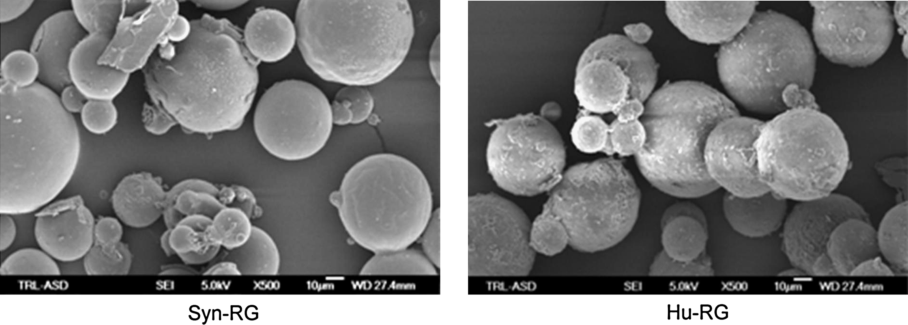
Scanning electron microscopy images of Syn-RG (left) and HU-RG (right) MSs. MSs, microspheres; RG, recombinant gelatin.
In vitro MS degradation assay
The enzymatic degradation time in vitro of RG MSs was determined with an enzymatic degradation assay. Briefly, 50 mg of gelatin MSs was dispersed in 10 mL of phosphate buffer pH 7.4 (Fluka, Sigma Aldrich, Zwijndrecht, The Netherlands) and incubated at 37°C with 0.1 mg of Clostridium histoliticum enzyme (Sigma Aldrich, Zwijndrecht, The Netherlands) for 48 h. Disappearance of the MSs was followed visually.
Cell isolation and culturing
Human skin fibroblasts (PK-84) were used for in vitro cytotoxicity assays. The fibroblasts were cultured at 37°C at 5% CO2 in Roswell Park Memorial Institute (RPMI) medium (cat. no. BE12-115F; BioWhittaker, Lonza Verviers, Belgium) supplemented with 10% fetal bovine serum (cat. no. CH30160.03; HyClone, PerBio, Etten-Leur, The Netherlands), 1% L-glutamine (cat. no. BE17-605E; BioWhittaker), and 1% penicillin/streptomycin (cat. no. 15140-122; Gibco, Breda, The Netherlands), and passaged 1:10 twice a week.
Adipose-derived stem cells (ADSCs) were isolated from human adipose tissue that was dissected from the abdominal wall after resection. ADSCs were isolated from fat tissue as previously described by Oedayrajsingh-Varma et al. 22 Cells were cultured at 37°C at 5% CO2 in Dulbecco's modified Eagle's medium (cat. no. BE12-604F) supplemented with 10% fetal bovine serum, 1% L-glutamine, and 1% penicillin/streptomycin, and passaged 1:3 once a week.
Cytotoxicity assay
Cytotoxicity was assessed with an [3-(4,5-dimethylthiazol-2-yl)-5-(3-carboxymethoxyphenyl)-2-(4-sulfophenyl)-2H-tetrazolium] (MTS) assay. Briefly, PK-84 cells (10,000 cell per well) were seeded in a 96-well plate. After 24 h the medium was replaced by extracts of the MSs. Extracts were prepared by shaking the MSs in cell culture medium for 24 h at 37°C. Then, 48 h after addition of the extracts, the mitochondrial substrate MTS was added. Formation of the formazan product was measured at 490 OD on a spectrophotometer after ∼1 h.
Culturing of ADSCs on MSs
MS suspensions in culture medium were prepared from Syn-RG and Hu-RG MSs. In a six-well low adherence plate 2 mL MS suspension was added to the wells. Per well, 400,000 ADSCs were added, resulting in a seeding density of 16,000 cells per cm2 MS surface area. ADSCs and cells were observed every day and photomicrographs were taken after 2 and 7 days. After 2 days, the MSs were collected by centrifugation for 2 min at 2000 rpm. The MSs were dissolved in fresh medium and put in a new six-well low-adherence plate for another 6 days.
Animals
Animal experiments were carried out with male AO rats (Harlan Nederland, Horst, The Netherlands) weighing 300 ± 50 g. The animals were housed under standard laboratory conditions: a regular light–dark cycle, and laboratory chow and acidified water ad libitum. All animal experiments were approved by the Local Committee on Animal Experimentation.
Surgical procedures
The gelatin MSs were dispersed in saline to a final concentration of 10% (w/v) and subcutaneously injected. Briefly, the animals were anesthetized using isoflurane/N2O/O2 and their backs were shaved and disinfected. Subsequently, 0.2 mL of each gelatin suspension was injected subcutaneously on the back of the rat. The materials and the surrounding tissue were explanted under anesthesia after 14 or 28 days (n = 5 per time point); thereafter, rats were killed by cervical dislocation.
Histology
After explantation, half of the explant was fixed in 2% glutaraldehyde solution prior to embedding in Technovit 7100, according to manufacturer's instructions. Sections of 2 μm were stained with toluidine blue and analyzed by light microscopy.
Masson's trichrome staining, which stains collagen blue and cells red, was performed on cryosections (5 μm) according to a standard protocol. 23
Immunohistochemistry
For immunohistochemical evaluation of the presence of macrophages and blood vessels in explants, sections of 5 μm were cut at −25°C and fixed with acetone. Sections were preincubated with 10% rabbit serum and subsequently incubated with antibodies against, respectively, CD68 (Serotec, Oxford, United Kingdom) for the detection of macrophages and giant cells, collagen IV (cat.no. ab6311-100) for the detection of blood vessels, collagen III (cat.no. ab6310-100; Abcam, Cambridge, United Kingdom) for the determination of possible ECM deposition, and CD3 for the detection of T-cells. Thereafter, endogenous peroxidase activity was removed by H2O2 treatment, which was followed by incubation with appropriate secondary antibodies (Dako, Glostrup, Denmark). Slides were then stained with 3-amino-9-ethylcarbazole, counterstained with hematoxylin, and embedded in Kaisers glycerin.
Polymerase chain reaction
Total RNA was extracted from explants using the RNeasy® microkit (cat. no. 74004; Qiagen, Hilden, Germany). The RNA yield was quantified by measuring the absorbance at 230, 260, and 280 nm. Samples with a 260/280 ratio between 1.8 and 2.2 were taken for further use. cDNA was prepared from 100 ng RNA using reverse transcriptase M-Mul-V (Fermentas, St. Leon Rot, Germany). Finally, polymerase chain reaction was carried out with 10 ng cDNA per reaction for genes using the primers listed in Table 2.
In situ zymography
Cryosections (5 μm) were incubated with 50 μL dye-quenched (DQ)-gelatin solution. DQ-gelatin is a gelatin that is heavily loaded with fluorescein molecules, which quenches the fluorescence. Cleavage of the gelatin by gelatinases in the tissue sections will result in increased fluorescence. The sections were incubated at 37°C for 2 h. The DQ-gelatin solution consisted of 100 mg/mL DQ-gelatin in 50 mM Tris/HCl buffer pH 7.4, which also contained 16 mM CaCl2, 0.05% Brij-35, and 5 mM phenylmethanesulfonyl fluoride (PMSF). After incubation, the sections were washed with tap water, subsequently incubated with Triton X-100 and 4′,6-diamidino-2-phenylindole, and finally mounted in citifluor. As negative controls, slides were incubated without the substrate and with substrate plus 20 mM ethylenediaminetetraacetic acid. The latter blocks MMP activity. As a positive control, slides were incubated with p-aminophenylmercuric acetate plus the substrate. p-Aminophenylmercuric acetate will activate all MMPs in the section, and this will thus show the maximum MMP activity possible in the sections. Digital fluorescence photomicrographs were obtained using a Leica DM RXA (Rijswijk, The Netherlands) fluorescent microscope equipped with a Leica DC350 FX camera and Leica Qwin Pro image analysis software.
Statistical analysis
All data were expressed as the mean ± standard deviation. The data presented in Figure 3B were subjected to a nonparametric Mann–Whitney U-test, because these data were not normally distributed. Data in Figure 4B were subjected to a two-sided Student's t-test. Differences are considered significant at p < 0.05.
Results
In vitro characterization
The size of the MSs after preparation was determined by scanning electron microscopy. The average diameter after preparation of the Syn-RG and Hu-RG MSs was, respectively, 67 ± 25% and 93 ± 25% μm.
Crosslinking percentages of the amino groups were 48% and 82% for Syn-RG and Hu-RG MSs, respectively, as assessed by an assay using trinitrobenzene sulfonate.
The MSs were subjected to enzymatic degradation by bacterial collagenase at 37°C for 48 h. Syn-RG MSs did not degrade, whereas approximately 75% of the Hu-RG MSs had degraded after 48 h (data not shown).
Syn-RG and Hu-RG MSs were not cytotoxic (data not shown) as assessed by MTS assay.
Cell adhesion to the MSs was investigated via light microscopical examination of the MSs after 1, 4, and 8 days of culturing. It can be seen in Figure 2A–F that Syn-RG as well as Hu-RG support the adherence of ADSCs. At day 0, cells attached to the MSs can be seen as little bulbs on the surface area of the MSs (denoted by arrows). After 4 days, the cells had formed bridges between adjacent MSs (denoted by an asterisk), and the MSs and adhered cells had arranged into a single large cluster (Fig. 2C, D). After 8 days, the single large cluster of cells and MSs had remained unchanged compared to day 4 (Fig. 2E, F). Figure panels 2G and 2H represent Hu-RG and Syn-RG MSs incubated without cells, respectively. These MSs form a layer of MSs on the bottom of the well and do not form clusters.
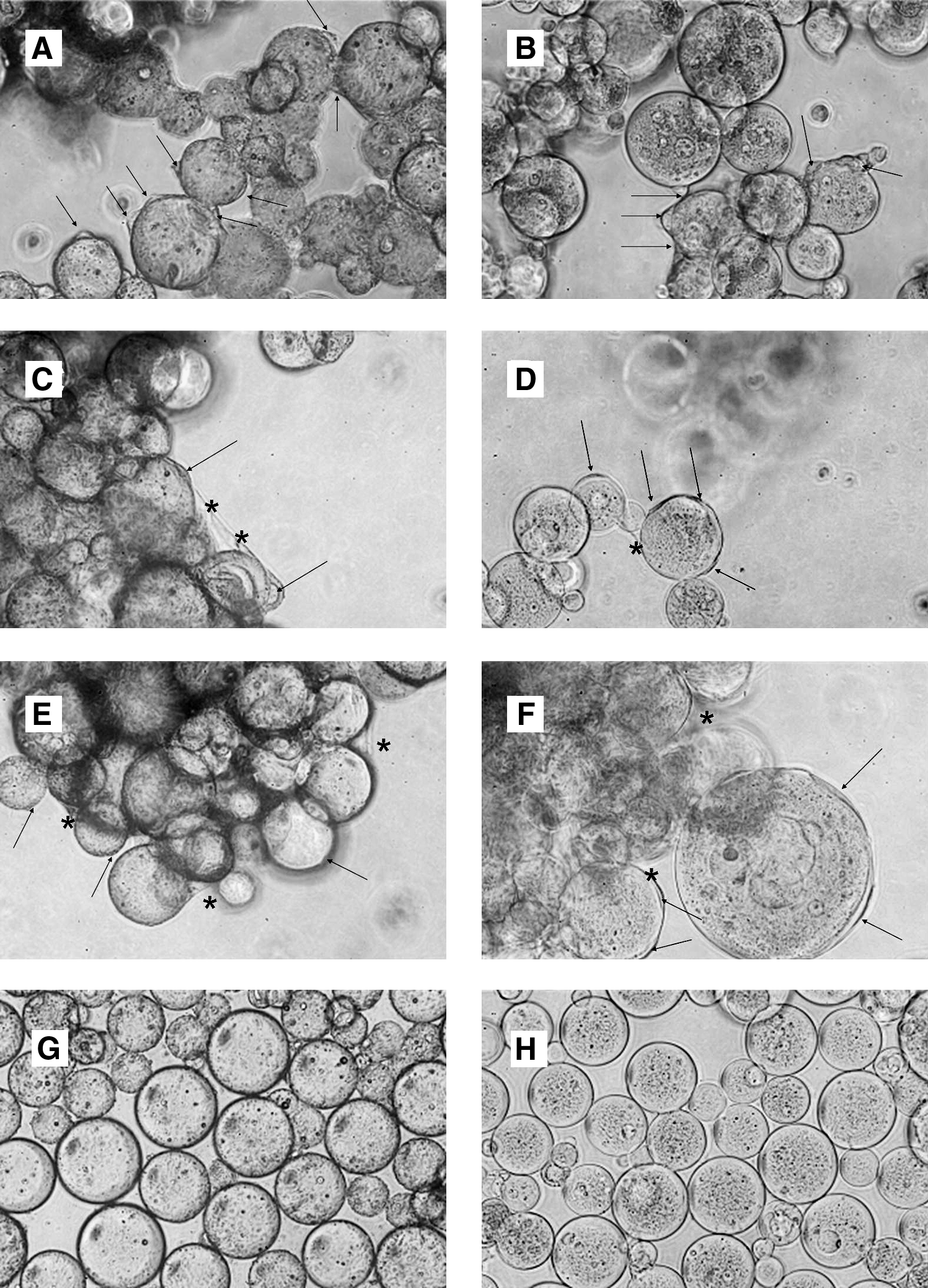
Photomicrographs of the adipose-derived stem cells seeded on Hu-RG (
In vivo tissue response
To study the in vivo tissue response, the MSs were suspended in physiologic saline (10% w/v) and injected subcutaneously on the back of the rats. After explantation and histological examination of the explants, distinct differences were observed in the tissue reaction to Syn-RG and Hu-RG MSs (Figs. 3A, B, 4A, B and 5A–M).
(
(

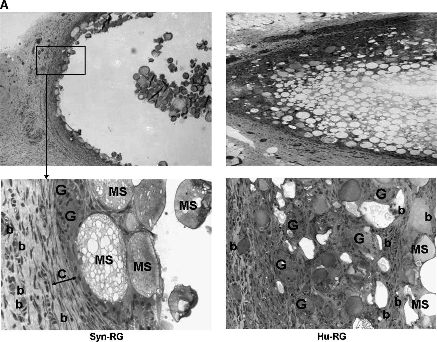
Staining of Syn-RG and Hu-RG explants after 14 days in vivo. Toluidine blue staining for a histologic overview (
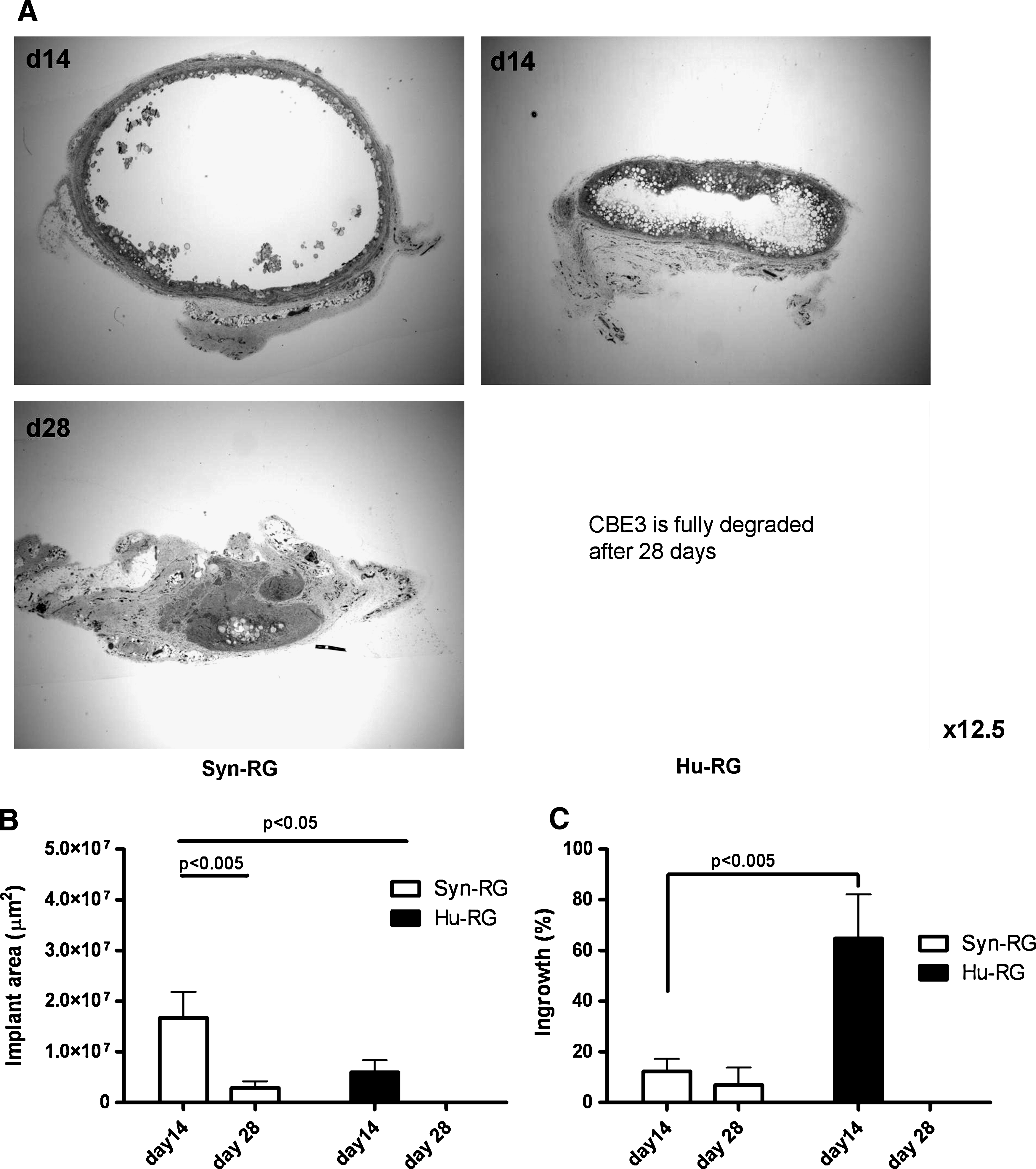
At 14 days after implantation, Hu-RG explants (60%) showed more ingrowth than Syn-RG explants (15%) (Fig. 4A, B, p < 0.005). Cells infiltrated deeper into the Hu-RG explants compared to the Syn-RG explants, where infiltration of cells was limited to the rim of the explants. Histology (Fig. 3A) and immunohistochemical staining (Fig. 5) revealed that the majority of the infiltrated cells were macrophages and fibroblasts. After infiltration, part of the macrophages had fused to giant cells. Significantly more macrophages and giant cells were present in Hu-RG explants compared to Syn-RG explants (Fig. 3B, p < 0.05). Polymorphonuclear leukocyte (PMNs) were not observed in explants of both types of RG MSs at day 14. The number of lymphocytes tended to be higher in Syn-RG than Hu-RG explants (not significant, data not shown).
In all Hu-RG explants, the formation of new blood vessels was seen between the MSs in the ingrown parts of the explant (Hu-RG, Fig. 5J), whereas most newly formed blood vessels in Syn-RG (Fig. 5I) explants were observed only at the rim of the explant.
Besides vascularization, the ingrowth was also accompanied by the deposition of newly formed ECM. Masson's Trichrome staining revealed that collagen bundles had deposited (light blue strands) between the Hu-RG MSs in ingrown areas of the explants (Fig. 5D). The previously mentioned minimal ingrowth at the rim of Syn-RG explants correlated with the observed minimal deposition of newly formed ECM in this area (Fig. 5C). When the composition of the newly formed ECM was examined for the presence of specific collagens, both collagen I (data not shown) and collagen III (Fig. 5E, F) appeared to be deposited in a comparable pattern. Both collagens were deposited in the ingrown areas in the Hu-RG and Syn-RG explants.
At 28 days after implantation, Hu-RG explants were fully degraded, whereas Syn-RG explants were partially degraded. The faster degradation of Hu-RG MSs might be due to the presence of considerably more macrophages and giant cells in the Hu-RG explants compared to Syn-RG explants (Figs. 3A, B, and 5G, H). Macrophages and giant cells can degrade the MSs via a two-step pathway: enzymatic degradation by collagenases and gelatinases, that is, MMPs, and by subsequent phagocytosis of the enzymatic degradation products. To investigate whether MMPs were involved in the degradation of the MSs, we first examined the mRNA expression levels of MMP-2, MMP-9, and MMP-14. In addition, also the mRNA levels of the tissue inhibitor of MMP (TIMP)-1 were examined. At day 14, all MMPs and TIMP-1 were detected, but no differences in mRNA expression of MMP-2 (gelatinase A), MMP-9 (gelatinase B), MMP-14 (activator of MMPs), and TIMP-1 were found when the expression levels in Syn-RG and Hu-RG explants were compared (Fig. 6).
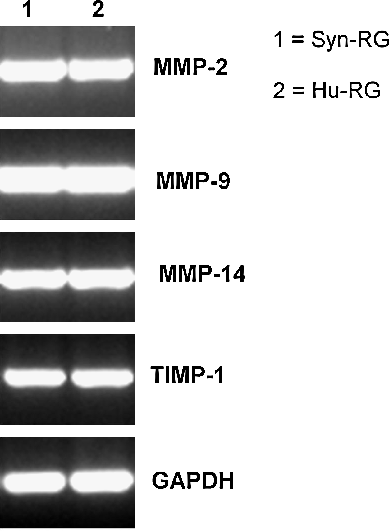
mRNA levels of MMP-2, MMP-9, MMP-14, TIMP-1, and GAPDH in Syn-RG and Hu-RG explants 14 days after implantation. MMP, matrix metalloproteinase; TIMP, tissue inhibitor of MMP; GAPDH, glyceraldehyde 3-phosphate dehydrogenase.
Although no differences in mRNA expression were observed, the in situ gelatinase assay and fluorescence microscopy showed that Hu-RG explants displayed a higher gelatinase activity than Syn-RG explants (Fig. 7C, F vs. 7I, L). This difference was most pronounced when the gelatinase activity between the MSs was observed; however, a slight difference in gelatinase activity in the rim of the explants could be seen. These results indicate that Hu-RG MSs are degraded faster than Syn-RG MSs.

In situ gelatinase activity in sections of Syn-RG and Hu-RG explants (magnification: ×200). (
Discussion
Our aim was to develop MSs prepared from RGs for the delivery of cells and or factors to augment TR. We describe the characterization and the tissue reaction to and degradation of two types of RG MSs, Syn-RG and Hu-RG.
Upon implantation, scaffolds or biomedical devices induce an inflammatory tissue response known as the foreign body reaction (FBR). The FBR is the primary reaction of the nonspecific immune system towards implanted foreign materials and is characterized by three distinct phases as described by Luttikhuizen et al. 24 Essentially, these phases are characterized by differences in inflammatory cells such as PMNs, macrophages, (myo)fibroblasts, and others, and their role in the FBR. Macrophages are important, for example, for the turnover of ECM through the secretion of proteolytic enzymes like MMPs and the secretion of transforming growth factor β that promotes ECM deposition by (myo)fibroblasts. When examining the tissue reaction towards the two types of RG MSs, we focused on parameters of the FBR that differed between the two types of MSs.
Clear differences in cellular ingrowth, ECM deposition, and degradation rate of the MSs were observed. Hu-RG MSs were fully degraded after 28 days, whereas Syn-RG MSs were only partially degraded (Fig. 4A). After 14 days, on average 60% of each Hu-RG explant was infiltrated by cells (Fig. 4B), which was associated with stroma formation, that is, deposition of ECM and neovascularization between the MSs in this area. In contrast, on average, only 15% of each Syn-RG explant was infiltrated by cells. Deposition of new ECM between these MSs and angiogenesis was restricted to the rim of Syn-RG explants (Fig. 5). Ingrowing cells in both explants appeared to be mainly fibroblasts and macrophages. Part of the macrophages fused to giant cells locally. Degradation of the MSs took place both by enzymatic degradation and phagocytosis. Although gene expression of MMPs was observed in both types of implanted RG MSs (Fig. 6), gelatinase activity was higher in Hu-RG explants than in Syn-RG explants (Fig. 7F, L).
The major difference between Hu-RG and Syn-RG is the presence of RGD moieties in Hu-RG, which promote cell adhesion. 25 In vivo, cells, like macrophages and fibroblasts, can bind and adhere to the RGDs via the α5β1 and αvβ3 integrins on their membranes (Reviewed in Ref. 26 ). Indeed, Hu-RG implants contained higher numbers of infiltrated macrophages and fibroblasts, which might be attributed to the RGD moieties. Significantly higher numbers of infiltrated macrophages (Fig. 3B) and giant cells were present in these explants. This is in accordance with our earlier observations that RGDs might not only up-regulate macrophage infiltration but also promote macrophage fusion to giant cells. 27
TR requires the transient presence of the consisting scaffold, yet this scaffold should be replaced by newly formed ECM upon scaffold degradation. Both types RG MSs met with these demands though in a different fashion. In Hu-RG explants, considerably more new ECM has been deposited than in Syn-RG explants (Fig. 5C–F). Since fibroblasts are the main producers of ECM and since it has been reported that fibroblasts produce ECM components upon binding to RGDs through integrin αvβ3, 28 the presence of RGDs in Hu-RG might explain the larger amount of new ECM in these explants.
Macrophages and giant cells can degrade the MSs via a two-step pathway: enzymatic degradation and subsequent phagocytosis of the degradation products. The infiltrated macrophages29,30 and formed giant cells 30 are both capable of producing MMPs. These enzymes play an important role in ECM turnover and remodeling. Two members of this MMP protein family are the gelatinases MMP-2 and MMP-9, which are capable of easily digesting not only gelatin, but also ECM components like collagen IV, V, and XI, laminin, elastin, and aggrecan core protein. 31
Although differences were observed in the number of infiltrated MMP-producing cells between Syn-RG and Hu-RG MSs, no differences in expression levels of mRNA for MMP-2, -9, and -14 were detected (Fig. 6). However, Hu-RG explants appeared to have a higher gelatinase activity in situ than Syn-RG explants, which could be anticipated, as Hu-RG has 4 to 5 times more cleavage sites for enzymatic degradation than Syn-RG. The results of the in vitro enzymatic degradation assay (data not shown) also support this observation.
The distinct degradation profiles and the difference in stroma formation between Syn-RG and Hu-RG MSs determine for a great deal their suitability for specific TE purposes. Syn-RG is less infiltrated by cells and degraded slower than Hu-RG and may be applied as a long-term instructive formulation, for example, as a slow-release depot for the sustained release of growth factors or cytokines. In contrast, Hu-RG MSs are faster infiltrated by cells and display a faster degradation profile, and stroma formation is rapidly initiated. These characteristics render Hu-RG MSs more suitable for relative short-term TE applications.
In many TE applications, cell-loaded scaffolds are used. We therefore also examined the cell-binding capacity of both types of RG MSs by seeding ADSCs on the MSs. ADSCs express integrins α5β1 and αvβ3, enabling them to bind to sequences like RGD. In vitro, Hu-RG but also Syn-RG supported cell adhesion. Apparently, the presence of RGDs in Hu-RG was not decisive for the ability of cells to adhere to the MSs in vitro. It appears that ADSCs are able to bind to Syn-RG via mechanisms other then via integrin binding.
Both types of MSs could therefore also be applied as delivery vehicles for (stem) cells, for example, in cardiac TR after myocardial infarction. Studies have been performed in which (stem) cells were used to improve cardiac function after myocardial infarction, 6 but a generally observed disadvantage was the quick disappearance of the cells from the site of injection. 32 Injection of cells attached to MSs could increase the local retention of delivered cells after administration. Before use in cardiac TE though, the tissue reaction toward the MSs in the heart should be examined because the tissue reaction was found to depend on the implant location. 33
In summary, these novel MSs, Syn-RG and Hu-RG MSs, proved to be suitable candidates for TR. Currently, we examine the use of these cell-loaded MSs for treatment of cardiac disease. In addition, incorporation of growth factors and cytokines in the MSs will be examined to obtain (slow)-release formulations that are applicable in several TR areas.
Conclusion
Our study shows that MSs prepared from RGs are suitable for use in TR. Syn-RG and Hu-RG MSs showed an appropriate tissue reaction after subcutaneous implantation in rats. We discussed the differences in tissue reaction between Syn-RG and Hu-RG and anticipated that these differences are likely the result of Hu-RG having RGDs in its sequence, whereas Syn-RG does not. At present, we are examining the application of the RG MSs as a delivery vehicle for drugs and cells. This study indicates that our novel RG MSs are promising candidates for guiding TR by means of drug and/or (stem) cell delivery.
Footnotes
Disclosure Statement
Sebastiaan G. Kluijtmans and Jan B. Bouwstra are both working at Fuji Manufacturing Europe B.V. who financed this research.