
Abstract
In this study, adipose-derived stem cells (ASCs) were cocultured with nucleus pulposus (NP) cells using a porous membrane to investigate the effect of NP cell phenotype on ASC chondrogenic differentiation. Human NP cells were collected from 14 patients and classified into two groups (normal vs. degenerative) depending on the level of type II collagen, aggrecan (AGG), type I collagen, and bax gene expression. Human ASCs were then cocultured with each group of NP cells on porous membranes in the absence of chondrogenic supplements. After 2 weeks, real-time-polymerase chain reaction results showed that ASCs cocultured with normal NP cells had much higher type II collagen and AGG gene expression than ASCs cocultured with degenerative NP cells. The production of AGG was also observed only in the group cocultured with normal NP cells. Additionally, coculture of ASC pellets with normal NP cells promoted the production of AGG as compared to coculture of ASC monolayer with normal NP cells. These data demonstrate that a coculture system using porous membranes can induce ASC differentiation into NP cells without chondrogenic supplements. Further, the phenotype of cocultured NP cells significantly influences the extent of ASC differentiation.
Introduction
There have been several approaches using bioactive molecules and/or three-dimensional (3D) scaffolds that provide biomimetic environments for embedded cells, thus promoting cell proliferation, differentiation, and matrix formation.7–9 ASCs cultured with differentiation medium containing transforming growth factor-beta (TGF-β), insulin-like growth factor (IGF)-1, dexamethasone, insulin, transferrin, and selenium supplement, and ascorbic acid can differentiate into chondrocytes. 10 However, the chemical agents in the medium may cause cytotoxicity and the growth factors are relatively expensive.11–13 To resolve these problems, a coculture system has been suggested.14–16 A recent study with human bone marrow-derived mesenchymal stem cells (BMSCs) and NP cells showed that cocultured NP cells could induce the differentiation of BMSCs without exogenous factors. 14 Additionally, Kp-MSCs (immortalized mesenchymal stem cells) cocultured with hPi-GFP (immortalized chondrocytes) differentiated into chondrocyte-like cells. 16 These reports demonstrated that a coculture system could enhance the secretion of cytokines from coculture cells and stimulate stem cell differentiation.
Phenotypic changes in the coculture cells are thought to be due to intercellular communication through connexin and gap junctions between the coculture cells.17,18 Yamamoto et al. also showed that direct cell-to-cell contact between cocultured NP cells and BMSCs significantly increases the viability of NP cells along with cytokine and growth factor production in the BMSCs. Compared to systems that do not permit contact between the coculture cells, direct cell-to-cell contact significantly upregulated TGF-β, IGF-1, epidermal growth factor, and platelet-derived growth factor related to metabolism and proliferation of NP cells. 19 However, no previous study has shown how the phenotype of NP cells may affect the differentiation of cocultured ASCs.
The main purpose of our study was to investigate the phenotype of NP cells from different patients and examine how human ASCs are influenced by the NP cell phenotype in a coculture system using porous membranes. For the experiment, human NP cells were collected from different patients, divided into normal and degenerative groups according to the expression of specific genes, including COL2, AGG, COL1, and bax. Cells in each group were then cocultured with ASCs using porous membranes under the condition of the direct cell-to-cell contact coculture. In the following experiment, pellets of human ASCs coated with hydrogel were compared to human ASCs cultured on the membrane surface to investigate whether changes in culture conditions can affect human ASC differentiation when they were cocultured with normal and degenerative NP cells without direct cell-to-cell contact.
Materials and Methods
Isolation and characterization of NP cells and ASCs
After obtaining patient informed consent and approval of the CHA University Ethics Committee, NP cells were separated from discarded NP tissue from 14 subjects who underwent spinal surgery for bursting fractures (n=3) or lumbar disc herniation (n=11). The tissue samples were washed three times using Hank's balanced salt solution (HBSS; WelGENE) containing 2% (v/v) antibiotics–antimycotics (Gibco BRL) for 15 min. Then, the tissue was digested with 0.05% (w/v) type 2 collagenase (Sigma-Aldrich) for 6 h. The digested mixture was strained with cell strainer (40 μm pore size; Becton Dickinson), centrifuged for 5 min at 1000 RPM, and washed with HBSS twice to remove any remaining collagenase. The washed NP cells were seeded in culture plates and maintained in Dulbecco's modified Eagle's medium (DMEM; Gibco BRL) containing 10% (v/v) fetal bovine serum (FBS; Wisent) and 1% (v/v) penicillin/streptomycin (P/S; Gibco BRL) until the cells were confluent. A total of 300,000 NP cells at passage 0 were dissolved in TRIzol (Gibco BRL) and frozen at −70°C until further evaluation of gene expression and phenotype.
The phenotype of NP cells from 14 donors was evaluated right after primary culturing. The cells were divided into two groups according to the gene expression of bax, COL1, AGG, and COL2. Each gene expression was normalized to the expression of GAPDH. The relative ratio of bax and COL1, known markers of NP cell dedifferentiation, was assigned a negative value. The relative ratio of COL2 and AGG, known markers of NP cells, was assigned a positive value. The negative and positive values of four different genes expressed by NP cells from each patient were added. Based on the total value, NP cells having either positive or negative value were pooled together and defined as a normal and degenerative NP cell group, respectively. Human ASCs were isolated from human subcutaneous adipose tissues and then cultured as previously described previously. 20
Coculture of NP cells and ASCs on porous membranes
A coculture system for ASCs and NP cells using porous membranes was established according to a previously described method. 20 Briefly, the porous membrane of a transwell insert (1 μm pore polyethylene terephthalate membrane; Becton Dickinson) was coated with a 0.2% (v/v) gelatin solution (Sigma) to promote cell attachment. For two-dimensional (2D) cell-to-cell contact in the coculture, the transwell cell culture insert (5000 cells per cm2) was placed inversely into a 50 mL beaker containing 35 mL of DMEM [10% (v/v) FBS and 1% (v/v) P/S] and a Teflon ring was placed on top of the inverted insert for fixation. Next, the NP cells were seeded on the bottom surface of the inverted membrane and cultured for 24 h. The insert with NP cells was turned upright and transferred into a six-well plate containing DMEM (10% (v/v) FBS and 1% (v/v) P/S) and ASCs were then placed on the top surface of a membrane inside the insert. The cells were cultured with medium in the absence of differentiation factors for 2 weeks.
In the 3D coculture group, ASCs pellets were prepared and cocultured in the transwell with NP cells. Specifically, 100,000 ASCs were put into a 15 mL conical tube (SPL) and centrifuged for 5 min at 500 g to form an ASC pellet. The ASC pellet was then coated with fibrin gel using a fibrin gel kit (Green Cross) according to the manufacturer's instruction. The ASC pellet was coated with 25 μL of the diluted fibrinogen solution, and the mixture was subsequently cross-linked by adding 25 μL of the diluted thrombin solution. The coated ASC pellet was placed on the top of membrane and then transferred into a six-well plate in which NP cells were seeded (5000 cells per cm2) and cultured for 2 weeks. ASCs were also cultured as monolayer on the porous membrane (20,000 cells per membrane) in the transwell for comparison.
Real-time-polymerase chain reaction analysis
Cultured cells were lysed using TRIzol reagent. Total RNA was extracted and reverse-transcribed using an RT-PCT kit (Bioneer). Sequences of the primers (Bioneer) used are shown in Table 1. Polymerase chain reaction (PCR) was performed in AccuPower Hotstart PCR PreMix (Bioneer), containing 10 pM of each primer. These reactions were performed using a MyGenie 96 Thermal block (Bioneer), with a denaturation step for 5 min at 95°C, followed by 25–35 cycles for 30 s at 95°C, 45 s at 57°C, and 60 s at 70°C, and extension at 70°C for 10 min. The PCR products were loaded on a 1.2% (w/v) agarose gel for electrophoresis at 100 V. After 30 min, the agarose gel was stained with ethidium bromide. Relative gene expression in each sample was evaluated by examining the gels under UV light.
Characterization of normal and degenerative NP cells
Senescence of normal and degenerative NP cells was observed by a β-galactosidase assay kit (Cell Signaling Technology) according to the manufacturer's instruction. Cytokine secretion by normal and degenerative NP cells was examined by a cytokine antibody array (RayBiotech Inc.) according to the manufacturer's instruction and cytokine signal spots were detected by chemiluminescence. Secretion of TGF-β1 and bone morphogenetic protein (BMP)-2 was evaluated by an enzyme-linked immunosorbent assay (ELISA) using a Quantikine ELISA kit (R&D system) according to manufacturer's protocol.
Alcian blue staining and Goldner's trichrome
ASCs grown on the porous membrane were fixed with 4% (w/v) paraformaldehyde (PFA; Yakari Pure Chemicals) after the 14 days. The fixed samples were washed with phosphate-buffered saline (PBS) and stained with 0.5% Alcian blue (Sigma) in 3% acetic acid (pH 2.5) for 30 min for glycosaminoglycan (GAG), and rinsed with distilled water. For the 2D cell-to-cell contact coculture, NP cells on the bottom of the membrane were removed with a scraper (SPL) to prevent the cells from being stained for GAG. NP cells and ASCs encapsulated by hydrogel were fixed with 4% (w/v) PFA and embedded in paraffin. Specimens were sectioned (5 μm thick) and stained with a Alcian blue solution and Goldner's trichrome solution (Sigma). Images were acquired using a light microscope equipped with a video camera.
Immunohistochemistry
ASCs cultured on the porous membrane were washed by PBS and subsequently fixed with 4% (w/v) PFA. Fixed ASCs were washed with PBS, permeabilized for 10 min in 0.25% Triton X-100 in PBS, and blocked with 1% bovine serum albumin in PBS for 30 min to prevent nonspecific binding of proteins. The cells were incubated with monoclonal anti-AGG antibody (Abcam) at 4°C overnight, and then stained with anti-mouse FITC-conjugated secondary antibody (Abcam). Hydrogel-coated ASC pellets in the 3D coculture system were fixed with 4% (w/v) PFA and then embedded in paraffin. Specimens were sectioned to a thickness of 5 μm and immunostained by the same method given above. Stained ASCs were observed with a confocal laser scanning microscope (LSM 510 META; Carl Zeiss Microimaging Inc.).
Results
NP cells characterization
NP cells derived from 14 different donors were characterized by real-time (RT)-PCR as shown in Figure 1A. The cells from each donor showed different levels of bax, COL1, AGG, and COL2 gene expression. Based on the ratio of each gene expression normalized by GAPDH, NP cells were scored as shown in Figure 1B according to the method described in the Materials and Methods section. Cells from donors 7, 13, and 14 had scores close to 1, whereas ones from Donor 4 had the lowest score (<−0.5). In this study, NP cells with positive values or negative values were separately pooled and defined as the normal group or degenerative group, respectively.
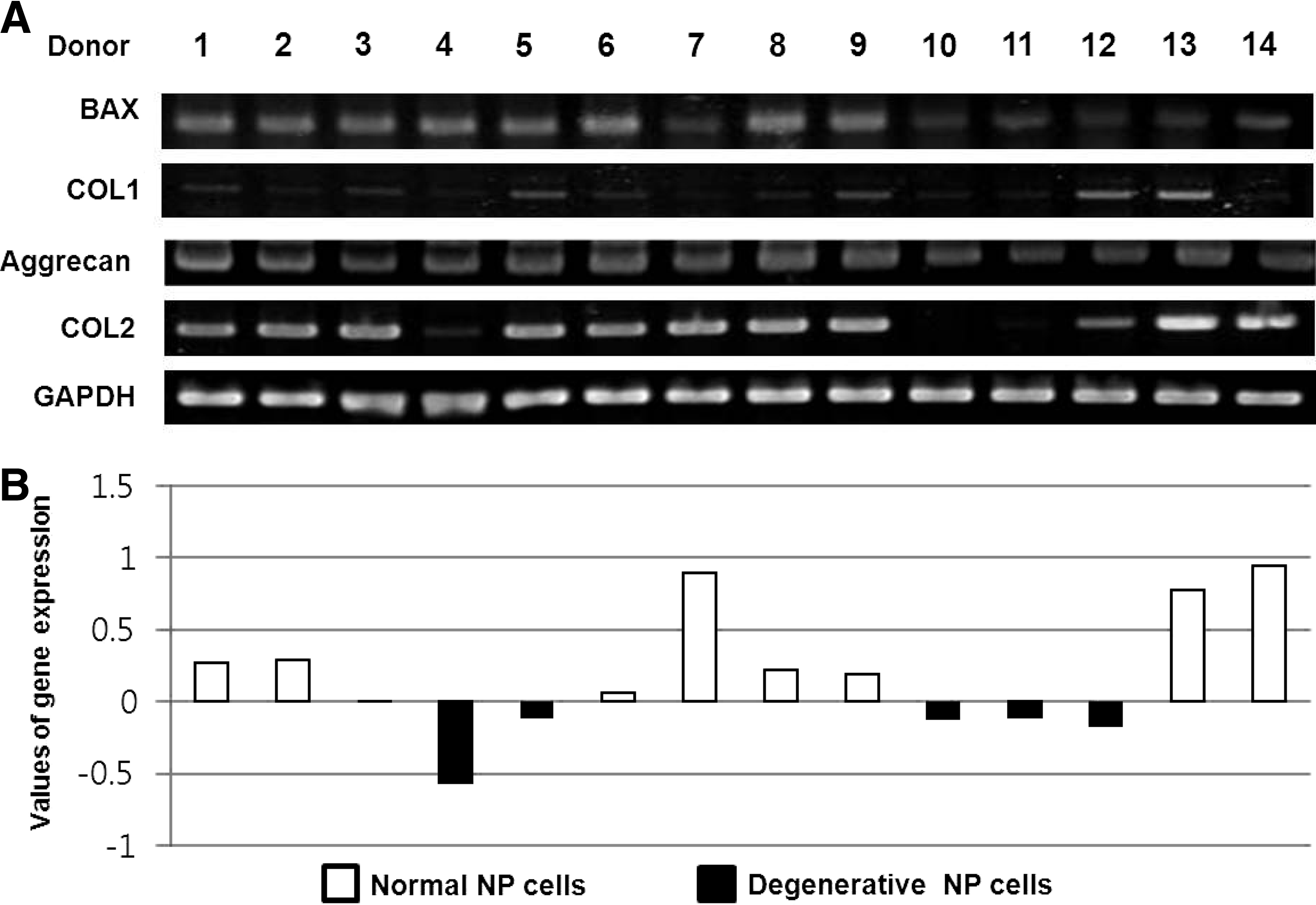
NP cells from 14 different patients.
After the NP cells were divided into two groups, they were tested for gene expression, senescence, cytokine expression, and cytokine secretion. Figure 2A shows the RT-PCR results confirming that the gene expression patterns in both groups were different. The normal group had much higher COL2 expression and less COL1 and bax expression than the degenerative group. According to the β-galactosidase assay (Fig. 2B), more senescence cells were found in the degenerative group than in the normal group. The expression of cytokines such as BMP-4, -6, -7, TGF-β1, β2, β3, and interleukin (IL)-6 was compared between the two groups. Figure 2C shows the cytokine antibody array analysis results of a normal and a degenerative NP cell group. It appeared that the cytokine expression patterns of both groups were similar, but the expression of IL-6, an anti-inflammatory cytokine, was much higher in the degenerative group than the normal group. The secretion levels of TGF-β1 (a chondrogenesis-inducing factor) and BMP-2 (an osteogenesis-inducing factor) were also compared by ELISA analysis (Fig. 2D). TGF-β1 secretion in normal NP cells was higher than in degenerative NP cells, whereas BMP-2 secretion showed the opposite trend.
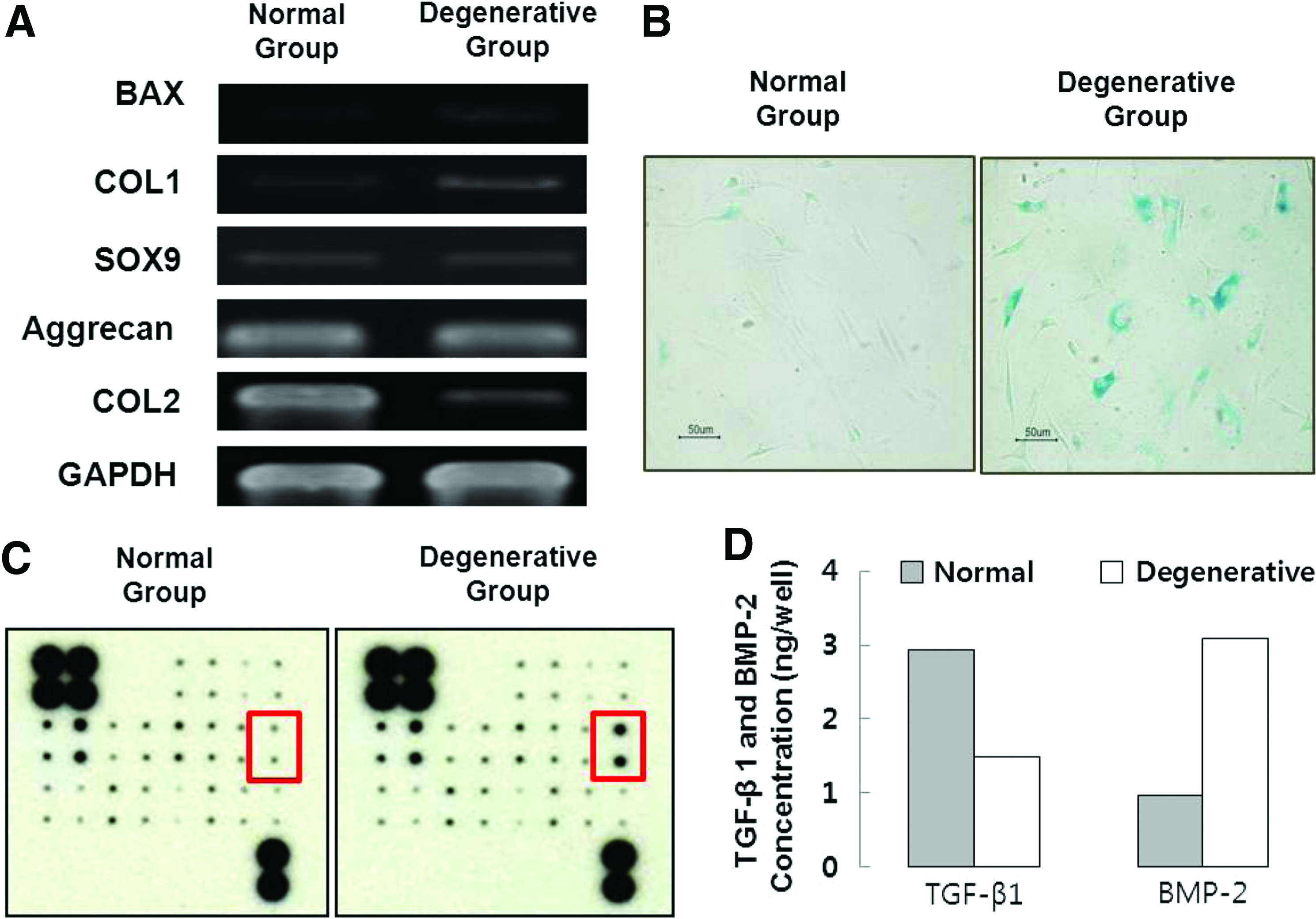
Characterization of NP cells from a normal and a degenerative group.
Comparison of ASCs cocultured with normal or degenerative NP cells under direct cell–cell contact
Figure 3A shows a schematic representation of the different ASC coculture conditions. Briefly, ASCs were cocultured with either the normal group or degenerative group under direct cell–cell contact and then examined using RT-PCR and Alcian blue staining. Figure 3B shows the RT-PCR results from ASCs cocultured with the normal and degenerative group. Gene expression of SOX9, AGG, and COL2 (markers of cells with phenotypes of NP cell) was only found in ASCs cultured with normal NP cells. However, ASCs cultured with degenerative NP cells did not express any genes related to the phenotype of NP cells.
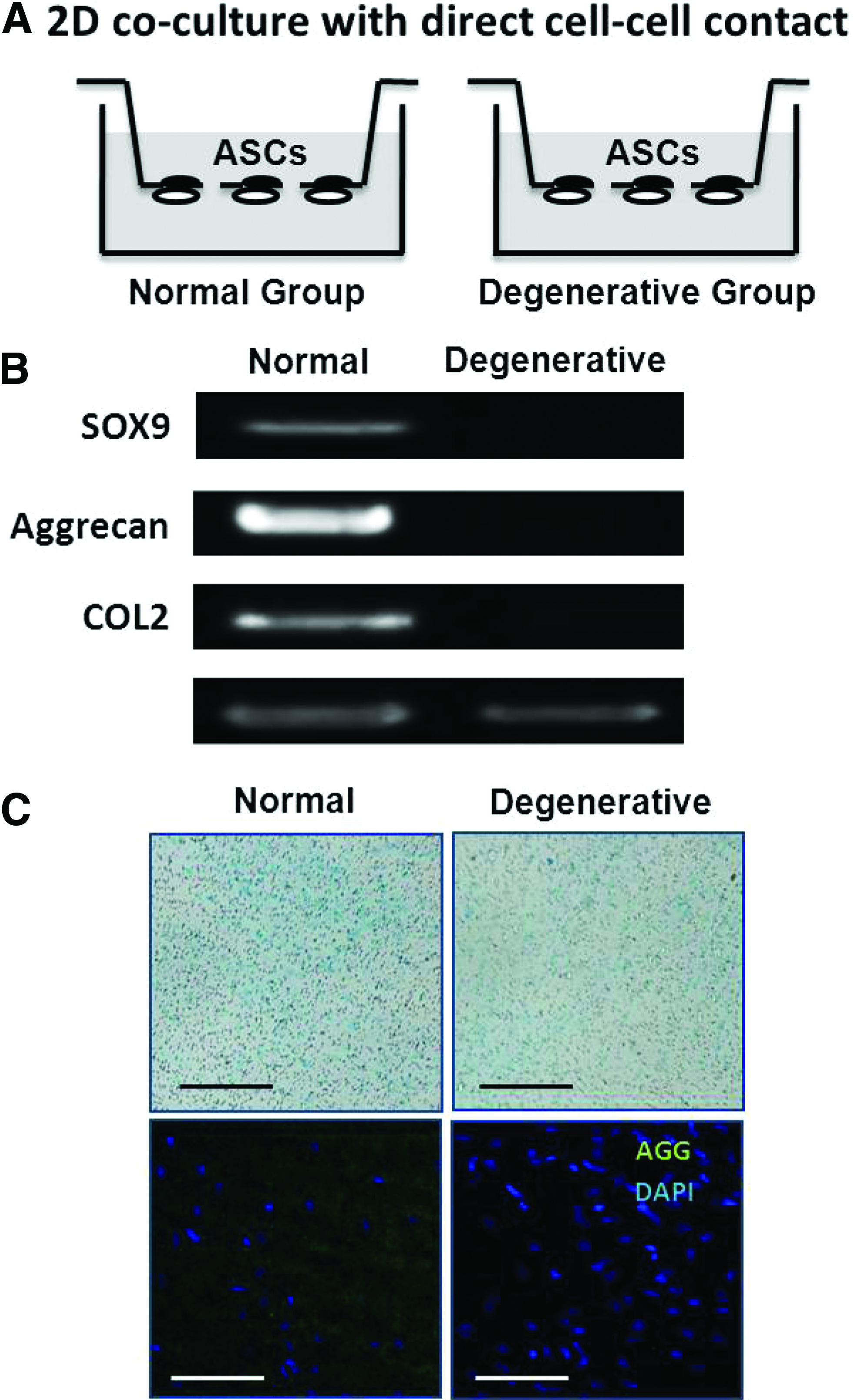
ASCs cultured with normal and degenerative NP cells on a porous membrane.
Each group of cells was stained with Alcian blue for GAG and underwent immunohistochemistry staining to observe AGG production (Fig. 3C). The staining results from the ASCs concur with the gene expression data. Higher GAG production was observed in ASCs cocultured with normal NP cells compared to those cocultured with degenerative NP cells. Immunohistochemistry results also confirmed that AGG production was only observed throughout ASCs cocultured with normal NP cells.
Comparison of ASC pellets and monolayers cocultured with normal or degenerative NP cells without direct cell-to-cell contact
Figure 4A shows a schematic representation of the 2D and 3D culture systems of ASCs grown in the absence of cell-to-cell contact. ASCs were retrieved after 14 days of culturing. The cells then were evaluated with RT-PCR analysis and immunohistochemistry. RT-PCR results showed that all groups cultured with either normal NP cells or degenerative NP cells did not express COL2 in the absence of direct cell-to-cell contact (Fig. 4B). However, the expression of AGG and SOX9 in ASCs cultured in the 2D culture system was higher compared to ASCs in the 3D culture system. Additionally, ASCs from both culture systems show higher expression of AGG when cocultured with normal NP cells. Qualitative images of cells stained with an anti-AGG FITC-conjugated secondary antibody showed that only ASCs cocultured with normal NP cells produced AGG. In particular, a difference in staining intensity was apparent between groups of ASCs cocultured under the 3D conditions.
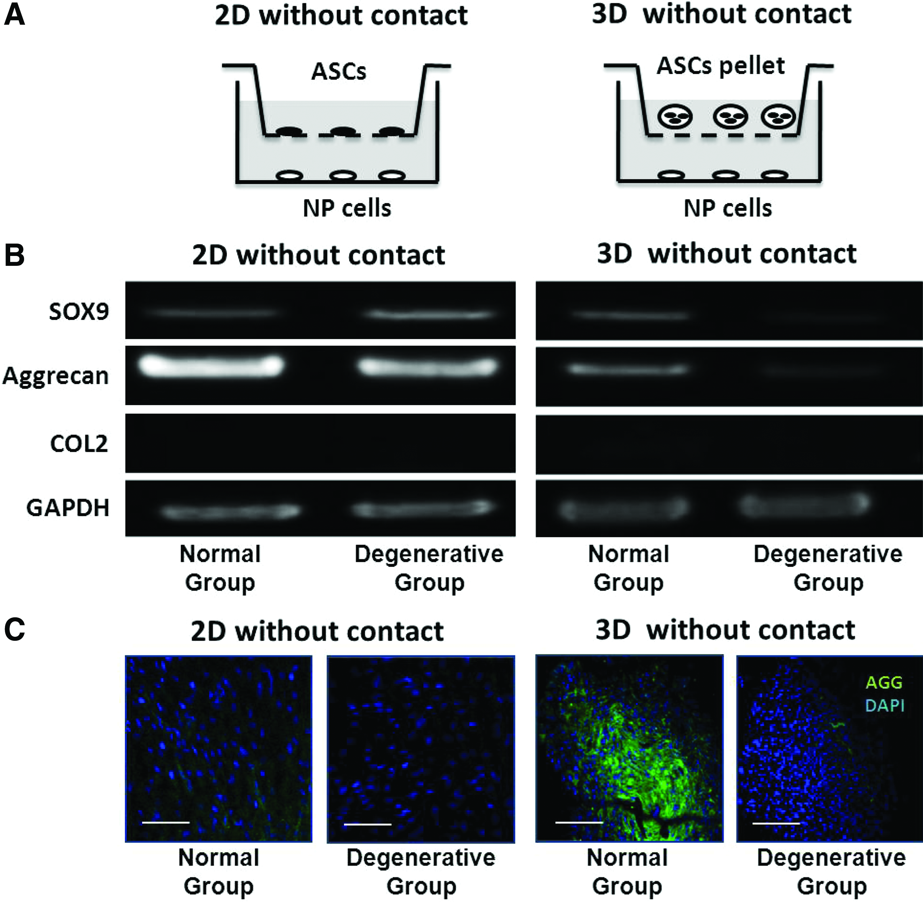
ASCs cultured with normal and degenerative NP cells on two different coculture systems.
Discussion
It has been suggested that NP cells could be activated by MSCs when cocultured. This would produce significant increases in proliferation and PG synthesis of NP cells, demonstrating the potential of coculture systems for treating degenerative IVD through tissue engineering.19,21 Another study also showed that coculturing NP cells and MSCs could initiate the differentiation of MSCs into NP-like cells, suggesting the possible use of activated MSCs for treating IVD diseases. 14
However, it is likely that autologous NP cells from patients with degenerative IVD diseases obtained for the purpose of cell therapy would not have the same phenotype as NP cells from healthy individuals. Therefore, the aims of this study were to assess the phenotype of NP cells from patients with IVD damage, and to examine how the phenotype of NP cells (normal vs. degenerative) would affect the differentiation of human ASCs into NP cells in a coculture system. We also examined the effect of two different coculture conditions (2D vs. 3D) on ASC differentiation into NP cells.
For this study, NP cells were collected from 14 donors and pooled separately into two groups depending on the expression level of genes related to the NP cell-like phenotype. As expected, the normal group exhibited higher chondrogenic and lower osteogenic properties compared to the degenerative group (Fig. 2A). The expression of genes related the NP cell phenotype were much lower in ASCs cultured with degenerative NP cells compared to ASCs cultured with normal NP cells (Fig. 3B). It appeared that normal cells had positive effects on ASC chondrogenic differentiation, whereas degenerative NP cells exerted negative effects; this was particularly evident in cultures with direct cell-to-cell contact. We strongly believe that this finding could give us a starting point for establishing cell therapies for IVD diseases using human ASCs. This is because most cases requiring the transplantation of stem cells would occur when patients have IVD degeneration similar to the in vitro cocultures of ASCs with degenerative NP cells. Therefore, the simple implantation of ASCs would not be as effective as expected for IVD regeneration. Instead, it would be necessary to obtain predifferentiated ASCs before implantation or provide environments similar to normal IVD tissues since ASCs cocultured with normal NP cells showed high expression of genes associated with the NP cell phenotype.
We also compared a 2D coculture system to a 3D coculture system that prevented cell-to-cell contact to examine how different coculture methods would affect ASC differentiation. In general, a 3D environment has been known to be more effective for cartilage regeneration compared with 2D culturing.22–24 However, our study showed that ASCs grown under 3D coculturing conditions had lower expression of genes associated with the NP cell phenotype than ASCs in 2D coculture conditions (Fig. 4B). This might be due to the different cell number ratio of ASCs to NP cells used in the 2D and 3D culture systems. The formation of an ASC pellet used for the 3D system required 100,000 ASCs, whereas only 20,000 ASCs were used in the 2D culture system. Meanwhile, the number of NP cells used for both 2D and 3D coculturing was equal. Thus, amount of cytokine-secreted NP cells might be insufficient to induce differentiation of ASCs under 3D coculture conditions. However, immunohistochemistry images indicated that the 3D pellet culture increased AGG production of ASCs compared to the 2D monolayer system. It could be because 3D conditions of ASC pellets may promote protein accumulation more than ASC monolayer cultures. However, further study is necessary to elucidate the effect of a 3D environment in the coculture system.
Conclusion
In this study, we showed that coculturing ASCs with NP cells on porous membranes promoted the differentiation of ASCs into NP cells. In particular, the chondrogenic differentiation of ASCs was enhanced when the cells were cultured with normal NP cells irrespective of the culture conditions. This demonstrated the important role of the cocultured cell phenotype. Additionally, the comparison of culture conditions (ASC pellet vs. an ASC monolayer) suggested that the spatial arrangement of the culture system may affect ECM production. Our findings demonstrate that ASC responses depend upon the cocultured NP cell phenotype and culturing conditions. These results could also be used to identify basic conditions for developing a suitable ASC coculture system for treating IVD diseases with tissue engineering methods.
Footnotes
Acknowledgments
This study was supported by the Mid-Career Researcher Program through a National Research Foundation of Korea (NRF) grant funded by the Korean Government (MEST) (no. 2009-0086518) and by Parts and Materials Technology Development Program (funded by the Ministry of Knowledge Economy, MKE, Korea) (No.0801DG-10033).
Disclosure Statement
No competing financial interests exist.