
Abstract
Calcium phosphate cement (CPC) can fill complex-shaped bone defects and set in situ to form a scaffold with intimate adaptation to neighboring bone. The objectives of this study were to determine (1) the effects of fiber length and alginate microbead volume fraction on CPC mechanical properties, and (2) the effect of cell seeding density of human umbilical cord mesenchymal stem cells (hUCMSCs) on their proliferation and osteodifferentiation on CPC. Adding microbeads to CPC degraded the strength. However, increasing the fiber length improved the mechanical properties. Strength and elastic modulus of CPC-microbead-fiber scaffold matched those reported for cancellous bone. When the cell seeding density was increased from 50k to 300k, the cell viability, osteodifferentiation, and bone mineral synthesis also increased. When the seeding density was further increased to 500k, the osteodifferentiation and mineralization decreased. Hence, the 300k seeding density was optimal for CPC-microbead-fiber under the specified conditions. At day 8, alkaline phosphatase (ALP) gene expression of hUCMSCs with seeding density of 300k was threefold the ALP at 150k, and 200-fold the ALP at 50k. At day 14, osteocalcin and runt-related transcription factor 2 with cell seeding density of 300k was fourfold those at 50k. At day 14, mineralization by hUCMSCs at seeding density of 300k was 5-fold the mineralization at 150k, and 25-fold that at 50k. In conclusion, the effect of stem cell seeding density on CPC was determined for the first time. At low cell densities, cell viability and mineralization increased with seeding density. However, a higher seeding density was not necessarily better, and an optimal seeding density on CPC resulted in the best osteodifferentiation and mineralization. The stem cell-seeded CPC-fiber scaffold with excellent osteodifferentiation and mineralization is promising for orthopedic and craniofacial applications.
Introduction
Human umbilical cords are inexhaustible and can be collected at a low cost.17–20 Human umbilical cord MSCs (hUCMSCs) showed multipotent characteristics and differentiated into adipocytes, osteoblasts, chondrocytes, neurons, and endothelial cells.17–24 They behaved as primitive MSCs, exhibited a high plasticity and developmental flexibility, 19 appeared to cause no immunorejection, and were not tumorigenic.19,20 However, little were reported on hUCMSC delivery via bioactive scaffolds for bone repair. Recently, hUCMSCs were combined with calcium phosphate cement (CPC) for bone tissue engineering.25,26
Hydroxyapatite and other calcium phosphate bioactive ceramics are important for bone repair because of their similarity to bone minerals.27–29 However, for a prefabricated bioceramic implant to fit into a bone cavity, the surgeon needs to machine the graft to the desired shape and drill the surgical site to make them fit, which increases bone loss, trauma, and surgical time. 5 In contrast, CPCs can be injected to fill complex-shaped bone defects and then harden in situ to form a scaffold with intimate adaptation to neighboring bone.30–35 Another advantage is that the CPC paste can be shaped in situ to achieve a high esthetics for craniofacial repairs. A recent in vitro study demonstrated that hUCMSCs in a CPC scaffold synthesized threefold more bone mineral than the gold-standard hBMSCs. 36 Another study showed that hUCMSCs on a CPC-fiber scaffold expressed higher alkaline phosphatase (ALP) and osteocalcin (OC) gene expression than those without fibers. 37 The presence of degradable fibers in CPC not only enhanced the osteogenic differentiation of stem cells, but also improved the load-bearing capability of the scaffold. 37 Further, alginate hydrogel microbeads were incorporated into CPC that could potentially encapsulate and deliver various growth factors. In previous studies, a microbead volume fraction of 50% was used in CPC. 26 However, the effect of microbead volume fraction in CPC on the load-bearing properties needs to be determined.
Cell seeding density is important for cell proliferation, differentiation, and extracellular matrix (ECM) synthesis.38–44 Previous study showed that a higher cell density enhanced biosynthesis of ECM such as collagen.38,39 Higher seeding density also yielded higher ALP and more mineralization. 40 Other studies determined optimal seeding density in vitro in a collagen gel 41 and in vivo in a goat model. 42 Varying the cell seeding density was used as a strategy to change the paracrine signal distance, thereby to optimize the cell–biomaterial microenvironments and enhance the osteogenic signal expression. 43
However, a literature search revealed no report on the effect of stem cell density seeded on CPC. Accordingly, the objectives of this study were to develop a load-bearing CPC scaffold by determining the effects of fiber length and alginate microbead volume fraction in CPC on the mechanical properties, and to investigate the effect of initial hUCMSC seeding density on cell proliferation and osteogenic differentiation on CPC-fiber scaffold for the first time.
Materials and Methods
CPC-fiber-microbead scaffold
The CPC powder consisted of a mixture of tetracalcium phosphate (TTCP), Ca4(PO4)2O, and dicalcium phosphate anhydrous (DCPA, CaHPO4). TTCP was synthesized from a reaction between DCPA and CaCO3, and ground to obtain TTCP particles of 1 to 80 μm, with a median of 17 μm. DCPA was ground to obtain particles of 0.4 to 3.0 μm, with a median of 1.0 μm. TTCP and DCPA were mixed at a 1:3 molar ratio to form the CPC powder. CPC liquid consisted of chitosan lactate (Vanson) dissolved in water at a chitosan/(chitosan + water) mass fraction of 15%. 44 Chitosan and its derivatives are natural biopolymers that are biodegradable and osteoconductive, 45 and could impart fast-setting to CPC. 46
Alginate is a natural polysaccharide extracted from seaweed, is noncytotoxic, and can encapsulate cells by forming a cross-linked gel under mild conditions without harming the cells.8,26 Alginate hydrogel microbeads were made because they could potentially encapsulate growth factors. The present study focused on the effect of cell seeding density, without investigating the delivery of growth factors. As described in a recent study, 26 a 1.2% sodium alginate solution was prepared by dissolving alginate (ProNova) in saline. The alginate solution was fed to a bead-generating device (Var J1, Nisco), which produced microbeads with a mean diameter of 207 μm. 26
Due to its relatively high strength, a degradable suture fiber (Vicryl, polyglactin 910; Ethicon) was used to reinforce CPC. 47 This suture consisted of small filaments braided into a bundle, with a bundle diameter of 322 μm. Previous studies showed that this suture fiber provided substantial reinforcement to CPC for about 4 weeks, which could support new bone formation, and then the fibers degraded and created long cylindrical macropores in CPC suitable for cell infiltration and tissue ingrowth.46,47
Mechanical testing
Two groups of specimens were fabricated for mechanical testing. The purpose of the first group was to investigate the effect of alginate microbead volume fraction in CPC on mechanical properties. A previous study mixed the microbeads with CPC paste at a microbead volume/specimen volume fraction of 50%. 26 In the present study, the following microbead volume fractions in CPC were examined: 0%, 20%, 40%, 50%, 60%, and 70%.
The aim of the second group was to examine the effect of fiber length on mechanical properties. In a previous study, the suture fiber was cut to a length of 3 mm so that the CPC-fiber paste was injectable. 26 Longer fibers could be desirable because they could provide better reinforcement to CPC, especially in filling larger bone defects where injectability is not required, but load-bearing capability is important. In this study, fiber lengths of 3, 6, and 10 mm were tested. A fiber volume/specimen volume fraction of 20% was used following a previous study. 26 The alginate microbead volume fraction was fixed at 50% for the second group.
The CPC paste was mixed with the microbeads and fibers and placed in amold of 3×4×25 mm. The specimens were set at 37°C in a humidor for 4 h, and then immersed in water for 1 day. A three-point flexural test was used to fracture the specimens on a Universal Testing Machine (MTS) using a span of 20 mm at a crosshead speed of 1 mm/min. 48 Flexural strength was calculated via S=3FmaxL/(2bh 2 ), where Fmax is the maximum load on the load–displacement (F-d) curve, L is span, b is specimen width, and h is thickness. Elastic modulus was calculated via E=(F/d) (L 3 /[4bh 3 ]). Work-of-fracture (toughness) was calculated as the area under the F-d curve divided by the specimen's cross-sectional area.46,47 For fiber specimens that had noncatastrophic fracture, a cut-off displacement of 2 mm was used to calculate the work-of-fracture values.46,47 Six specimens were tested for each condition (n=6).
Culturing hUCMSCs
hUCMSCs were obtained commercially (ScienCell). Umbilical cords of healthy babies born by normal term delivery were used, and hUCMSCs were harvested from the Wharton's jelly of the cords following procedures described previously.17,22 The use of hUCMSCs was approved by the University of Maryland. Cells were cultured in a low-glucose Dulbecco's modified Eagle's medium with 10% fetal bovine serum and 1% penicillin/streptomycin (Invitrogen). Passage 4 hUCMSCs were used. The osteogenic media contained 100 nM dexamethasone, 10 mM β-glycerophosphate, 0.05 mM ascorbic acid, and 10 nM 1 α,25-Dihydroxyvitamin (Sigma).21,24,26
Live/dead staining of hUCMSCs
Different seeding densities of hUCMSCs were tested on CPC containing 50% microbeads and 20% fibers with a 3 mm length, which is designated as “CPC-microbead-fiber.” CPC-microbead-fiber disks of 2 mm thickness and 12 mm diameter were fabricated and sterilized in an ethylene oxide sterilizer (Andersen). The following cell seeding densities were tested: 50,000, 150,000, 300,000, and 500,000 cells, each of which were diluted into 2 mL of osteogenic media and added to a well containing a CPC disk. A 24-well plate was used. The cell seeding density is denoted as 50k, 150k, 300k, and 500k, where 1k=1000. The first three densities followed those of a previous study. 43 The 500k density was added to further increase the seeding density. The culture was incubated for 1, 4, 8, and 14 days. This followed a previous study 43 that used 1, 4, and 8 days. The day 14 was added to prolong the culture period.
Cells were live/dead stained and viewed by epifluorescence microscopy (Eclipse TE300; Nikon). Staining was done with 2 mL of Tyrode's Hepes buffer containing 2 μM calcein-AM and 2 μM ethidium homodimer-1 (Molecular Probes). 26 Three randomly chosen fields of view were photographed from each disk. Five disks yielded 15 photos for each condition. NLive is the number of live cells, and NDead is the number of dead cells. The percentage of live cells=NLive/(NLive + NDead). The number of live cells attaching to the specimen was divided by the area to obtain: Live cell number per area=NLive/A.
Osteogenic differentiation
Quantitative real-time reverse transcription polymerase chain reaction (qRT-PCR, 7900HT; Applied Biosystems) was used. hUCMSCs attaching to CPC-microbead-fiber scaffold were cultured for 1, 4, 8, and 14 days in osteogenic media. The total cellular RNA of the cells were extracted with TRIzol reagent (Invitrogen) and reverse-transcribed into cDNA using a High-Capacity cDNA Archive kit. TaqMan gene expression assay kits, including two predesigned specific primers and probes, were used to measure the transcript levels of the proposed genes. They included human ALP (Hs00758162_m1), OC (Hs00609452_g1), collagen type I (Coll I, Hs00164004), runt-related transcription factor 2 (Runx2, Hs00231692_m1), and glyceraldehyde 3-phosphate dehydrogenase (GAPDH, Hs99999905). Relative expression for each gene was evaluated using the 2−ΔΔCt method. 49 Ct values of target genes were normalized by the Ct of the TaqMan human housekeeping gene GAPDH to obtain the ΔCt values. The Ct of hUCMSCs cultured on tissue culture polystyrene (TCPS) in the control media for 1 day served as the calibrator.26,43
Mineral synthesis via hUCMSCs
First, hUCMSCs were seeded on TCPS, and cultured in control media or osteogenic media for 14 days. Alizarin Red S (ARS) staining was used to observe bone mineralization. 50 Cells on CPC-microbead-fiber specimens were fixed with 10% formaldehyde and stained with ARS (Millipore) which stained calcium-rich deposits by the cells into a red color. The reason for this step was to establish the ARS staining for hUCMSCs on TCPS and to compare the effect of the osteogenic media with control media.
Second, mineral synthesis via hUCMSCs on CPC-microbead-fiber disks was examined. After 14 days in osteogenic media, the hUCMSC-CPC disks were stained with ARS. An osteogenesis assay (Millipore) was used to extract the stained minerals and measure the Alizarin Red concentration at OD405, following the manufacture's instructions. CPC-microbead-fiber specimens with the same compositions but without hUCMSCs were measured as control. The control's Alizarin Red concentration was subtracted from the Alizarin Red concentration of the corresponding scaffold with hUCMSCs, to yield the net mineral concentration synthesized by the cells.
One-way and two-way analysis of variance (ANOVA) were performed to detect significant (α=0.05) effects of the variables. Tukey's multiple comparison procedures were used to group and rank the measured values, and Dunn's multiple comparison tests were used on data with non-normal distribution or unequal variance, both at a family confidence coefficient of 0.95.
Results
Figure 1 shows the effect of alginate microbead volume fraction on the mechanical properties of CPC scaffold. Each specimen contained 20% fibers at a length of 3 mm. Typical load–displacement curves are shown in panel Figure 1A. In Figure 1B, the flexural strength decreased from 8.4±1.8 MPa without microbeads, to 2.3±0.4 MPa at 50% microbeads, and to 1.5±0.3 MPa at 60% microbeads (p<0.05). Similarly, in Figure 1C and D, the work-of-fracture and elastic modulus also decreased significantly (p<0.05) with increasing the microbead volume fraction.
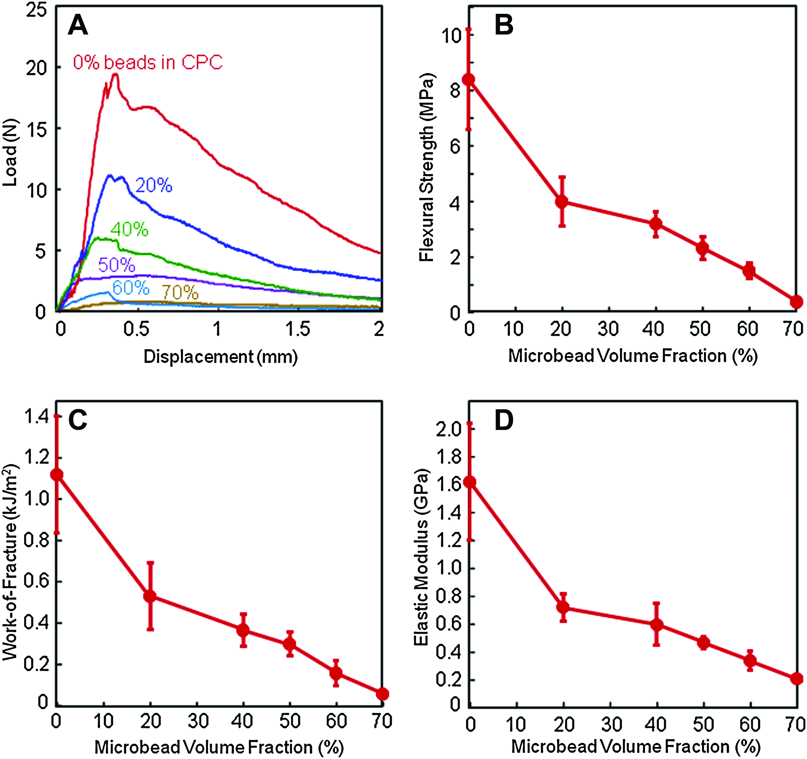
Effect of alginate microbead volume fraction on the mechanical properties of CPC composite scaffold.
Figure 2 shows the effect of fiber length on the mechanical properties of CPC. Each specimen contained 50% of hydrogel microbeads. In Figure 2A, the load-bearing capability increased when longer fibers were used to reinforce CPC. In Figure 2B, the strength of (1.8±0.4) MPa for CPC without fibers was indicated by the square symbol on the left axis. The strength increased to (4.9±0.5) MPa with 6-mm fibers, and to (6.1±0.5) MPa using 10-mm fibers (p<0.05). In Figure 2C, work-of-fracture was increased by nearly 20-fold using 10-mm fibers, compared with CPC without fibers. In Figure 2D, the elastic moduli of the CPC scaffold with fibers were significantly higher than that without fibers (p<0.05), whereas fiber length did not affect the modulus (p>0.05).
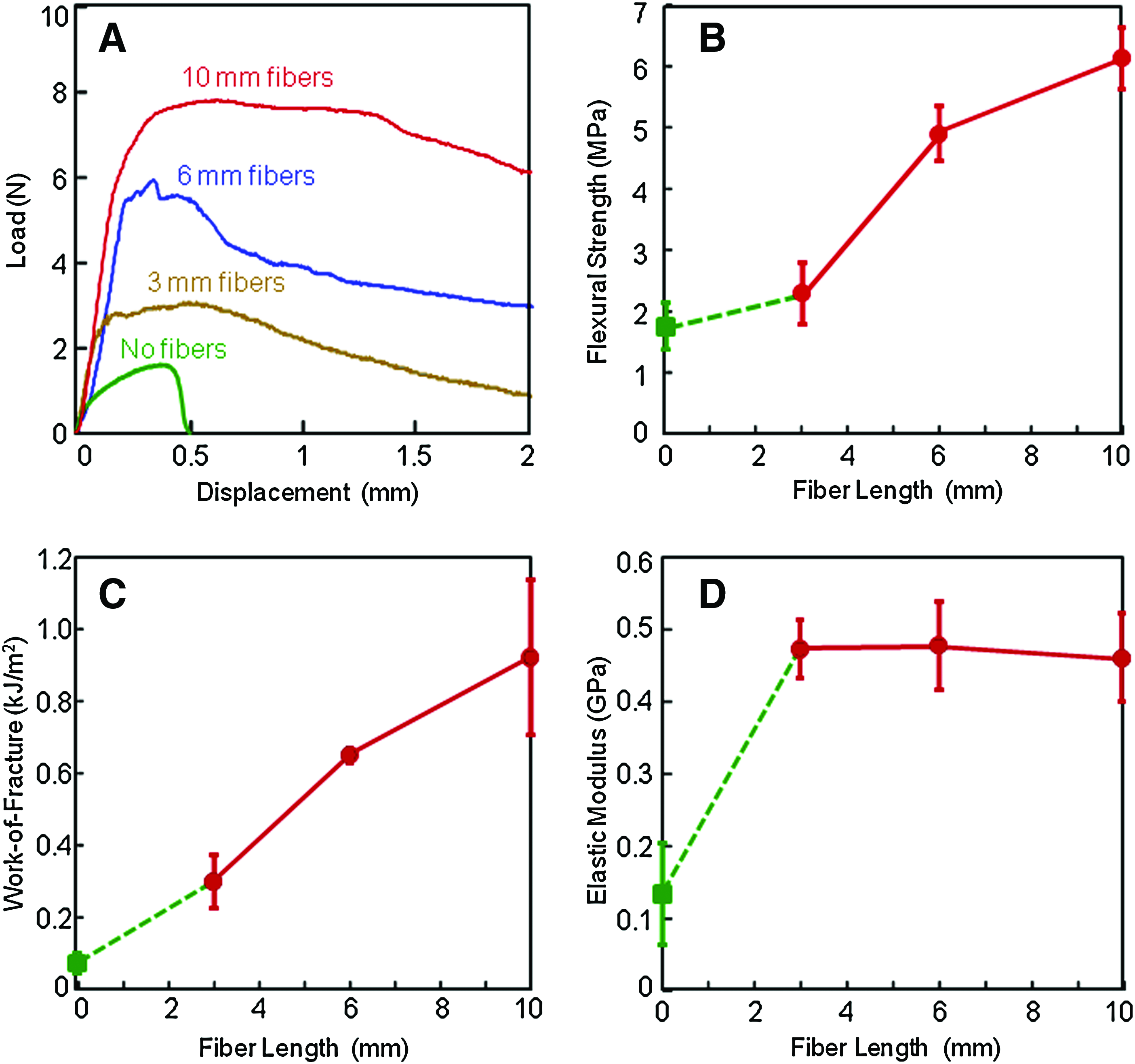
Effect of fiber length on the mechanical properties of CPC composite scaffold.
Figure 3 shows photos of live hUCMSCs (stained green) seeded on CPC-microbead-fiber disks at various initial seeding densities. hUCMSCs proliferated on CPC-microbead-fiber scaffold over time and increased rapidly in numbers. For example, specimens that received a cell seeding density of 150k became nearly confluent at day 14. Specimens that received a seeding density of 300k and 500k cells became confluent at day 8.

Photos of live cells (stained green) attaching on CPC-microbead-fiber scaffold. Each column had the same initial seeding density, which was indicated in the top photo. The culture time is labeled on the left side. The CPC specimen contained 50% microbeads and 20% fibers with 3 mm length (designated as CPC-microbead-fiber). Live cells appeared to have adhered and attained a normal, polygonal morphology on CPC-microbead-fiber scaffold. Dead cells were stained red and were very few on all disks (photos not shown). hUCMSCs proliferated on CPC-microbead-fiber specimens over time and increased rapidly in numbers. hUCMSCs, human umbilical cord mesenchymal stem cells. Color images available online at www.liebertonline.com/tea
Figure 4 plots (A) the percentage of live cells, and (B) live cell number per specimen area. Two-way ANOVA showed significant effects (p<0.05) of seeding density and culture time, with a significant interaction between these two parameters (p<0.05). In Figure 4A, at day 1, the percentage of live cells increased from 72% to 87% when the seeding density was increased from 50k to 500k. The percentage of live cells improved over time, and by day 14, it was above 95%. In Figure 4B, the live cell number per area increased rapidly with increasing the initial cell seeding density, as well as with culture time. At day 14, the live cell number per area plateaued when the initial cell seeding density was increased from 300k to 500k, as a confluent layer of cells was formed. Due to proliferation, the live cell number per area was increased from day 1 to 14 by five- to sixfold, for cell seeding densities of 50k to 300k. At an initial cell seeding density of 500k, the live cell number per area was increased from day 1 to 14 by threefold.
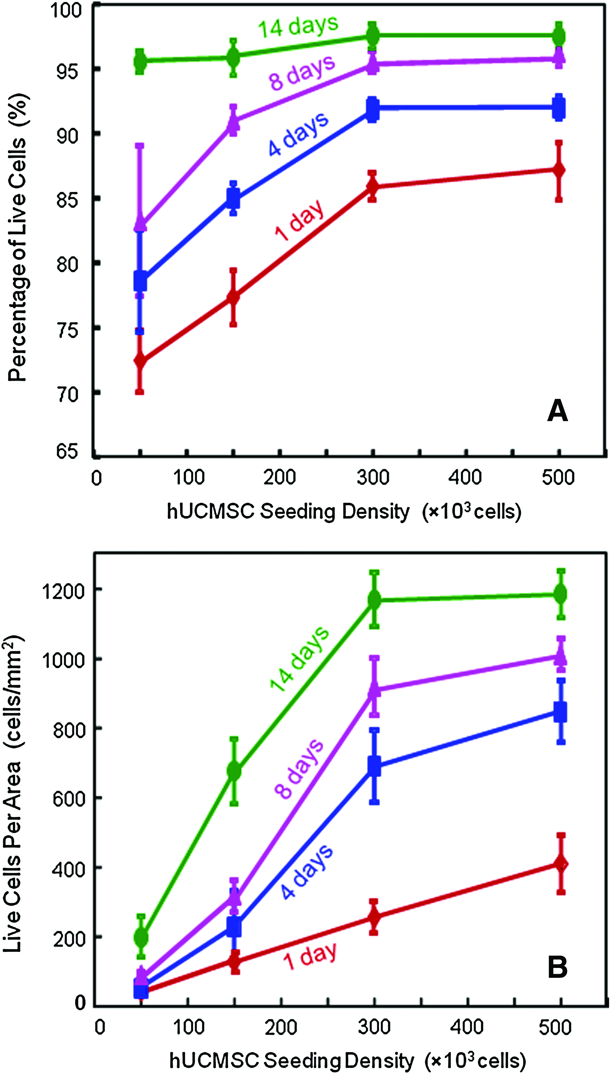
Viability of hUCMSCs attaching on CPC-microbead-fiber scaffold.
Figure 5 plots the RT-PCR results for (A) ALP, (B) OC, (C) Coll I, and (D) Runx2 gene expression. In Figure 5A, at 50k cell seeding density, the ALP was minimal at day 1 to 8, and then increased at day 14 (p<0.05). The ALP for all other seeding densities was higher at day 1. However, the highest ALP peaks were observed at day 8 for 300k and 500k seeding densities. At day 8, the ALP at a cell seeding density of 300k was 3-fold the ALP at a seeding density of 150k, and 200-fold the ALP at seeding density of 50k. In Figure 5B, OC peaked at day 14. OC expression at a seeding density of 300k was fourfold that at 50k. OC for 150k and 500k seeding densities was higher than for 50k (p<0.05). In Figure 5C, Coll I was greatly increased at day 14, and it was similar among different cell seeding densities at day 14 (p>0.05). In Figure 5D, Runx2 was increased at day 8 and 14. At day 8, Runx2 for 300k and 500k seeding densities was higher than that for 150k and 50k (p<0.05). At day 14, Runx2 at 300k seeding density was the highest among all groups (p<0.05). Runx2 at a cell seeding density of 300k was fourfold the Runx2 at 50k.
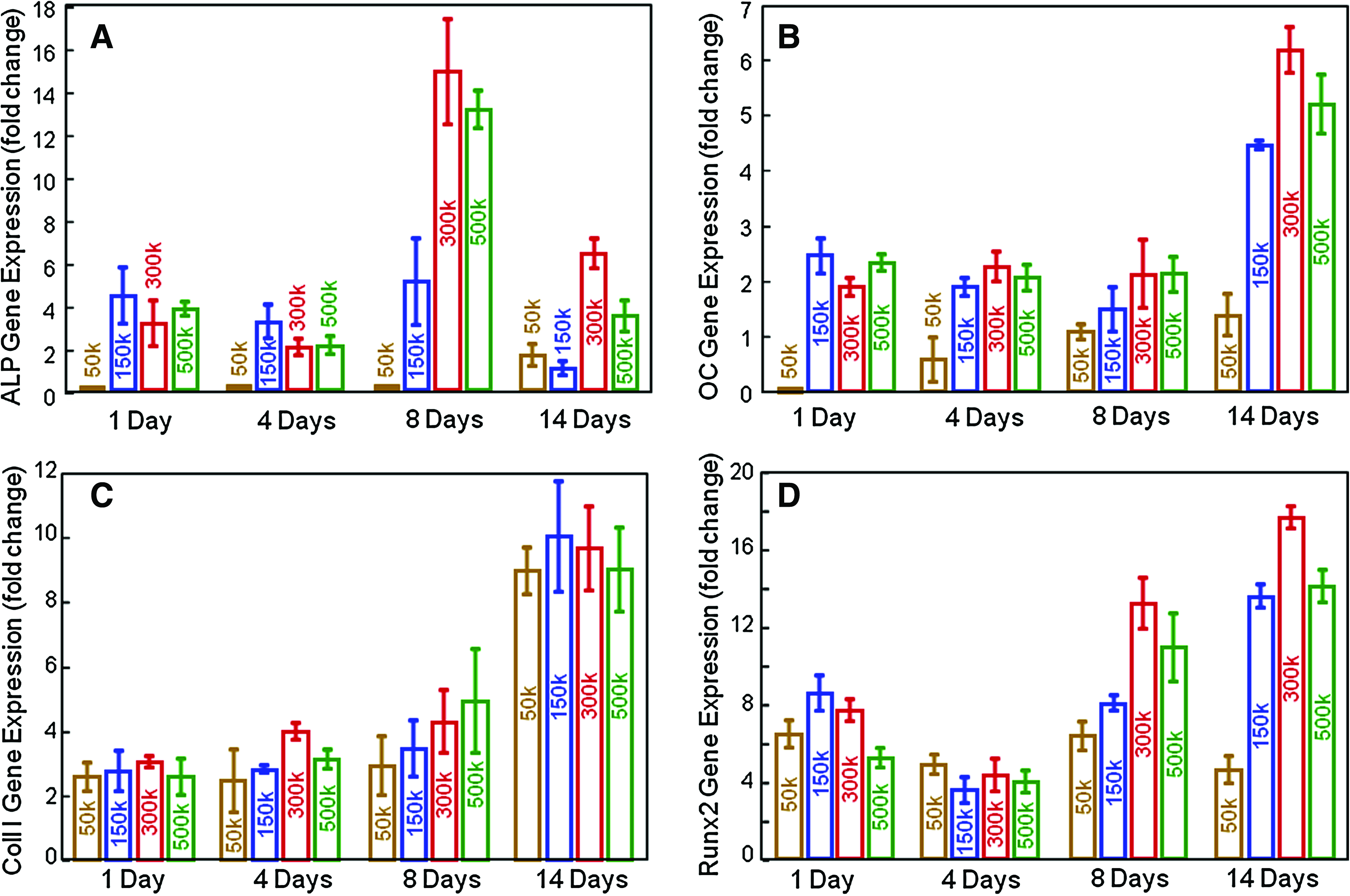
Osteogenic differentiation. The numbers in each plot indicate the different hUCMSC seeding densities on CPC-microbead-fiber scaffold, where 1k=1000. Real-time reverse transcription polymerase chain reaction on gene expression for
In Figure 6A and B, hUCMSCs were cultured on TCPS for 14 days in control media and osteogenic media, respectively. There was no ARS staining in Figure 6A, but obvious minerals in Figure 6B. This indicates that there was mineral synthesis by the hUCMSCs in the osteogenic media.
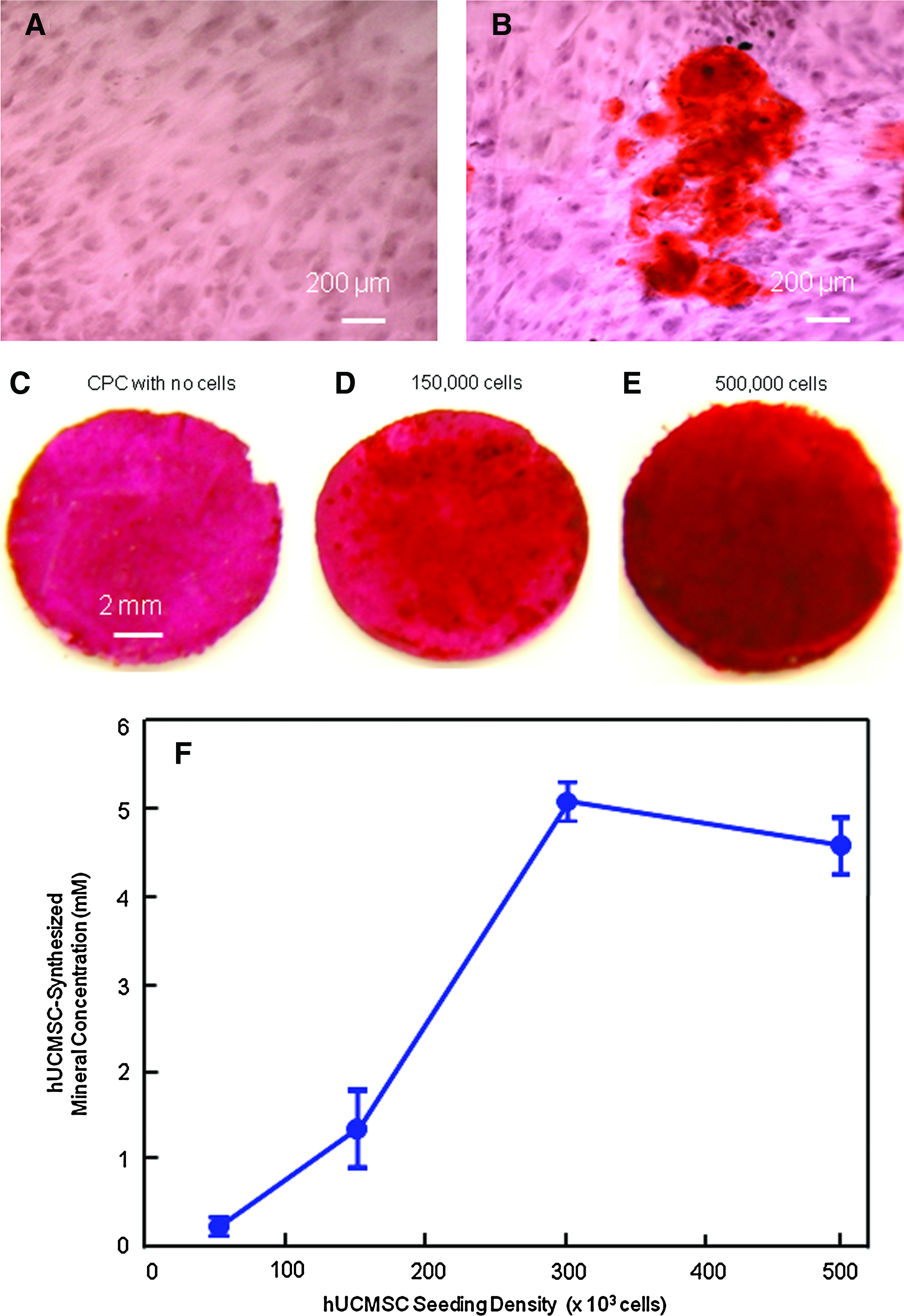
Mineral synthesis by hUCMSCs.
Since the CPC-microbead-fiber disk consisted of minerals, the disk without cell seeding in Figure 6C also stained a red color. However, when hUCMSCs were seeded on CPC-microbead-fiber scaffold for 14 days in osteogenic media, the red color became darker and thicker with increasing cell seeding density. Examples are shown in Figure 6D with 150k, and Figure 6E 500k cell seeding density. There was a layer of new mineral matrix synthesized by the cells that covered the CPC-microbead-fiber disk. The thick matrix mineralization formed on the cell-scaffold construct covered not only the top surface, but also the peripheral areas at the sides of the construct, especially at 300k and 500k seeding densities. The mineral concentration synthesized by the hUCMSCs was measured by an osteogenesis assay and plotted in Figure 6F. The mineralization increased with seeding density from 50k to 300k (p<0.05). The mineralization at seeding density of 300k and 500k was not significantly different from each other (p>0.05).
Discussion
In this study, the effects of hUCMSC seeding density on cell proliferation, osteogenic differentiation, and mineral synthesis on CPC-microbead-fiber scaffold were investigated for the first time. Various scaffold materials have been previously investigated for tissue engineering applications. Previous studies examined the effects of cell seeding density on bone tissue engineering by seeding cells on coralline hydroxyapatite, 51 poly lactide-co-glycolide (PLGA) scaffolds, 52 TCPS, 40 titanium fiber mesh, 53 PLGA-polycaprolactone (PCL) scaffolds, 54 PCL-tricalcium phosphate (TCP) scaffolds, 55 collagen gel, 41 hydroxyapatite-TCP scaffold, 42 and poly(propylene fumarate) scaffold. 43 These scaffold materials were shown to be suitable in supporting cell attachment and proliferation.41–43,51–55 Several previous studies developed composite scaffolds by combining a bioceramic (such as TCP) with a polymer (such as PCL),42,55 with the benefit of overcoming the hydrophobicity of the polymer and the brittleness of the ceramic. The purpose was to utilize the best of both worlds, by combining the mechanical toughness of the polymer with the bioactivity, hydrophilicity, and osteoconductivity of the bioceramic.
Using TTCP and DCPA, the CPC paste can be sculpted during surgery to conform to the defects in hard tissues, and then self-hardens to form resorbable hydroxyapatite. 30 In several in vivo studies, CPC was shown to have excellent osteoconductivity, was resorbed and replaced by new bone, and was highly promising for a wide range of clinical applications.31,32 In 1996, CPC was approved the Food and Drug Administration for repairing craniofacial defects in humans, thus becoming the first CPC available for clinical use. 32 However, due to its brittleness and low strength, the “use of CPC was limited to the reconstruction of non-stress-bearing bone,” 31 and “none of the indications include significant stress-bearing applications.” 32 In addition, the effect of stem cell seeding density on CPC has never been investigated. Therefore, the present study aimed at using fibers to reinforce CPC, and to study the effect of hUCMSC seeding density on CPC for the first time. The purpose was to determine the effects of hUCMSC seeding densities of 50k, 150k, 300k, and 500k. The purpose was not to investigate the effect of different materials such as the difference between CPC and TCPS. In preliminary studies, it was noticed that the hUCMSCs cultured on TCPS and on CPC for 14 days in osteogenic media had made similar amounts of minerals as examined via ARS. However, TCPS is not resorbable, whereas CPC is resorbable and can be replaced by new bone.31,32 In addition, a recent study demonstrated that CPC had excellent hUCMSC attachment and proliferation, although only a single cell seeding density was investigated. 25
The present study investigated a CPC-microbead-fiber scaffold, which could be injected or placed as a paste into a bone defect with intimate adaption to bond to neighboring bone, be shaped easily to achieve esthetics for craniofacial repairs, and then set in situ. The microbeads could deliver growth factors, and could degrade away to release the growth factors while creating macropores in CPC, a topic of future study. Previous studies reported that hygrogels had strengths of about 0.1 MPa,56,57 thus concluding that “hydrogel scaffolds…do not possess the mechanical strength to be used in load-bearing applications.” 58 Hence, the addition of hydrogel microbeads into CPC weakened the CPC (Fig. 1), as the microbeads served as voids in CPC. However, adding fibers greatly improved the strength and toughness of the CPC-microbead scaffold (Fig. 2). While the 3-mm fibers only slightly increased the strength, they raised the work-of-fracture (toughness) by fourfold, compared with CPC-microbead without fibers. A previous study showed that the CPC-microbead paste with 3-mm fibers was readily injectable through a 10-gauge needle. 26 The present study showed that fibers of 6 and 10 mm lengths increased the strength of the CPC-microbead scaffold to 5–6 MPa. CPC with these fibers could be useful in filling large defects where mechanical properties are important to avoid scaffold fracture. The fiber reinforcement mechanisms are that the fibers deflect the cracks and bridge the cracks, and consume energy during fiber pullout thereby resisting crack opening. 59 Longer fibers are more effective than short fibers or particulates at deflecting and bridging the cracks and consuming energy during fiber pullout. With 50% hydrogel microbeads, the strengths of CPC-microbead-fiber scaffolds of 5–6 MPa exceeded the reported strength of 3.5 MPa for cancellous bone, 60 and overlapped the 2–11 MPa for commercial porous hydroxyapatite implants prefabricated by sintering at high temperatures. 61 In addition, the in situ-setting CPC-microbead-fiber scaffolds had elastic moduli of about 0.4–0.5 GPa, similar to the reported elastic modulus of about 0.3 GPa for cancellous bone, although cancellous bone is much weaker than cortical bone. 62 It should be noted that these are very preliminary results, and the distribution of fibers in the CPC paste needs to be examined and improved. Therefore, short fibers of 3 mm length could be used for injectable applications, whereas longer fibers such as 10 mm could be used in a noninjectable paste for maximum reinforcement when placing the paste into large defects. The volume fraction of alginate microbeads of 50% appeared to be appropriate in maintaining good strength and toughness for the CPC scaffold. These microbeads could deliver cells and growth factors, and then could degrade to create 50% macropores in CPC.
ALP is an enzyme expressed by MSCs during osteogenesis and is a well-defined marker for their differentiation.43,63 It has been observed that the genetic expression of ALP is upregulated at an early stage of osteogenic differentiation. As the cascade of events for the differentiation proceeds, other bone markers such as OC become upregulated, whereas the ALP decreases. A number of studies have described an ALP increase and then a decrease, with an ALP peak occurring between day 4 and 16 of culture.43,63 For example, one study showed that the ALP of MSCs cultured under flow perfusion showed an ALP peak at day 8, and then the ALP decreased at day 16. 63 Another study measured the expression of ALP, which was minimal at day 1, greatly increased at day 4, and then decreased at day 8. 43 In addition, the OC peaked at a later time than ALP. A previous study observed that the OC expression peaked at day 8, later than the day 4 for ALP. 43 These results are consistent with the present study, which showed that for the hUCMSCs on the CPC-microbead-fiber scaffold, the ALP gene expression peaked at day 8 and then decreased at day 14, whereas the OC, collagen I, and Runx2 expression peaked at day 14.
Previous studies on the effects of cell seeding density fall into two groups. Group one showed that higher seeding density promoted biosynthesis, yielded higher expressions such as ALP and OC, and resulted in more bone mineralization.39,40,43,53 Group two indicated that the higher cell seeding density might not be advantageous, and an intermediate, optimal seeding density yielded the best ECM formation and tissue regeneration.41,42,51,54,55 When one starts with a low cell seeding density and gradually increase it, the cell functions and biosynthesis will be enhanced due to cell–cell interactions, 41 increased intercellular signaling via endogenous signal molecules, 43 and increased secretion by neighboring cells of ECM for other cells to attach to. This trend continues until an optimal cell seeding density is reached for a specific cell-scaffold construct. Once the cell seeding density exceeds the optimal density, contact-inhibition via gap junctional intercellular communication between adjacent cells starts to suppress cell proliferation. This, in combination with limited nutrients to be shared by many more cells, hypoxia of overly crowded cells, and insufficient waste removal, results in a decrease in cell function. If the cell seeding densities examined were lower than or up to the optimal seeding density, one would only observed an increasing trend in cell function with increasing cell seeding density. Further, a previous study showed that osteogenic differentiation such as ALP and OC gene expression at day 4 were enhanced by a lower cell seeding density, whereas mineralization at day 8 was enhanced by a higher cell seeding density. 43 Therefore, these previous studied showed that (1) if the cell seeding densities were below the optimal density, then increasing the cell seeding density resulted in an increase in cell function such as bone marker expressions and ECM production; (2) when the cell seeding density exceeded the optimal density for a specific scaffold type, a further increase in cell seeding density resulted in a decrease in cell function and tissue regeneration; and (3) the results on the effect of cell seeding density depended not only on the range of cell seeding density that was investigated in a specific study, but also on the particular bone marker and ECM component that were measured.
In the present work, the percentage of live cells was improved with increasing the cell density. At day 1, the percentage of live cells was increased from 72% at a cell seeding density of 50k, to 87% at a seeding density of 500k. Over time, the cells proliferated, the percentage of live cells increased, and the difference in the percentages of live cells shrank between different cell seeding densities. At initial cell seeding densities of 50k, 150k, and 300k, the number of live cells per area was increased by five to sixfold at day 14 compared with that at day 1. However, at a higher cell seeding density of 500k, the number of live cells per area was increased only by threefold from day 1 to 14, likely due to the formation of confluent cells with contact inhibition. The RT-PCR results showed that the 300k seeding density was the best for osteogenic differentiation of hUCMSCs on CPC-microbead-fiber scaffold. This was corroborated by mineral staining, which showed that the 300k hUCMSC density synthesized the most bone minerals. The staining on CPC without cells was subtracted from those with hUCMSC seeding. However, there might still be influence of the CPC substrate on the color observation of the cell-synthesized minerals. Therefore, while the ranking in mineral concentration in Fig. 6F should still hold, caution should be used in taking the data as absolute results.
These results showed that a low cell seeding density such as 50k resulted in a poor viability, low osteogenic differentiation, and minimal mineralization. It appeared that the cells needed each other and relied on each other to enhance attachment and viability, manifested by the cells being drawn to other cells to form clusters on CPC (e.g., B, F, and M in Fig. 3). This was further manifested by the fact that as the cell numbers increased, either by a higher initial seeding density or by cell proliferation over time, the percentage of live cells increased (Fig. 4A). This indicates that the cells survived better when the number of neighboring cells increased. Therefore, two competing factors appeared to be operative. One factor was that the cells relied on each other for cell–cell interactions and intercellular signal molecules, with more cells synthesizing more ECM to enhance each other's viability and function. This factor was dominant at low to medium cell densities. The other factor was that over-loaded cells faced contact-inhibition, limited nutrients, hypoxia, and insufficient waste removal. This factor could start to set in at high cell densities. The seeding density of 300k on CPC-microbead-fiber scaffold appeared to be optimal, yielding a high percentage of live cells, fast proliferation, the highest bone marker expression, and the most mineralization. An even higher cell seeding density of 500k resulted in lower bone marker expression and slightly less mineralization. In the present study, the cells were diluted into 2 mL of media and added to a well of a 24-well plate with CPC disks of 12 mm in diameter and 2 mm in thickness; hence, the optimal cell seeding density would change when these culture parameters change. In addition, the use of hUCMSCs for tissue engineering is still in its early stage, and several issues, including immunorejection, still need to be investigated. It is expected that for autologous applications, the future could hold great promise for those who have stored their cords at birth. For allogenic uses, tissue typing as is done with organ transplant could likely be performed when hUCMSC banks become more common in the future. The present study indicates that a higher hUCMSC seeding density is not necessarily better, and an optimal seeding density should be determined for a specific stem cell-scaffold construct to enhance cell proliferation, differentiation, and ECM synthesis.
Conclusions
This study investigated the effects of hUCMSC seeding density on CPC for the first time. hUCMSCs proliferated well on CPC scaffold containing alginate hydrogel microbeads and degradable fibers. The CPC-microbead-fiber scaffold had mechanical strength and elastic modulus that approximated those of cancellous bone. While a short fiber length of 3 mm could be used for reinforcement of injectable CPC, longer fibers with lengths such as 10 mm could provide higher reinforcement for noninjectable CPC to be placed in large defects. Adequate strength and toughness were achieved for fiber-reinforced CPC with 50% of microbeads, which could deliver cells/growth factors and then degrade to create 50% macroporosity in CPC. When the cell seeding density was increased from 50k to 300k, the cell proliferation, osteogenic differentiation, and bone mineral synthesis also increased. However, when the seeding density was further increased to 500k, the bone marker gene expression and mineralization slightly decreased. The seeding density of 300k on CPC-microbead-fiber scaffold appeared to be optimal, yielding a high percentage of live cells, fast proliferation, the highest bone marker expression, and the most mineralization. The stem cell-seeded CPC-fiber scaffold with excellent osteodifferentiation and mineralization is promising for orthopedic and craniofacial applications. Further study is needed in animal models to investigate bone regeneration in vivo via CPC constructs laden with various stem cell densities.
Footnotes
Acknowledgments
We are indebted to Drs. L.C. Chow and S. Takagi at the Paffenbarger Research Center, and Dr. Carl G. Simon at the National Institute of Standards and Technology for discussions and help. This study was supported by NIH R01 grants DE14190 (H.X.), Maryland Stem Cell Fund (H.X.), and the University of Maryland Dental School.
Disclosure Statement
No competing financial interests exist.