
Abstract
Tissue-engineered heart valves (TEHV) have been proposed as a promising solution for the clinical needs of pediatric patients. In vivo studies have shown TEHV leaflet contraction and regurgitation after several months of implantation. This has been attributed to contractile cells utilized to produce the extracellular matrix (ECM) during TEHV culture. Here, we utilized such cells to develop a mature ECM in a fibrin-based scaffold that generates commissural alignment in TEHV leaflets and then removed these cells using detergents. Further, we evaluated recellularization with potentially noncontractile cells. A tissue-engineered leaflet model was developed with mechanical anisotropy and tensile properties comparable to an ovine pulmonary valve leaflet. No change in tensile properties occurred after decellularization using 1% sodium dodecyl sulfate and 1% Triton detergent treatment. Cell removal was verified by DNA quantitation and western blot analysis for cellular proteins. Histological and scanning electron microscope imaging showed no significant change in the ECM organization and microstructure. We further tested the recellularization potential of decellularized leaflets by seeding human mesenchymal stem cells (hMSC) on the surface of the leaflets and evaluated them at 1 and 3 weeks in two culture conditions. One medium (M1) was chosen to maintain the MSC phenotype while a second medium (M2) was used to potentially differentiate cells to an interstitial cell phenotype. Cellular quantitation showed that the engineered leaflets were recellularized to the highest concentration with M2 followed by M1, with minimum cell invasion of decellularized native leaflets. Histology showed cellular invasion throughout the thickness of the leaflets in M2 and partial invasion in M1. hMSC stained positive for MSC markers, but also for α-smooth muscle actin in both media at 1 week, with no presence of MSC markers at 3 weeks with the exception of CD90. These results show that engineered leaflets, while having similar tensile properties and collagen content compared to native leaflets, have better recellularization potential.
Introduction
While in vivo outcomes of TEHV have been meaningful in developing the technology, the primary limitation to long-term success has been a shortening of the leaflets, which has been attributed to tissue contraction induced by the transplanted cells.12,13 TEHV made from synthetic polymers have shown mild-to-moderate regurgitation at explants. 9 In a study by Gottlieb et al., this was most obvious after implantation for 6 months. 9 TEHV made from fibrin have also shown similar results. In a study by Flanagan et al., TEHV seeded with autologous smooth muscle cells showed shortened leaflets within 4 weeks after implantation. 13 Similar results were also observed in our investigation with using a fibrin-based TEHV seeded with dermal fibroblasts, where valve functionality was compromised at 4 weeks and lost by 8 weeks. 12 In this study, we also demonstrated recruitment of putative progenitor cells (CD44 positive and CD45 negative) that were not obviously contractile (smooth muscle actin [SMA] negative) within the leaflet tissue after 8 weeks of implantation, 12 indicating potential for recellularization of a decellularized TEHV with noncontractile cells.
Overall, the in vivo studies to date clearly show that while in vitro TEHV development requires tissue cells to create the extracellular matrix (ECM), there is a clear need to remove these cells in a mature TEHV before implantation to obviate tissue contraction and achieve recellularization with more quiescent cells similar to those found in normal heart valve leaflets. This problem is most pronounced using fibrin as the scaffold, where cell-induced compaction of fibrin gel to induce commissural alignment has been exploited.14,15 While having mechanical anisotropy in engineered leaflets is advantageous, the cell-induced compaction that induces alignment in vitro can also lead to overcompaction and shortening of the leaflets in vivo, as these transplanted cells remain in a contractile phenotype. 12 To this end, we investigated decellularization of engineered valve leaflets after in vitro maturation to remove the fibroblasts while retaining the functional ECM. While this is a new approach for TEHV, 16 decellularization of native valves has been investigated.17–21
A decellularized TEHV potentially gives excellent initial performance; however, nonviable tissue valves are prone to degradation and calcification, especially in pediatric patients. 22 To address this issue, recellularization of decellularized native valves has been extensively investigated.18,19,23,24 While several studies have been successful, overall recellularization has shown to be challenging, requiring 4–6 weeks in vitro culture. 17 Here, we also evaluated the recellularization potential of engineered leaflets in comparison to native leaflets with human mesenchymal stem cells (hMSC) and the effects of invading hMSC on the engineered leaflets. hMSC have been studied extensively as a cell source for cell therapy25–27 and engineered tissues, including heart valves,8,9,28–30 because of their multilineage differentiation potential31–33 and potential as an allogeneic cell source.34,35 Research has also shown that MSC differentiation can be controlled to potentially create noncontractile cells, 36 which would serve as an ideal cell source to mitigate leaflet contraction seen previously with implanted TEHV.9,12,13
Materials and Methods
Cell culture
Neonatal human dermal fibroblasts (nhDF; Clonetics) were maintained in a 50/50 mixture of the Dulbecco's modification of Eagle's medium and Ham's F12 cell culture medium (DMEM/F12; Cellgro) supplemented with 15% fetal bovine serum (FBS; Thermo-Fisher Scientific), 100 U/mL penicillin, and 100 μg/mL streptomycin. Cells were incubated at 37°C in 100% humidity and 5% CO2, passaged at ∼90% confluency, and harvested for use at passage 9. hMSC (Lonza) were maintained in mesenchymal stem cell basal medium (MSCBM) (Lonza) and used at passage 5. hMSC were characterized by immunostaining for known MSC markers CD29, 44, 90, and 105 (data not shown). hMSC were also stained for αSMA before seeding on decellularized tissue and found to be negative, and their trilineage potential was characterized by differentiation to osteoblasts, adipocytes, and chondrocytes (data not shown).
Engineered leaflet preparation and culture
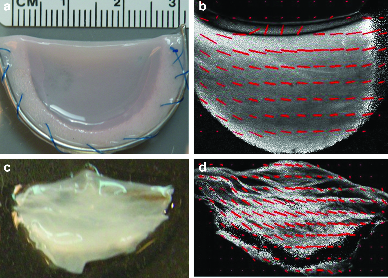
nhDF-seeded fibrin gel was formed by adding thrombin (Sigma) and calcium chloride in 20 mM HEPES-buffered saline to a suspension of nhDF in fibrinogen (Sigma). All components were kept on ice before mixing. The final component concentrations of the cell suspension were as follows: 4 mg/mL fibrinogen, 1.1 U/mL thrombin, 5.0 mM Ca2+, and 1 million cells/mL. Cell suspensions were mixed and poured in a six-well tissue culture plastic plate. In each well, a leaflet mold was previously placed, which consisted of a C-shape 312-stainless (Mc Master Inc.) wire attached to a porous polypropylene scaffold (Small Parts, Inc.) to create an attachment for the fibrin gel (Fig. 1a). The six-well plate was pretreated with 5% Pluronic F-127 (Sigma) solution for 1 h.
Photographs and polarized light images of the engineered leaflet
After injecting 5 mL of the fibrin-forming cell suspension into the mold cavity, the molds were placed in a humidified incubator and maintained at 37°C, 5% CO2 for 15 min to allow for gelation. Subsequently, 5 mL of DMEM supplemented with 10% FBS, 100 U/mL penicillin, 100 μg/mL streptomycin, 2 μg/mL insulin, 50 μg/mL ascorbic acid, and 1×nonessential amino acids (Cellgro) was added. The medium was completely changed three times per week. One day after casting, the molds were detached from the well, and six molds were transferred to a larger jar with 100 mL of supplemented DMEM and incubated on a shaker for 5 weeks. The total culture duration was selected based on prior studies.14,15 The final dimensions of each leaflet were 15 mm in circumferential width, ∼10 mm in radial length, and ∼300 μm in thickness.
Decellularization
After 5 weeks of culture, leaflets were rinsed in phosphate-buffered saline (PBS) and incubated on a shaker for 24 h with 1% sodium dodecyl sulfate (SDS; Sigma). The SDS solution was changed three times, at 30 min, 1 h, and 4 h. The samples were rinsed in PBS and incubated with 1% Triton X-100 (Sigma) for 30 min. The leaflets were extensively washed with PBS for 24 h and incubated in deoxyribonuclease enzyme (Sigma) in the DMEM supplemented with 10% FBS for 4 h. After decellularization treatment, samples were fixed in 4% paraformaldehyde for histological analysis or seeded with hMSC for recellularization studies. Similarly, fresh ovine pulmonary valve leaflets were harvested and identically treated to create decellularized native leaflets.
Uniaxial tensile testing
Tissue strips cut from each leaflet sample (engineered and native) of dimension 2×10 mm were tested for tensile properties in both the circumferential and radial directions. The thickness of each strip was measured using a 50 g-force probe attached to a displacement transducer. Tissue strips were placed in compressive grips, attached to the actuator arm and load cell of an Instron MicroBionix (Instron Systems), and straightened with an applied load of 0.005 N. This position was used as the reference length of the strip. After six cycles of 0%–10% strain conditioning at 2 mm/min, strips were stretched to failure at the same rate. True strain was calculated based on the natural log of the tissue length divided by the reference length. The stress was calculated as force divided by the initial cross-sectional area. The tangent modulus (E) was determined as the slope of the linear region of the stress–strain curve before failure. The peak stress was defined as ultimate tensile strength (UTS).
Histology, fiber alignment imaging, and scanning electron microscope imaging
Leaflet circumferential strips were fixed in 4% paraformaldehyde, embedded in OCT™ (Tissue-Tek), and frozen in liquid N2. Sections of 9-μm thickness were stained with Lillie's trichrome and picrosirius red stains. Images were taken at 10×magnification using a color CCD camera. For picrosirius red-stained sections, images were taken with the sections placed between crossed-plane polarizers. Fiber alignment was measured using a polarized light imaging method. 37
Samples for scanning electron microscope (SEM) imaging were fixed using a modified Karnovsky's solution of 0.1 M sodium cacodylate containing 2% paraformaldehyde (PFA), 2.5% glutaraldehyde, and 2 mM CaCl2 for 3 h at room temperature with continual mixing. Samples were postfixed with 1% OsO4 in cacodylate buffer for 1 h, and then dehydrated in a graded ethanol series. Samples were freeze-fractured in liquid N2 to expose the cross-section region. Specimens were further dehydrated with absolute ethanol, critical point-dried, mounted on aluminum stubs, and sputter-coated with platinum to an approximate thickness of 2 nm. Images were acquired with a Hitachi S-900 Field Emission SEM at 3×10⁵ keV.
Collagen and cell quantification
The collagen mass content was quantified using a hydroxyproline assay previously described 38 assuming 7.46 mg of collagen per 1 mg of hydroxyproline. The tissue sample volume was calculated using the measured length, width, and thickness of the strips (as described above for uniaxial testing). Collagen concentration was calculated as collagen mass per unit volume. The cell content was quantified with a modified Hoechst assay for DNA assuming 7.7 pg of DNA per cell. 39 The cell concentration was calculated as the number of cells per unit volume.
Recellularization
Subsets of native and engineered leaflets were seeded with MSC to evaluate potential for recellularization and a change in properties due to invading cells. One side of each leaflet was seeded with 50,000 cells to form a subconfluent monolayer. The leaflets were incubated for additional 1 and 3 weeks in the supplemented DMEM (M2, same as used for growing leaflets) with potential to differentiate the cells and in the MSCBM (M1, same as used for hMSC expansion) to potentially maintain the MSC phenotype. Harvested leaflets were evaluated for mechanical, biochemical, and histological assessment.
Immunostaining
Histology sections of engineered leaflets were stained for αSMA (Sigma, A5228), CD29 (Stemgent, 09-0025), CD44 (R&D Systems, 965616), CD90 (R&D Systems, 965609), and CD105 (Abcam, ab49228). Sections stained for CD105 were treated with 0.1% Triton X before blocking to expose antigens. All samples were blocked with 5% normal donkey serum for 1 h before incubation in the primary antibody at 1:200 dilutions overnight. Samples were then stained with a cye2 fluorescent-labeled secondary antibody (Jackson Immuno lab) at 1:200 dilution and nuclear Hoechst stain. Sections were imaged at 20×magnification using an upright microscope (Leica).
Western blotting
Western blotting was used to assess remnant cellular protein after decellularization. Tissue strips were flash-frozen in liquid N2 at harvest. The proteins were extracted as previously described. 40 For each sample, 20 μg of total protein was boiled in a reducing sample buffer and separated by SDS–polyacrylamide gel electrophoresis. The proteins were transferred to nitrocellulose (Whatman) using a wet transfer buffer (10% methanol, 2.2 g/L CAPS, pH 11). The blot was incubated in a blocking solution (5% dry milk and 0.1% Tween-20 in PBS) for 1 h and then incubated with a primary antibody overnight at 4°C (mouse monoclonal anti-β-actin; Sigma, at 1:1000; rabbit monoclonal β-2-macrogloublin antibody; Abcam, at 1:1000). The blot was then probed with horseradish peroxidase-conjugated secondary anti-IgG (Jackson Immuno Lab) at a dilution of 1:5000 and developed using enhanced chemiluminescence.
Statistics
For all experiments, n=3 or higher sample number was used. Statistical significance of differences between groups was determined using Student's t-test for two treatments and one-way analysis of variance for more than two treatments with the Tukey post hoc test in GraphPad Prism® software for Windows. Any reference to a difference in the Results and Discussion sections implies statistical significance at the level p<0.05. In all cases, where the difference was significant with nonoverlapping error bars, no symbols are used. Where the difference was statistically significant, but with overlapping error bars, a paired symbol is used.
Results
Tissue-engineered leaflet model
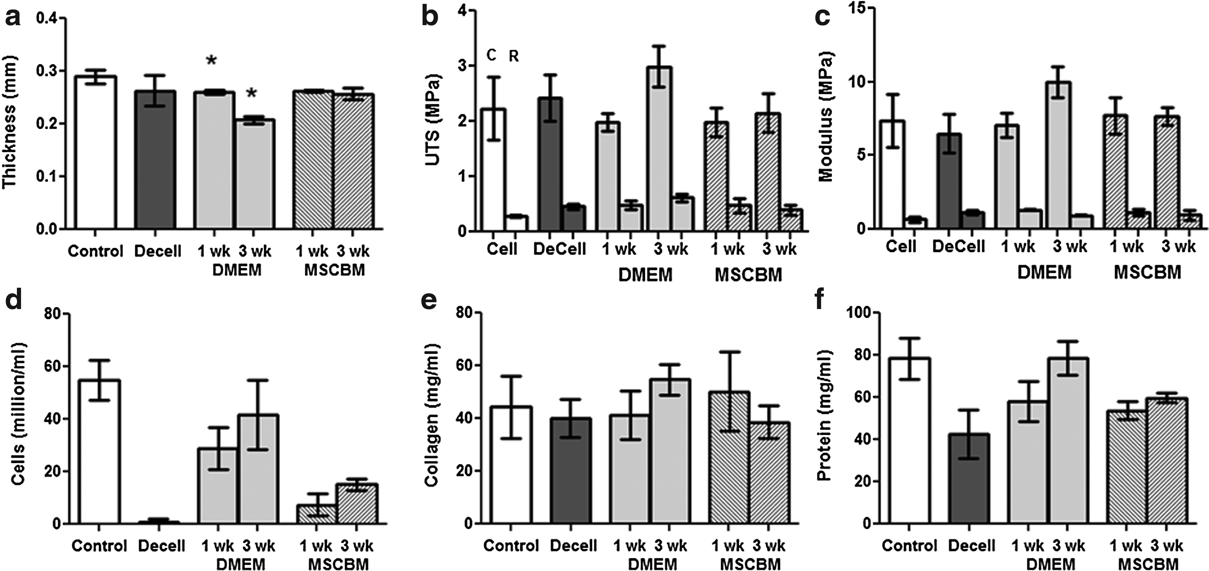
The tissue-engineered leaflet model was developed to study decellularization and recellularization of engineered tissue. Leaflets were fabricated by casting fibrin gel with entrapped nhDF in a C-shape mold placed in a well of six-well plate. The free edge of the gel compacted freely to generate a tissue in the shape of a leaflet (Fig. 1a) with dimensions matching native leaflets (Fig. 1c). Polarized light imaging was used to evaluate the fiber alignment in engineered and native leaflets and showed strong anisotropy of fibers with dominant alignment in the circumferential direction (Fig. 1b, d). Engineered and native leaflets were also evaluated for mechanical properties in both the circumferential and radial directions. There was no difference in the thickness of engineered leaflets (294±80 μm) from native leaflets (336±81 μm) (Fig. 2a). The UTS of engineered leaflets (C: 2.2±1.1 MPa, R: 0.28±0.05 MPa) was also not different from native leaflets (C: 2.0±0.2 MPa, R: 0.35±0.1 MPa) in both the circumferential and radial directions (Fig. 2b). However, the modulus was higher for engineered tissue in the circumferential direction (C: 7.4±3.5 MPa, R: 0.65±0.28 MPa) compared to native tissue (C: 3.2±0.8 MPa, R: 1.3±0.24 MPa) (Fig. 2c). The mechanical anisotropy of engineered and native leaflets was calculated by taking the ratio of the modulus in the circumferential and radial directions. There was no difference between the engineered (4.99±1.6) and native (4.2±2.6) leaflets.
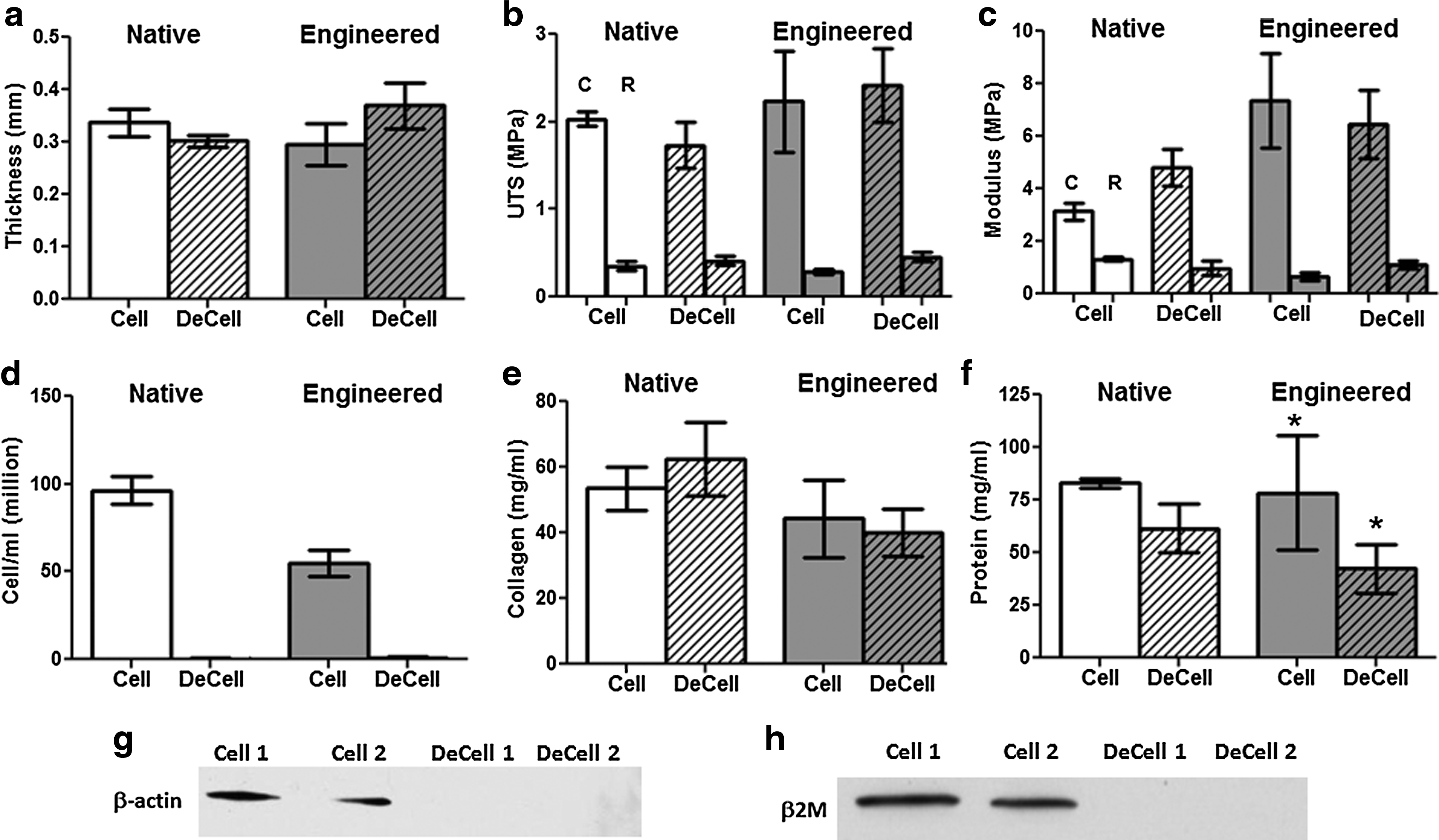
The cell concentration was lower in engineered leaflets (55±8 million/mL) compared to native leaflets (97±8 million/mL) (Fig. 2d). However, there was no difference in collagen and total protein content (Fig. 2e, f).
Effect of decellularization on tissue properties
No change in thickness, UTS, modulus, or mechanical anisotropy occurred after decellularization of engineered or native leaflets (Fig. 2a–c). Cellular DNA was reduced by 99.98% in engineered leaflets and 99.99% in native leaflets after decellularization. The total reduction in tissue protein concentration was 36 mg/mL in engineered leaflets and 21 mg/mL in native leaflets, reflecting removal of cellular proteins. No change in collagen content was observed after decellularization. Complete cellular removal was further verified by western blotting for the cellular membrane protein β-actin and MHC type 1 subprotein β-2-macrogloublin, neither of which were detectable after decellularization (Fig. 2g, h).
Collagen microstructure and content after decellularization
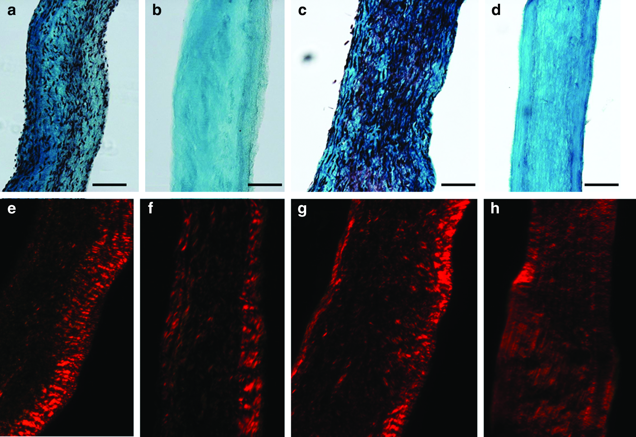
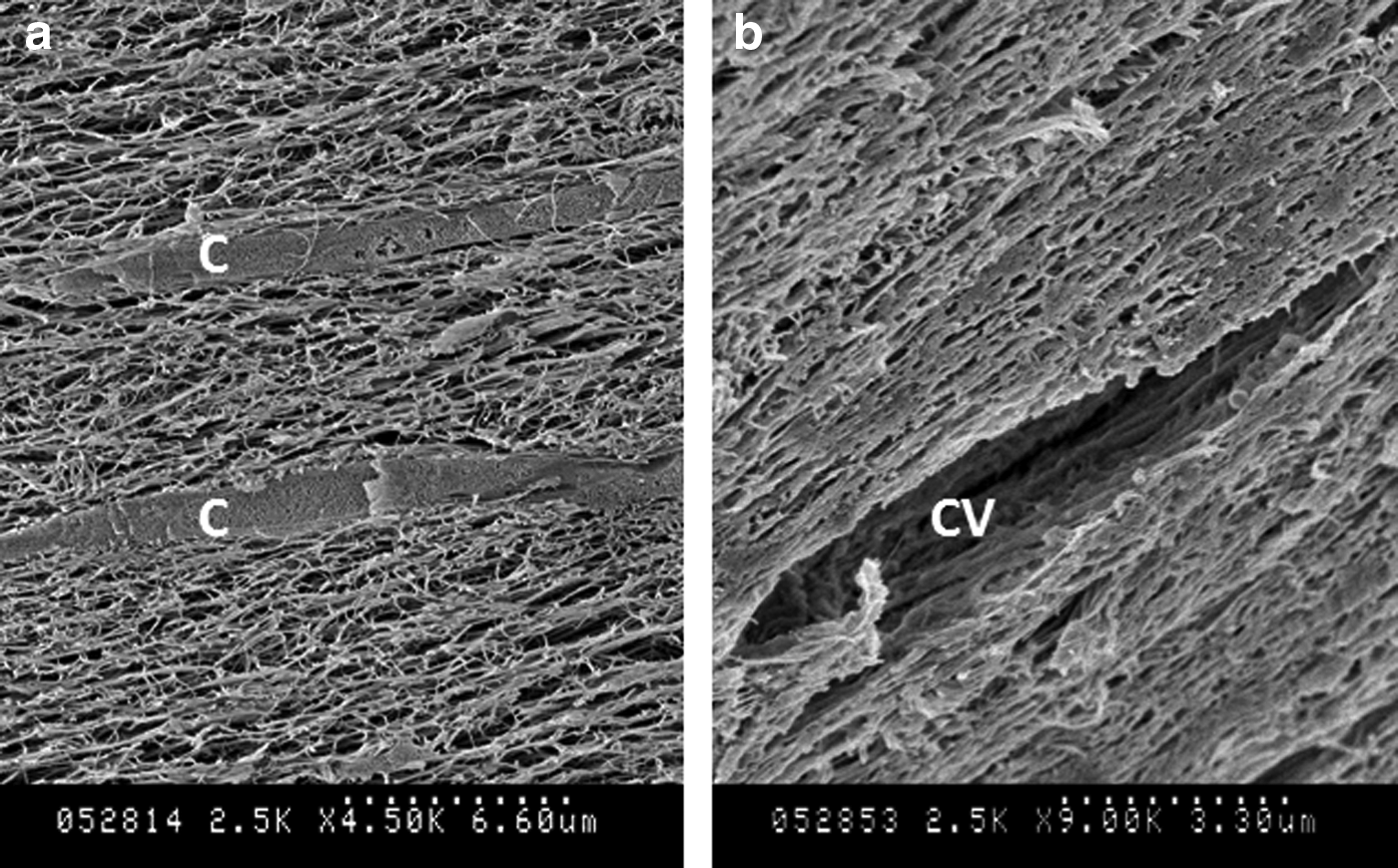
Trichrome and picrosirius red staining were used to assess organizational changes in the ECM after decellularization. Trichrome staining showed removal of cells after decellularization (Fig. 3a–d) with picrosirius red staining showing a little change in brightness (Fig. 3e–h), which indicates bundling and maturity of the collagen fibers.41,42 The microstructure was further evaluated using SEM imaging. Figure 4a shows nhDF surrounded by ECM fibers in engineered leaflets. After decellularization, no visible change in fiber orientation was observed; however, cavities with the size of a cell were evident (Fig. 4b). These cavities were less frequent than the number of cells, suggesting collapse of the fiber network after decellularization and/or SEM preparation of samples. There is some change in microstructure apparent in the decellularized sample, where the fibers appear aggregated relative to the untreated sample. However, this may represent an artifact of sample shrinkage during critical point drying and/or charging during imaging, both of which were more pronounced for the decellularized sample, rather than a true change in microstructure.
Trichrome and Picrosirius red images of native leaflet before
Scanning electron microscope (SEM) image of TEHV leaflet
Engineered leaflet recellularization
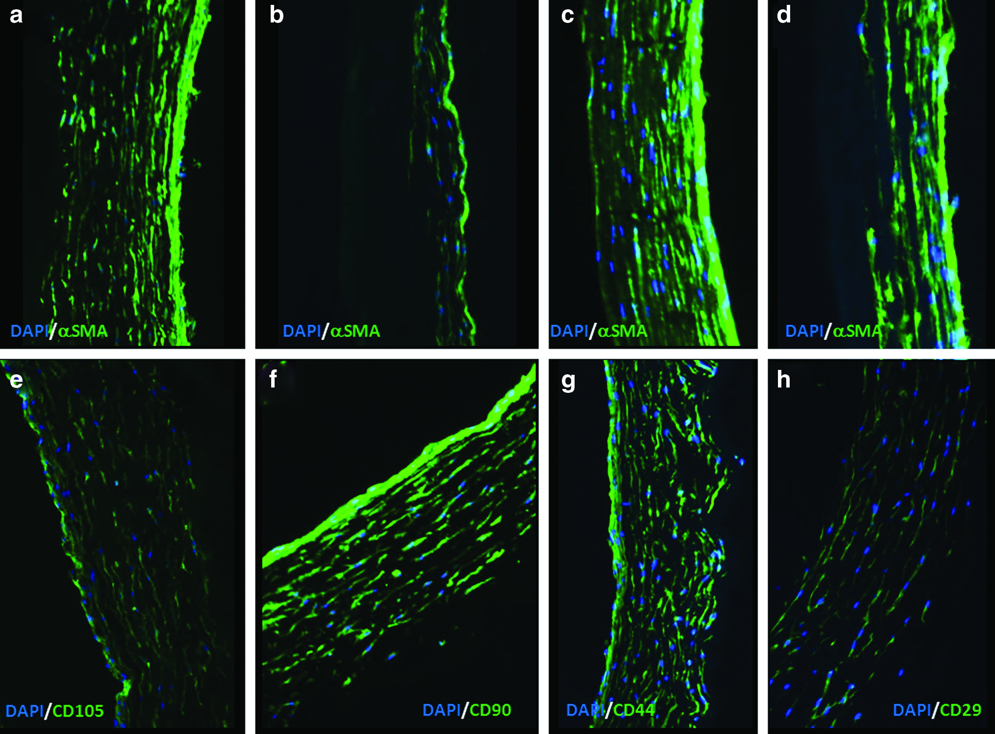
Decellularized leaflets seeded with hMSC were harvested after 1 and 3 weeks of culture. Uniform invasion of cells throughout the thickness of the leaflets was observed with DMEM at 1 and 3 weeks (Fig. 5a, c). However, in MSCBM, only partial invasion was seen at 3 weeks (Fig. 5b, d). The invading cells had elongated in the same orientation as ECM fibers (circumferential) with positive staining for αSMA in both DMEM and MSCBM. Immunostaining for MSC markers showed positive staining for CD29, CD44, CD90, and CD105 in both media at 1 week. Figure 5e–h shows stained sections for leaflets in the DMEM at 1 week. However, at 3 week, in both media, cells were only positive for αSMA and CD90 and were negative for calponin, CD29, CD44, and CD105 (Fig. 6).
αSmooth muscle actin immunostaining of recellularized leaflet after

Immunostaining of recellularized leaflets at 3 weeks with
No change in width, length, or thickness of the samples was observed after 1 week in both culture conditions (DMEM and MSCBM) (Fig. 7a). However, at 3 weeks, leaflet thickness in the DMEM decreased by 21% (Fig.7a). Samples were mechanically tested, and no difference in UTS or modulus existed after 1 week of recellularization. At 3 weeks, only in the DMEM, both circumferential UTS and modulus increased (Fig. 7b, c). Biochemical analysis showed no difference in collagen or protein concentrations at 1 week, with increased values in the DMEM after 3 weeks (Fig. 7e, f). After 1 week in the DMEM, the cell concentration in the engineered leaflet was 53% (29±8 million/mL) compared to the cellularity before decellularization (55±8 million/mL) and 30% compared to a native leaflet (96±8 million/mL). After 1 week in the MSCBM, the cell concentration in the engineered leaflet was 13% compared to the cellularity before decellularization. The cellularity increased in both media after 3 weeks (Fig. 7d).
Native leaflet recellularization
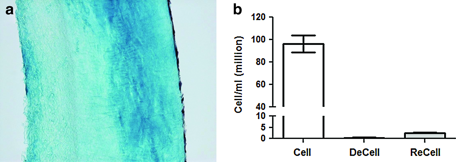
Similar to the engineered leaflets, native leaflets were also decellularized and seeded with hMSC and cultured for 1 week in the DMEM. Histology revealed very few cells invading the tissue (Fig. 8a). DNA quantitation showed the cell concentration at 2% of the native value after 1 week of culture (Fig. 8b).
Discussion
To date, TEHV based on synthetic and biological scaffolds have been implanted in ovine models. Long-term implantation has resulted in regurgitation caused by sustained leaflet contraction.9,12,13 Excessive contraction of engineered tissues generally and engineered valve leaflets specifically has been reported for both synthetic and biopolymer scaffolds. To mitigate excessive contraction, several in vitro studies have investigated the dependence on cell type, 43 scaffold stiffness, 44 and use of drugs to inhibit contraction.45–47 While these studies have been promising in vitro, significant challenges exist in their in vivo application. Another potential approach is to remove the contractile cells, which deposit the ECM to convert the scaffold into a tissue, once the TEHV has matured in vitro and subsequently replaced them with phenotypically noncontractile cells. Here, we have investigated this approach by decellularizing engineered valve leaflets developed from fibrin gel entrapped with fibroblasts, which yields commissurally aligned tissue in vitro with minimal remnant fibrin. While a decellularized TEHV could be implanted to allow for host cell recellularization, a lack of cells postimplant to repair and remodel the ECM during valvular cycles could lead to tissue failure via ECM deterioration. Hence, we further evaluated recellularization potential in vitro by seeding hMSC on the surface of decellularized engineered leaflets and assessing cellular invasion into the tissue after 1 and 3 weeks.
Overall, this study shows that tissue-engineered valve leaflets grown in vitro can be decellularized without substantial alteration of microstructure or loss of tensile mechanical properties. Research of engineered arteries has already shown that engineered tissue can be decellularized without major microstructural changes, with promising in vivo performance.48,49 The total protein lost in engineered leaflets was higher than native leaflets. Since no change in the collagen concentration occurred, the engineered leaflets were immunostained for fibrin, laminin, and fibronectin after decellularization, showing some loss of remnant fibrin and laminin (data not shown). While not quantitated, the loss of remnant fibrin and other cell-produced noncollagenous proteins could account for the relatively higher protein loss in the engineered leaflets.
Although decellularizing TEHV to create off-the-shelf valve replacements is a new idea, 16 several groups have used a similar approach with native valves. However, the primary reason for decellularizing the native valve has been to remove immune-reactive cells. 50 Studies with human, porcine, and ovine valves have been performed.18,20,21,24 Extensive in vitro and in vivo immune-reactive properties have been evaluated to ensure that the ECM in such xenografts is immune compatible. 50 Despite much effort, a study reported by Simon et al. showed death of three pediatric patients (75%) in a clinical trial of decellularized porcine valve implants. 51 In a more recent study, Ruffer et al. reported early intervention in six patients (38%) after implantation of decellularized porcine valves. 52 A larger trial with decellularized porcine valves in 93 patients showed freedom from conduit failure to be 60% at 24 months and below 25% after 36 months. 53 In this study by Perri et al., there was no single cause of failure, but histology showed the presence of inflammatory giant cells in the explanted valves. While immune compatibility of decellularized native valves can potentially be resolved, another challenge with tissue valves in pediatric patients is the elevated risk of ECM degradation and calcification. 22 Decellularized porcine valves implanted in the ovine pulmonary position were shown to have a 50% reduction in tensile properties, acid-soluble collagen content, and sulfated GAGs after 3 months in vivo. 18 The first report of a decellularized TEHV recently appeared, 16 wherein TEHV were made from PGA mesh infiltrated with a suspension of ovine vascular cells in a fibrin-forming solution and then cultured in a bioreactor, as previously described. 10 Decellularization was achieved using overnight treatment in PBS containing Triton X-100 and sodium deoxycholate, followed by treatment with Benzonase. A thinning of the leaflets occurred in their study, unlike our study. Similar to our study, there was no change in UTS, modulus, collagen content, or gross histological appearance.
To address the potential failure of decellularized valves both in vitro and in vivo recellularization has been investigated. The hypothesis is that once a valve is fully recellularized, it will be able to repair, remodel, and grow with the patient similar to a healthy host valve. To date, recellularization of decellularized native leaflets has been poor and has required 4–6-week bioreactor culture period to achieve substantial recellularization. 17 With decellularized porcine valves, only 2% of the cell concentration compared to the native valve was achieved after 4 weeks of static culture. 19 Other recellularization approaches include dynamic in vitro culture20,54 and conjugating decellularized valve tissue with CD133 antibody to recruit cells in vivo. 18 It remains to be seen how recellularized native-valve leaflets remodel long-term in vivo.
In this study, we evaluated the recellularization potential of tissue-engineered leaflets after 1 and 3 weeks of in vitro culture. The recellularized leaflets were incubated in our standard fibrin-based TEHV culture medium (DMEM, M2) and standard hMSC culture medium (MSCBM, M1). M1 was utilized to potentially maintain the MSC phenotype as the serum in MSCBM has been selected by the vendor to maintain the MSC phenotype in a culture flask. In addition, M2 was chosen to evaluate potential differentiation of MSC into interstitial cells as well as their invasion. In both media, histology and DNA quantitation showed much higher cell invasion in engineered leaflets than native leaflets at 1 week. In the best invasion conditions for these studies, which occurred with M2, we observed recellularization of decellularized engineered leaflets at 30% compared to 2% with decellularized native leaflets, relative to the ovine leaflet cell concentration, after 1 week. After 3 weeks, the cellular concentration was at 16% in M1 and at 43% in M2. While the exact cause of this difference was not investigated, it is hypothesized that the more homogenous ECM and lower collagen concentration in the engineered leaflets allowed for better invasion than the native leaflets. Considering no change was observed in tensile properties or dimensions of the engineered leaflets after 1 week of recellularization, the engineered leaflets provide a significant advantage over native decellularized leaflets for clinical application. At 1 week, invading cells were positive for MSC markers in both media, even though the level of invasion and resulting cell concentrations were different. The cells were also positive for αSMA, indicating potential onset of differentiation. At 3 weeks, cells were negative for CD29, 44, and 105. Further testing showed that while invading cells expressed αSMA, they were negative for calponin at 3 weeks. The results show that while the medium type influenced the level of cellular invasion, which generally reflects migration and proliferation, neither type prevented the differentiation of MSC. At 3 weeks, tissue thickness decreased in M2 by 21%, indicating potential compaction. While no shortening in the area of the leaflet was seen, further assessment of the traction/contractile properties of the invading cells in both media is warranted.
We have shown in previous studies of tissue similarly formed from nhDF in fibrin gel that supplementing the medium with transforming growth factor (TGF)-β induces elastin deposition, from an otherwise negligible amount to 10% of the ovine pulmonary valve leaflet value. 55 However, the medium used for leaflet culture before decellularization was not supplemented with TGF-β, so the elastin deposited in these constructs was likely negligible. While no direct test of elasticity was performed here, similarly prepared tubular constructs have been cyclically pressurized for 7 weeks without evidence of dilatation 56 and evidenced more elastic behavior when cultured in a medium supplemented with TGF-β. 55 How elastin would be distributed in these engineered leaflets when cultured with TGF-β and the consequent effects on cell invasion remain to be determined.
Overall, the current study indicates that while the medium type may modulate the rate of recellularization, which can be extensive, it may not be possible to prevent MSC differentiation during recellularization. While in vivo performance has yet to be evaluated, generating a decellularized TEHV with short culture duration for autologous cell seeding or potentially faster in vivo recellularization is a very attractive property for the next-generation TEHV. Similar to this study, Dijkman et al. seeded MSC onto the surface of their decellularized TEHV, but only reported results after 3 days of incubation, at which time no substantial invasion was evident; the phenotype of the cells was not reported. 16
Conclusions
Tissue engineering has been utilized to develop a completely biological heart valve. While several implant studies have been performed in animal models, the majority have failed due to hypercontractility of the transplanted cells and resultant leaflet shortening and loss of coaptation. In this study, we used decellularization to remove contractile cells from an engineered tissue with dimensions, mechanical properties, and anisotropy comparable to native pulmonary leaflets. The decellularization process had no effect on collagen content of the engineered leaflets. After 1 week of in vitro culture with seeded hMSC, cells invaded throughout the thickness of the decellularized engineered leaflets. In comparison, very few cells invaded the native leaflets, demonstrating favorable recellularization potential of the engineered leaflets. The proposed approach could provide a viable method for creating off-the-shelf engineered leaflets with promising recellularization potential. 16
Footnotes
Acknowledgments
The authors acknowledge Naomi Ferguson for cell culture, Sandy Johnson for SEM preparation and imaging, Lee Meier for biochemical and DNA quantitation, Martina List and Minna Chen for technical assistance, and Experimental Surgical Services for providing the ovine valves. Funding was provided by NIH R01-HL083880 and NIH R01-HL107572 to R.T.T.
Disclosure Statement
No competing financial interests exist.