
Abstract
Successful engineering of a small-diameter vascular graft is still a challenge despite numerous attempts for decades. The present study aimed at developing a tissue-engineered vascular graft (TEVG) using autologous outgrowth endothelial cells (OECs) and a hybrid biodegradable polymer scaffold. OECs were harvested from canine peripheral blood and proliferated in vitro, as well as identified by immunofluorescent staining. Electrospun hybrid chitosan/poly(ɛ-caprolactone) (CS/PCL) nanofibers were fabricated and served as vascular scaffolds. TEVGs were constructed in vitro by seeding OECs onto CS/PCL scaffolds, and then implanted into carotid arteries of cell-donor dogs (n=6). After 3 months of implantation, 5 out of 6 of TEVGs remained patent as compared with 1 out of 6 of unseeded grafts kept patent. Histological and immunohistochemical analyses of the TEVGs retrieved at 3 months revealed the regeneration of endothelium, and the presence of collagen and elastin. OECs labeled with fluorescent dye before implantation were detected in the retrieved TEVGs, indicating that the OECs participated in the vascular tissue regeneration. Biomechanical testing of TEVGs showed good mechanical properties that were closer to native carotid arteries. RT-PCR and western blot analysis demonstrated that TEVGs had favorable biological functional properties resembling native arteries. Overall, this study provided a new strategy to develop small-diameter TEVGs with excellent biocompatibility and regeneration ability.
Introduction
C
Over the past 50 years, clinical studies have indicated that the endothelial cells (ECs)-seeded grafts have high patency rate in human cardiac artery and lower extremity artery bypass grafting.9,10 Despite such successful results, the clinical application of the ECs-seeded graft was still hampered by the lack of a convenient source of ECs. 11 During the last decade, fortunately, the discovery of endothelial progenitor cells (EPCs) has opened up vistas to possible TEVGs with these cells. Recently, two subsets of EPCs were indentified after an in vitro culture-mediated expansion: the early EPCs and outgrowth endothelial cells (OECs). The later subset was considered a good candidate for vascular regenerating cell therapy, as it had much higher proliferative potential than early EPCs and marked similarity to fully differentiated ECs with regard to cellular morphology, marker expression, and the potential to form capillary-like structures.12,13 Beyond that, the procedure of using OECs was much less invasive than the surgical harvest of ECs from large veins or tissue microvessels. 14 It was, therefore, anticipated that OECs harvested from peripheral blood mononuclear cells (PBMCs) and proliferated effectively might serve as a very promising cell source for vascular tissue engineering. 15
The vascular scaffold is another important component of TEVG, which serves as cell carriers, provides structural support, and contributes to the ultimate success of engineered grafts. Native extracellular matrix (ECM) is a molecular complex made up of proteins and polysaccharides, and it comprises three-dimensional hierarchical fibrous structures of nanometer-scale dimensions, which play important roles in regulating cell behaviors. 16 Thus, it is appreciated that synthetic nanofibers with dimensions and biocompatibility similar to natural ECM could be excellent scaffolds for TEGVs. 17 More recently, electrospinning process has gained considerable attention in connection with the fabrication of nanofibrous scaffolds, which can mimic the nano-scaled geometry and topology of ECM structures.18–20 At present, both synthetic polymers, including polylactide, poly(lactide-coglycolide), poly(ɛ-caprolactone) (PCL), and polyurethane, and natural proteins such as collagen, elastin, gelatin, and chitosan (CS) have been electrospun into ultrafine fibrous scaffolds.21–27 Hybrid electrospun scaffolds based on blends of synthetic and natural polymers can enhance both physical properties and biological functionality.23,27 Up to date, some effort has been made to develop hybrid scaffolds that recapitulate the ECM for vascular tissue engineering; however, the possibility of those hybrid electrospun scaffolds used for TEVGs has rarely been studied in vivo. 28
In this study, we attempted to develop small-diameter TEVGs by seeding OECs onto the electrospun CS/PCL scaffolds. For that purpose, OECs were harvested from canine peripheral blood and proliferated in vitro, then the endothelial lineage phenotype of the OECs was identified. CS/PCL nanofibers were fabricated and served as vascular tissue scaffolds. Thereafter, TEVGs were constructed in vitro and implanted into cell-donor canine models for 3 months, and the vascular tissue regeneration and graft patency rate were examined, which will eventually be needed to demonstrate its clinical applicability.
Materials and Methods
Fabrication and morphologies of nanofibrous scaffolds
Electrospun scaffolds were fabricated using PCL (DaiGang Co. Ltd.) and chitosan (DaiGang Co. Ltd.) in concentrated acetic acid. Briefly, PCL was dissolved in glacial acetic acid to prepare 7 wt% solution, and chitosan was dissolved in 80% acetic acid solutions to prepare 7 wt% solution. Both solutions were then combined at a ratio of 1:1, and the final acetic acid concentration in the mixture was 90%. The mixtures were slightly warmed and stirred to form homogeneous solutions. Then, the solution was delivered through a 20-gauge blunt tip needle (inner diameter 0.58 mm) attached to a 5 mL syringe at a flow rate of 25 μL/min. This flow rate was maintained with a syringe pump (Silugao Co. Ltd.). A DC voltage (High DC power supply) of 3 kV/cm was applied between the syringe needle and the plate covered by aluminum foil on which electrospun fibers were deposited. For fabricating the tubular-shaped construct, a mandrel with a stainless steel rod (3 mm of diameter) was used. It was fixed at a distance of 10 cm from the needle and rotated at approximately 150 rpm. After the electrospinning process had been completed, the electrospun samples were dried under a vacuum at 40°C overnight to remove acetic acid and water possibly remaining after electrospinning. The electrospun scaffolds were observed under a scanning electron microscope (SEM, Model S-2260N; Hitachi Co. Ltd.). The nanofibers diameter and alignment were measured from the SEM micrographs by random counting of 50 fiber diameters and fiber angles using image analysis software (Image J, Bethesda, MD). The porosity was determined by mercury intrusion porosimetry (Micromeritics) according to the manufacturer's protocol.
Isolation and culture of OECs
Canine PBMCs were isolated using Ficoll-Paque Plus (Amersham Biosciences) density-gradient centrifugation of adult canine peripheral blood. 29 Then, the cells were re-suspended in microvascular growth complete medium-2 (EGM-2 MV; Cambrex) supplemented with 10% fetal bovine serum. The PBMCs (1×10⁷/well) were immediately plated on collagen type I (BD Biosciences)-coated 35-mm diameter tissue culture plates (BD Biosciences). After 24 h of culture, non-adherent cells were discarded, and fresh medium was applied. Medium was changed daily for 7 days and then every other day until the first passage. 30 The morphological changes of adherent cells were visualized with Olympus phase-contrast microscopy over culture.
Characterization of OECs
Cultured cells were detached in non-enzymatic cell dissociation medium (Sigma-Aldrich) to preserve cell membrane markers. Cells were incubated for 30 min at 4°C with monoclonal antibodies against CD31-PE, CD144-PE, CD34-PE (BD Bioscience), CD14-FITC, CD45-FITC (Immunotech), and CD133-PE (R&D Systems) according to each manufacturer's protocol. Quantitative fluorescence analysis was performed using a fluorescence-activated cell sorting (FACS) flow cytometer and WinMDI software (Becton Dickinson).
To further confirm the OECs phenotype, the attached EC-liked cells were incubated with DiI-labeled acetylated LDL (DiI-acLDL, 10 μg/mL; Molecular Probes) at 37 °C for 1 h. The cells were then fixed with 4% paraformaldehyde for 30 min at 37 °C and incubated with FITC-labeled Ulex europeus agglutin (FITC-UEA-I; 10 μg/mL; Sigma) for 4 h at 37 °C. After being stained, the samples were observed with a phase-contrast fluorescent microscope. For the analysis of capillary tube formation, detached OECs were seeded onto four-well plates (40,000 cells/well) precoated with 300 μL Matrigel (BD Biosciences), and the wells were observed after 24 h in culture.
Construction of TEVGs
The construction of TEVGs was divided into two steps: static seeding and dynamic culture. For static seeding, the cultured OECs were harvested and diluted with 1 mL of the EGM-2 culture medium, and they were labeled with 1 μg/mL of a fluorescent cell tracker, CM-DiI (Molecular Probes) for in vivo cell tracing. 6 The cell suspension (1.0×10⁶ cells/mL, 0.5 mL) was added into CS/PCL scaffold (length, 50 mm; inner diameter, 3 mm) and subsequently immersed in the culture medium for 6 h at 37°C. Another cell suspension was then added in the same manner, and the graft was rotated 90° around its longitudinal axis. After repeating this procedure 4 times (total seeded cells, 2.0×10⁶ cells), the graft was incubated for another 24 h to ensure complete cells attachment. After 2 days of incubation in EGM-2 culture medium statically, the OECs-seeded graft was obtained and the dynamic culture was followed as previously reported. 11 In brief, the cell-seeded graft was connected to a circulatory loop system, which consisted of a roller pump (Zhisun Instrument Co., Ltd.) upstream of the graft, and an outflow reservoir downstream of the graft. The graft was tied to the circulatory loop, and the circulatory loop was filled with culture medium. Medium flowed from the roller pump through the graft and into the outflow reservoir. Medium in the outflow reservoir was pumped up with the roller pump and recirculated. To simulate physiological flow rates, the pump setting was 70 strokes/min, which yielded a flow rate of 60 mL/min. At this flow rate, the calculated shear stress at the graft was 30 dyn/cm2. The entire system was installed in an incubator at 37°C in a humidified environment with 5% CO2. After 7 days of perfusion, the graft was retrieved for laser confocal microscopy (LCM), SEM observation, and subsequent animal implantation. The percent of OECs coverage was calculated through a planar morphometric analysis of SEM.
Surgical implantation
The TEVGs (n=6) were implanted as carotid artery interposition grafts in cell donor dogs. Anesthesia was induced with an injection of intramuscular ketamine (30 mg/kg) and intravenous pentobarbital (30 mg/kg), and then maintained with isoflurane and oxygen. Through a longitudinal mid-neck incision, bilateral common carotid arteries were exposed. Before arterial clamping, heparin (100 U/kg) was administered intravenously. The grafts were placed as an end-to-end anastomosis to the common carotid arteries using a 6-0 prolene suture. The non-seeded CS/PCL scaffold grafts (n=6) were implanted in a similar manner in the contralateral side as controls. The length of both graft segments was approximately 4–5 cm. The arterial flow was re-established, and the closure was sutured by layers. All animals received aspirin (100 mg daily) postoperatively. The implanted graft patency was monitored with a handle Doppler probe (HP Sonos 4500; Philips) every week.
Morphologic studies of explanted grafts
After 3 months of implantation, all the grafts were explanted for histological examination. Midportion segments of the grafts were fixed with 10% (vol/vol) buffered formaldehyde solution and dehydrated with a series of ethanol. The samples were embedded in paraffin, sectioned 4 mm in thickness, and stained with H&E and Masson trichrome method. To evaluate graft re-endothelialization, samples were incubated with HRP-conjugated Polyclonal anti-von Willebrand factor (vWF) antibody (DakoCytomation), followed by visualization with diaminobenzidine (Sigma). Scanning and transmission electron microscopic (JEOL 100CX; Jeol USA) studies were conducted using tissues processed as previously described. 31
For the detection of CM-DiI-labeled cells and vWF-positive cells, the midportion tissue sections were stained immunofluorescently for vWF using FITC-conjugated anti-goat IgG secondary antibodies (Jackson ImmunoResearch Laboratories), and analyzed using a confocal microscope (LSM510; Carl Zeiss). To further confirm the contribution of seeded OECs for the re-endothelialization of TEVGs, ECs were extracted from these explanted TEVGs tissues following standard collagenase digestion procedure, then plated in 24-well tissue culture treated plates, and incubated in a humidified 5% CO2 incubator at 37°C. ECs obtained after extraction generally adhere to culture plates within 6 to 8 h, non-adherent cells were removed after overnight culture, and adherent populations were expanded under low-density conditions (seeding density 1000 cells/cm2 and allowed to grow for approximately 60–70% confluency) in EBM-2 media. After that, the cells were fixed with paraformaldehyde and permeabilized for 15 min, then treated with vWF using FITC-conjugated anti-goat IgG secondary antibodies, and DNA was stained using an aqueous solution of 2,6-diamidino-4-phenylindole (DAPI) in the mounting medium (1 μg/mL for 5 min). Stained cells were imaged with the LCM.
Biomechanical evaluations of explanted grafts
The TEVGs retrieved at 3 months were opened up and carefully cut into rectangular strips (20×5 mm) along the tube axis. Uniaxial tensile testing of TEVGs was carried out in tension mode using ramp force procedure in a DMA machine (Thermal Analysis Instruments, Inc). An 18 N load cell with a ramp force of 0.1 N/min was applied. The stress versus strain curves were generated (n=6), and the tensile modulus was determined from the slope of the curve within 10% strain. The fresh carotid artery and CS/PCL scaffold served as control groups.
RT-PCR and western blot analyses
Total RNA was extracted from the mid-portion segments of the retrieved TEVGs by Trizol reagent (Invitrogen). Reverse transcription reaction was performed with 5 μg of pure total RNA using ReverTra Ace-α-TM and oligo (dT)20 primers (Toyobo). Synthesized cDNA was amplified by PCR using the primers shown in Table 1. 32 For amplification, the following thermocycler program was used: 94°C for 2 min, 94°C for 30 s, annealing 60°C for 30 s, 72°C for 30 s, and 72°C for 10 min, repeated for 40 cycles from step 2 to step 4. The PCR products were resolved on 1.5% agarose gels (containing ethidium bromide at 0.5 μg/mL) and revealed at UV light. The densities results were analyzed by Quantity One software (Bio-Rad). The band intensities of vWF, endothelial NO synthase (eNOS), and KDR cDNA were normalized as a ratio to ribosome s17 cDNA in the same sample.
eNOS, endothelial NO synthase; vWF, von Willebrand factor.
Total proteins were extracted from the mid-portion segments of the retrieved TEVGs by lysis buffer (pH 7.4) containing 25 mM Tris base, 0.4 M sodium chloride, 0.5% (w/v) sodium dodecylsulfate, and a protease inhibitor cocktail (Sigma). Total protein concentrations in each sample were determined by Bradford protein assay. Equal amounts of protein were denatured by boiling at 95°C for 5 min, reduced in sodium dodecyl sulfate (SDS) sample loading buffer with 2-mercaptoethanol, followed by electrophoresis in 8% SDS-polyacrylamid gel. The protein was transferred to polyvinylidene difluoride membranes (Bio-Rad) and immunoblotted using human eNOS antibody (Pierce) at a dilution of 1:1000. Donkey anti-rabbit conjugated with horseradish peroxidase (Santa Cruz Biotechnology) was used as the secondary antibody at a dilution of 1:2000. Antibody binding was detected on x-ray films using an enhanced chemiluminescence method. β-actin on the same membranes was used as a constitutive marker. Quantity One software (Bio-Rad) was used to determine the densities' results.
Statistical analysis
Quantitative data were expressed as mean±SD. Statistical analysis was performed by unpaired Student t test using SAS software (version 9.1; SAS Institute). A p-value of less than 0.05 was considered statistically significant.
Results
Fabrication and characterization of CS/PCL scaffolds
CS/PCL tubular scaffolds with an inner diameter of 3 mm and a wall thickness of about 100 μm were fabricated under our optimized electrospinning conditions described earlier. Scaffolds dimensions (length and inner diameter) could be readily adjusted by choosing mandrels with different sizes to collect nanofibers. Wall thickness of the tubular nanofiber scaffolds could be controlled by electrospinning time at the rough relationship of 50 μm/h. A representative gross image of electrospun CS/PCL scaffold was shown in Figure 1A, and the inner diameter of the scaffold was 3 mm. SEM of the tubular CS/PCL scaffolds showed a highly interconnected porous structure throughout the wall. The inner and outer surfaces of the scaffolds were composed of a randomly oriented and nano-scale fibers network (Fig. 1B–F). The average diameter of CS/PCL fibers was 550±120 nm with a distribution in the range of 200–800 nm, and the average porosities were about 85%±4.1%. The average angle of fiber deviation is 35°±26° when the collector was rotated at 150 rpm.
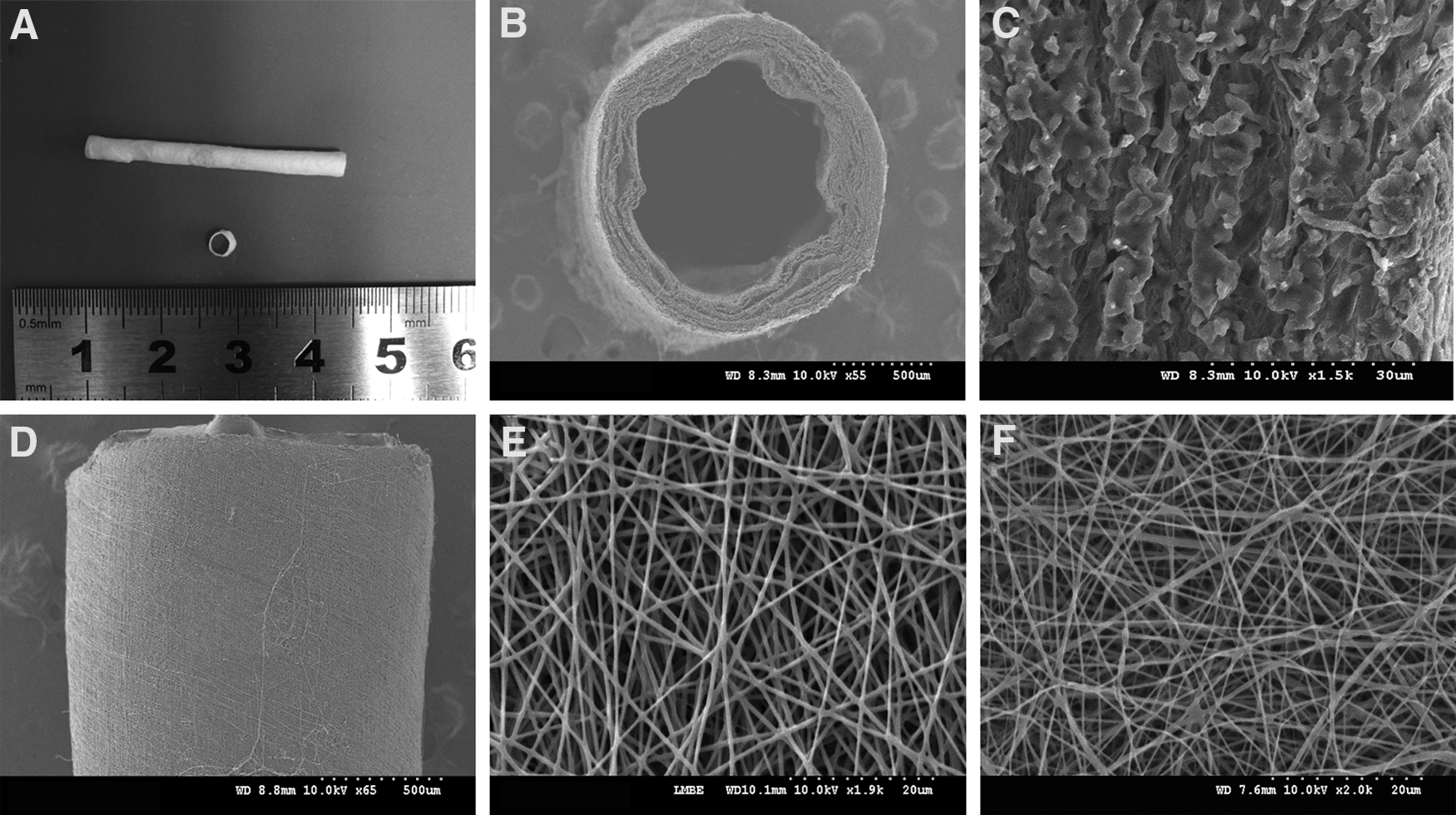
The morphology of electrospun nanofibrous chitosan/poly(ɛ-caprolactone) (CS/PCL) scaffolds.
Morphological observation and characterization of OECs
The OECs appeared after 2–3 weeks as small colonies in cultures of mononuclear cells (MNCs) from the peripheral blood consisting of single flattened cells (Fig. 2A), which develop cobblestone-like cell morphology over time (Fig. 2B). The cells were also assembled into primitive vascular tube-like structures when plated in Matrigel (Fig. 2C). After 3–4 weeks of culture, nearly all of the cobblestone-like cells (>95%) showed double-positive results in the LDL-uptake assays and in their capacity to bind the UEA-lectin (Fig. 2D–F). In addition, the FACS studies showed that the OECs expressed the endothelial markers CD31 and CD144 at a high level (Fig. 2G, H), and expressed CD34 on their surface, but at a low level (Fig. 2I). They did not express the hematopoietic-cell-specific surface antigens CD45, CD14 (Fig. 2J, L) or hematopoietic precursor antigen CD133 (Fig. 2K).

Characterization of outgrowth endothelial cells (OECs) derived from canine peripheral blood mononuclear cells (PBMCs).
Construction of TEVGs in vitro
When highly proliferative OECs were seeded onto small-diameter CS/PCL scaffolds, OECs adhered tightly to the luminal surfaces of the grafts (Fig. 3a, d). After 2 days of static culture, the cells spread out fully and possessed a sustainable growth capability (Fig. 3b, e). After the seeded grafts had been translocated to the closed circulatory loop apparatus and co-cultured for 7 days, OECs elongated and aligned in the direction of flow, and adhered tightly to each other to maintain the high integrity of their confluent monolayer structure (Fig. 3c, f), which was similar to that of native artery. The following planar morphometric analysis revealed that the ratio of OECs layer relative to the total luminal surface was 81.4%±7.9%.
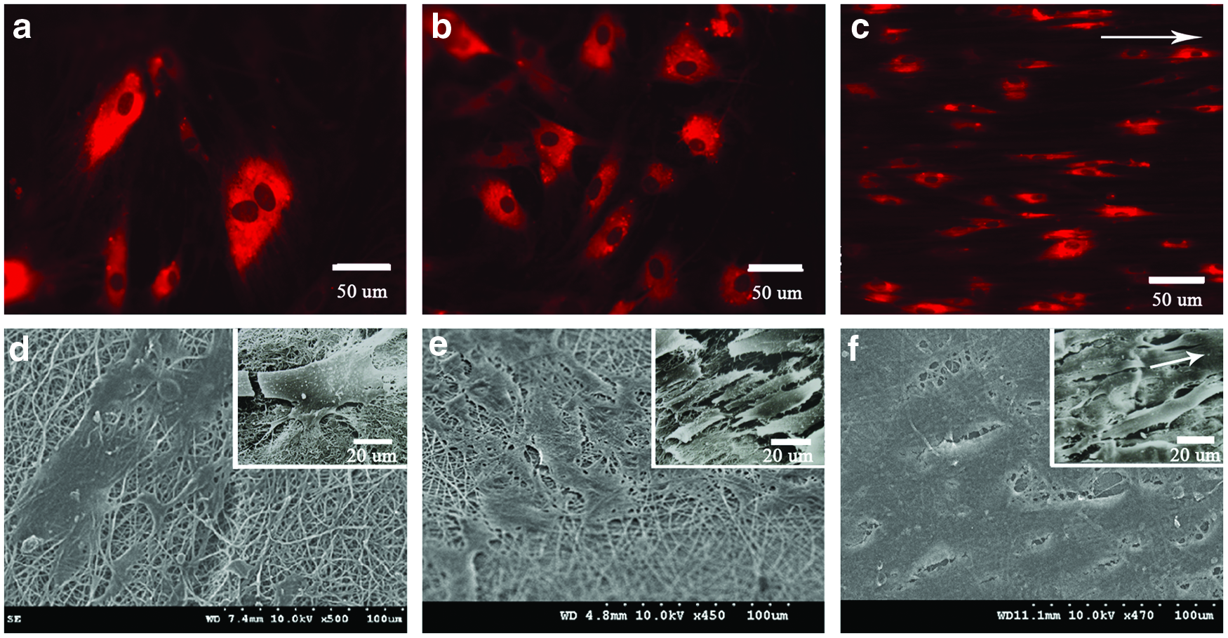
The morphology of highly proliferative OECs seeded on the luminal surface of scaffolds.
Graft patency
Segments of the carotid arteries in cell-donor dogs were replaced by seeded (n=6, TEVGs) and unseeded (n=6, controls) scaffolds. To determine the graft patency, the animals were periodically investigated by arterial Doppler ultrasound after implantation. Patency rate of the vascular grafts was significantly improved by OECs seeding. Five out of six TEVGs maintained patency for approximately 3 months; however, only one out of six controls was still patent, one TEVG and four controls were observed with thrombotic occlusion within the first month, and one control occluded in the second month after implantation (Fig. 4).
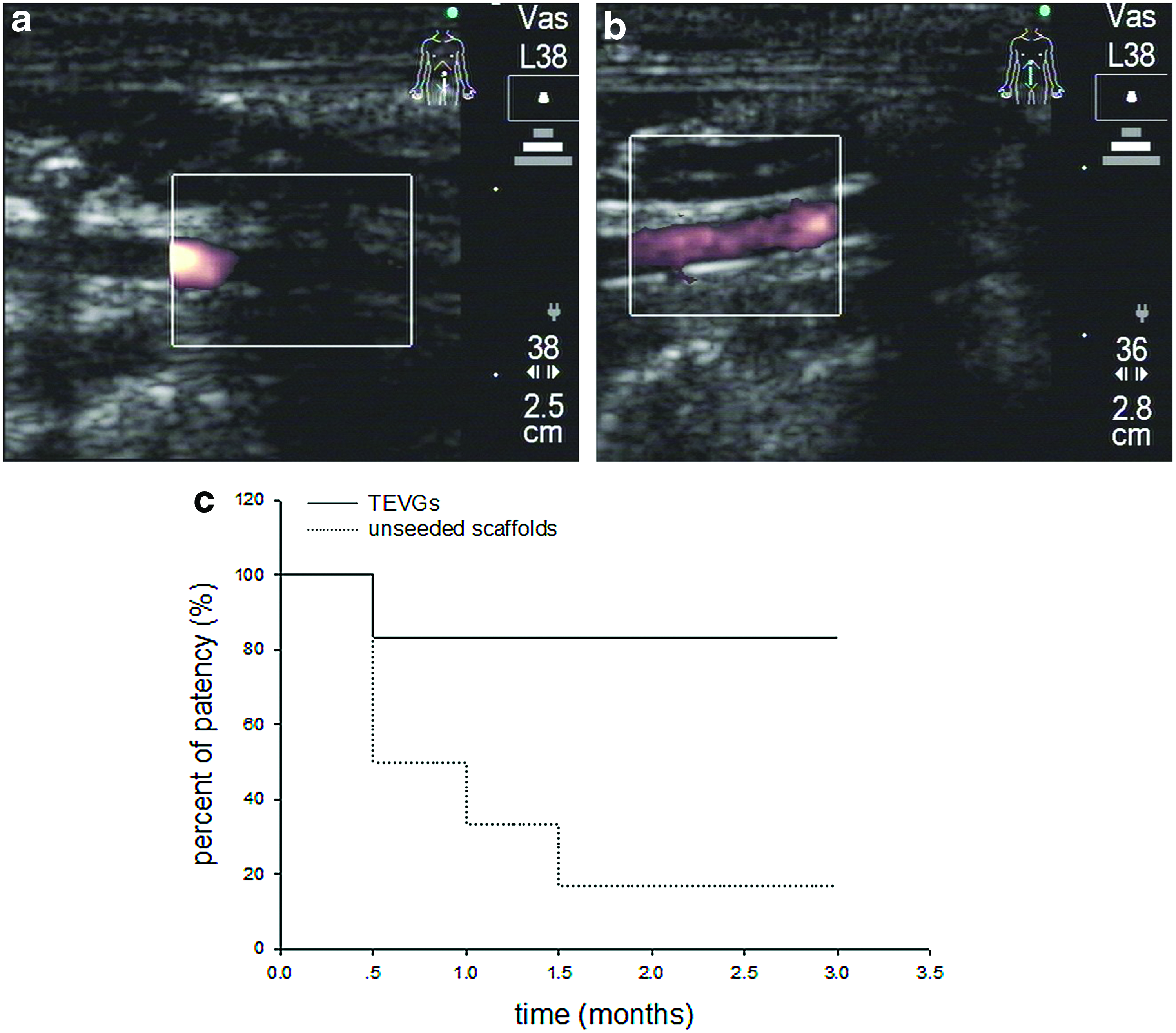
Duplex Doppler ultrasound examination of
TEVGs regeneration in vivo
The carotid arterial angiographies were performed after 12 weeks of implantation, which showed that bilateral grafts were patent in 1 out of 6 dogs, both occluded in 1 out of 6 dogs, and in 4 out of 6 dogs, the TEVGs were patent, but the contralateral unseeded grafts were occluded (Fig. 5a). The surgical view showed a normal vessel-like appearance of the TEVGs without matrix rupture or deformation (Fig. 5b, c). Histology of the TEVGs indicated that tissue regeneration proceeded well, and the neoarterial walls had a thickness of approximately 300 μm at 3 months after implantation (Fig. 5d–f). H&E-stained sections showed a flattened cell monolayer lining the luminal surface and apparent cellular infiltration in media of TEVGs (Fig. 5d). Masson trichrome staining showed typical three layers of structures regeneration (endothelium, media, and adventitia), but without obvious intimal hyperplasia observed (Fig. 5e). Positive staining for vWF of the cells overlying the intimal layer was properly denoted ECs (Fig. 5f). SEM showed that the luminal surfaces of the TEVGs were covered with a confluent monolayer of cobblestone-like cells orienting in a parallel manner to the direction of arterial flow (Fig. 5g), and TEM confirmed the typical morphologies of collagen and elastin (Fig. 5h, i). The OECs labeled with CM-DiI before seeding were identified in the intimal and the medial parts of the TEVGs explanted at 3 months (Fig. 5i). The monolayered cells on the luminal surface of the TEVGs were also positive for the vWF-related antigen via immunofluorescent staining (Fig. 5k). The merged images of vWF and CM-DiI double-positive cells showed that endothelium formed from seeded OECs was present in the luminal surface of TEVGs (Fig. 5l). Similar results were further confirmed from immunofluorescent staining of ECs isolated and cultured from 3-month TEVGs, which showed DAPI, vWF, and CM-DiI triple-positive images (Fig. 6a–d).

The morphology of TEVGs retrieved after 3 months of implantation.
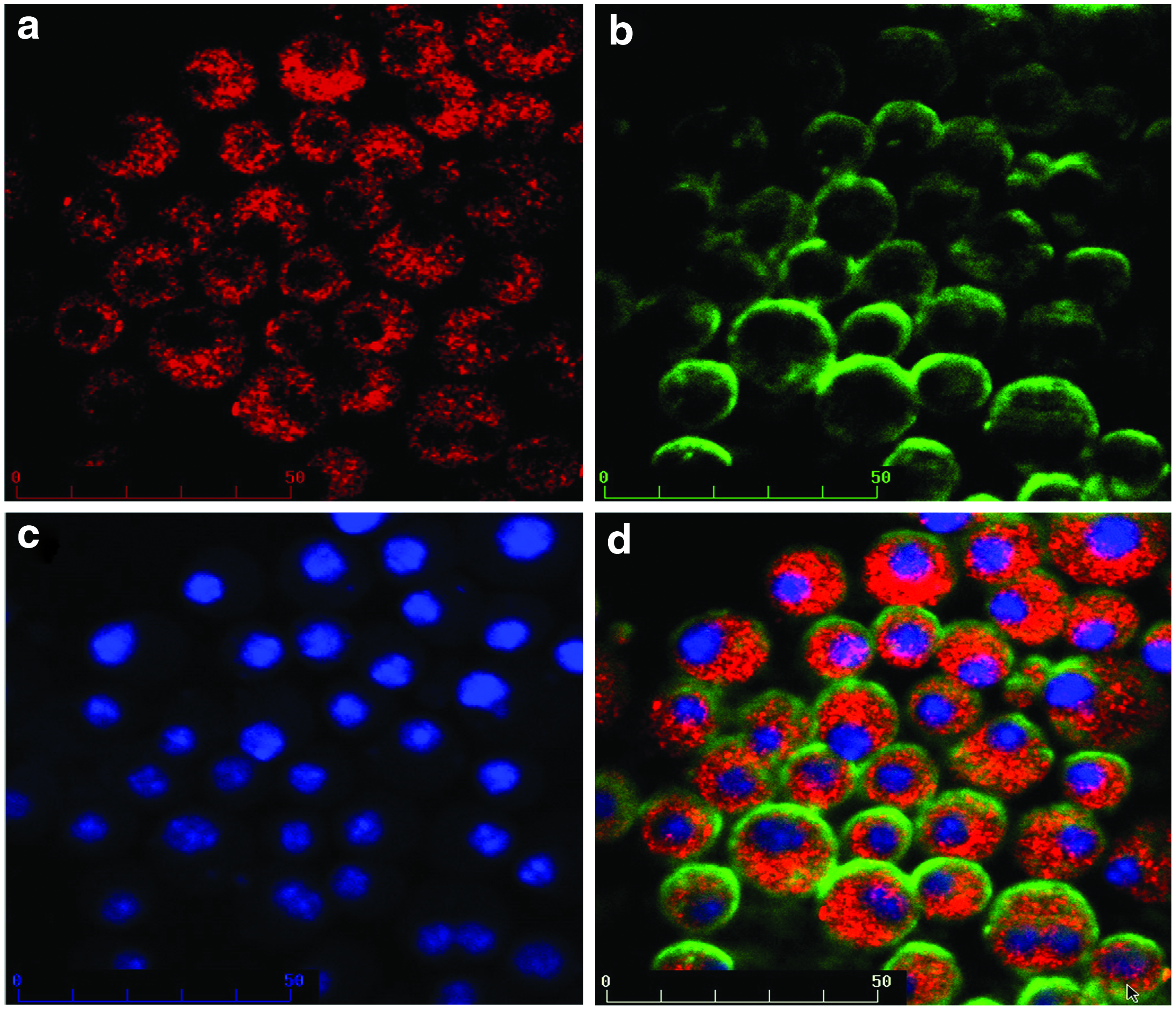
Immunofluorescent staining of ECs isolated from TEVGs retrieved after 3 months of implantation. The staining showed that the ECs were
Biomechanical evaluation
The stress-strain behaviors of the electrospun CS/PCL scaffolds, TEVGs, and native carotid artery were shown in Figure 7, and the tensile strength and ultimate strain obtained from 6 independent tests were summarized in Table 2. The results showed that the CS/PCL scaffold had a tensile strength of 1.53±0.23 MPa and a modulus of 2.33±0.81 MPa with a failure strain value of 50.4%±4.2%. OECs seeding and co-culture in vitro for 7 days improved the scaffold mechanical properties and resulted in differing mechanical behavior. the TEVG retrieved 3 months after implantation exhibited an average tensile strength of 3.06 MPa and a tensile modulus of 9.80 MPa with a strain at break equal 85.9±9.3, comparable to that of native arteries.
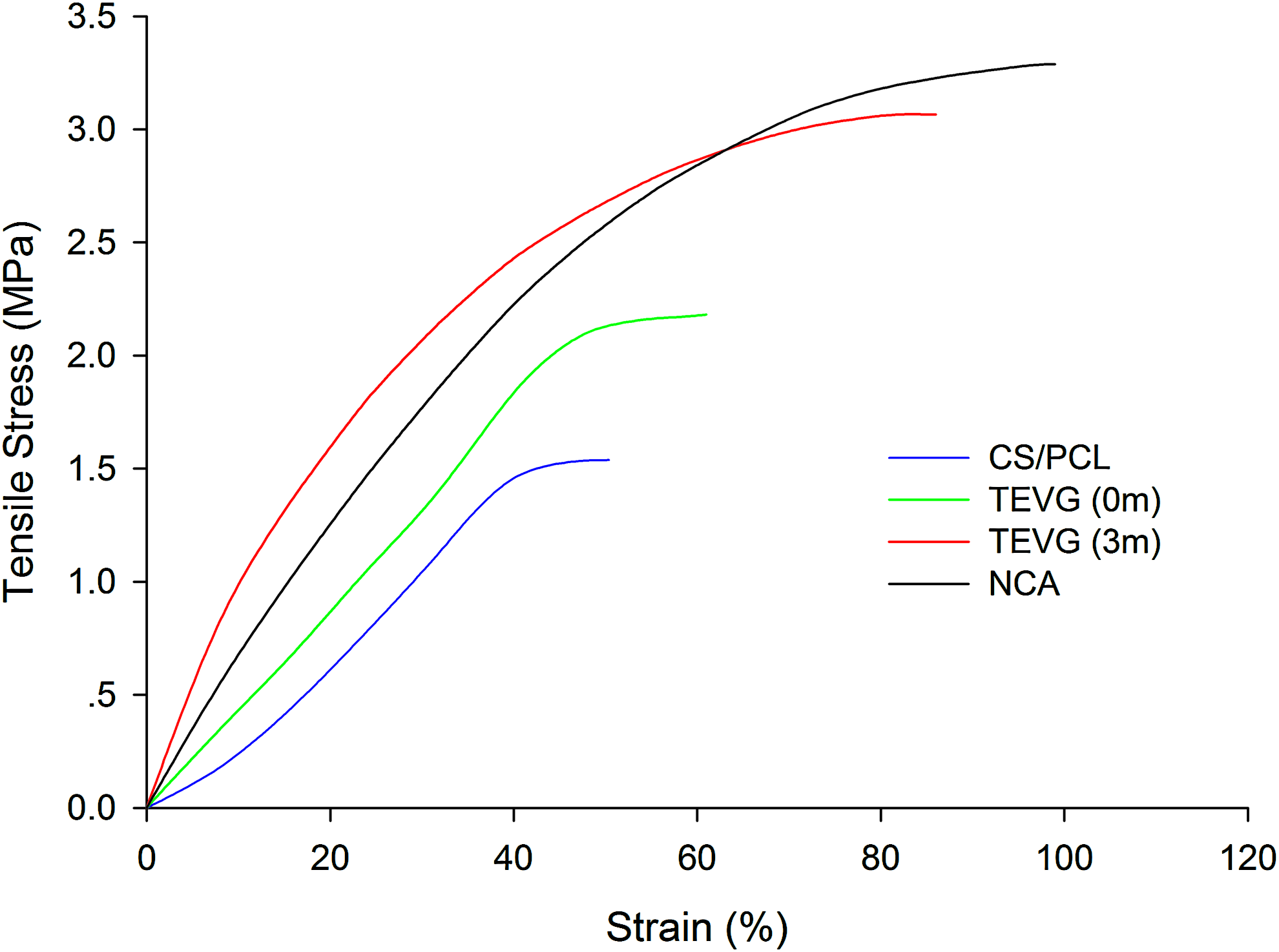
The stress-strain curves of electrospun CS/PCL scaffolds, TEVGs before implantation, TEVGs retrieved after 3 months, and native carotid artery. CS/PCL, electrospun CS/PCL scaffold; TEVG (0m), TEVG before implantation; TEVG (3m), TEVG retrieved after 3 months; NCA, native carotid artery. Color images available online at www.liebertpub.com/tea
CS/PCL, electrospun CS/PCL scaffold; TEVG (0m), TEVG prior to implantation; TEVG (3m), TEVG retrieved after 3 months; NCA, native carotid artery; TEVG, tissue engineered vascular graft; CS/PCL, chitosan/poly(ɛ-caprolactone).
Gene expression of the TEVGs
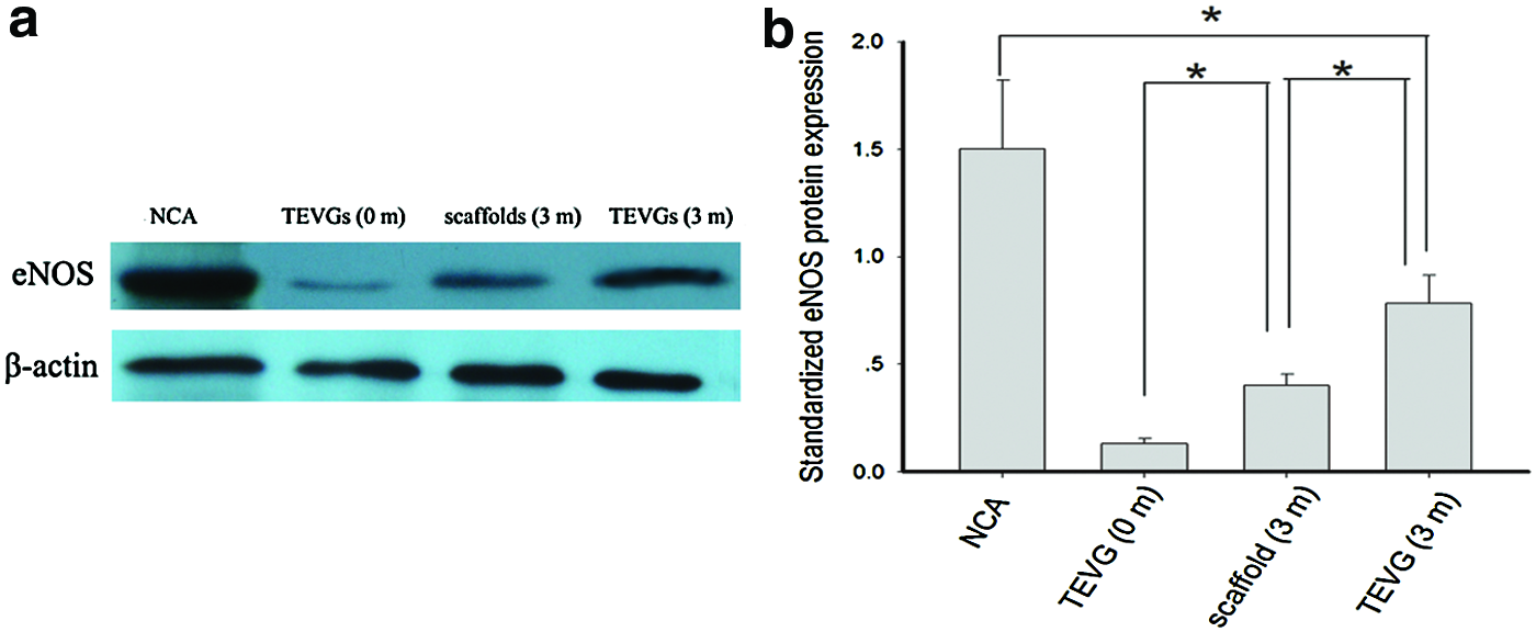
Semi-quantitative RT-PCR analysis showed that mRNA expression of vWF, eNOS, and KDR was higher in the TEVGs than in the control grafts (Fig. 8a). It was shown that the ratios of vWF, eNOS, and KDR to ribosome s17 were significantly higher (p<0.05) in the TEVGs than in the control grafts (Fig. 8b–d), indicating enhanced endothelialization in the TEVGs. The ratios of vWF and KDR to ribosome s17 in the TEVGs were not significantly different (p>0.05) from those of native carotid artery (Fig. 8b, d). Western blot analysis for eNOS showed that eNOS protein expressed in the TEVGs was significantly higher (p<0.05) than in the control grafts, but still significantly lower than native carotid artery (p<0.05) from native carotid artery (Fig. 9).
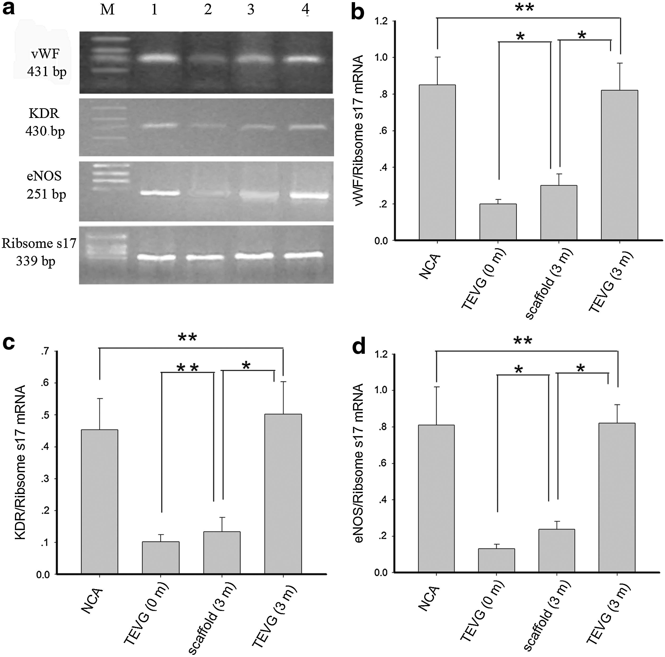
Semi-quantitative RT-PCR analysis.
Discussion
Small-diameter TEVGs are typically composed of cells and scaffolds. The choice of cells and scaffolds is paramount, and the optimal combination is unclear as of yet. In this study, we have demonstrated the feasibility of seeding autologous OECs onto composite electrospun CS/PCL scaffolds to construct novel TEVGs.
Recently, electrospinning-based tissue engineering has been intensively studied and especially focused on searching for optimum combinations of biopolymeric and synthetic materials, and yielding scaffolds with acceptable material properties and biocompatibility. Chitosan (CS) has been used in a number of biomedical applications, such as drug-delivery systems, wound dressings, and nerve regeneration agents, due to its high biocompatibility, low toxicity, and antibacterial properties.33,34 However, less flexibility in regulating the mechanical properties and biodegradation limits its usage. 35 PCL is an aliphatic polyester that degrades slowly and possesses high tensile and elongation properties, 23 but it has hydrophobicity, neutral charge contribution, limited bioregulatory activity, and susceptibility to bacteria-mediated degradation, 36 which have greatly limited its application as tissue engineering scaffolds. Therefore, blending the bioactive functions of CS with the good mechanical properties of PCL to generate a new hybrid material might show an improvement in biological, mechanical, and degradation properties compared with each individual component. As expected, previous reports proved that CS/PCL nanofibrous scaffolds possessed nanoscale microporous, excellent biocompatible and biomechanical properties, and could serve as ideal scaffolds for tissue engineering. 37 We hereby applied electrospinning techniques to fabricate composite CS/PCL scaffolds; the results showed that CS/PCL scaffolds were easily fabricated by electrospinning, the diameters of the CS/PCL nanofibers ranged from 550±120 nm with a distribution in the range of 200–800 nm, and the porosities were more than 80%. The scaffolds exhibited an average tensile strength of 1.53 MPa and a tensile modulus of 2.33 MPa with a failure strain value of 50.4%±4.2%, which would be proper for vascular tissue engineering scaffolds. 38
A reliable cell sourcing is equally vital for the successful tissue engineering of vascular grafts. Recently, increasing evidence has proved that OECs had significant proliferative potential to an endothelial lineage and contributed to the process of endothelium repair.12–14 With a comparison of early EPCs, OECs have a higher proliferative potential and vessel formation properties and, therefore, are considered the best candidate for angiogenic cell therapy. 15 In the present study, we harvested OECs from canine PBMCs, and after 2–3 weeks of culture and proliferation, the induced cells presented a typically “cobblestone-shaped” structure. The majority of the cells expressed endothelial markers CD31 and CD144, and were capable of taking up LDL particles and UEA from the media simultaneously. By using tube formation assays on Matrigel matrix, we further confirmed that the OECs possessed a high capacity to establish primitive vascular tube-like structures. These findings were in good agreement with recent reports on this subset of EPCs. 39
Thereafter, we successfully seeded OECs onto the lumen of the scaffolds and cultured under pulsatile strain and stress conditions. The observed good OECs' attachment and confluent morphology on the lumen of CS/PCL scaffold might be due to several cues presented by both scaffold and OECs: (1) high porosity, high spatial interconnectivity, and high surface area to volume ratio of scaffold; (2) natural polysaccharide of scaffold used to replace glycosaminoglycan, which was the main component of natural ECM; and (3) the high attachment and proliferation potential of OECs. We speculated those provided OECs a similar condition as the natural-basement membrane where OECs grew onto a natural environment. Our previous study found that aligned electrospun nanofibers were not only biocompatible with OECs but also actually promoted and guided their sustained proliferation. 17 Veleva et al. demonstrated that electrospinning significantly improved the cytocompatibility of the surfaces to OECs. 40
The following in vivo implantation demonstrated that the novel TEVGs could be easily sutured and there was no blood leaking or deformation. After 3-month implantation, TEGVs have shown an improved patency compared with non-seeded grafts (5/6 vs. 1/6), and most of the occluded grafts were found within the first month after implantation, due to thrombus formation. We hypothesized that both excellent biocompatibility of CS/PCL scaffold and dynamic OECs seeding contributed toward reducing the in vivo thrombogenicity, and improved the early patency rate of TEVGs. One TEVG occlusion might be due to the detachment of OECs from the luminal surface after implantation or incomplete endothelialization in vivo. The histological analysis of the patent TEVGs retrieved at 3 months revealed a high degree of reendothelialization, two major ECMs (collagen and elastin), and typical three layers of structures regeneration. The results also showed that CM-DiI-labeled cells were detected in the luminal surfaces of the explanted TEVGs after 3 months of implantation, which was consistent with the findings of previous study reporting that cells labeled with CM-DiI could be observed for approximately 130 days in vivo. 41 The merged images of vWF and CM-DiI double-positive cells suggested survival of the seeding OECs and their contribution to endothelium formation. Other studies hypothesized that the seeded stem or progenitor cells could not only directly differentiate into the vascular cells, but also function via a paracrine mechanism to recruit additional host cells that work together to form neovessels.42,43
Moreover, the endothelial function was assessed for approximately 3 months through gene and protein expression analysis. It was found that TEVGs expressed a much higher level of vWF, KDR, eNOS, mRNA, and eNOS protein than the control non-seeded grafts, but the expression of vWF, KDR, and eNOS mRNA was comparable to native carotid artery, which demonstrated that endothelial function was obtained during in vivo remodeling. It is well known that vWF is an adhesive glycoprotein that is synthesized exclusively in ECs and megakaryocytes, and it plays a central role in hemostasis by mediating the adhesion of an initial platelet plug to the sub-endothelium of injured blood vessels and serves as the carrier for factor VIII in plasma. 44 KDR is the principal mediator of the angiogenic effects of VEGF, potent in stimulating ECs proliferation and migration, and its importance is highlighted by the failure of flk-1 null mice to develop organized blood vessels. 45 The expression of eNOS is critical for functional TEVGs, which catalyzes the production of NO, a potent vasodilator that inhibits SMCs proliferation and migration which are responsible for intimal hyperplasia as well as platelet aggregation and adhesion.46,47 Taken together, the present study demonstrated that OECs seeding might enhance endothelialization and reduce intimal hyperplasia of TEVGs, and maintain similar physiological functions of normal vessel, subsequently indicating improvement of graft patency.
Since electrospinning of degradable polymers to form scaffolds with precise fibrillar architectures has become increasingly popular, the biodegradation and remodeling after implantation have become critical issues in electrospinning-based TEVGs. It is possible that the nanoscaled material will degrade faster than large bulk materials, because the high surface area to volume ratio of the nanofibers provides a large contact area between the materials and the water or enzymes involved in the degradative process. 48 Bölgen et al. reported degradation of PCL nanofiber in vitro over 6 months and in vivo over 90 days. 49 PCL nanofibers with thinner diameters or in the presence of lipase experienced faster degradation. 50 PCL and CS may also affect each other's degradation processes. The degradation products, lactic acid and hydroxyhexanoic acid for PCL and glucosamine for CS, will all affect the pH value, thus further affecting the degradation speed of CS/PCL. In this study, although it was not possible to accurately predict the speed of degradation of CS/PCL nanofibers, we could reasonably conclude that these novel TEVGs maintained a good balance between degradation and remodeling on 3 months after implantation, based on the findings of host cells infiltrating and collagen and elastin generating to new matrix materials while a few nanofibers observed any more, as well as the good mechanical properties of the TEVGs after 3 months which were closer to those of native carotid arteries. In other words, CS/PCL scaffolds may provide growth potential in vivo through autologous vascular tissue remodeling after matrix biodegradation, but the precise biodegradative behavior needs to be further studied.
In conclusion, seeding OECs onto electrospun CS/PCL scaffolds might be an attractive approach for tissue engineering small-diameter vascular grafts, and this technique seems to be able to generate essentially normal vessel walls within a few months. To our knowledge, this is the first report on small-diameter TEVGs that are constructed by seeding OECs onto electrospun CS/PCL scaffolds, and our findings provide strong evidence for the validity of our protocol. However, there are many questions that remain as to how seeded OECs proliferate, differentiate, and arrange themselves in an appropriate fashion to constitute a new tissue, and what the detail of the electrospun CS/PCL scaffolds degeneration and remodeling is. Another general problem is that for off-the-shelf availability such as when an emergency bypass needs to be performed, growth of an artificial vessel and preparation for implantation would take too much time if autogenous OECs are to be implemented. Finding answers and solutions to these questions would permit us to obtain even better TEVGs.
Footnotes
Acknowledgments
The authors are grateful to Wei Wang from the Department of Vascular, Division of Vascular Ultrasound for performing the professional duplex ultrasound examination. This project was supported by the National Natural Science Foundation of China (Grant No. 81201210 and 81270396) and the Science Project of Jiangsu Health (Grant No. Z201308).
Disclosure Statement
No competing financial interests exist.