
Abstract
Background:
The chondrogenic potential of adipose-derived stem cells (ASCs) has been previously demonstrated, although several reports have indicated that ASCs produce less cartilage-specific matrix than bone marrow-derived mesenchymal stem cells. In this study, we intended to improve chondrogenic phenotypes of ASCs using hydrostatic pressure (HP), without utilizing any growth factors other than the transforming growth factor-β1.
Methods:
Human ASCs (CD13+, 44+, 90+, 14−, 31−, 34−) were harvested and cultured. After three passages, the cells were suspended in 0.3% neutralized collagen type I solution and injected into semipermeable membrane tubes, from which 66 pouches were constructed. After a day of incubation, the 66 pouches were divided into three groups. Group HP1: Pouches were incubated for 1 week with treatment of cyclic HP at 0–0.5 MPa (4.93 atm), 0.5 Hz, with a medium replenishment rate of 0.1 mL/min at 37°C, 3% O2, and 5% CO2 in air using a bioprocessor. This was followed by 3 weeks with no HP and without pouches. Group HP2: Pouches were incubated for the first and third week (2 total weeks) with the same condition of Group HP1. No HP was applied in the second and fourth week. Group AP: Pouches with one end opened were incubated without HP. We evaluated the cell constructs histologically and immunohistochemically, as well as for specific gene expression.
Results:
Accumulation of the matrix in the HP1 and HP2 groups was much denser than AP groups, particularly after 2 weeks. Cell numbers in the HP groups increased gradually in the middle zone and peaked at 1 week after incubation, maintaining their numbers for the entire course on the surface layer of the construct. In the genomic study results, COL 2A1, COL 10A1, ACAN, SOX9, MMP3, and MMP13 were upregulated and COL 1A1, ITGB1, and PCNA were downregulated by HP. There were no significant differences between HP1 and HP2 gene expression.
Conclusion:
It was suggested that HP is especially beneficial in the early stage of chondrogenesis of ASCs. Moreover, the expression profile of genes related to chondrocyte differentiation/proliferation was significantly enhanced by HP loading compared with the AP control.
Introduction
C
It has been recognized that chondrocytes, particularly those in articular cartilage, can be proliferated and maintained under physiologically sound mechanical stresses, including hydrostatic pressure (HP)19,20 and shear stress.21,22 Thus, the mechanical stresses seem to be useful to produce a cartilage-like construct in vitro for cell-based therapy. We have reported that a pressure perfusion culture system was useful to construct three-dimensional (3D) engineered cartilage using bovine articular chondrocytes,23–25 human dermal fibroblasts, 26 and human ASCs with a collagen gel/sponge scaffold.27,28 We attempt to take advantage of the effects of repetitive HP application to promote more biosynthesis of cartilage ECM by chondroinduced ASCs.
In this study, we clarified the effects of repetitive HP application on ASCs chondroinduced with TGF-β1. We compared two different HP application protocols: once (early differentiation stage) versus twice (early and late differentiation stage). We evaluated the compatibility of cell and exogenous material, properties of proliferation, gene expression profiles of typical cartilage molecules, and production of cartilage ECM both histologically and immunohistologically.
Materials and Methods
Isolation and culture of human ASCs
Under an approval of an IRB protocol at Brigham and Women's Hospital, Boston, Massachusetts, discarded human adipose tissue (∼5 g) was harvested from three different donors (abdominal subcutaneous adipose tissues from 37-, 43-, and 44-year-old females). They were rinsed extensively with phosphate-buffered saline (PBS; Invitrogen, Carlsbad, CA). The tissue was finely minced and digested with 30 mL collagenase type I (0.075%; Worthington Biochemical Corporation, Lakewood, NJ) dissolved in Dulbecco's modified Eagle's medium (DMEM)/Ham's F-12 (Invitrogen) 50:50 with gentle shaking for 30 min at 37°C using a 50-mL conical tube. The digested cell suspension was then diluted with an equal volume of growth medium: DMEM/Ham's F-12 with 10% fetal bovine serum (FBS; Invitrogen) and 1% antibiotics–antimycotics (Invitrogen). After centrifugation for 5 min at 390 g, the cell pellet was resuspended with the growth medium, and viable cells were counted with Trypan blue. Five hundred thousand cells were seeded to a 100-mm tissue culture dish and incubated in the growth medium at 37°C and 5% CO2. This primary culture was defined as passage 0 (P0). The cells were expanded in the growth medium until 80–90% confluence (∼5–7 days in culture), before undergoing three passages (P3).
Characterization of ASCs
Five hundred thousand P3 cells were harvested with 0.025% trypsin-EDTA (Invitrogen) and incubated with fluorescein isothiocyanate-, phycoerythrin-, or allophycocyanine-conjugated cell surface antibodies: CD13, CD14, CD31, CD34, CD44, CD45, and CD90 (BD Biosciences, San Diego, CA) for 60 min at 4°C. After incubation, the cells were washed thrice with PBS supplemented with 1% FBS followed by resuspension in 0.3 mL of cold PBS with 1% FBS for characterization. Expression of cell surface protein was characterized using a FACSCalibur flow cytometer with CELLQuest acquisition software (Becton-Dickinson, Franklin Lakes, NJ). Data analysis was performed using Flow Jo software (Tree Star, Ashland, OR).
Three-dimensional cell culture
The P3 cells (7×10⁷ cells) were harvested with trypsin and suspended in 3.5 mL of DMEM/Ham's F-12. 1×10⁶ cells were suspended in 50 μL of cold neutralized 0.3% collagen type I solution (PureCol; Advanced BioMatrix, San Diego, CA) and injected into a semipermeable membrane tube (PVDF, 500 kDa; Spectrum Labs, Rancho Dominguez, CA). The tube was incubated at 37°C for 1 h for gelation and sealed with stainless steel clips for making pouches. 29 The cells and collagen formed a cell/collagen gel construct within a pouch. A total of 66 pouches were prepared for this study.
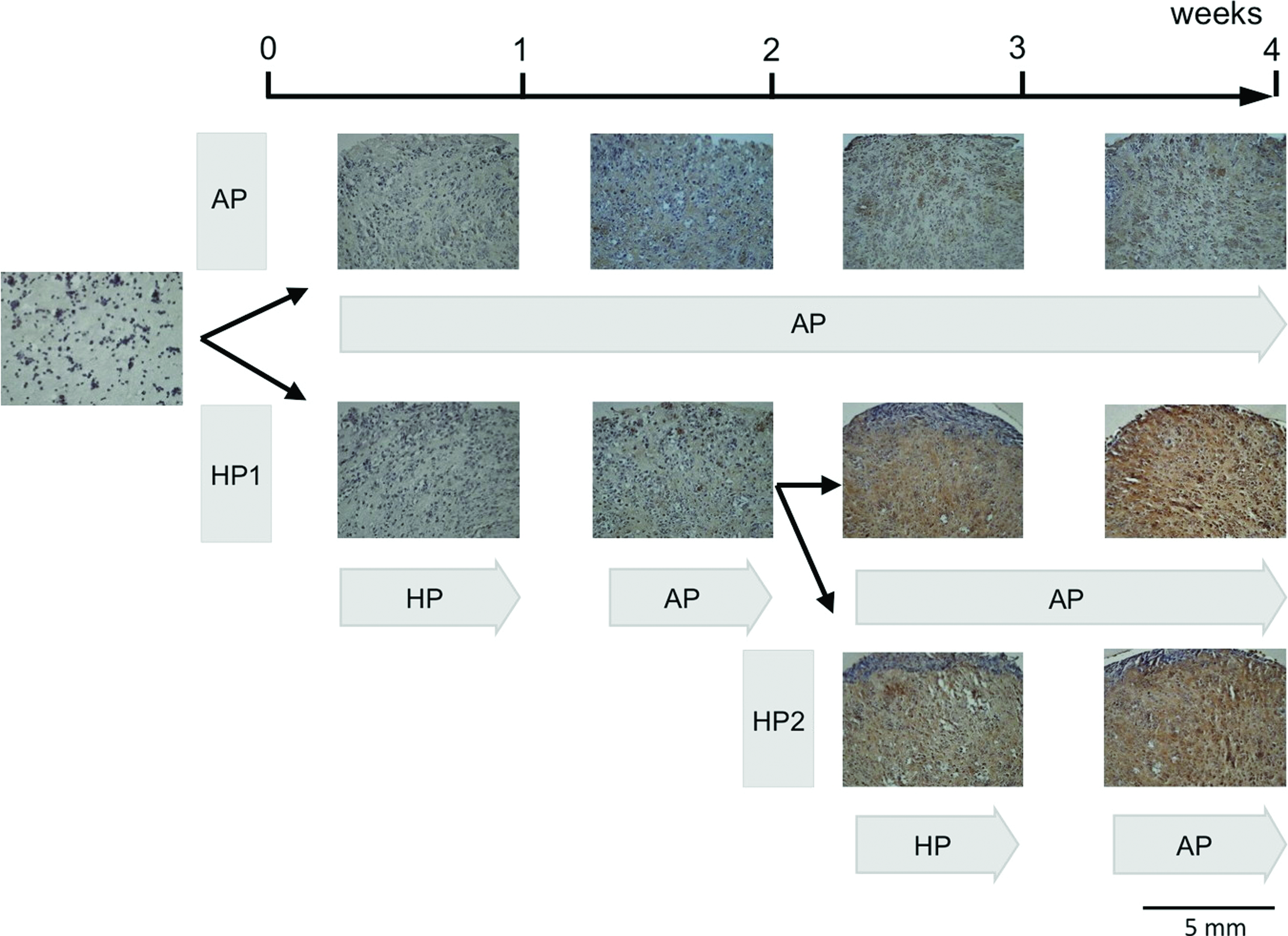
After a day of incubation in the growth medium at 37°C and 5% CO2 in air, six pouches were harvested for samples of day 0. The 60 remaining pouches were divided into three groups for 4-week treatment protocols (Fig. 1). Samples of HP1 at 1 and 2 weeks were overlapping with those of HP2. Specific HP settings were based on our previous works.27,28
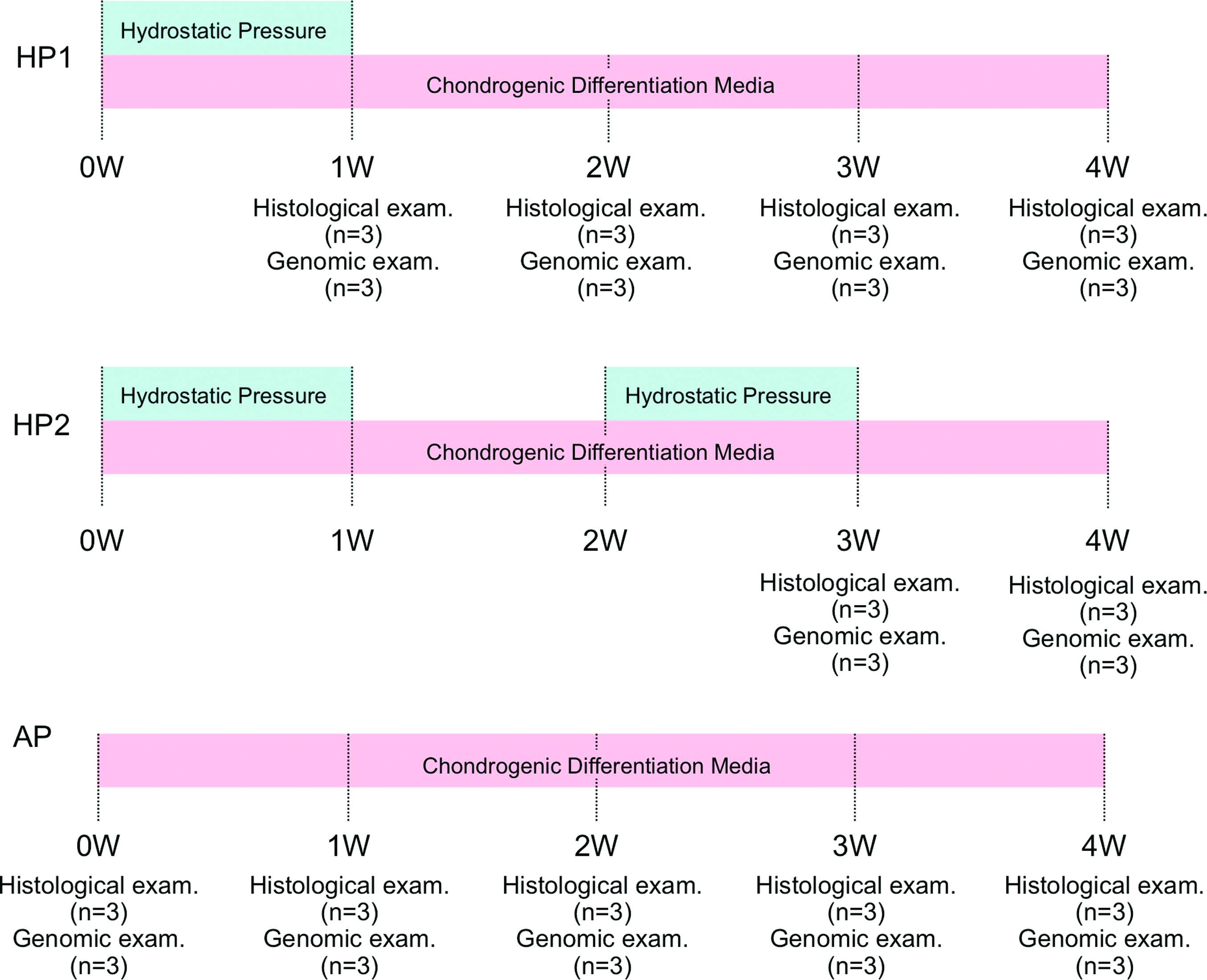
Experimental design. Pouches containing adipose-derived stem cells (ASCs) and collagen gel constructs were divided into three groups: (1) AP, (2) HP1, and (3) HP2. The pouches were incubated in a differentiation medium defined as DMEM/Ham's F-12 with 2% FBS and 1% antibiotics–antimycotics, 10 ng/mL transforming growth factor–β, 1×10⁻⁷ M dexamethasone, 50 μg/mL ascorbic acid 2-phosphate, 1 mM sodium pyruvate, and 40 μg/mL l-proline. After a day of incubation in the growth medium at 37°C and 5% CO2 in air, six pouches were harvested for samples of day 0. The 60 remaining pouches were divided into three groups for 4-week treatment protocols. Samples of 1 and 2 weeks were shared with HP1 and HP2 groups. AP, atmospheric pressure; HP, hydrostatic pressure; DMEM, Dulbecco's modified Eagle's medium. Color images available online at www.liebertpub.com/tea
Group AP: Pouches with one end opened were immersed in a differentiation medium and incubated at atmospheric pressure (AP) at 37°C, 3% O2, and 5% CO2 for all 4 weeks.
Group HP1: Pouches were incubated for week 1 with treatment of cyclic HP at 0–0.5 MPa (4.93 atm), 0.5 Hz, with a medium replenishment rate of 0.1 mL/min at 37°C, 3% O2, and 5% CO2 in air using a bioprocessor (TEP-P03; Purpose, Shizuoka, Japan), followed by AP for weeks 2–4. The constructs were removed from pouches and incubated in nonadherent dishes.
Group HP2: Pouches were incubated for weeks 1 and 3 (total 2 weeks) with the same condition of Group HP1. The constructs were removed from pouches at week 1 followed by incubation in nonadherent dishes for another week at AP. Those constructs were placed within the pouches and incubated for 1 week (week 3) with the same condition of HP1 followed by incubation at AP for another week (week 4).
In every group, the cells were incubated in a differentiation medium defined as DMEM/Ham's F-12 (Life Technologies, Carlsbad, CA), with 2% FBS, 1% antibiotics–antimycotics (Life Technologies), 10 ng/mL TGF-β1 (R&D Systems, Minneapolis, MN), 1×10⁻⁷ M dexamethasone (Sigma-Aldrich, St. Louis, MO), 50 μg/mL ascorbic acid 2-phosphate (Sigma-Aldrich), 1 mM sodium pyruvate (Sigma-Aldrich), and 40 μg/mL l-proline (Sigma-Aldrich). Concentrations of these agents were tested on chondrogenesis in our previous works.27,28
Histological and immunohistochemical evaluation
Cell/collagen gel constructs were harvested from pouches and fixed with 2% paraformaldehyde solution, then embedded in either glycol methacrylate (JB-4®; Polysciences, Warrington, PA) or paraffin (Fig. 2). The samples embedded with the glycol methacrylate were cut into 10-μm-thick sections and stained with 0.2% toluidine blue O (Fisher, Waltham, MA) at pH 4.0. Immunohistological staining was performed using a Vectastatin ABC kit (Vector Laboratory, Burlingame, CA) and a 3,38-diaminobenzidine (DAB) kit (Vector Laboratory). Paraffin sections at 5-μm thickness were dewaxed with xylene and then rehydrated in a graded series of alcohol to PBS. After rinsing with PBS, the sections were blocked with 3% normal horse serum at room temperature for 20 min in a humidified chamber. For collagen type II staining, the sections were incubated with a rabbit anti-human collagen type II antibody diluted 1:50 (Chemicon, Temecula, CA) with 1% normal horse serum for 60 min at room temperature. After three rinses with PBS, the sections were incubated with the biotinylated goat anti-rabbit IgG antibody (Vector Laboratory) followed by manufacturer instructed application of the ABC kit. Color was developed with DAB. Nuclei were counterstained with hematoxylin.
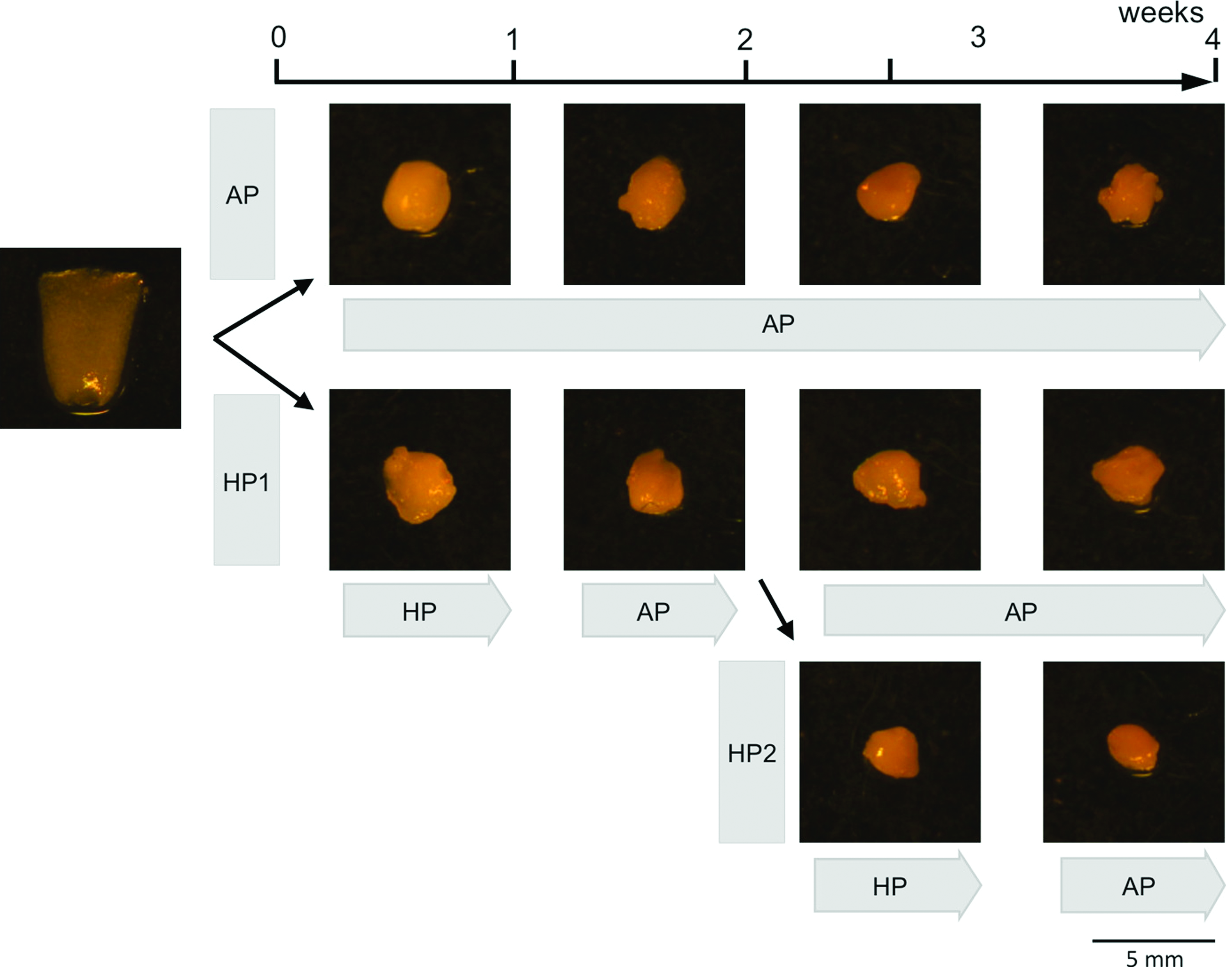
Cell constructs of chondroinduced ASCs incubated within a pouch. Photographs represent cell constructs at each time point. The photograph of 0 W (at the day to start cell culture) shows an initial shape of the cell construct embedded in a semipermeable membrane tube (composed of a pouch afterward). The cell constructs become smaller spheres in shape with time. Color images available online at www.liebertpub.com/tea
Semiquantitative histological evaluation
Toluidine blue-stained sections were used for evaluating cellularity in the constructs (Fig. 3A). The total number of nuclei of the constructs in each surface zone (SZ) and middle zone (MZ) was counted. Three fields in each zone of 40× images were randomly chosen.
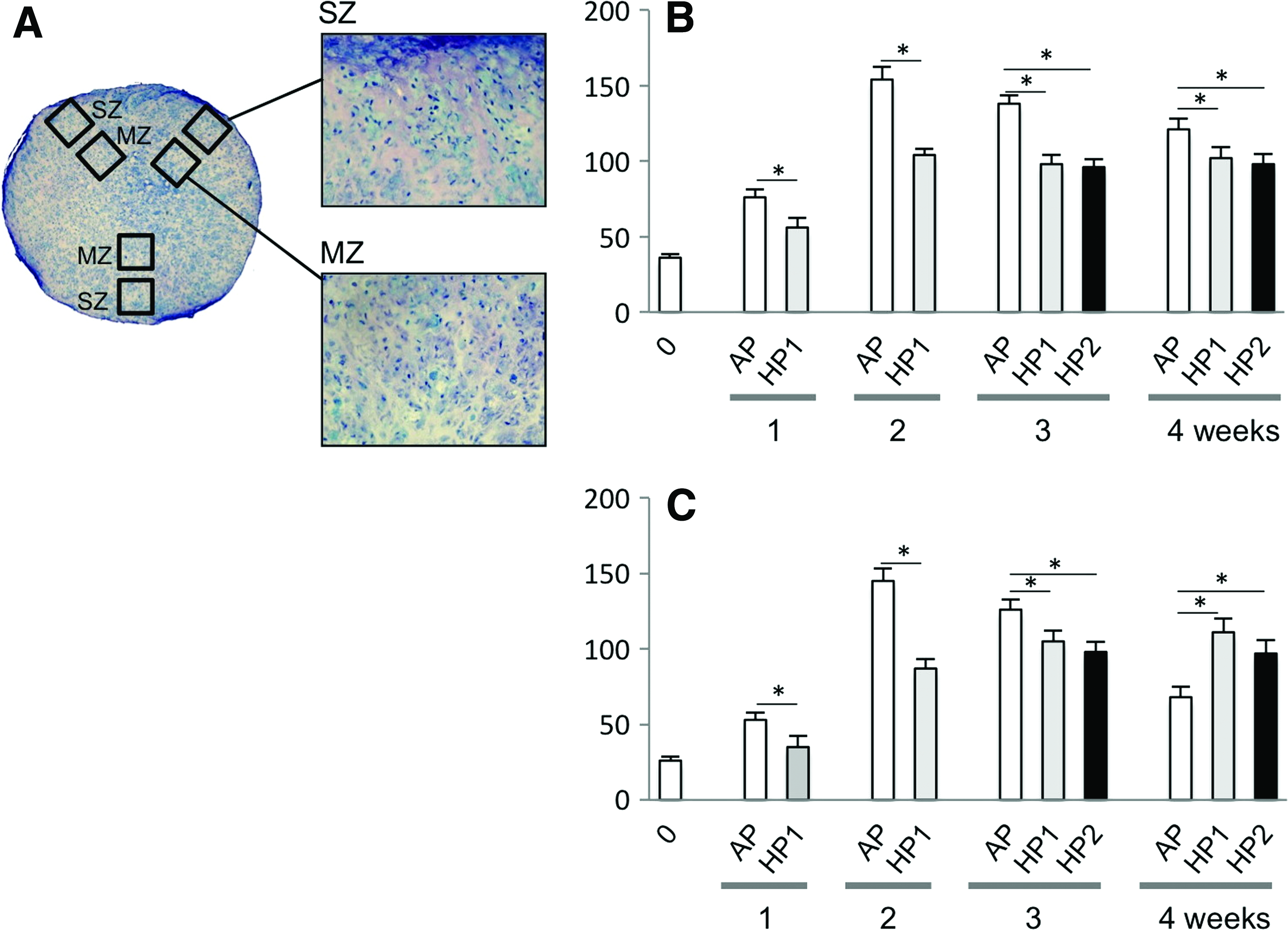
Cell number within a cell construct.
Real-time reverse transcriptase–polymerase chain reaction
Freshly collected samples (three constructs in each group at each time point) were rinsed with PBS and total RNA was extracted using the RNeasy mini kit (Qiagen, Chatsworth, CA), following the manufacturer's instructions. Briefly, samples were finely homogenized with a handheld homogenizer and QIA shredder with buffer RLT, including beta-mercaptoethanol. After adding 70% ethanol and mixing well, samples were centrifuged using the RNeasy spin column. Then, buffers RW1 and RPE were sequentially added into the column and centrifuged. Total RNA was extracted with RNase free water. Total RNA was quantified using the NanoDrop (NanoDrop Technologies, Wilmington, DE) method.
Complementary deoxyribonucleic acid (cDNA) was synthesized using a SuperScript III First-Strand Synthesis System for reverse transcriptase–polymerase chain reaction (RT-PCR) (Invitrogen Life Technologies, Carlsbad, CA). Total RNA (<1 μg) was mixed with random hexamers (50 ng/μL) and dNTP (10 mM), then incubated at 65°C for 5 min. Tubes were cooled on ice and RT buffer (10×), MgCl2 (25 mM), DTT (0.1 M), RNaseOUT (40 U/μL), and SuperScript III RT (200 U/μL) were added, giving a final volume of 21 μL. Samples were then incubated at 25°C for 10 min, 50°C for 50 min, 85°C for 5 min, and cooled on ice. Then, Escherichia coli RNase H (2 U/μL) was added and incubated at 37°C for 20 min.
Real-time RT-PCR was performed in an ABI Prism7300 system (Applied Biosystems, Foster City, CA) using the RT 2 SYBR Green/ROX qPCR master mix (SA Biosciences, Frederick, MD), with primers designed for this study (Table 1). Amplifications of the cDNA samples were performed in triplicate in 96-well plates at a final volume of 20 μL at 40 PCR cycles, consisting of a denaturation step at 95°C for 15 s and an anneal/extension step at 60°C for 1 min. Fluorescence measurements were used to generate a dissociation curve with the system software program v1.4 (Applied Biosystems). Relative quantity (RQ) of each gene was determined from the standard curve. Signal levels were normalized to the expression of a constitutively expressed gene, GAPDH, and shown as a relative ratio.
Real-time reverse transcriptase–polymerase chain reaction (RT-PCR) was performed in an ABI Prism 7300 system (Applied Biosystems) using the RT2 SYBR Green/ROX qPCR master mix (SA Biosciences) with these primers.
Statistical analysis
Cell number data were analyzed using one-way analysis of variance followed by Dunnett's test for comparing all culture conditions versus AP control with p<0.05 being considered significantly different (GraphPad InStat ver. 3.00, San Diego, CA). mRNA expression was considered statistically significant (95% confidence) when the minimum and maximum values of the RQ did not overlap when comparing the values of the groups.30–32
Results
Characterization of human ASCs
By flow cytometric analysis of cultured cells, the percentage of positive cells for (1) stromal associated markers CD13, 44, and 90 was 98.8±0.6, 95.9±1.4, 96.4±2.3, respectively; (2) macrophage and neutrophilic granulocyte marker CD14 was 0.5±0.2; (3) endothelial-associated marker CD31 was 2.6±0.8; (4) stem cell-associated marker CD34 was 4.4±0.6; and (5) pan-hematopoietic marker CD45 was 0.2±0.1. These results were consistent with a previous report of human ASCs cultured with identical growth media. 18 Although primary ASCs are a heterogeneous cell population, this heterogeneity decreased after three passages in culture. 18
Macroscopic, histological, and immunohistochemical evaluation
Macroscopically, a cell/gel construct at day 1 was the same shape as a semipermeable membrane pouch (Fig. 2). However, after 1 week of incubation, the constructs shrunk and became an irregular sphere-like shape. The size of spheres over 4 weeks of incubation was not significantly different among groups.
Histologically, cells were distributed throughout the construct (Fig. 3A). The cell numbers in the AP groups increased by 2 weeks of incubation and decreased thereafter on both the SZ and the MZ in the cell/gel constructs (Fig. 3B, C). In contrast, the cell numbers in the HP groups increased on the MZ by 3 weeks and SZ by 2 weeks after incubation and maintained their numbers thereafter. However, the cell numbers at the SZ and MZ groups were not statistically different between HP1 and HP2. An accumulation of the pericellular and extracellular metachromatic matrix stained with toluidine blue was seen in all groups and increased over 4 weeks (Fig. 4). Accumulation of the matrix in the HP1 and HP2 groups was denser than AP groups, particularly after 2 weeks. Immunohistochemical evaluation revealed that there was no difference between groups at 0 and 1 week in the accumulation of type II collagen. However, the accumulation of type II collagen in all groups increased with time after 2 weeks, with HP groups showing greater production than AP groups at each time point (Fig. 5).
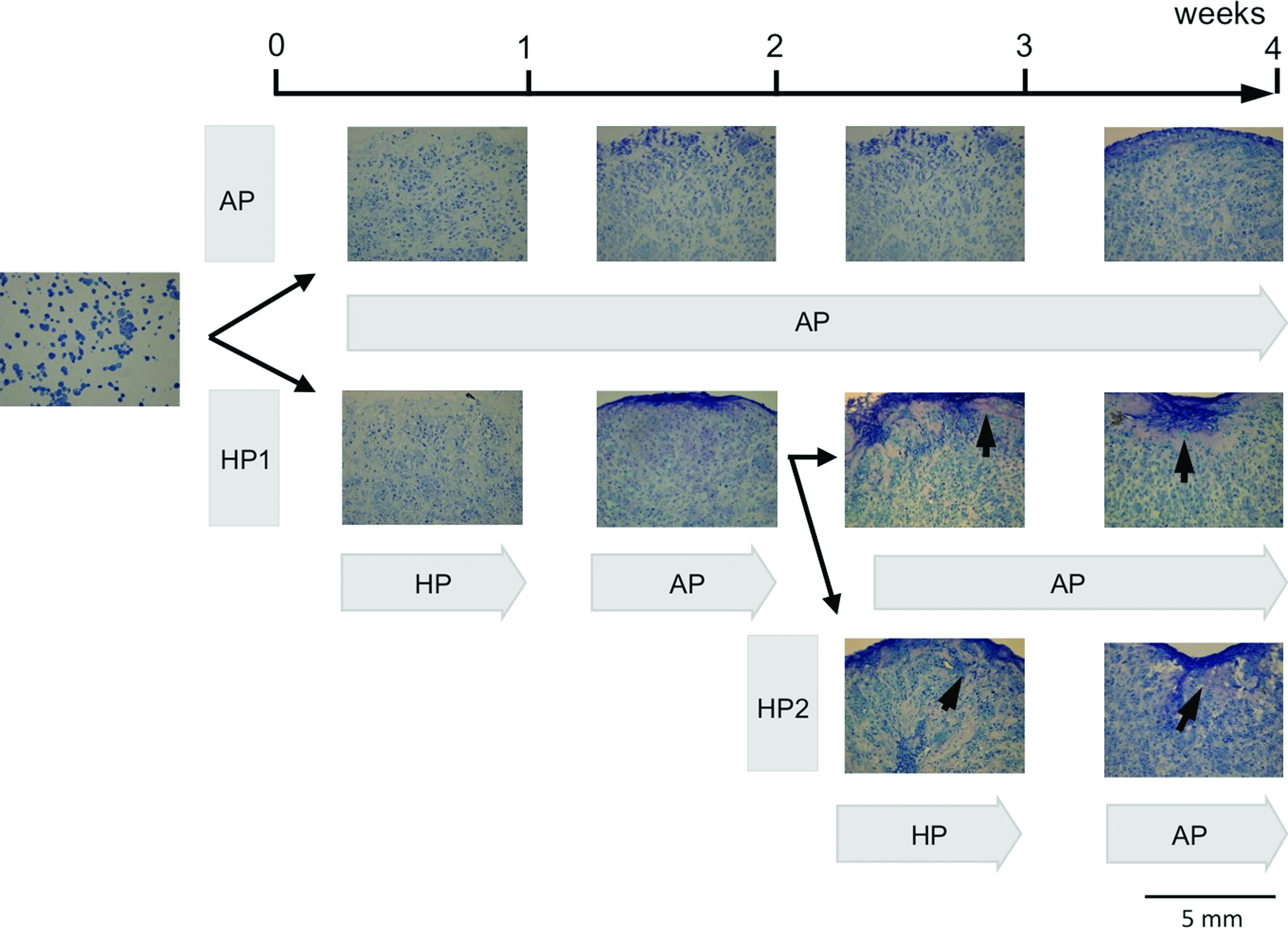
Photomicrographs of histology of chondroinduced ASCs/collagen gel stained with toluidine blue O. The 10-μm sections were stained with toluidine blue O that indicates metachromatic matrix negatively charged cartilage matrix. AP, at atmospheric pressure; HP1, with treatment of hydrostatic pressure during the first 1 week; HP2, with treatment of hydrostatic pressure during the first 1 week and the third 1 week. Arrows show an accumulation of the pericellular and extracellular metachromatic matrix stained with toluidine blue. Color images available online at www.liebertpub.com/tea
Photomicrographs of immunohistochemistry of chondroinduced ASCs/collagen gel stained with the collagen type II antibody. The 5-μm sections were stained with the collagen type II antibody and 3,38-diaminobenzidine, shown in brown. Nuclei were counterstained with hematoxylin shown in blue. HP1, with treatment of hydrostatic pressure during the first 1 week; HP2, with treatment of hydrostatic pressure during the first 1 week and the third week. Color images available online at www.liebertpub.com/tea
Genomic evaluation
Relative gene expression of chondrogenic genes, including collagen type II (COL2A1), collagen type X (COL10A1), aggrecan (ACAN), and Sox 9 (SOX9), were measured. In addition, collagen type I (COL1A1) was included as an estimating characteristic of fibrotic cartilage and estimation of matrix quality by comparing COL1A1 with COL10A1. We also included matrix metalloproteinase-3 (MMP3) and -13 (MMP13) as indicators of cartilage matrix degradation. Integrin β1 (ITGB1) and proliferation cell nuclear antigen (PCNA) were included to estimate characteristics of cell adhesion and proliferation.
In the results, two distinct trends were seen (Fig. 6). First, gene expression of COL2A1, COL10A1, ACAN, SOX9, MMP3, and MMP13 increased with time. Second, gene expression of COL1A1, ITGB1, and PCNA was decreased at 1 week compared with the 0 week control, but then peaked after week 2, and then decreased once again. With regard to the comparison between the AP and HP groups, there were significant differences (95% confidence), especially 3 and 4 weeks after incubation in all genes. However, the HP1 and HP2 group in all genes showed a similar expression profile.
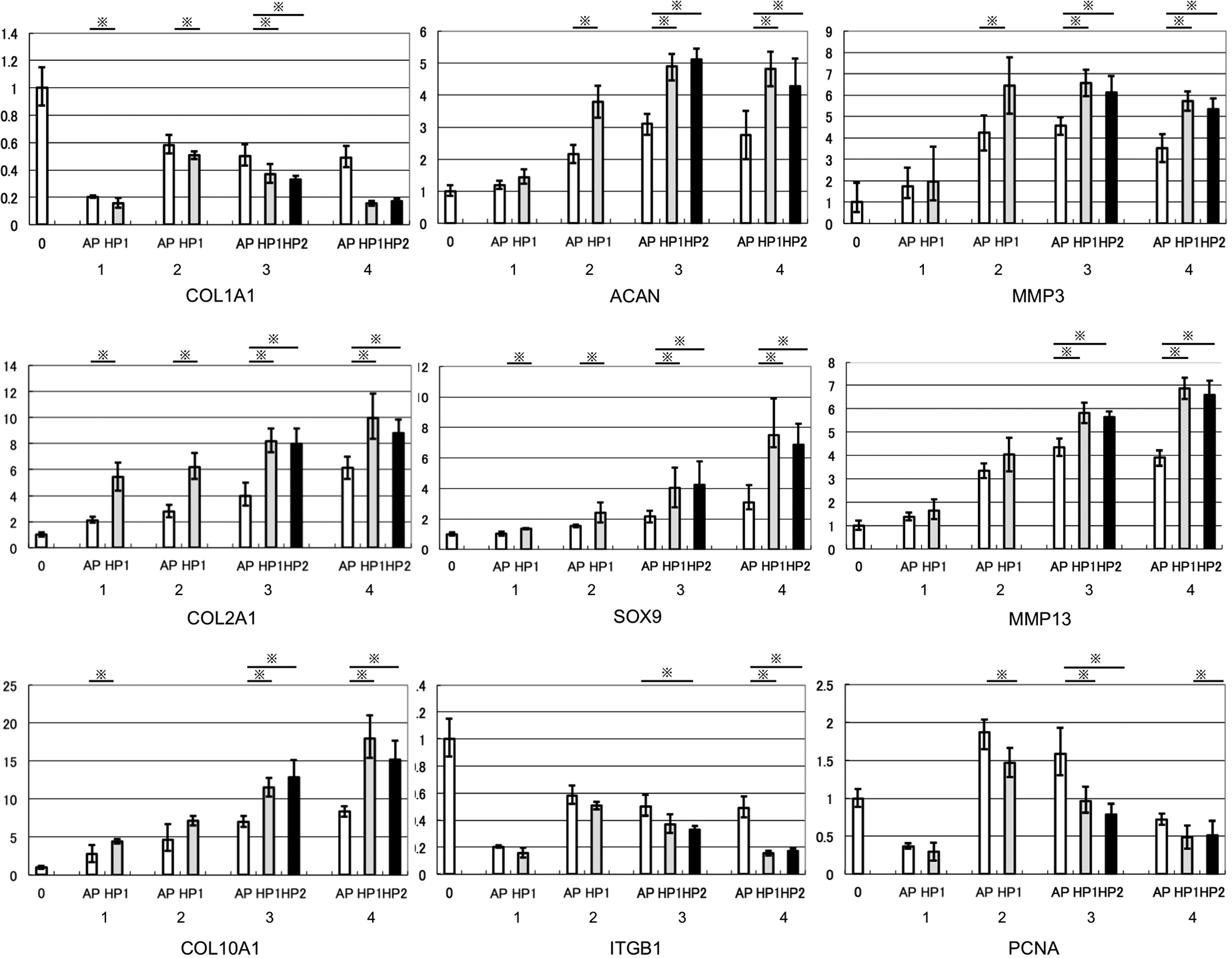
Relative gene expression profile. COL 2A1, COL 10A1, ACAN, SOX9, MMP3, and MMP13 were upregulated with HP treatment and COL 1A1, ITGB1, and PCNA were downregulated with HP. There were no significant differences between HP1 and HP2 treatment. mRNA expression profiles of anabolic and catabolic molecules by chondroinduced ASCs exposed to HP1 and HP2 compared to AP control. Abbreviations indicate ACAN, aggrecan core protein; COL1A1, collagen type I; COL2A1, collagen type II; COL10A1, collagen type X; MMP3, matrix metalloproteinase-3; MMP13, matrix metalloproteinase-13; PCNA, proliferating cell nuclear antigen; ITGB1, integrin beta-1; SOX9, SRY-box 9. External bars indicate maximum relative quantity (RQ) and internal bars indicate minimum RQ. mRNA expression was considered statistically significant (95% confidence) when the minimum and maximum values of the RQ did not overlap when comparing the values of the groups. * shows significant difference (95% confidence).
Discussion
Recently, it was reported that BMSCs have more chondrogenic potential than ASCs in vitro.15,17,33,34 However, the ASC benefits from the ability to be harvested from fat tissue with minimally invasive techniques. Thus, we motivated to gain more information on mechanisms of chondrogenesis by ASCs and tried to manipulate their differentiation. Chondrogenic factors include soluble differentiation factors, cell scaffold materials, and physical factors. TGF-β1 is a well-known soluble chondrogenic factor and, as such, has widely been used for chondroinduction. However, increases in concentrations of the TGF-β1 did not improve chondrogenesis of ASCs. Meanwhile, bone morphogenic protein-6, of the same superfamily, induced expression of TGF-β receptor-I. 18 Combining either TGF-β3/BMP-6 35 or Activin/BMP-2 36 improved chondrogenesis of ASCs. Thus, a combination of multiple differentiation molecules may improve the chondrogenic potential of ASCs.
Moreover, we expected synergistic effects of soluble factors and insoluble materials. It has been reported that chondrogenesis of ASCs was stimulated by insoluble materials, for example, type II collagen-hyaluronan composite scaffolds, 37 poly (ethylene glycol)-poly (L-alanine) diblock copolymer thermogel, 38 and genipin-cross linked cartilage-derived matrix. 39 The characteristics of these materials are not easily manipulated, so the improvement of materials was rather unrealistic. In our previous study, bovine chondrocytes embedded in a collagen gel increased accumulation of cartilage ECM with time. 23 Thus, we intended to reproduce the same collagen gel for culturing ASCs.27,32 In addition, we had the additional option of using physical factors to promote chondroinduction of ASCs. Physical factors, ultrasound, 40 electromagnetic fields, 41 and hyperbaric oxygen 42 have been suggested as chondrogenic-enhancing factors of ASCs. We attempted to stimulate chondroinduction of ASCs using combinations of TGF-β1, collagen gel, and HP.27,28 We developed a HP/perfusion culture system to stimulate chondrogenesis by bovine and human articular chondrocytes with cyclic HP at 0.5 MPa, 0.5 Hz. 29 Beyond these our previous works, we tested two different HP loading protocols and measured additional gene expression, including catabolic genes, because of apparent gel shrinkage.
Upregulation of COL2A1, COL10A1, ACAN, SOX9, MMP3, and MMP13 was significantly higher by HP than by AP. Conversely, COL1A1, ITGB1, and PCNA were significantly lower by HP (period of HP for 1 week) than by AP at 1 and 2 weeks, followed by a decline after that. MMP3 encodes an enzyme, which degrades fibronectin, laminin, collagens III, IV, IX, and X, and cartilage proteoglycans, and MMP13 cleaves collagen type II more efficiently than type I and III. Thus, it was speculated that chondroinduced ASCs produced cartilage ECMs, but subsequently broke them down with at least MMP3 and 13. Both the anabolic and catabolic turnover should thus be recognized in the chondroinduced ASC construct in vitro. HP application promoted synthesis and accumulation of cartilage ECMs that were seen histologically and immunohistologically within the construct. Although catabolic molecules were also stimulated, the absolute values of the expression were not comparable as their activities.
SOX9 is an essential transcription factor for the induction of cartilage phenotypes, including collagen type II and aggrecan, during skeletal development. 43 Thus, HP stimulated the transcription pathway that influences differentiation of ASCs with TGF-β. COL10A1 was found at the periphery of hypertrophic chondrocytes and upregulated by mechanical stresses. 43 Thus, HP promoted collagen type X maturation of regeneration of chondroinduced chondrocytes, 43 although its upregulation is a common feature in chondrogenesis in vitro. 23
HP also inhibits the PCNA and ITGB1, typical markers of cell proliferation and cell adhesion, and collagen type I, which is a minor cartilage molecule. We speculate that HP promoted the remodeling of the cell construct by chondroinduced ASCs, while suppressing cell proliferation and migration. The PCNA expression was consistent with cell number data (Fig. 3B, C). Because cell constructs had shrunk, we measured ITGB1 expression under the premise that ITGB1 overexpression indicated that the cells caused construct contraction. However, the profile was opposite. Thus, it was speculated that the constructs were degenerated with catabolic molecules, for example, MMPs, rather than just contraction of the cell construct.
With regard to the differences in molecular expression between different times of HP application (once vs. two 1-week applications), the levels of mRNA expression of chosen molecules were not significantly different between the two application protocols. It is suggested that HP impacts an early stage of chondrogenesis of ASCs by TGF-β1. Thus, 1-week HP loading was enough to instruct chondroinducing ASCs. One strategy for producing a chondrocyte ECM-rich construct could be the inhibition of MMP activity by promoting inhibitors, for example, tissue inhibitor for metalloproteinase. In addition, HP can be used for promoting the remodeling of a cartilage-like construct by loading at a later time, for example, after 4 weeks of culture.
In summary, the distinct profiles support our interpretation of the effects of HP, which promoted chondrogenic differentiation of ASCs (Fig. 7). With regard to the matrix degradation, both MMP3 and MMP13 were also upregulated by HP. Although comparing levels of gene expression has qualitative limitations, the anabolic and catabolic turnover could be worked in a coordinated manner in the course of cartilage tissue formation. These combined metabolic processes result in the remodeling of the 3D ECM structure in cartilage.
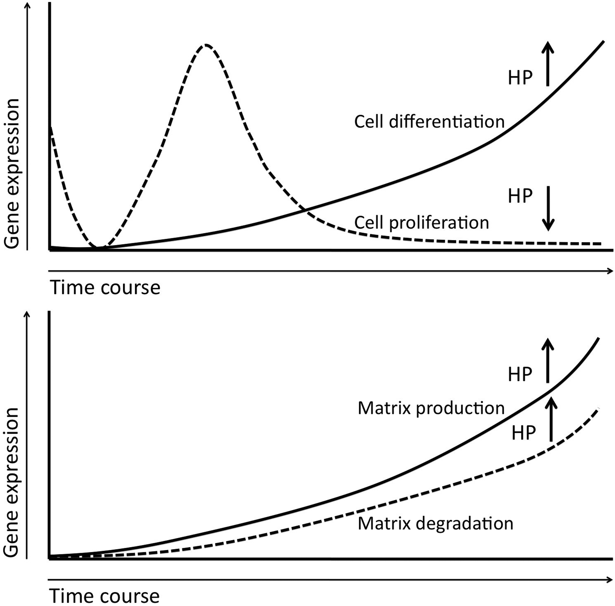
Scheme of the effect of HP on cell differentiation/proliferation and matrix production/degradation.
Authors' Contributions
Rei Ogawa: Collection and/or assembly of data, data analysis and interpretation, and manuscript writing (primary author); Dennis P. Orgill: Financial support and manuscript writing; George F. Murphy: Data analysis and interpretation; Shuichi Mizuno: Conception and design, material and methods, collection and/or assembly of data, and manuscript writing (senior author).
Footnotes
Disclosure Statement
No competing financial interests exist.