
Abstract
Three-dimensional (3D) cell culture platforms are increasingly utilized due to their ability to more closely mimic the in vivo microenvironment compared to traditional two-dimensional methods. Limitations of currently available 3D materials include lack of cell attachment, long polymerization times, and inclusion of undefined xenobiotics, and cytotoxic cross-linkers. Evaluated here is a unique hydrogel comprised of polyelectrolytic complex (PEC) fibers formed by hyaluronic acid and chitosan (CT). When hydrated with fetal bovine serum containing human mesenchymal stem/stromal cells (hMSCs), a hydrogel with an elastic modulus of 264±38 Pa formed in seconds with cells distributed throughout the matrix. Scanning electron microscopy showed a lattice-like meshwork of PEC fibers forming irregular compartments. hMSCs showed 48% viability during the first 24 h, with cell populations thereafter reaching a steady state for 14 days. hMSCs in the matrix were induced to differentiate to chondrogenic, osteogenic, and adipogenic phenotypes. Emergent features, at days 56 and 70, consisted of chondrogenesis on the surface of hydrogels induced to osteogenic and adipogenic phenotypes. Results indicate that this matrix may be useful for tissue engineering and disease modeling applications.
Introduction
T
While each type of hydrogel has been found to have utility in various applications, including 3D cell culture, wound healing, regenerative medicine, disease modeling, and cellular therapies, among others, each has advantages and limitations.2,3 In this regard, hydrogels formed from natural materials such as HA, CT, collagen, and fibrin are typically mechanically weak limiting their usefulness for many cell culture and tissue engineering applications. Nevertheless, there is great interest in the development of natural hydrogel materials due to their inherent biological activity and biocompatibility. Therefore, in our efforts to design an improved hydrogel material we focused on the use of HA and CT, as they are both naturally occurring and biocompatible biopolymers. Our goal was to produce a material that retains the native conformation of bioactive polymers while improving mechanical properties such that the hydrogel would serve a wide range of applications.
HA is a linear polysaccharide found in the ECM during embryonic development and wound healing.
4
It is composed of repeating β1,4 linked disaccharide units of β1,3 linked
By taking advantage of the polyanionic nature of HA and the polycationic nature of CT in aqueous solution a unique hydrogel material comprised of polyelectrolytic complex (PEC) fibers, termed Cell-Mate3D, was produced. PEC fibers formed by HA and CT give the matrix structural integrity and elastic properties without chemical or UV cross-linking, allowing each component to remain in its native and biologically relevant state.24,25 Here, we characterize structural features and rheological behavior of this novel material. We also evaluate its ability to maintain viability and enable growth of hMSCs, and allow for the differentiation of these cells into adipogenic, chondrogenic, and osteogenic phenotypes.
Materials and Methods
Preparation of hyaluronic acid/chitosan (HA-CT) dry blend (Cell-Mate3D™)
Sodium hyaluronan (HA-Na) (original MW=1600–1800 kDa; polydispersity index (PDI)<4.0) and CT protonated with formic acid (CTNH3+) (original MW=400–600 kDa; PDI<3.0) were used in this study. HA (Lifecore Biomedical, Chaska, MN) was used as received. CT (NovaMatrix; FMC Biopolymers, Princeton, NJ) was received as a base at 85–87.5% degree of deacetylation and protonated with formic acid to 100% of available amine groups. Protonated chitosan-base was prepared as a 0.1% (w/v) solution, filter sterilized (0.2 μm), and aseptically filled into 120 cc sterile vials. The CT solution was lyophilized, reduced to small leaflets and mechanically blended with small particles of HA-Na at the mass ratio of HA-Na=1.0: CT=1.44; carrying a charge ratio of CT-n+=2.0: HA-Na-n−=1.0.
Expansion of hMSCs and preparation of hMSC-loaded Cell-Mate3D
Commercial bone marrow-derived hMSCs (Lot Nos. 6881 and 6890; Sciencell, Carlsbad, CA) were expanded in plate culture according to the manufacturer's instructions. Both cell lots were derived from the same donor. Cells were expanded in commercial Mesenchymal Stem Cell Medium (MSCM) growth medium (Cat. No. 7500; Sciencell) on tissue culture flasks coated with poly-l-lysine (Cat. No. 0413; Sciencell) at 2 μg/cm2 before embedding in Cell-Mate3D.
The HA-CT dry blend was brought to room temperature and mixed by vigorous vortexing. The dry blend was hydrated by drip loading a cell suspension in fetal bovine serum (FBS) (Thermo Fisher Scientific, Waltham, MA) under continuous agitation. Five hundred microliters of the FBS cell suspension containing 19 million hMSCs was used to hydrate 35.5 mg of the dry blend. The cell-loaded matrix was transferred into a 1 cc syringe barrel by centrifuging at 1000 G through a funnel apparatus. This process produced a 400–500 μL cylinder of cell-embedded matrix that was then extruded from the syringe barrel, cut to 100 μL sections, and placed into MSCM growth medium.
Time course of hMSCs in maintenance medium
Each hMSC-loaded Cell-Mate3D matrix piece (prepared as above) was placed in one well of a six-well tissue culture dish containing 5 mL of commercial MSCM growth medium (Sciencell). One sample was fixed immediately (day 0) in fresh 10% neutral buffered formalin (NBF) (Acros, Waltham, MA) and the remaining samples were collected and fixed at D1, D3, D9, and D14. Cell density (MSC/unit area) was calculated for each iteration (n=3) and each time point by hand counting intact cell nuclei in 10 high magnification fields in hematoxylin and eosin (H&E)-stained sections. Beginning with a randomly selected start point and direction on the sections, sequential, nonoverlapping images were acquired for 10 consecutive fields in each hydrogel construct. Images were obtained using an Olympus BH-2 microscope equipped with a Spot Insight 4 camera and Spot Advanced software (Diagnostic Instruments, Inc., Sterling Heights, MI). Cell densities in the D0 samples for each of the three iterations of the experiment were then set as 100% and the D1–D14 values calculated as a percent of the D0 sample. This normalized the differences in loading densities in the three experiments. Sections from each time point were also immunohistochemically stained for cleaved caspase-3 and Ki67. Positive labeled cells and unlabeled cells for each stain and time point were counted in sufficient random, nonoverlapping, high magnification fields such that at least 100 cells were counted for each sample. Values for cleaved caspase-3 and Ki67 were then expressed as percent labeled cells at each time point.
hMSC differentiation assays
For chondrogenic differentiation assays (n=3), Cell-Mate3D matrix loaded with hMSCs were placed in MSCM growth medium (Sciencell) for 24 h. Chondrogenic differentiation was then induced using StemPro Chondrogenesis differentiation medium (Cat. No. A10071-01; Life Technologies, Carlsbad CA). Samples were fixed for histology on D14 of culture. As a control, 250,000 hMSCs were pelleted into the bottom of a 15 mL conical tube in 0.5 mL of MSCM growth medium (Sciencell). After 24 h, growth medium was replaced with StemPro Chondrogenesis medium (Life Technologies). For each condition, medium was replaced every 3–4 days. On D14 the pellet was fixed for histology. Two additional Cell-Mate3D samples were prepared as above but harvested on D56 and D70 respectively.
For osteogenic differentiation assays (n=3), Cell-Mate3D matrix loaded with hMSCs were placed in MSCM growth medium (Sciencell) for 24 h. Osteogenic differentiation was then induced using StemPro Osteogenesis differentiation medium (Cat. No. A10072-01; Life Technologies). As a control, hMSCs were seeded in plate culture at 5,000 cells per cm2 in growth medium and after 24 h growth medium was replaced with StemPro Osteogenesis medium (Life Technologies) according to the manufacturer's instructions. For each condition, medium was replaced every 3–4 days. On D28–D30 samples were fixed for histology. Two additional samples were prepared as above but harvested on D56 and D70 respectively.
For adipogenic differentiation assays (n=3), Cell-Mate3D matrix loaded with hMSCs were placed in MSCM growth medium (Sciencell) for 24 h. Adipogenic differentiation was then induced using StemPro Adipogenesis differentiation medium (Cat. No. A10070-01; Life Technologies). As a control, hMSCs were seeded in plate culture at 40,000 cells per cm2 with growth medium. The next day, growth medium was replaced with StemPro Adipogenesis differentiation medium (Life Technologies). For each condition, medium was replaced every 3–4 days. On D14 samples were fixed, processed, and evaluated for lipid content with Oil Red O stain (Millipore, Billerica, MA) and by immunohistochemistry for PPAR-γ.
Fixation and processing of Cell-Mate3D matrix
For light microscopy, samples were placed in 10% neutral buffered formalin and fixed for 1 h (plate cultures) or 3.5 h (Cell-Mate3D constructs). Following fixation samples were transferred to 70% ethanol solution and processed for routine paraffin embedding, sectioning at 4 μm, and stained as indicated.
For scanning electron microscopy (SEM), samples were fixed in 2.5% glutaraldehyde (Electron Microscopy Sciences, Hatfield, PA) in 0.1 M cacodylate buffer (Electron Microscopy Sciences) for 3 h, washed in 0.1 M cacodylate buffer pH 7.4 (3×), and treated with 1% osmium tetroxide (aqueous; Electron Microscopy Sciences) pH 7.4 for 1 h followed by washes in 0.1 M cacodylate buffer pH 7.4 (3×). The samples were then dehydrated in a graded series of ethanol solutions up to 100% ethanol. Samples were then subjected to critical point drying, mounted onto a metal stub with double-sided carbon tape, and sputter coated with gold and palladium. Samples were examined with a Hitachi S3500N scanning electron microscope at an accelerating voltage of 12 kV.
Histochemical and immunohistochemical stains
All samples were routinely stained with H&E. Preparations differentiated toward cartilage were additionally stained with Alcian blue by standard protocols, and preparations differentiated toward bone were stained by the von Kossa method. Before immunohistochemical staining, en face decalcification was done on the paraffin blocks of the D56 and D70 osteogenic and adipogenic differentiation assays for 10 min in Decalifying Solution (Newcomer Supply, Middleton, WI). For immunohistochemical staining, sections were cut at 4 μm, deparaffinized, and rehydrated, followed by incubation with 3% hydrogen peroxide to quench endogenous peroxidase activity and 15 min in serum-free protein block (Dako, Glostrup, Denmark). Sections were then subjected to appropriate antigen retrieval methods (if needed) and incubated with the primary antibody at room temperature for 60 min as follows: PPAR-γ (clone C26H12; Cell Signaling, Danvers, MA), 1:200, citrate antigen retrieval; Ki67 (clone MiB-1; Dako), 1:50, citrate antigen retrieval; type II collagen (clone CIIC1; Hybridoma Lab, Baltimore, MD), 1:10, pepsin antigen retrieval; type X collagen (Abcam, Cambridge, MA), 1:400, trypsin antigen retrieval; aggrecan (Cat. No. AF1220; R&D Systems, Minneapolis, MN), 1:200, no antigen retrieval; Runx2 (Cat. No. ab23981; Abcam), 1:200 dilution, no antigen retrieval; osteocalcin (Clone No. 190125; R&D Systems), 1:400, no antigen retrieval; and cleaved caspase-3 (Cell Signaling), 1:50, EDTA antigen retrieval. Detection for mouse antibodies was performed with Mouse Envision+ kit (Dako), goat primary antibodies with PROMARK (Biocare Medical, Concord, CA), and rabbit primary antibodies with Rabbit Envision kit (Dako). Sections were then counterstained with Mayer's Hematoxylin (Sigma-Aldrich, St. Louis, MO).
Measurement of mechanical properties
Mechanical properties of the Cell-Mate3D matrix were characterized using an AR-G2 rheometer (TA Instruments, New Castle, DE). Cell-Mate3D constructs containing hMSCs were prepared as described above. One hundred microliter sections were loaded onto the peltier plate, maintained at 37°C, and the 8 mm parallel plate was lowered to a gap of 1,200 μm. The Cell-Mate3D matrix was coated with a thin layer of dimethylpolysiloxane to prevent evaporation. Oscillation stress sweeps were performed at a constant frequency to select a % strain within linear viscoelastic regime of the matrix. Frequency sweeps were then performed from 0.1 to 100 rad/s with 1% strain at 37°C. For all measurements n=3 (three independent experiments performed on different days, each with a single replicate).
Statistical analysis
For each time course experiment, the average cell density was calculated for D0, D1, D3, D9, and D14, respectively. Cell counts were then converted to percentages with percentage at day 0 set to 100%. Percentage at day X was calculated as 100%×(count at day X)/(count at D0). For cleaved caspase-3 and Ki67 IHC stain quantification, means and standard deviations of the cell counts (expressed as percentages) were calculated for each time point (D0, D1, D3, D9, and D14). For each data set, analysis of variance (ANOVA) F test was performed to evaluate whether the means were significantly different across different days. To assess which means differed from which other means, we performed multiple comparisons with Tukey–Kramer adjustment for the p-values to account for multiple testing. A p-value <0.05 was considered significant.
Results
Morphologic assessment of Cell-Mate3D matrix
Under the conditions described, the hydrated mixture of HA and CT almost instantaneously formed a semisolid, translucent, malleable, and extrudable hydrogel-hydrocolloid (Fig. 1a). When immersed in cell culture medium and maintained under sterile conditions in a 37°C incubator, the hydrogel-hydrocolloid continued to retain structural integrity after 10 weeks. Light microscopic examination of formalin-fixed, Epon-embedded 1 μm thick sections stained with methylene blue revealed a regular structure of hyaluronan strands attached to a chitosan backbone (Fig. 1b). SEM revealed a lattice-like meshwork of PEC fibers (Fig. 1c, d). Spaces between the PEC fibers as observed in SEM images were of variable size up to ∼50 μm in greatest dimension. In the hydrated state, cells, media, and unreacted HA and CT occupy these spaces.
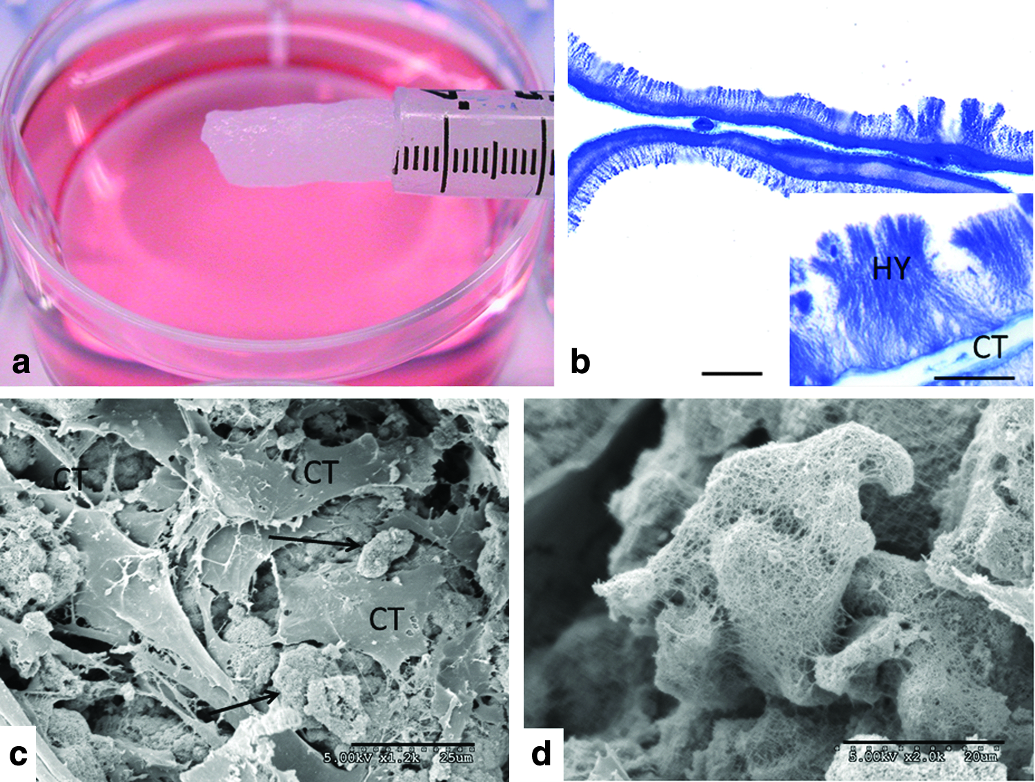
Structural features of the Cell-Mate3D hydrogel.
Rheological study of hMSC embedded Cell-Mate3D
Rheology was used to measure the storage and loss modulus of the Cell-Mate3D matrix with embedded hMSCs (n=3). After 120 min, the mechanical properties stabilized, with a storage modulus of 71±10 Pa and a loss modulus of 51±8 Pa (Fig. 2). Because measurements were obtained within the linear viscoelastic regime, G′ and G" could be used to calculate the elastic modulus, E, by the following equation:
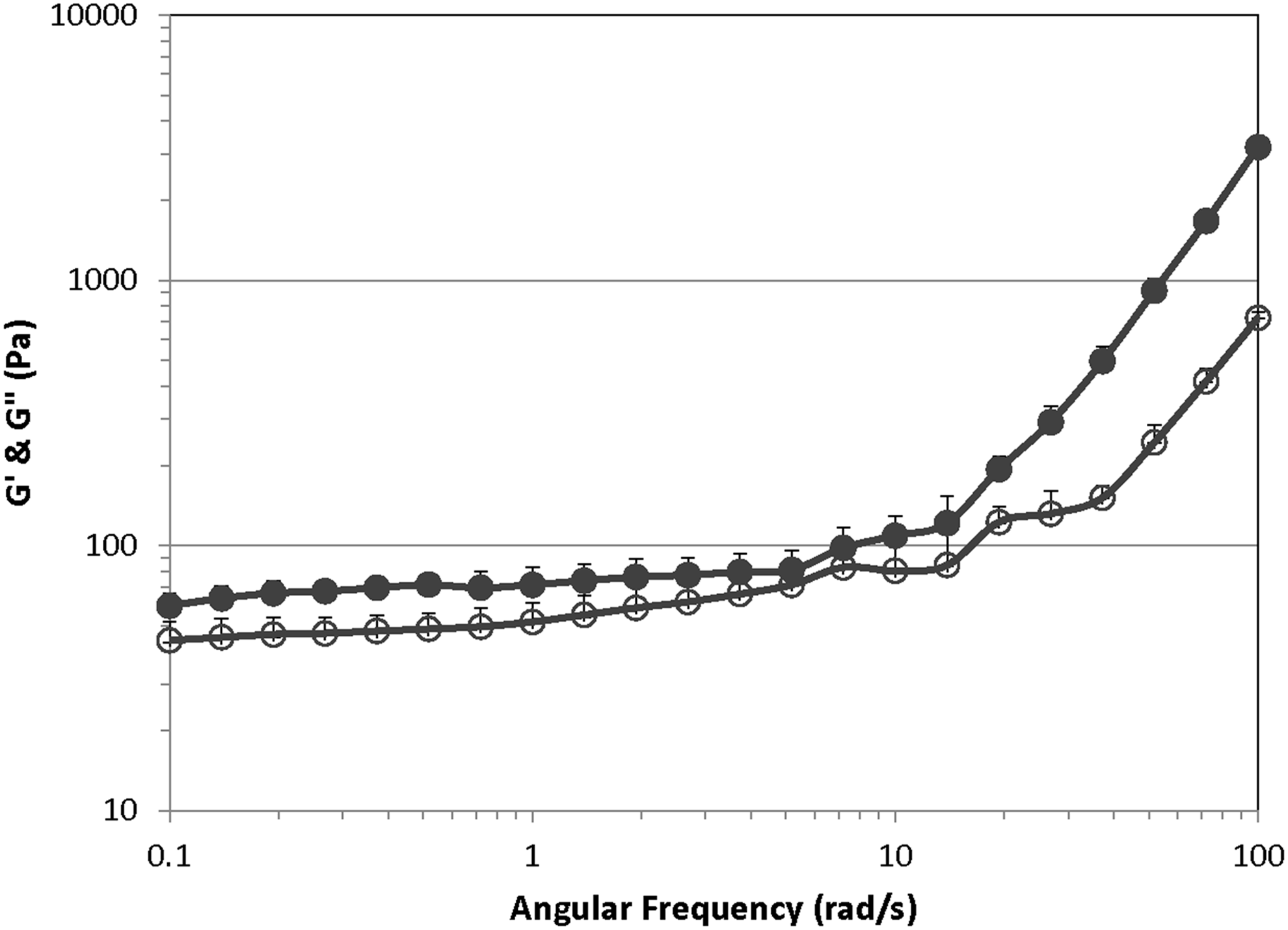
Oscillatory rheology frequency sweeps of Cell-Mate3D gels hydrated with fetal bovine serum (FBS) containing hMSCs. Frequency sweep measurements were made from 0.1 to 100 rad/s with 1% strain at 37°C. Filled symbols show the storage modulus, G′, and open symbols show the loss modulus, G″. Data represented as mean±SE (n=3).
The elastic modulus of Cell-Mate3D matrix with embedded hMSCs was 264±38 Pa.
hMSC culture in Cell-Mate3D hydrogel and time course assessment
At D0 histologic sections of preparations cultured in MSC growth medium showed a high cell density within the hydrogel with cells distributed throughout the construct. Cells were arranged in loose aggregates or scattered throughout the matrix without substantial gaps in distribution. Cells at this time point had rounded morphology and did not appear to be attached to the matrix surfaces (Fig. 3). Samples from D1 to D3 showed markedly reduced cell density with generally a loss of appearance of cell clusters. The hMSCs in these samples were now arranged on the surfaces of the matrix and had assumed a flattened to spindle-shaped morphology (Fig. 3). The D14 samples were similar in histologic features to the D1 and D3 samples but now secondary cell to cell structural arrangements were becoming evident in some regions.
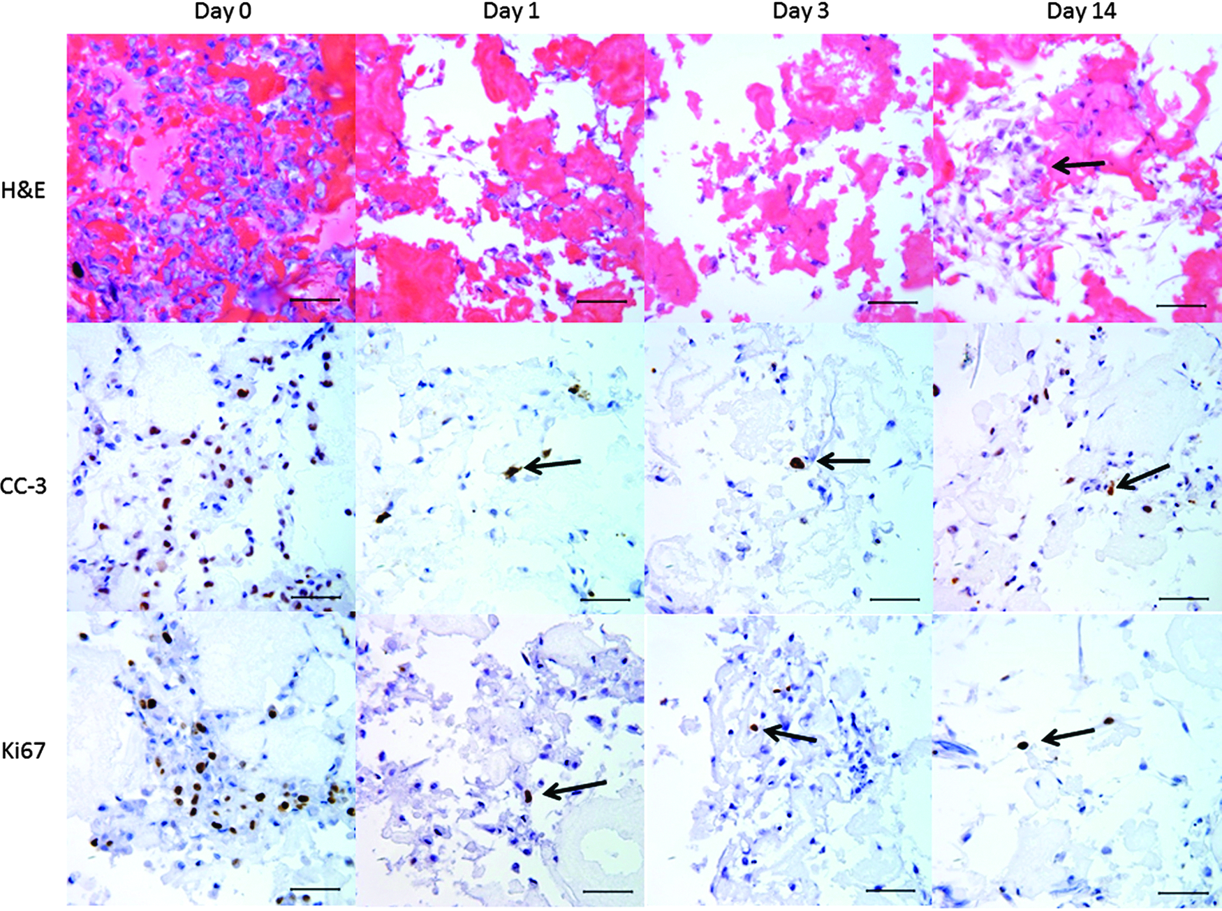
Histology of time course study of hMSC in Cell-Mate3D and maintained in MSC growth medium. On hematoxylin and eosin (H&E)-stained sections a relatively high cell density can be seen on day 0 with diminished cell density on days 1, 3, and 14. Cells on day 0 were arranged in loose clusters, had rounded morphology, and were not associated with matrix fibers. At later time points cells were typically attached to the matrix and had a more flattened to spindle morphology. On day 14 additional cell to cell arrangements independent of the matrix could also be found (arrows). Immunohistochemical stains for cleaved caspase-3 showed large numbers of labeling cells (brown label) on day 0 with diminished labeling cells on days 1, 3, and 14 (arrows). Similarly immunohistochemical stains for Ki67 showed greatest numbers of labeled cells on day 0 and fewer labeled cells on days 1, 3, and 14 (arrows) Scale bars=50 μm.
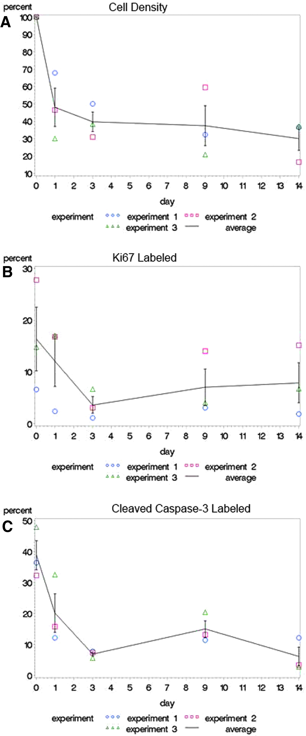
Quantification of cell density in the Cell-Mate3D matrix from D0 to D14 showed significant differences in cell density among the samples (ANOVA F test p<0.0001) (Fig. 4). Specifically, there were significantly lower cell densities on D1, D3, D9, and D14 versus D0 (Tukey-Kramer p≤0.002 for all pairwise comparisons) with mean cell density dropping to 48% of D0 values by D1 and to 30–40% on D3–D14. Comparisons between cell densities on D1–D14 were nonsignificant (Tukey–Kramer p>0.05). Corresponding to the decrease in cell numbers between D0 and later time points there was also a significant difference among samples in numbers of cells labeled with the apoptosis marker cleaved caspase-3 (ANOVA F test p<0.0001). Specifically, there were significantly greater numbers of cleaved caspase-3-positive cells in the D0 versus D1–D14 samples (Tukey–Kramer p<0.0001) and significantly more labeled cells in the D1 samples versus D3 (p=0.002) and D14 (p=0.0009) showing a declining expression of cleaved caspase-3 over time (Fig. 4B). All other pairwise comparisons did not significantly differ. Analysis of cells labeled with the cell division marker Ki67 showed significantly different values across the time points (ANOVA F test p=0.009); however, in pairwise comparisons only the D0 versus D3 comparison was statistically significant (Tukey–Kramer p=0.007). Thus, the levels of Ki67 labeling achieved a constant level following an initial drop from D0 levels (Fig. 4C).
Differentiation of hMSCs in Cell-Mate3D
On D14, the hMSC-loaded Cell-Mate3D constructs directed toward chondrogenic differentiation (n=3) showed marked alteration of cell morphology and ECM. The cells exhibited rounded morphology and were surrounded by pale eosinophilic (not shown) and Alcian blue-positive matrix consistent with cartilage differentiation (Fig. 5). Cartilage differentiation was nonuniform and was most evident in the outer one-third of constructs where there was increased ECM formation and remodeling of the Cell-Mate3D hydrogel material. Furthermore, cells and ECM in all D14 chondrogenic constructs were strongly positive for aggrecan, while both collagen II and collagen X (Fig. 5) showed strong intracellular staining and lesser amounts of staining in the surrounding ECM. These features were similar to those found for the hMSCs that underwent conventional pellet differentiation (Supplementary Fig. S1; Supplementary Data are available online at www.liebertpub.com/tea). Samples from D56 to D70 showed modest increases in aggrecan, type II collagen, and type X collagen labeled ECM in comparison to D14 samples (Fig. 5). In contrast, D14 controls (hMSCs in Cell-Mate3D in MSC growth medium) showed a minimal increase in Alcian blue matrix, and by immunohistochemistry they were negative for type II collagen and aggrecan, with positive cellular and minimal matrix staining for type X collagen suggesting early cartilage differentiation.
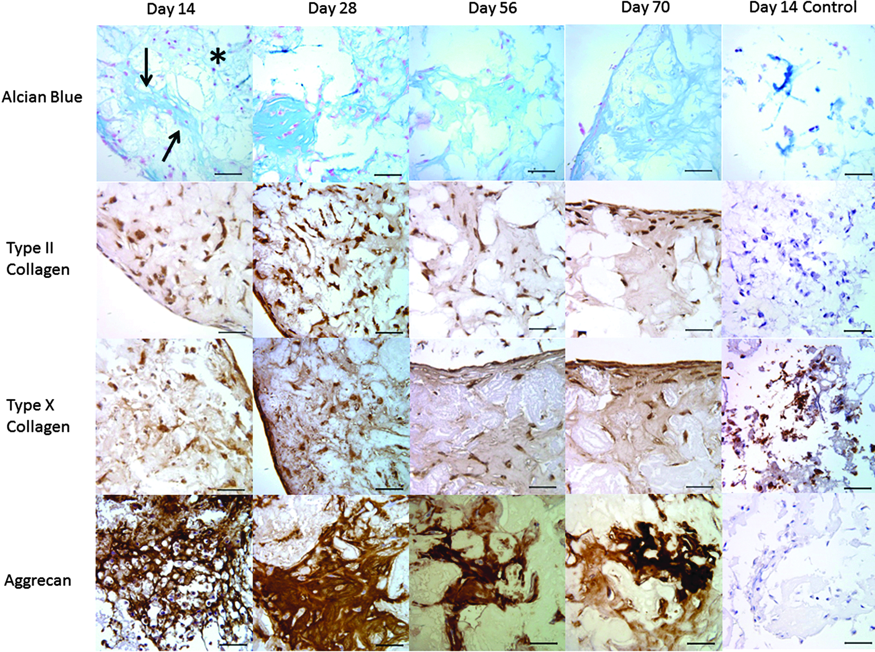
Histology of chondrogenic differentiation assays of hMSC in Cell-Mate3D. On day 14, samples showed extensive remodeling of the Cell-Mate matrix by differentiating hMSC at the periphery of the construct with increased extracellular Alcian blue-positive matrix formation (arrows) consistent with cartilage matrix. Central areas (*) do not show this distinct remodeling and retained the native structure of the hydrogel matrix. Histologic samples from chondrogenic differentiation assays on days 56 and 70 showed continued viability of the cells with modest increases in Alcian blue-positive matrix compared to day 14. Immunohistochemical stain for the cartilage marker aggrecan showed strong labeling of extracellular matrix (ECM) in subsurface regions of the constructs at all time points. Immunohistochemical stains for type II and type X collagens on day 14 samples showed strong cellular staining in most cells near the periphery of the constructs but little matrix staining. On days 56 and 70 there was strong cellular staining for both markers and weak staining of the ECM. Day 14 controls (hMSC in Cell-Mate3D in MSC growth medium) showed a minimal increase in Alcian blue matrix, and by immunohistochemistry were negative for type II collagen and aggrecan, with positive cellular and minimal matrix staining for type X collagen suggesting early cartilage differentiation. Scale bars=50 μm.
By D28–D30 the hMSC-loaded Cell-Mate3D constructs directed toward osteogenic differentiation (n=3) showed markedly increased optical density of the hydrogels, which could be seen by light microscopy as dark serpentine regions in the constructs (not shown). Histologically, many cells in all constructs were small with angular profiles and were arranged on surfaces of pale eosinophilic to basophilic ECM and bore close resemblance to osteoblasts (Fig. 6). At all time points von Kossa stains showed marked ECM mineralization consistent with bone differentiation, which tended to increase over time (Fig. 6). At all time points a majority of cells showed positive staining for Runx2. Osteocalcin was minimally seen in cells on D14 but showed strong staining in a majority of cells at D28. On D56 and D70 within the body of the hydrogel construct the majority of cells continued to show strong osteocalcin immunoreactivity but, additionally, there was also prominent staining of the ECM further confirming osteogenic differentiation. D14 controls (hMSC in Cell-Mate3D in MSC growth medium) showed negative von Kossa stains, rare osteocalcin-positive cells but no matrix staining, and weak to moderate Runx2 staining in the majority of cells, indicating little osteogenic differentiation in growth medium. Typical osteogenic differentiation with positive von Kossa and Runx2 immunohistochemical stain was found with conventional plate osteogenic differentiation assays of these same hMSCs (n=3) (Supplementary Fig. S1). Osteocalcin stains on these control differentiation assays were negative (not shown).
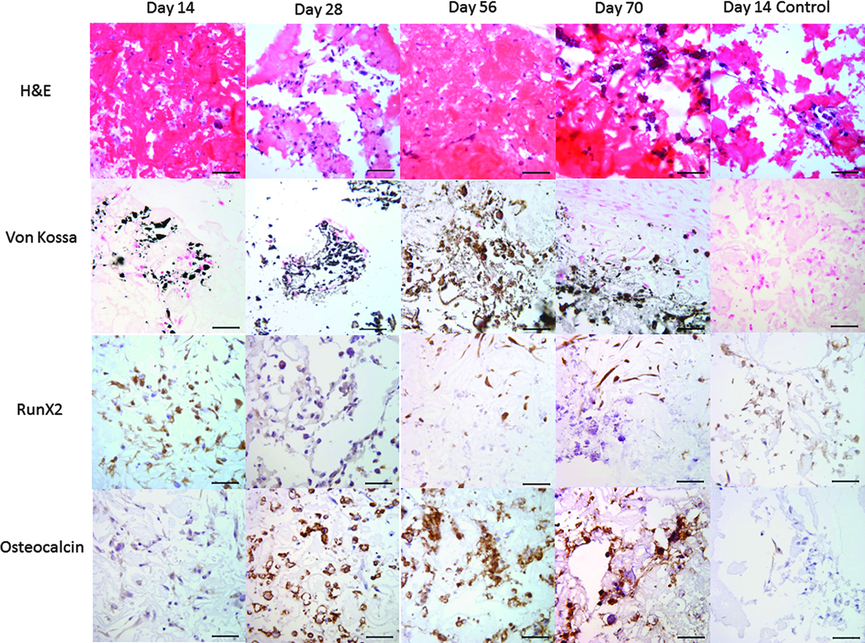
Histology of osteogenic differentiation assays of hMSC in Cell-Mate3D. Cells in these constructs were small with angular profiles and were arranged on surfaces of pale eosinophilic to basophilic ECM and bore resemblance to osteoblasts. Von Kossa stains showed widespread mineral deposition, which increased over time. Immunohistochemical stains for Runx2 showed moderate to strong intracellular staining of the majority of cells at all time points. Immunohistochemical stains for osteocalcin showed only weak intracellular staining on day 14 with increasing staining at day 28 and widespread cellular and ECM staining on days 56 and 70 consistent with osteogenic differentiation. Day 14 controls (hMSC in Cell-Mate3D in MSC growth medium) showed negative von Kossa stains, rare osteocalcin-positive cells but no matrix staining, and weak to moderate Runx2 staining in the majority of cells, indicating little osteogenic differentiation. Scale bars=50 μm.
On D14 the hMSC-loaded Cell-Mate3D constructs directed toward adipose differentiation (n=3) showed a minority population of scattered cells that contained lipid droplets, with similar findings on D56 and D70 (Fig. 7). Furthermore, at all time points, many of the cells containing lipid also showed nuclear immunohistochemical staining for PPAR-γ consistent with adipocyte differentiation (Fig. 7), although at a relatively low level. D14 controls (hMSC in Cell-Mate3D in MSC growth medium) showed rare cells with nuclear PPAR-γ staining but these cells did not contain appreciable lipid droplets (Fig. 7).
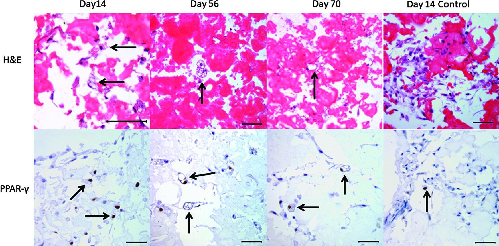
Histology of adipogenic differentiation assays of hMSC in Cell-Mate3D. H&E stained samples at all time points showed scattered cells with one or more round, clear cytoplasmic vacuoles consistent with lipid vacuoles (arrows). Many of the lipid-containing cells showed nuclear immunohistochemical staining for PPAR-γ (arrows) consistent with adipose differentiation. Day 14 controls (hMSC in Cell-Mate3D in MSC growth medium) showed rare cells with nuclear PPAR-γ staining but these cells did not contain appreciable lipid droplets. Scale bars=50 μm.
In addition to the directed differentiation within the hydrogel constructs described above, an additional phenomenon was observed on the surfaces of the days 56 and 70 osteogenic and adipogenic differentiation assays, whereby there was the formation of a distinctly different tissue (Fig. 8). In all instances, this region was populated by oval to fusiform cells in roughly parallel alignment with the surface of the construct and separated by abundant Alcian blue-positive matrix, which was also strongly immunoreactive for aggrecan. The majority of cells in these regions were also positive for type II and type X collagens and some weak ECM staining for these markers was also seen in the tissue on the D56 and D70 osteogenic differentiation assays. In all instances, a minority population of cells in these regions was weakly positive for Runx2 but osteocalcin was not detected in the surface tissue. Some lipid-containing cells with nuclear labeling for PPAR-γ were also present in the surface tissue of the adipogenesis-induced hydrogels but not in the osteogenesis-induced hydrogels.
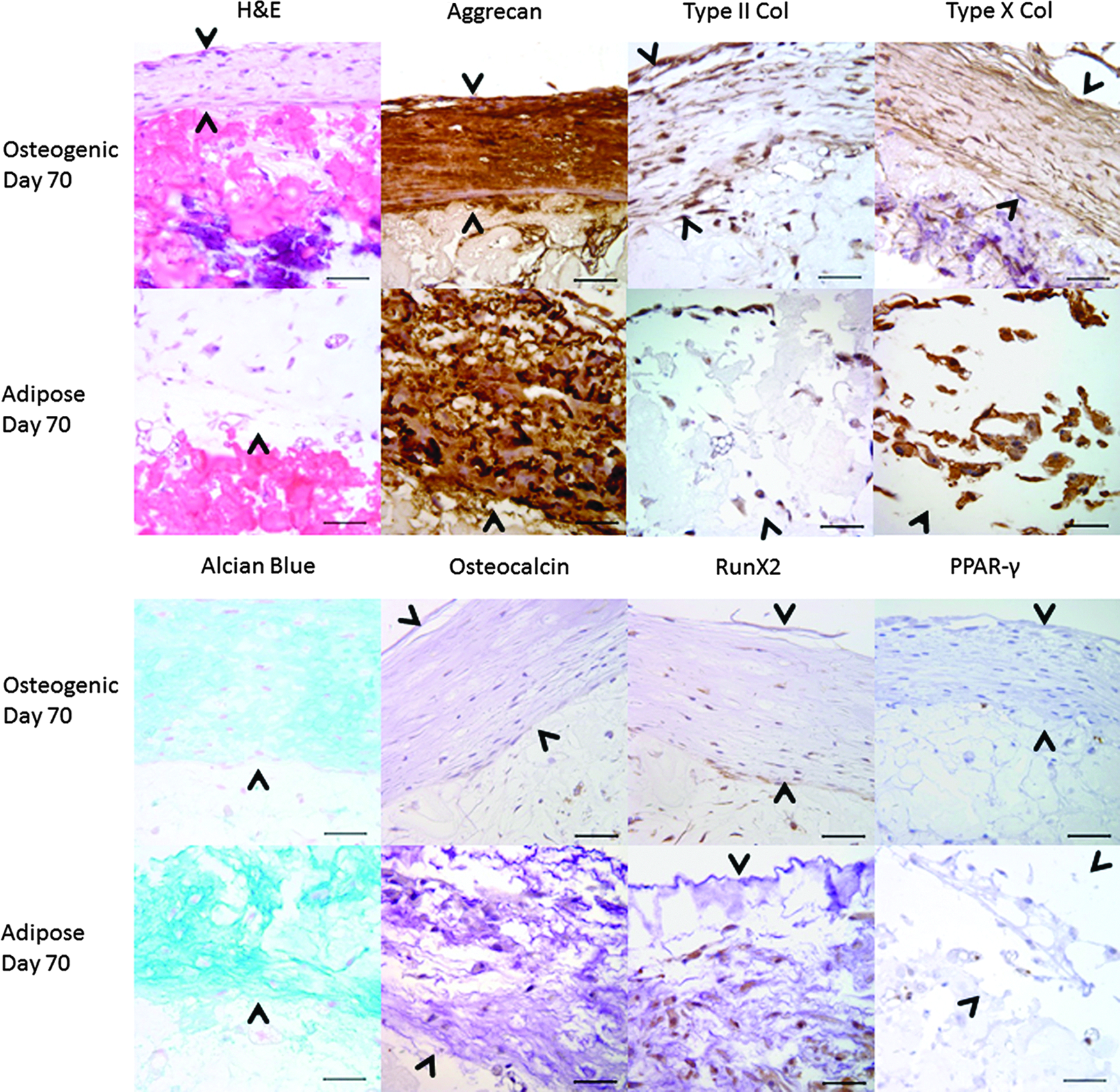
Histology of surface tissue on day 70 of osteogenic and adipogenic differentiation assays of hMSC in Cell-Mate3D. H&E-stained sections show formation of a distinct surface tissue under both conditions, which histologically differs from the differentiation observed in the underlying hydrogel construct (arrow heads delineate these regions). This surface tissue was readily distinguished from the underlying hydrogel matrix, which is bright red in the H&E sections. The surface tissue was comprised of oval to fusiform cells separated by abundant Alcian blue-positive matrix, which was also strongly positive on immunohistochemical stains for aggrecan. Cells in this tissue were also strongly positive for both type II and type X collagens with some ECM staining for these components in the tissue from the osteogenic differentiation. The tissue was also notably negative for osteocalcin and showed only weak cellular staining in a few cells for Runx2. In the adipose differentiation there were also some lipid-containing cells with positive PPAR-γ nuclear labeling within this surface tissue. Scale bars=50 μm.
Discussion
A wide array of 3D scaffolding materials, including a variety of hydrogel materials have been employed in tissue engineering applications and in vitro cell culture. All have advantages and limitations, the latter of which include a requirement for cytotoxic cross-linkers, long polymerization times, structural rigidity, and inability of cells to be distributed throughout and attach to the matrix. Here, we demonstrate the formation and utility of a novel 3D cell culture matrix comprised of HA and CT, termed Cell-Mate3D. This construct is unique in that its mechanical properties result from the formation of insoluble PEC fibers.24,25 Consequently, cross-linking of either component is not required to yield structural integrity appropriate for in vitro 3D cell culture. The stability of this material is demonstrated by its continued integrity after 10 weeks in culture. In addition to its inherent chemical stability, it is also possible that embedded cells provide additional integrity to the construct through CD44-mediated interaction with HA.26–30 Furthermore, electrostatic forces between the negatively charged cell membrane and the positively charged NH3 groups on CT may further stabilize the matrix. The attachment of hMSC to the matrix, likely via the CD44 HA receptor, was evident in the morphologic transition from D0 to D1 and beyond as the cells rearranged from loose unattached clusters to a flattened and spindle-shaped morphology with the cells intimately associated with the matrix surfaces. This was also illustrated by SEM imaging, where it was possible to see cells surrounded by and associated with a web-like array of fibers.
Mechanical properties of the ECM have been shown to significantly impact cellular response and are an important factor in directing cellular differentiation.31–34 Under conditions utilized in this study, using FBS as the hydration fluid, which contained 19 million cells per 500 μL, we found that Cell-Mate3D exhibited an elastic modulus of 260 Pa. Thus, this approach yielded a compliant or soft construct, which would tend to emulate the matrix of the blastula or the central nervous system35,36 and be less favorable to the differentiation of MSCs toward adipose, cartilage, or bone tissue. Nevertheless, as demonstrated in the differentiation assays, the matrix compliance did not prevent hMSC differentiation to directed cell types. Assessment of pluripotent stem cell or neural progenitor cell behavior in the Cell-Mate3D matrix was beyond the scope of this study but may prove to be an additional application of this matrix.
An important feature of the hydrogel setup employed in the Cell-Mate3D system is the ability to incorporate cells and other components into the hydration fluid. As shown in this study, this results in cells immediately incorporated in the hydrogel and dispersed throughout the matrix, although a completely uniform distribution was not achieved. A potential concern of embedding cells into the matrix was maintaining cell viability during and after the transfer. While the cause is unclear, the viability data presented here indicate that significant cell death via apoptosis did indeed occur in the first 24 h after embedding. Subsequently, cell populations plateaued and maintained a relatively steady state (Fig. 4). The steady state appeared to be balanced by levels of apoptosis (cleaved caspase-3 expression) and mitosis, (Ki67 expression), which dropped from initially high levels to plateaus, and then maintained for the 2-week duration of the study. Thus, the initial cell loading density was not maintained but resulted in a cell density of about 30–40% of initial levels. Therefore, while the Cell-Mate3D system does not introduce cytotoxic cross-linking agents into the system, hMSC viability from D0 to D1 in Cell-Mate3D was low compared to other PEC and covalently cross-linked hydrogel constructs reported in the literature.20,37–39 Although the mechanism for this loss of viability was not specifically addressed in the current study, contributing factors may include cellular disruption occurring during the vortexing step in preparation of the hydrogel construct, cell damage incurred during the poly-electrolytic complexation of HA to CT, and formulation of the hydration fluid. Further studies are in progress to evaluate each of these factors and to make adjustments to optimize hMSC viability in the system.
However, even with the apparent initial cell loss and the low elastic modulus of the cell embedded Cell-Mate3D construct, we demonstrated directed chondrogenic, osteogenic, and adipogenic differentiation within the matrix similar or superior to typical plate (osteogenic and adipogeneic) or micromass pellet (chondrogenic) differentiation assays of the same hMSC population. The system was robust in this regard and resulted in appropriate differentiation in every case. Furthermore, differentiation toward both cartilage and bone progressed over time with continued cell viability in all of the differentiation assays for the duration of the study (70 days).
Interesting emergent features formed in the adipogenic and osteogenic differentiation assays that were not present in correlating two-dimensional (2D) culture. Tissue that formed on the surface of osteogenic and adipogenic constructs had morphologic, histochemical, and immunohistochemical resemblance to cartilage despite the continuance of only osteogenic or adipogenic culture medium. The mechanisms underlying this off-target cartilage differentiation were not addressed in this study, but causative mechanisms might include: (1) an increase in the intrinsic matrix stiffness over time, (2) increased rigidity of the constructs due to contractile forces exerted by the embedded cells on the scaffold material, (3) elaboration of cytokines or growth factors from embedded cells, (4) release of bioactive constituents from the hydrogel material such as HA, and (5) combinations of these factors. In both cases, the long-term osteogenic and adipogenic Cell-Mate3D constructs produced intriguing structures exhibiting increased complexity compared to 2D cell culture conditions, which may represent a form of tissue organoid.
In conclusion, Cell-Mate3D's ability to maintain cell viability and permit the differentiation of hMSCs into chondrogenic, adipogenic, and osteogenic phenotypes, including the formation of emergent tissue-like features, suggests that it may serve as a flexible platform for additional applications. Furthermore, the ability to easily incorporate additional cell types, ECM components, growth factors, or other desired factors into the fluid of hydration would facilitate the development of more complex in vitro tissue models. Cells that express the CD44 receptor or otherwise interact favorably with HA, such as MSCs, embryonic stem cells/induced pluripotent stem cells, and cancer stem cells may be particularly well suited to this matrix. We therefore suggest that Cell-Mate3D could be utilized as a tool for additional 3D cell culture applications in tissue engineering, regenerative medicine, and cancer stem cell biology.
Footnotes
Acknowledgments
The authors would like to acknowledge the technical assistance of Paula Overn, Josh Parker, and Lindsey Harper in the preparation, histochemistry, and immunohistochemistry of histological samples.
Disclosure Statement
J.H.B., B.A.L., and Y.W.C. are employees of Bioactive Regenerative Therapeutics, Inc., which provided the Cell-Mate3D materials for this study. No competing financial interests exist for any of the other authors.
References
Supplementary Material
Please find the following supplemental material available below.
For Open Access articles published under a Creative Commons License, all supplemental material carries the same license as the article it is associated with.
For non-Open Access articles published, all supplemental material carries a non-exclusive license, and permission requests for re-use of supplemental material or any part of supplemental material shall be sent directly to the copyright owner as specified in the copyright notice associated with the article.