
Abstract
We designed a sheet-like bone substitute capable of adapting to different geometries and becoming a standard tissue-engineered process for bone surgery. Preosteoblastic cells were seeded on to a monolayer of calcium phosphate granules and cultured in a flat parallelepipedic cell culture chamber for 1 month. From the various diameters of the granules examined, the 80–200 μm group exhibited the most homogeneous performances regarding both biological (cell morphology, viability, differentiation, and simple metabolic activity) and mechanical (cohesion and stress-strain behavior) properties. This sheet was easy to handle after extraction from the culture chamber and showed versatile geometry and flexibility, making it easy to use for surgeons, especially for small defects of the maxillofacial area.
Introduction
T
Despite a large number of studies with a variety of cells, materials, shapes, or bioreactors, to date there is still no consensus on the best process leading to adequate bone substitutes based on tissue engineering.6–9 Various materials have been studied and considered as possible scaffolds for in vitro bone ingrowth, such as ceramics,10,11 polymers,12,13 natural biomaterials such as silk or chitosan14–16 or to a lesser extent metals, 7 and composites of these different groups. 17 Relevant shapes for the scaffold have also been debated, with different studies on granules and microspheres, 18 fibres,19,20 hydrogels, 21 or engineered complex structures.22,23 In addition, a bioreactor's ability to mimic in vivo environmental conditions is still an unsolved issue, with a wide variety of systems using mechanical stretching, flow perfusion, spinner flasks, rotating wall vessels,24–26 or specifically designed chambers. 27
Targeted applications may be helpful for making choices from among all the possibilities. Besides intrinsic mechanical and biological properties (micro- and macroporosity,28,29 pore interconnectivity, 30 osteoconduction, and osteoinduction 31 ), specific clinical requirements also have to be satisfied by the target substitute: it must be adaptable, and fit with all geometries of the bone mass loss in the surgical area, for instance, the complex geometry of the oral and maxillofacial area; it must have optimal cell viability and differentiation, and be stretched or folded during surgery without damage, thanks to the relevant mechanical properties. 32 From this point of view, current solutions have limitations in terms of their mechanical and biological behavior, 7 the vascularization of the scaffold, 33 and the cost for a standard bioreactor process. 34
Following a previous study of these clinical requirements, 35 we selected a set of parameters for a new in vitro-built bone substitute to be applied in bone surgery. A monolayer of calcium phosphate (CP) granules was selected as the scaffold, inside a specific flat cell culture chamber with a large culture surface. CP ceramics have been widely used for bone and dental tissue engineering 36 thanks to their relevant properties, especially osteoconduction and osteoinduction. 10 In addition, stacking granules may create the required porous interconnected structure and high surface-to-volume ratio, 2 and it showed better results than CP blocks. 37 This biomaterial was colonized by MC3T3-E1 preosteoblastic cells, a usual cell line to determine the potential of new scaffolds for proliferation, adhesion, and differentiation of bone tissue.38–40 No growth factor was added to the culture medium as we expected the system culture chamber+biomaterial to be efficient in promoting cell growth and differentiation. After 1 month of culture, both the biological and mechanical properties of the substitute were examined, depending on the CP granules' mean diameter. We focused particularly on the morphology and shape relevance of the bone substitute for clinical applications, especially the small geometry of defects in the maxillofacial area.
Materials and Methods
Cell culture chamber
We used the Clinicell25® culture cassette (Mabio-International) as a specific flat cell culture chamber to obtain the in vitro-built tissue. The geometry is a parallelepipedic chamber with a 24 cm2-culture surface for a 10 mL-volume filled with medium, with two Luer Lock connections (Fig. 1). It is gas permeable and plasma treated to promote cell adhesion. After 1 month of culture, the upper side of the chamber was opened to remove the tissue and to perform the analysis.
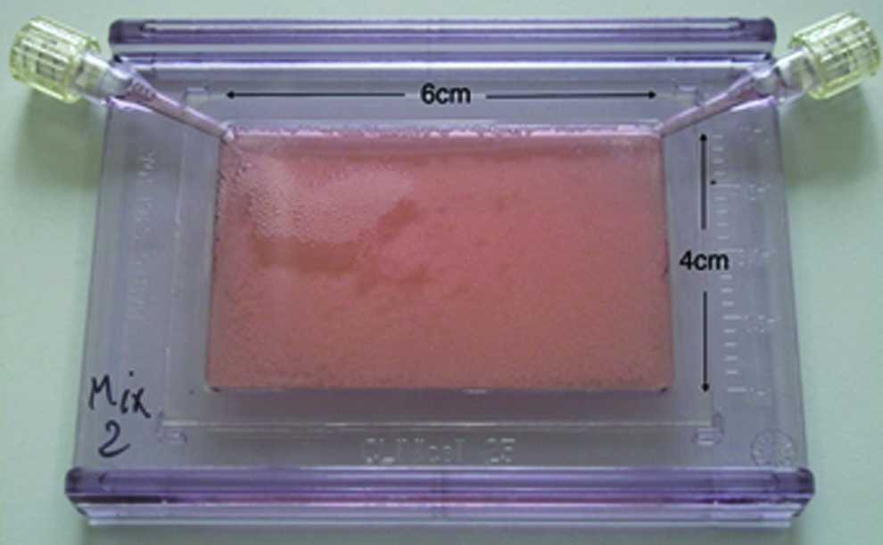
Geometry of the Clinicell25® cell culture chamber (Mabio International). Color images available online at www.liebertpub.com/tea
Biomaterial
CP granules (20% hydroxyapatite (HA) Ca10(PO4)6(OH)2, 80% β-tricalcium phosphate (TCP) Ca3(PO4)2; Biomatlante) were inserted as the scaffold material inside the culture chamber to obtain a single monolayer covering the whole surface. The layer was stable enough after 2 days of culture to prevent displacements of the biomaterial when the culture medium was changed. Different mean diameters of granule were investigated: <80, 80–200, 500–1000 μm, and mixed composition (Table 1). Hereafter, we refer to these groups as the S (small), M (medium), L (large), and Mix groups, respectively. A group without CP granules inside the culture chamber was studied as a control.
L, large; M, medium; S, small.
The cell culture chambers with CP granules were preincubated with complete medium for 48 h before cell seeding.
Cell culture
The murine preosteoblastic cell line MC3T3-E1 subclone 4 (ATCC® CRL-2593) was cultured in α-MEM Eagle medium with stabilized glutamine (Pan-Biotech) supplemented with 10% fetal bovine serum (FBS; Gibco Invitrogen), 1% Penicillin-Streptomycin (Gibco Invitrogen), 1% sodium pyruvate (Gibco Invitrogen), and maintained at 37°C and 5% CO2. To develop the substitute, cells were harvested at P24 and seeded in a specific cell culture chamber (10 million cells in each chamber) for 1 month with the same supplemented medium and 50 mg/L of ascorbic acid (Sigma Aldrich). The medium was changed every 2 or 3 days.
Scanning electronic microscopy observations
After the substitute was removed from the culture chamber, tissue morphology and cell adhesion were observed using scanning electronic microscopy (SEM; Philips XL30 ESEM-FEG). The samples were immersed in Rembaum solution (as described in Rajaraman et al. 41 ) for 24 h, rinsed with de-mineralized water and then coated with gold before observation.
Viability test
Cell viability was estimated by a Live/Dead® kit (Invitrogen) according to the manufacturer's protocol. Briefly, Calcein AM (1 μM) and Ethidium homodimer-1 (EthD-1, 1 μM) fluorescent dyes were respectively employed to stain viable and dead cells. The samples were observed using fluorescence microscopy (Leica microsystems), allowing us to qualitatively determine cell viability and morphology of leaving cells. Holes in the CP granule monolayer were observed with phase contrast. Cell nuclei were counterstained by Hoechst 33342 dye (0.5 μg/mL; Invitrogen) to estimate the viability rate. Image processing was carried out with the free software CellProfiler (www.cellprofiler.org). Briefly, color images were converted to grayscale images, then erosion was applied (a morphological operation to separate close nuclei from each other) and cells were counted based on size and intensity of the nuclei. At least five areas were randomly chosen for image acquisition on the sheet-like substitute for each group.
Metabolic activity test
The cell metabolic activity was quantitatively measured with the AlamarBlue® Assay (Invitrogen). One day per week, the culture medium was removed from the culture chamber in a dark room and replaced with culture medium complemented with 10% of AlamarBlue solution. The culture chamber was incubated for 45 min at 37°C. The complemented medium was then removed and fluorescence intensity was measured (excitation wavelength: 560 nm, emission wavelength: 595 nm). The changes in fluorescence correspond to the biochemical reduction of the AlamarBlue solution in the culture medium resulting from cell metabolic activity (redox reaction in the mitochondria). The fluorescence intensity of the complemented medium without contact with the cells was substracted from the results before the analysis.
Differentiation test
Alkaline phosphatase (ALP) activity, an early marker of osteoblast differentiation, was assessed by a histochemical semi-quantitative approach, using an ALP kit (Sigma-Aldrich) according to the manufacturer's protocol. Samples were observed with a binocular magnifier (Leica Microsystems).
An Enzyme-linked Immunosorbent Assay Kit (ELISA kit; Cloud-Clone Corp.) for osteocalcin (OCN) was also used according to the manufacturer's protocol to study the late-stage differentiation of samples. Briefly, the culture medium removed from the chambers at day 30 after 24 h of incubation was added in a plate precoated with an antibody specific to OCN. After incubation with the kit's reagents, color changes were measured with spectrofluorimetry at 450 nm and linked to the concentration of OCN with a standard curve. The results were compared to the protein total mass using a standard Bradford assay. Lysis was performed with RIPA buffer (Fisher Scientific), then Coomassie blue was added (Interchim) and absorbance was read at 570 nm. The standard curve was obtained with bovine serum albumin (Interchim).
Stress-strain tests
Using the Electroforce 3200® system (Bose), conventional tensile tests were conducted on preloaded tissue strips (1×3 cm) at the speed of 0.1 mm/s until failure. Forces versus displacement were recorded. In situ tests were performed with SEM observations and a specific tensile testing machine (Deben Microtest 300), with the same speed and sample's size as standard tensile tests, as a tool for understanding the relationship between the tissue's microstructure and its mechanical properties. To spot easily the region of interest, a cutout was made in the middle of the strip before mounting. Displacement was regularly stopped to perform image acquisition of the crack propagation and evaluate damage mechanism.
Statistic tests
At least six independent experiments were performed for each group (S, M, L, Mix, or control), then mean and standard deviation were calculated. Two-way analysis of variance with Tukey's test (metabolic activity) and unpaired t-test (viability, OCN production) were used to define the significance of the results.
Results
Cell culture chambers were filled with CP granules of a specific size (S, M, L, Mix) inserted as a monolayer. Ten million MC3T3-E1 cells were seeded in the medium-filled chamber. Cell colonization was assured via the follow-up of metabolic activity. After a 4-week incubation, each culture chamber was opened to collect the reconstructed tissue.
Tissue cohesion
After 1 month of culture in the chamber, we investigated the possibility of extracting a biohybrid sheet-like substitute made of bone cell tissue and CP granules from the chamber. The most cohesive sheet was the M group: it was possible for the substitute to be stretched or folded and for it to stay cohesive (Fig. 2). The S group offered the most heterogeneous results, with, in some tissue areas, the worst handling (no possibilities for removing the tissue from the chamber) and in other areas a tissue as cohesive as that of the M group. The other groups (L and Mix) had a sheet-like morphology inside the chamber, but the substitute lost its integrity once removed. In the control group (chamber without CP granules), cells organized in a layer that we were not able to remove.
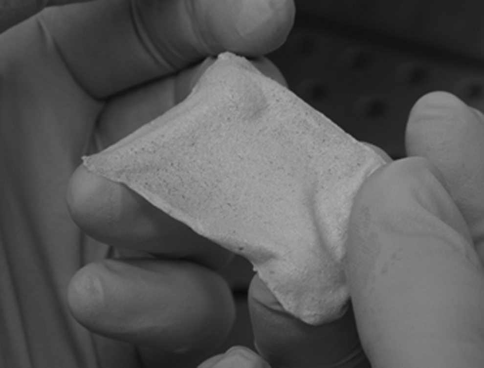
Tissue cohesion of the M group's sheet-like substitute removed from the cell culture chamber (cells on 80–200 μm calcium phosphate [CP] granules after 1 month of culture).
To explain these different tissue cohesions, SEM observations were conducted. The control group (Fig. 3A) showed a dense and continuous cell tissue. CP granules without cells can be seen on Figure 3B. We found the same homogeneous tissue covering the granule monolayer for the M and Mix groups (Fig. 3D, F). Concerning the L group, the cells were more likely to attach and grow inside the macroporosity of the granules (Fig. 3E). There was only heterogeneous tissue with cell spots when samples from the S group were observed (Fig. 3C).
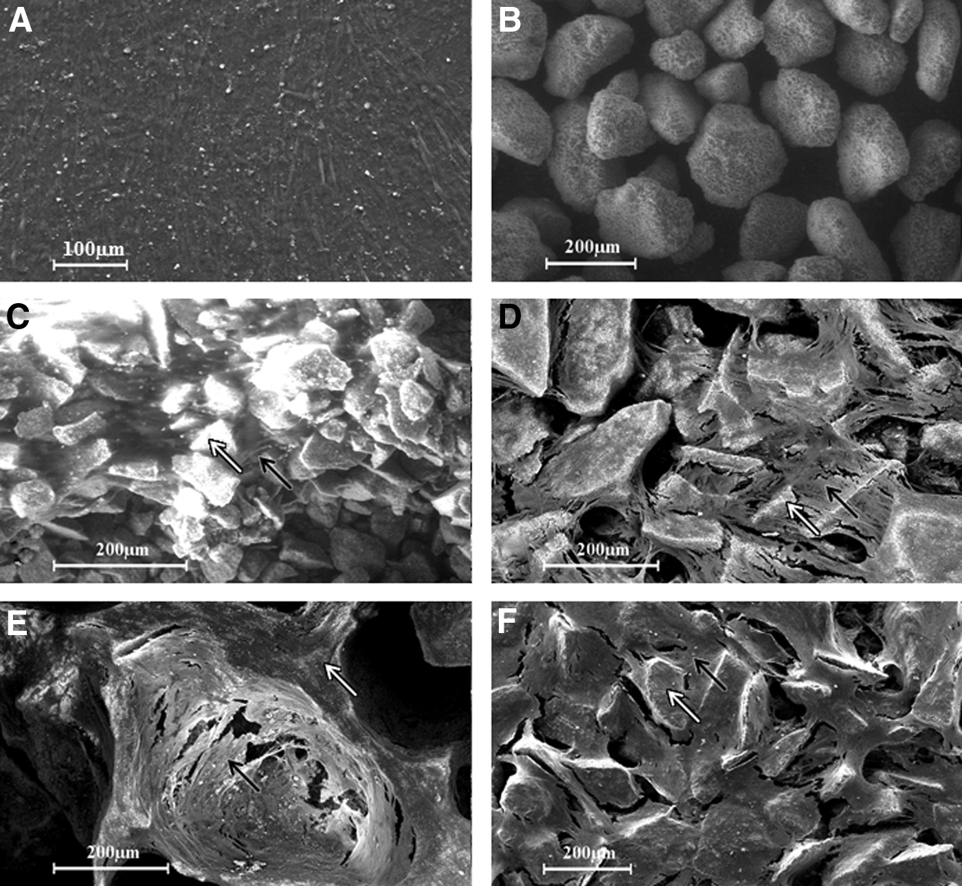
Scanning electronic microscopy (SEM) observations of the in vitro-built tissue after 1 month of culture. MC3T3 subclone 4 cells
Cell viability
We visualized granules colonization by living cells of each group under fluorescence microscopy using Calcein AM. As seen above with SEM observations, the control group showed a continuous cell tissue (Fig. 4A) that we found also on the CP granule layer for M and Mix groups (Fig. 4D, F, CP granules' autofluorescence can be seen on Fig. 4B). The S group's tissue was heterogeneous with only spots of living cells (Fig. 4C). We noticed few cell bridges between granules for the L group (Fig. 4E).
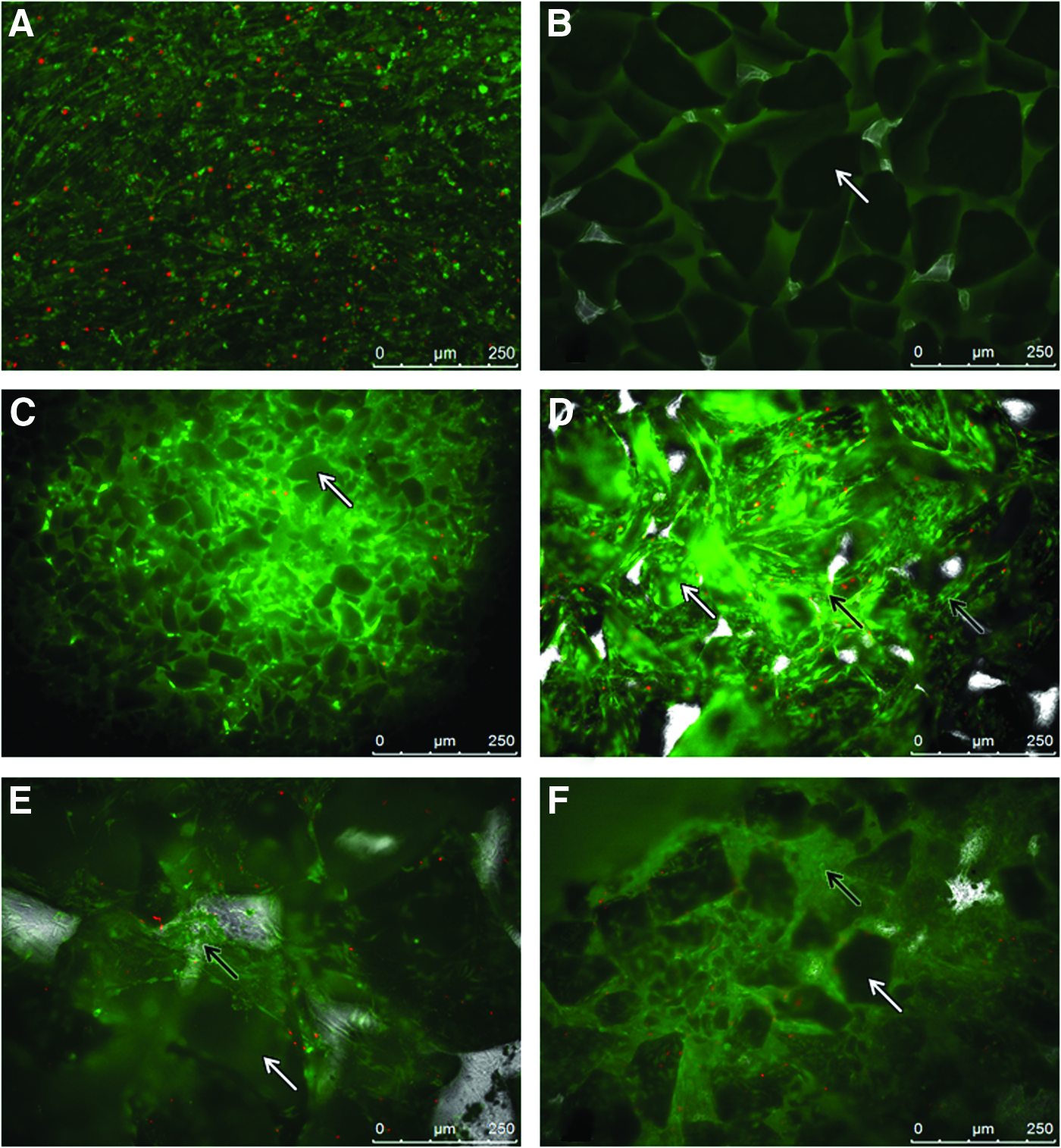
Fluorescence microscopy observations of the in vitro-built tissue after 1 month of culture. MC3T3 subclone 4 cells
Analysis of fluorescence microscopy images allowed us to evaluate the viability percentage from the ratio between dead cells' nuclei (visualized with EthD-1 dye) and all existing nuclei (Hoechst 44432 dye) (Fig. 5). The S and L groups showed the highest viability rates (76%±24% and 66%±25% respectively), but with relatively high standard deviations due to the heterogeneity of the tissue as seen above. Viability dropped to 59%±18% for the M group and to 50%±17% for the Mix group. Standard deviations were lower than in the S and L groups, due to better homogeneity. However, the viability rates of each group showed values not statistically different from control condition (62%±15%), suggesting that the CP granules did not induce specific cell mortality.
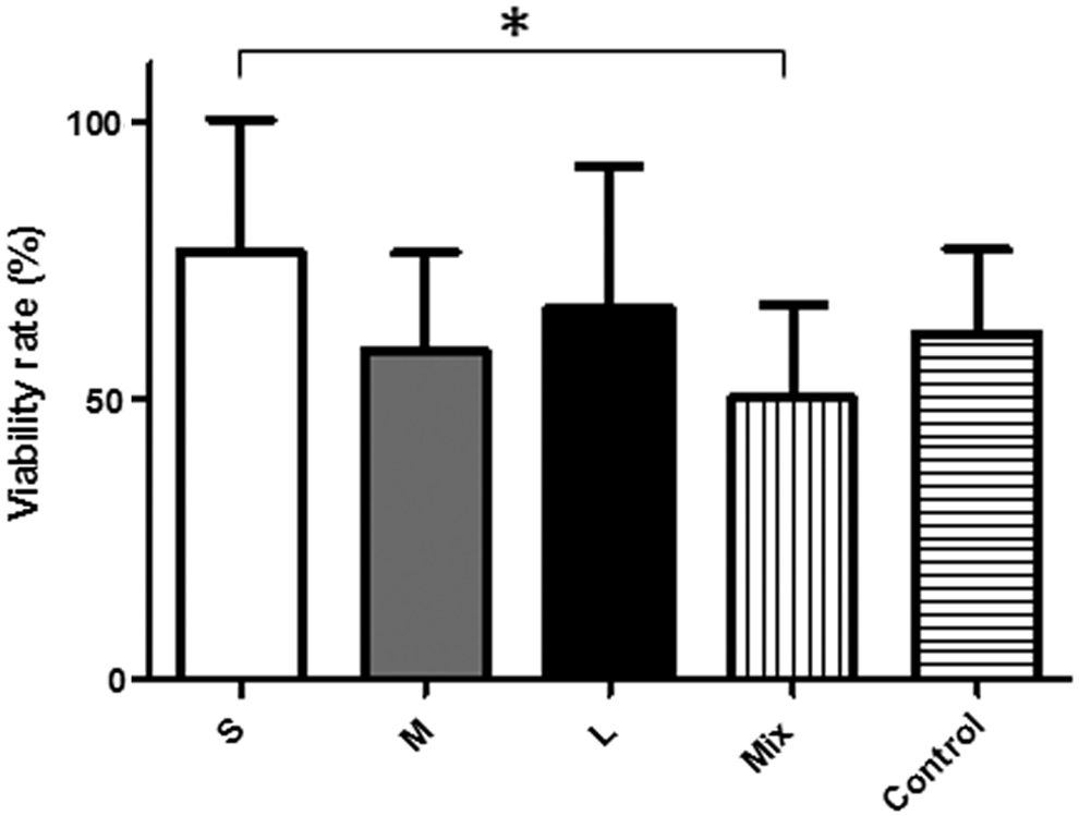
Viability rate of each group after 1 month of culture measured with fluorescence microscopy images treatment: S, M, L, Mix, and without CP granules (control). Ratio between the number of dead nuclei seen with EthD-1 dye and all nuclei seen with Hoechst 33342 dye with standard deviations (*p<0.05).
Metabolic activity
The metabolic activity of the cells in each group was investigated once a week throughout the culture period, using the AlamarBlue assay to evaluate mitochondrial reduction activity. The results, expressed with arbitrary units of the spectrofluorimeter, allowed us to estimate the cell viability and global proliferation of the rebuilt tissue.
To show the variation profiles during the culture, data were normalized by the fluorescence intensities recorded on day 7 (Fig. 6). We were so able to highlight changes in the metabolic activity throughout the month of culture and to perform a statistical study. Two main profiles were noticed: on the one hand, S and L groups showed increasing metabolic activity week after week, with the highest increase for the S group (statistically different from the control group at day 14, p<0.05 and day 21, p<0.0001). On the other hand, stable profiles were obtained for M and Mix groups, where metabolic activity appeared to remain constant, as it was noticed for the control too.
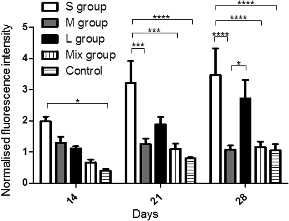
Metabolic activity for each group measured with Alamar Blue, mean and standard deviation for each group. Data were normalized from the results at day 7 by the spectrofluorimetry arbitrary unit (*p<0.05, ***p<0.001, ****p<0.0001).
To explain these two profiles, untreated data were compiled to show the evolution in metabolic activity between the first (Y axis) and the last (X axis) measurements, that is, between 7 and 28 days (Fig. 7). For each chamber, data points were included in elliptic domains where the major axis was defined using the linear regression line and the minor axis using the corresponding correlation coefficient (MS Excel software). It appeared that groups previously described with stable metabolic profiles (M and Mix groups) showed higher values of metabolic activity, except for the control group. Especially, most of the points of the M group were located near the diagonal, which means that metabolic activity was stable over time of culture, despite the variability.
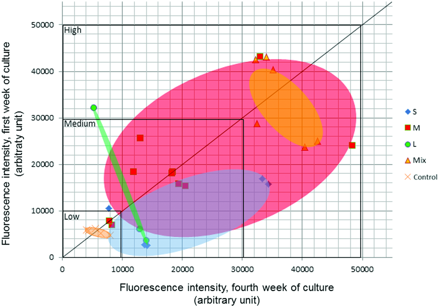
Metabolic activity for each group measured with Alamar Blue, evolution between the first and the fourth weeks of culture. Decreasing and increasing metabolic activities over time take place respectively in the upper left area and in the lower right area, and data on the x=y line stand for stable activity. Data points are included in elliptic domains for each group. The major axis of each domain was defined using the linear regression line and the minor axis using the corresponding correlation coefficient (MS Excel software). Color images available online at www.liebertpub.com/tea
Differentiation
Subclone 4 cells from the preosteoblastic MC3T3-E1 line are not able to produce ALP in conventional culture while they are not totally differentiated into osteoblast cells. 38 This enzyme was therefore a suitable marker for investigating the influence of CP granules' size on cell differentiation after 1 month of culture in the chamber. In addition, staining for ALP activity on the whole sheet-like substitute allowed us to identify the high differentiation areas and to characterize the homogeneity of the tissue.
After staining, the red areas were representative of significant ALP activity, whereas CP granules themselves remained white (Fig. 8B). The control group showed a homogeneous medium staining (Fig. 8A). The differentiation was higher with M group where the presence of 800–200 μm CP granules did not create heterogeneity compared to the control (Fig. 8E). A heterogeneous repartition of differentiated cell areas was obtained with the Mix group, in which ALP activity appeared at the edges of the substitute, where the cells were in contact with the chamber's edges (this area can be seen on Fig. 8F). The S group's substitutes also showed heterogeneous areas, with red spots in various locations (Fig. 8C), but without specific repartition. The L group (Fig. 8D) did not show any red areas, indicating that this size of CP granules did not promote the ALP activity.
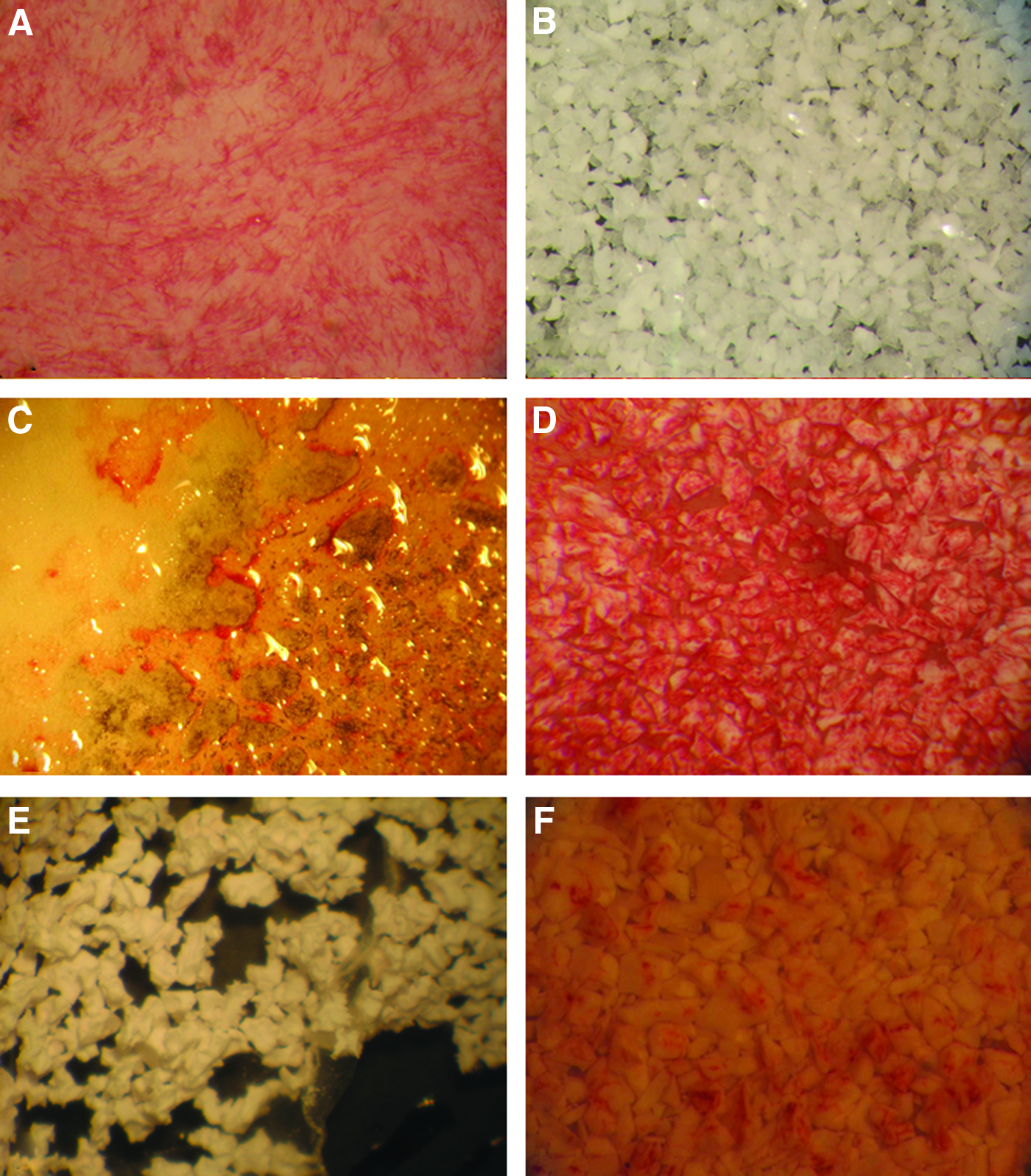
Alkaline phosphatase (ALP) staining on the in vitro-built tissue after 1 month of culture. MC3T3 subclone 4 cells
We investigated then the stage of differentiation of M group's samples, which showed the best results in terms of ALP activity and mechanical cohesion. Using ELISA kit and Bradford assay, we were able to detect the production of OCN by the cells inside the culture chamber (485±97 pg/μg/24 h, Fig. 9). This showed that the substitute reached the latest stage of osteoblast differentiation. 42 The same range was found, without significant difference, for the control group (554±120 pg/μg/24 h, Fig. 9), which offered no cohesion as seen above.
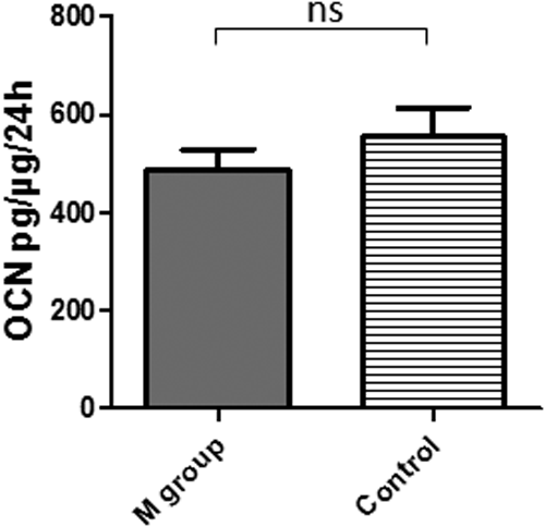
Osteocalcin (OCN) production rate at the end of the 1-month culture for M and control group (ns=no significant difference). Mass of OCN per mass of total proteins for 24 h.
Summary of biological characterization
Macroscopic and microscopic observations, along with the biological behavior described above, are summarized in Table 1. From all the groups, only the M group's substitutes remained cohesive and could be handled without breaking outside of the culture chamber (Fig. 2). The M group showed same levels of ALP activity, OCN production, and viability as the control group, and the CP granules homogeneously covered over the whole surface of the monolayer by the cell tissue as well (Fig. 4D). Its stable metabolic activity between the first and fourth week was coherent with the high differentiated state of cells (Figs. 6 and 7). Concerning the other groups, it was found that (1) the CP granules were not efficiently bound together by the cell tissue in the L group, because the cells grew inside the pores of the biggest granules (Fig. 3D), (2) the Mix group's samples revealed heterogeneous ALP activity, with a gradient from the center of the substitute (no differentiation) to its edges (medium differentiation) (Fig. 8D), and (3) the cell proliferation of the S group's samples was heterogeneous with spots of living cells (Fig. 4C).
Because the sheet-like substitute from the M group was cohesive and could be correctly handled, further studies were performed on this sample to investigate its mechanical behavior and to establish the relationship between biological response, tissue microstructure, and its macroscopic mechanical properties. Despite results close to M group's ones for some biological properties, the other groups were dismissed from the mechanical characterization due to their consistent lack of cohesion.
Mechanical behavior
The sheet-like substitute covered a surface area of ∼24 cm2 (as seen in Fig. 1), so at least three strips (3 cm by 1.5 cm) of this tissue could be obtained from each culture chamber dedicated to the mechanical study. Conventional tensile tests were performed as described above on 13 samples, leading to an analysis of the maximum stress as a function of strain, the mean breaking strain and the Young's modulus (Fig. 10). As the substitute developed from a single layer of 80–200 μm CP granules, a thickness of 200 μm was hypothesized to estimate the generated stress. The strips reached a maximum mean stress of 34±15 kPa for 28% of strain. The mechanical behavior appeared to be linear elastic and we could estimate a mean Young's modulus of 1.6±0.3 kPa for the reconstructed tissue (Fig. 10).
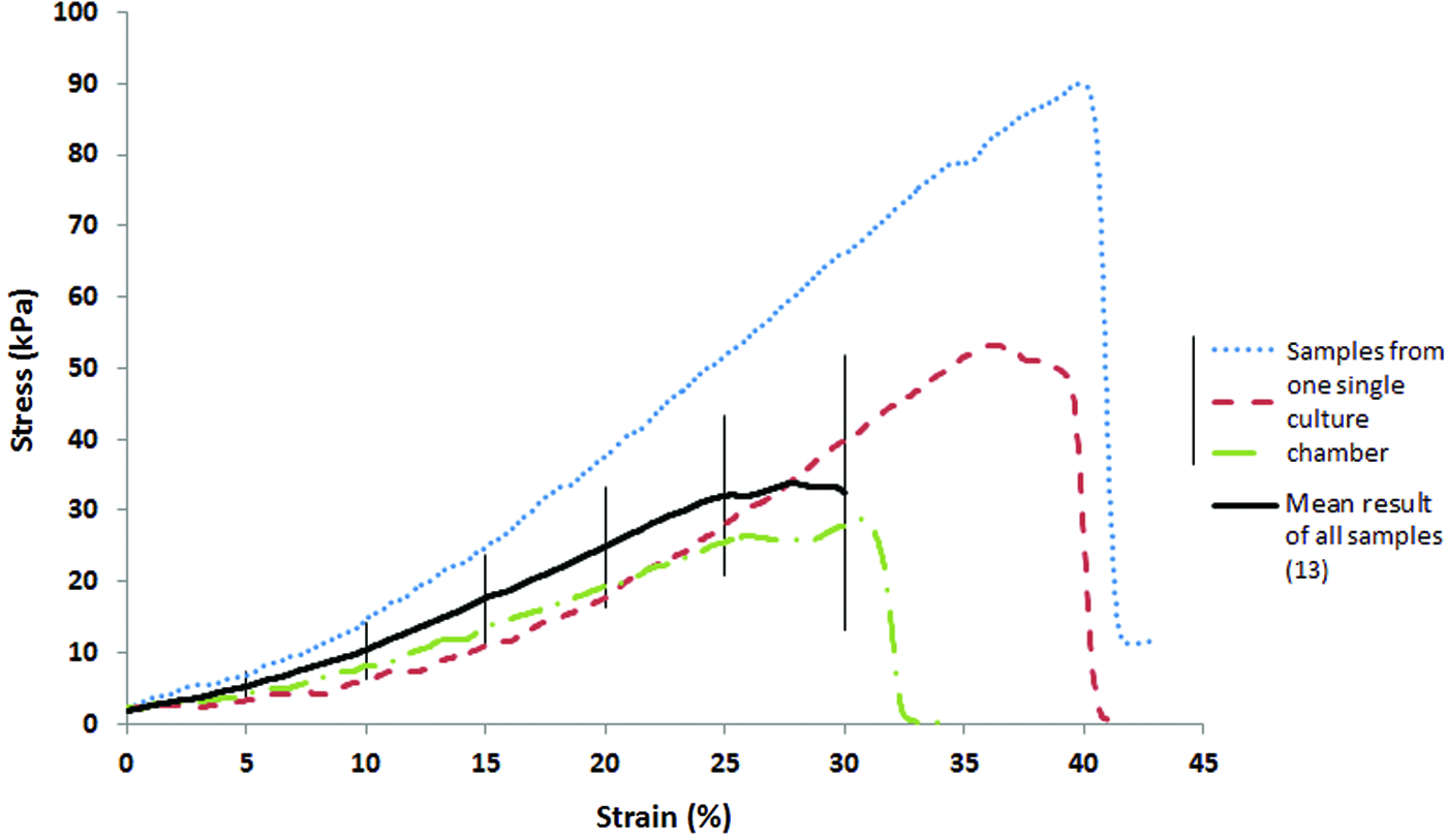
Measured stresses during tensile tests on in vitro-built tissue strips (1.5×3 cm), depending on the strain. Mean result of 13 samples from four culture chambers after 1 month of culture on 80–200 μm CP granules (M group) with standard deviation (black solid line) and results of 3 samples from one single culture chamber (blue, red, and green dashed lines). Mean calculation was stopped when the first failure appeared. Color images available online at www.liebertpub.com/tea
In situ tensile tests were also performed under SEM, using the specific module Deben Microtest 300 mounted on the SEM device (Philips XL30 ESEM-FEG), to observe failure propagation and local damage mechanisms, and more specifically the interactions between the CP granules and bone tissue. This analysis led to the conclusion that the reconstructed tissue presented a strong adhesion between the cellular matrix and the CP granules. Hence, the extracellular matrix (ECM) was identified as the part of the tissue that broke, without any displacement of the granules on either side of the tear (Fig. 11). There was no loosening of the granules within the substitute when tensed.
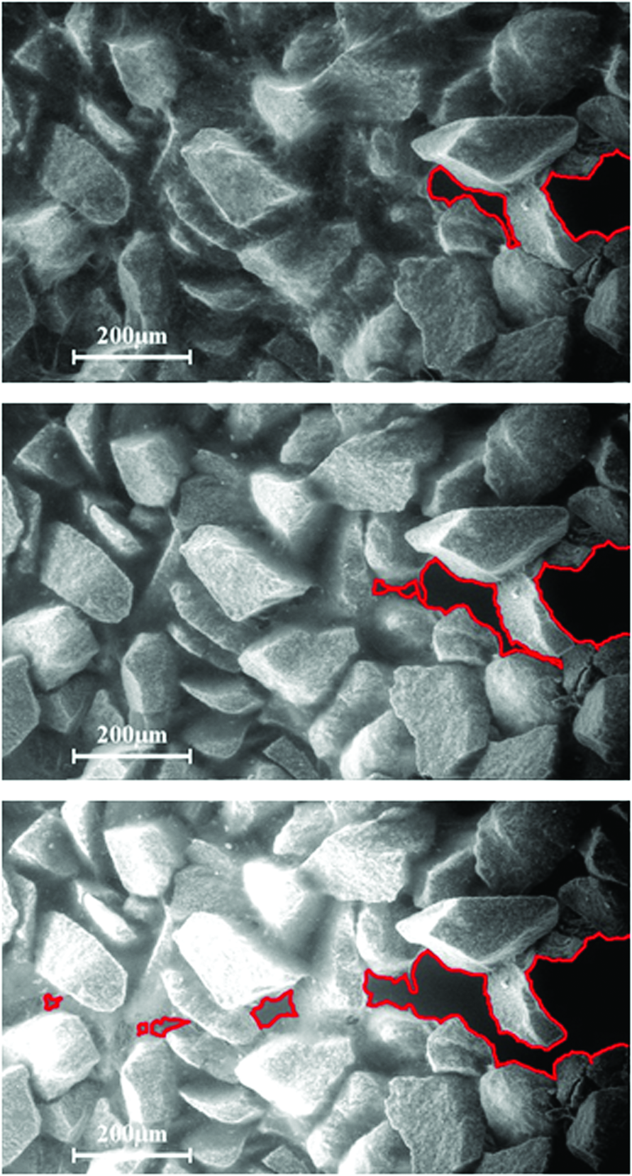
SEM observations of the in vitro-built substitute (M group) during a tensile test, at three different times (from top to bottom). The cell tissue broke (red lines) but the CP granules remained cohesive on both sides of the break. Color images available online at www.liebertpub.com/tea
Discussion
Bone can be affected by a lot of diseases 43 or damage caused by accidents. As previously stated, tissue engineering is considered to be a promising method for improving healthcare and repairs. 44 Our choices in terms of biomaterial, cell culture chamber, and tissue geometry were determined by the clinicians' expectations. CP ceramics are well known for their osteoconduction and osteoinduction properties, 10 especially the hydroxyapatite/tricalcium phosphate compounds. 45 Used as a granule monolayer in a parallelepipedic cell culture chamber, they led to a hybrid bone substitute with a flexible geometry, which could be relevant for several clinical fields, for instance maxillofacial surgery: it would be suitable for the small different complex shapes needed in this area. The culture chamber, able to be completely filled with medium and with an adequate surface, was specifically chosen to obtain such a geometry of rebuilt tissue. Moreover, when HA powders are used to fill bone defects, they are usually mixed with other matrices to avoid migration out of the implant region. 46 Because the granules are bound together by the cell tissue, our process could avoid this need of an additional substance.
In this study, we did not expect to mimic the exact mechanical properties of native bone, but we aimed to obtain a cohesive substitute with a relevant shape for clinical issues. The biological and mechanical properties of the in vitro-built tissue with different mean diameters of CP granules used as a scaffold were therefore investigated to determine their optimal size. Biological and mechanical behaviors are strongly bound together, with mechanical stimuli playing an important role in bone cell development, known as mechanotransduction. 47 These induced biological modifications in turn modify the mechanical properties of bone with changes in the production of tissue components and cell alignment. 47
The goal of the global process was to meet clinicians' expectations for both aspects. Changing the mean diameter of the CP granules modified the macroporosity of the monolayer, known to be an important factor for cell attachment and cell viability.48–51 In particular, for HA/TCP particles, Mankani et al. investigated in vivo bone formation for several size ranges. 52 Bone marrow stromal cells and HA/TCP particles were transplanted into mice for at least 4 weeks, and bone formation was estimated by semi-quantitative observations and histomorphometric methods. The behavior of the different groups of rebuilt tissues appeared to be clearly different, showing a strong link between particle size and the properties of the tissue. We observed the same tendency: the M group (80–200 μm) was more suitable for the target shape of a sheet-like substitute than the other ones. It showed homogeneous cell tissue, with CP granules bound together by the ECM, and high and homogeneous differentiation. In addition, it was the only group providing these positive results for all of the biological properties investigated (Table 1), with results closed to control's ones in terms of viability, cell tissue morphology, and OCN production, but with higher ALP activity and metabolic activity. After removal from the culture chamber and manipulations, M group's substitutes remained stable and cohesive. The lack of early development and/or homogeneous renewal noticed in the other groups led to a heterogeneous, nondifferentiated, and noncohesive tissue. These results were coherent with the work of Mankani et al., 52 who used the same size range as the M group to obtain the greatest bone formation.
It should be highlighted that, for the M group, the homogeneous and high differentiation shown by the ALP staining test was achieved with only acid ascorbic, but without other traditional added factors (such as beta-glycerophosphate or dexamethasone53,54) or specific differentiation medium, which is actually a common method.2,8,15,18 Our process, based on a parallelepipedic cell culture chamber and a CP granule monolayer, was thus efficient enough to promote the differentiation of the MC3T3 subclone 4 cells. It has been shown that the use of additional growth factors, such as transforming growth factor beta, bone morphogenetic protein, or fibroblast growth factors, may create complications during the clinical step, 55 with the need for accurate and precautionary validation for the patient's safety. 56 For these reasons, our factor-free process seems relevant for both industrial and clinical validations, with further studies needed concerning the use of FBS.
Because the M group's substitutes were stable and cohesive, we were able to investigate the mechanical properties of the biohybrid tissue. The study of breaking mechanisms under SEM observations showed a complete break but no loosening of the CP granules, which were maintained together by the ECM on both sides of the failure. Quantitatively, it was difficult to estimate which range of maximal stress and strain needs to be targeted. Native bone tissue effectively shows high variability in its mechanical properties in vivo, especially due to the constant remodeling 57 and the composite nature of the tissue: Guilak et al. 58 found a Young's modulus of 0.6 kPa for the cartilage ECM, and Rho et al. 59 estimated it at 10.4±3.5 GPa for trabecular bone with a mechanical method. During the tensile tests on tissue strips, low stress values (mean stress<35 kPa) and estimated Young's modulus (<2 kPa) were measured. These results thus showed an improvement compared to the ECM alone and the M group's substitutes remained cohesive during manipulations, but these values would not be adequate to avoid failures during clinical operations. For these reasons, the main goal of further studies should be the investigation of a new biomaterial with inherent mechanical resistance before cell growth. Sun and Gouk 60 found a Young's modulus of around 15 MPa for a commercial skin tissue matrix. This value could thus be a target threshold for a substitute, which would be resistant to handling by the clinician.
The mechanical cohesion showed that our sheet-like bone substitute could meet clinicians' expectations regarding the easy handling issue. The biological behavior should be confirmed with future in vivo trials (subcutaneous then intramuscular implantations on mice to investigate mineralization and bone integration). Recent in vivo studies about CP granules used alone without in vitro tissue engineering step suggest a relevant response of our substitute.61,62
Conclusion
In this study, we proposed a new bone tissue engineering method based on a CP granule monolayer and a specific cell culture chamber. We showed that our process, with an optimal mean diameter for the granules (80–200 μm), allowed us to generate a sheet-like bone substitute with properties close to clinicians' expectations, especially for maxillofacial applications, such as versatile geometry, flexibility, mechanical resistance, and elastic behavior, without the use of traditional growth factors. It has to be improved to guarantee better mechanical cohesion during surgery, but it is a promising construct for in vivo trials, which should confirm that it is an adaptable, autologous and factor-free alternative to grafts and biomaterials used alone.
Footnotes
Acknowledgments
The authors would like to thank Dr. Pierre Layrolle for providing the biomaterials (Biomatlante) and for initial discussions, Prof. Bernard Devauchelle (Head of Maxillofacial Surgery), and the team of Prof. Jean-Pierre Marolleau (Hematology department) at the University Hospital, Amiens, France, for fruitful discussions regarding surgeons' requests and regulations.
This research was supported by the Picardie region and the Equipex FIGURES. This project was cofinanced by the European Union and the European Regional Development Fund. Timothée Baudequin acknowledges the financial support of the CNRS and the Collegium INSIS-UTC.
Disclosure Statement
No competing financial interests exist.