
Abstract
At present, injuries or rupture of tendons are treated by surgical repair or conservative approaches with unpredictable clinical outcome. Alternative strategies to repair tendon defects without the undesirable side effects associated with the current options are needed. With this in mind, a tissue engineering approach has gained considerable attention as a promising strategy. Here we investigated a synthetic three-dimensional (3D) microenvironment able to interact with stem cells and inducing, via coupled biochemical and physical signals, their early commitment toward the tenogenic lineage. This multiphase 3D construct consisted of a braided hyaluronate elastic band merged with human bone marrow mesenchymal stem cells (hBMSCs) and poly-lactic-co-glycolic acid microcarriers loaded with human growth differentiation factor 5 (hGDF-5) by means of fibrin hydrogel. The multiphase structure allowed hBMSC culture under cyclic strain within a microenvironment where a controlled amount of hGDF-5 was regularly delivered. The cooperative biochemical and physical stimuli induced significantly increased expression of tenogenic markers, such as collagen type I and III, decorin, scleraxis, and tenascin-C, within only 3 days of dynamic hBMSC culture. This approach opens exciting perspectives for future development of engineered tendon tissue substitutes.
Introduction
T
Providing tissue-engineered substitutes grown in vitro and able to integrate with the host tissues is a promising alternative. 6 Scaffolds composed of various biomaterials manufactured via several fabrication techniques have been used for tendon tissue engineering (TE), but there is still the need for substitutes with adequate biological and mechanical properties able to provide support for cell attachment, proliferation, and differentiation.7,8 A combination of appropriate cells, biomaterials/scaffolds, biochemical cues/growth factors (GFs), and physical stimulation seems a successful approach to this aim. Tenocytes, fibroblasts, and stem cells (SCs) have all been tested as potential candidates for this purpose. 9 Adult mesenchymal SCs (MSCs) are multipotent cells able to differentiate into several lineages both in vitro and in vivo. Given their proliferation potential, biomolecular production, cell-to-cell signaling, and formation of appropriate extracellular matrix (ECM), MSCs appear as the most promising cell source to improve structural and biomechanical features of an injured tendon on autologous administration. 10 In particular, bone marrow MSCs (BMSCs) have been studied for tendon repair and regeneration, 11 and two clinical trials are investigating the efficacy of BMSCs in the treatment of refractory Achilles tendinopathy (Source: Australian New Zealand Clinical Trials Registry, Trial No. ACTRN12610000985088) and rotator cuff tears repair (Source: ClinicalTrials.gov, Trial No. NCT01687777). Several GFs, including insulin-like growth factor (IGF), platelet-derived growth factor (PDGF), basic fibroblastic growth factor (bFGF), bone morphogenetic proteins (BMP), transforming growth factor-beta (TGF-β), and vascular endothelial growth factor (VEGF), are involved in the activation and regulation of the cellular responses during tendon repair. 12 The inclusion of one or more of them into a TE protocol should improve/accelerate the engineered tissue fabrication by activating signaling cascades leading to the transcription of tendon tissue-specific genes.13,14 In particular, several authors reported the ability of growth differentiation factor 5 to induce genes consistent with tenogenic differentiation and significantly stimulate neotendon phenotype.15–18 On the contrary, its deficiency is reported to delay Achilles tendon healing in mice.19,20 Thus, it has been recently suggested that a sustained release of GFs inside a three-dimensional (3D) scaffold may provide an effective level of control when trying to influence cell phenotype, compared to simple addition of GFs into the culture medium. 21 Poly-lactic-co-glycolic acid (PLGA) microcarriers (MCs) were proposed as transient scaffold components to ensure sustained and controlled delivery of GFs.22,23
Mechanostimulation may also act as a significant input to maintain 24 or induce 25 the tendon phenotype. In particular, messenger RNA (mRNA) expression of type I and III collagen, and tenascin-C—all of them tenogenic markers—significantly increased in MSCs subjected to 10% stretching for 48 h; this effect persisted 48 h later. 26 Mechanical stimulus applied to braided nanofibrous scaffold also promotes tenogenic differentiation in vitro. 27
Several bioreactors have been used to impact mechanical stimuli to cells in culture.28,29 Some of them can also act as a stand-alone cell culture incubator able to transfer a controlled, recordable, and adjustable (cyclic) deformation to a 3D scaffold. 30
The choice of a suitable biomaterial to fabricate a scaffold to support cell growth is also a relevant issue. 31 Scaffolds should have appropriate mechanical properties to provide support, which is critical to the early phases of repair. In addition, biocompatibility of the substrate for cell attachment and proliferation, along with its biological signals, is especially important for SC-based approaches to tendon regeneration. 8 Biomaterials used for scaffold fabrication include both biomimetic synthetic polymers and biological molecules, 32 but an ideal biodegradable scaffold has not been identified yet. Scaffolds from hyaluronan polymer with braided fibers have been used; some weeks after surgery, these scaffolds produced improved biomechanical properties of the regenerated tendon tissue in the rotator cuff, and bolstered production of type I collagen in rabbits. 33 Also, PLGA scaffolds have been reported to improve tendon healing, both histologically and mechanically, and to facilitate the production of type I and III collagen fibrils improving both in vitro and in vivo tendon mechanical properties.34,35
Although each relevant signal, useful to induce tenogenic lineage commitment, has been studied individually, few protocols have proposed to exploit the synergic action. 36 Barber et al. 27 presented a protocol where hMSCs were cultured in the presence of mixed tenogenic GFs (BMP-2, FGF-2, and GDF-5) and stimulated with cyclic tensile strain, reporting their commitment into the tenogenic lineage, as evidenced by the significant upregulation of mainly scleraxis gene expression after 17 days in culture. Although this evidence corroborates the synergistic effect of supplying cooperative biochemical and mechanical signals to the cells, neither information about the specific contribution of a single GF and its active concentration within the engineered tissue construct nor the minimum time needed for obtaining significant commitment was presented and discussed.
To provide 3D microenvironment that ensures the controlled delivery of cooperative biochemical and mechanical signals to differentiating cells, GDF-5 was chosen, as the relevant biochemical signal. In this way, cells within a 3D microenvironment are stimulated with controlled mechanical deformation and a locally released, kinetically controlled biochemical signal, to induce their fast tenogenic lineage commitment. To this aim a multiphase 3D microenvironment was designed. It comprised (1) a braided hyaluronate elastic band, (2) PLGA MCs loaded with human GDF-5 (hGDF-5), and (3) human BMSCs (hBMSCs) merged together within a fibrin hydrogel and cultured under cyclic strain.
Materials and Methods
Supercritical emulsion extraction technology
PLGA MCs were obtained using supercritical emulsion extraction (SEE). This innovative technology allows the fast production of polymer MCs from multiple emulsions by supercritical extraction of the oily phase in a countercurrent packed tower operating in continuous mode. 37 In this experiment, unloaded and hGDF-5-loaded MCs were fabricated starting from a water/oil/water emulsion ratio of 1:19:80. In detail, recombinant hGDF-5 (PeproTech, Rocky Hill, NJ) was dissolved into 0.04% w/v human serum albumin (hSA; Sigma-Aldrich, Milan, Italy) containing polyvinyl alcohol (PVA) used as surfactant. This solution was added to the oily phase composed of ethyl acetate (EA, purity 99.9%) and PLGA (50:50, RESOMER® RG 504H, molecular weight: 38000–54000 kDa from Boehringer Ingelheim) at 10% w/w. The water/oil (w/o) emulsion was obtained after 90-s sonication with a digital ultrasonic probe operating at 50% of its amplitude (Branson Ultrasonics Corporation, Danbury, CT). The emulsion was then immediately poured into a 0.6% w/w EA-saturated aqueous Tween 80 solution, which is used as outer water phase to form the secondary emulsion by a high-speed homogenizer (mod. L4RT; Silverson Machines Ltd., Waterside, Chesham Bucks, United Kingdom) for 6 min at 10°C in an ice bath with a stirring rate of 2800 rpm. The emulsions were processed by SEE immediately after preparation. Operative pressure and temperature conditions in the high-pressure column were 8 MPa and 38°C, respectively, with an SC-CO2 flow of 1.4 kg/h and an L/G ratio of 0.1 w/w. 38 The suspension was collected continuously at the bottom of the extraction column, washed to eliminate the surfactant, and lyophilized. To obtain a cell culture-grade preparation, the washing steps were performed in the presence of a pen/strep (1% w/v) solution. Each run allowed the recovery of 98% of the loaded biopolymer and assured an excellent batch-to-batch reproducibility. Empty and loaded MCs were produced using the same process conditions.
Microcarrier morphology and size distribution
The droplets formed in the emulsion were observed using an optical phase-contrast microscope (Olympus, Tokyo, Japan). The shape and morphology of the MCs were investigated by field emission-scanning electron microscopy (FE-SEM; mod. LEO 1525; Carl Zeiss, Oberkochen, Germany). Samples of powder were placed on a double-sided adhesive carbon tape previously glued to an aluminum stub and coated with a 250 Å thick gold film using a sputter coater (mod.108 A; Agar Scientific, Stansted, United Kingdom). The scaffold structure was also fixed in 4% paraformaldehyde (PFA; Sigma-Aldrich) for 3 h, followed by an overnight incubation in 0.1 M sodium cacodylate/4% PFA (Sigma-Aldrich). The scaffolds were then dehydrated in a series of ethanol dilutions (75%, 90%, and 100%), embedded in paraffin (Fisher Scientific, Milan, Italy), and sectioned. The structure was then coated with gold as described above. Droplet size distributions (DSD) of the emulsion and particle size distributions (PSD) of the suspension were measured using a laser granulometer (mod. Mastersizer S; Malvern Instruments Ltd., Worcestershire, United Kingdom), based on dynamic light scattering (DLS). Sizes are expressed as volume mean size (MS, μm) with standard deviation (SD, ±μm).
hGDF-5 loading, release study, and diffusion coefficient calculation
hSA was used in the MC production process as a hGDF-5 stabilizer in the internal water phase of the double emulsion. The specific amount of hSA loaded into PLGA MCs was determined by dissolving 10 mg of dried powder in 600 μL of acetonitrile and sonicating it in 1400 μL of water. The remaining undissolved PLGA was separated by centrifugation at 2000 rpm for 2 min. The resulting clear supernatant solution was directly analyzed at room temperature (RT) by high-performance liquid chromatography (HPLC; Agilent Technologies, Inc., Santa Clara, CA). The amount of hSA in solution was calculated by HPLC-ultraviolet (UV) analysis using a calibration curve and then converted into the effective load in terms of amount (mg) of protein loaded into PLGA (g). Similarly, hGDF-5 loaded into PLGA MCs was extracted with the same procedure, monitored by enzyme-linked immunosorbent assay (ELISA)-based assay (PeproTech), and then calculated as μg/g (protein loaded/PLGA).
hGDF-5 release profiles were monitored in vitro, using the same ELISA-based assay. Microparticles of 20 mg were suspended in 2 mL of Dulbecco's modified Eagle's medium (DMEM), placed in an incubator at 35°C, and stirred continuously at 50 rpm. At fixed time intervals, the samples were centrifuged at 4000 rpm for 15 min and the supernatant was completely removed and replaced with fresh media to maintain sink conditions. Released hGDF-5 concentrations from collected samples were then measured with an ELISA (PeproTech). Release experiments were performed in duplicate (n = 2), and the proposed curve is the mean profile obtained.
hGDF-5 diffusion coefficient from the release data was calculated assuming a spherical geometry of the system, a constant diffusion coefficient, and a fixed initial molecule concentration. In this way, the diffusion controlled mass transfer equation can be expressed as the Equation (1):
where Mt represents the amount of the released molecule at a given time (t), M
where D0 is the diffusion coefficient of the molecules in the undegraded polymer (t = 0), and k is a constant that characterizes the dependence of the diffusivity on the molecular weight of the polymer. This parameter is not a universal constant, but depends on the physicochemical properties of the system, and a value of 5.9 10−10 cm2/kDa/s was used for data fitting. 40 The fitting procedure for D calculation was based on the minimization of the resulting differences between experimental and theoretical values (least squares method; R 2 value = 0.98).
hBMSC isolation and harvesting
The data presented in this article were obtained using hMSCs harvested from the bone marrow of five donors (two female and three male, age between 33 and 39; n = 3 including the static and the dynamic cultures with either empty or hGDF5-loaded MCs). Cells were used at passage 2. Written informed consent was obtained (Immuno-Haematology U.O.C. Ethics Committee Protocol No. 071/2015). hBMSCs were isolated and cultured as previously described with some modifications. 41 Briefly, mononuclear cells were enriched using a Ficoll-Paque PLUS gradient (GE Healthcare Life Sciences, Uppsala, Sweden), then seeded in T75 plastic flasks (106 MNCs/cm2) with the MesenCult™-SF Culture Kit (StemCell Technologies, Inc., Vancouver, Canada), and incubated at 37°C in 5% CO2 atmosphere and 95% relative humidity. The medium was changed every 3–4 days. Once the cell cultures reached 70–80% confluence (12–17 days for the first passage), the cells were dissociated with StemPRO accutase (Invitrogen) and replaced every 6–8 days at ∼80% confluence. Cells at first passage (P1) were used unless otherwise stated. To validate the purity of the hBMSCs at the end of the period of in vitro culture, FACS analyses were performed. Isolated cells were detached using StemPRO accutase, stained for MSC markers (CD105, CD90 and CD73) and hematopoietic SC markers (CD14, CD20, CD34, CD45; Becton Dickinson, Franklin Lakes, NJ), and analyzed by flow cytometry (FACSCalibur, Becton Dickinson Headquarters). hMSCs were characterized using the following directly conjugated monoclonal antibodies: mouse anti-human CD73 PE (phycoerythrin), clone AD2; mouse anti-human CD90 FITC (fluorescein isothiocyanate), clone 5E10; mouse anti-human CD105APC (allophycocyanin), clone 266; mouse anti-human CD14 APC, clone MφP9; mouse anti-human CD20 FITC, clone L27; mouse anti-human CD34 PE, clone 8G12; and mouse anti-human CD45 FITC, clone 2D1.
Multiphase 3D microenvironment preparation
The SC-based scaffold was prepared by hydrogelling a fibrin mesh on hyaluronate braided fibers (Hyalonect®; courtesy of Anika Therapeutics, Abano Terme, Italy) to embed hBMSCs and PLGA MCs (with/without [w/wo] hGDF-5) into a multiphase deformable tissue construct capable of biochemical signal delivery. To prepare each sample, a mixture of fibrinogen from human plasma (Sigma-Aldrich), aprotinin (Sigma-Aldrich), and DMEM (Lonza, Verviers, Belgium) supplemented with 10% heat-inactivated fetal calf serum (referred to as growing medium, GM) was added at a 1:1:1 ratio to 100 mg of PLGA microcarriers (56 μg/g w/wo hGDF-5) and then to an average of 5 × 10⁵ cells. The amount of loaded MCs was chosen to achieve a physiologically relevant level of hGDF-5 inside the scaffold during the first 3 culture days. 18 The cell/MC suspension was homogeneously pipetted into a mold (30 × 20 × 0.45 mm) to fill all the available volume in which the band was previously placed. After a quick addition of 100 U/mL thrombin (Merck-Millipore, Vimodrone, Italy), the mold was placed in a 37°C humidified incubator for 30 min to allow fibrin polymerization. As soon as the hydrogel was formed, the band was entrapped inside the hydrogel that was uniformly distributed on both sides of it. Then, engineered constructs were transferred from the molds to standard polystyrene culture plates, each containing 15 mL of the GM, and cultured in an incubator at 37°C in a 5% CO2 atmosphere and 95% relative humidity. After 24 h, the medium was changed, and the engineered constructs were subjected to static (referred to as static group, SG) or dynamic culture (referred to as dynamic group, DG).
Static and dynamic culture
The SG-engineered constructs (w/wo hGDF-5, referred to as SG+/SG−, respectively) were cultured statically within culture plates at 37°C in a humidified atmosphere containing 5% CO2 for 72 h. Mechanical stretch was applied to DG-engineered constructs in a custom-made bioreactor. 30 Briefly, the Hyalonect scaffold was clamped at both free ends, held by one motionless and one sliding arm (operated by a linear motor actuator), and assembled into the stand-alone culture chamber. After selection of stretching parameters using a custom-written software developed with LabVIEW v. 8.2 (www.ni.com/labview), the sliding arm of the apparatus moved to a pretensioning position, representing the maximal load. This load was then relaxed to a minimum value cycling between these two positions at the imposed frequency. In addition, continuous feedback signals provided by a load cell positioned on the fixed arm allowed to maintain the defined load on the scaffold in response to physical modifications in the growing pseudotissue in culture, by automatic adjustment of the pretensioning position. For the mechanical stimulation of DG, the engineered constructs (w/wo hGDF-5, referred to as DG+/DG−, respectively) were placed into the stand-alone culture chamber of the bioreactor and stretched at 1 Hz of frequency and 0.1 N of strain force (which is about 10% elongation in length). These operating parameters were set after mechanical testing of the mechanical behavior of the simple braided hyaluronate band and of the tridimensional multiphase construct. These measurements were performed according to the ASTM 1708 by a CMT 6000 dynamometer (SANS, Shenzen, China) equipped with a 1 kN load cell. Samples conditioned in DMEM for 1 h were shaped to obtain specimens having a gauge length (Lo) of 22 mm and a width (W) of 5 mm. For each sample, thickness (S) was measured with a thickness gauge brand at three different averaged points. Monoaxial deformation was applied to the sample at a speed of 22 mm/min, and force (F) and elongation (L) during traction were recorded. The value of force (F) provided by the instrument was divided by the sample area (A = W × S) to obtain the strength values (σ = F/A). The deformation values L during the run were compared to the initial length to obtain values of strain (ɛ = [L − Lo]/Lo); the ultimate tensile strength (σ max, expressed in MPa) was calculated as load to failure/cross-sectional area of the sample.
RNA isolation and gene expression profile
At the end of each SG or DG experiment, cells were collected, and the mRNA expression of type I and III collagen (Col1A1, Col3A1), decorin (DCN), scleraxis (SCX), and tenascin-C (TNC) was analyzed by RT-quantitative polymerase chain reaction (qPCR). Total RNA was extracted from hBMSCs seeded into the 3D construct of each experimental group using TRIzol® reagent (Life Technologies, Monza, Italy) and chloroform (Sigma-Aldrich) as previous described. 42 For each sample, 400 ng of total RNA was reverse-transcribed using the iScript® cDNA synthesis kit (Bio-Rad, Milan, Italy). Relative gene expression analysis was performed in a CFX Connect™ Real-Time PCR Detection System (Bio-Rad), using the SsoAdvanced™ Universal SYBR® Green Supermix (Bio-Rad) with the validated primers for Col1A1, Col3A1, DCN, SCX, and TNC (Bio-Rad), and following MIQE guidelines. 43 Amplification was performed in a 20 μL final volume, including 5 μL of complementary DNA (cDNA) as template. Specificity of the formed products was addressed performing a melting curve analysis. Triplicate experiments (two replicates each) were performed for each condition explored and data were normalized to glyceraldehyde-3-phosphate dehydrogenase (GAPDH) and hypoxanthine phosphoribosyltransferase 1 (HPRT1) expression (reference genes), applying the geNorm method 44 to calculate reference gene stability between the different conditions (calculated with CFX Manager software; M < 0.5). Fold changes in gene expression were determined by the 2−ΔΔCq method and are presented as relative levels versus hBMSCs cultured in static condition in the presence of unloaded MCs (SG–). Comparison between groups was performed using the unpaired Student's t-test. Differences were considered statistically significant when p < 0.05. 45
Results
A SC-based tridimensional multiphase construct was fabricated with the aim to test a reliable strategy to rapidly commit hBMSCs toward a tenogenic phenotype (Fig. 1). The cells used were 95% positive for CD73, CD90, CD105, and negative for CD14, CD20, CD34, CD45 before their use; their early induction of tenogenic markers was evaluated after 72 h of synergic stimulation using cyclic strain and locally released hGDF-5. Cyclic deformation of 3D multiphase construct was applied via a proprietary device, 30 set to transfer a mean load of 0.1 N over 1 Hz cycles to the scaffold throughout the entire experiment. The load parameters were applied after evaluating the tensile strength and the elastic modulus of the 3D multiphase construct.
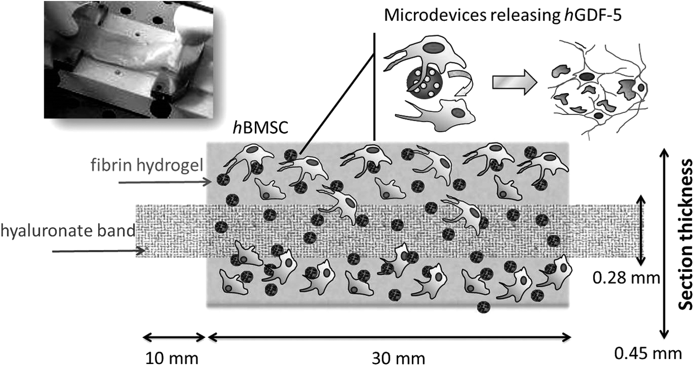
Image and schematic illustration of the 3D multiphase stem cell-based construct. The scaffold was formed by fibrin hydrogel coating hyaluronate braided fibers to embed hBMSCs and PLGA MCs (w/wo hGDF-5) into a multiphase deformable tissue construct capable of both biochemical signaling and mechanical strain delivery. The free ends of the hyaluronate band allowed the docking into the holders of the mechanical actuation system for cyclic load transfer. 3D, three dimensional; hBMSCs, human bone marrow mesenchymal stem cells; hGDF-5, human growth differentiation factor 5; MCs, microcarriers; PLGA, poly-lactic-co-glycolic acid; w/wo, with/without.
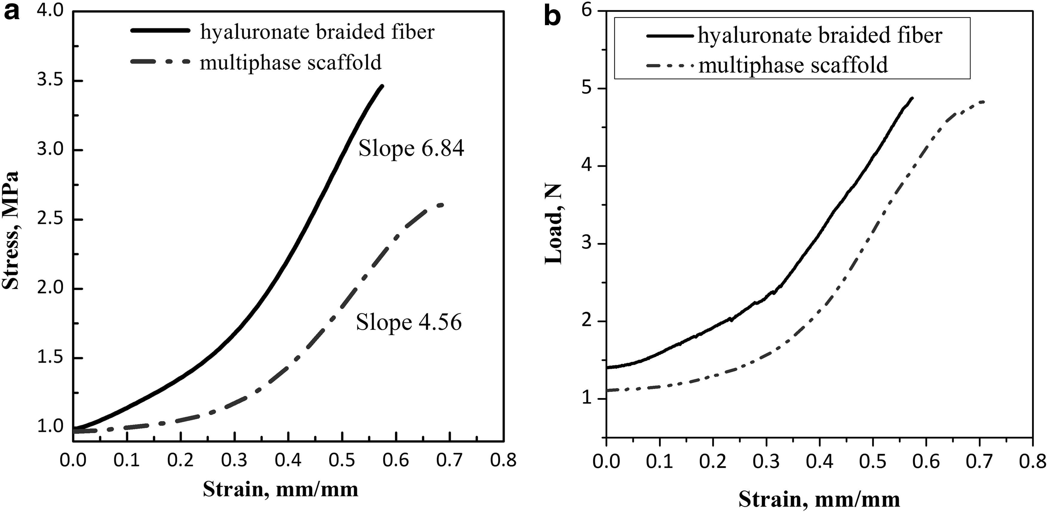
The curves of stress (MPa) versus deformation (dL/Lo, mm/mm) are plotted in Figure 2a. The load-bearing Hyalonect band thickness is 0.22 mm, whereas the multiphase SC-based construct showed a thickness of 0.45 mm. Both structures had a uniform behavior to failure. The values of maximum stress and tensile strength at failure are reported for each sample in Table 1, together with their elastic modulus. The braided band showed an ultimate tensile strength of 3.40 ± 0.03 MPa and an average elastic modulus (passive stiffness) of 6.84 ± 0.02 MPa with a percentage elongation at yield of 57%. The complete multiphase scaffold band showed an ultimate tensile strength of 2.15 ± 0.5 MPa and an average elastic modulus of 4.56 ± 0.7 MPa, with a percentage elongation at a yield of 70%. The load (expressed in Newton) versus strain plot is also given in Figure 2b. These data indicate that our 3D multiphase construct maintained a tensile strength and elastic modulus comparable with the braided hyaluronate core, and is suitable for the applied cyclic deformation protocol.
Stress–strain plot and elastic modulus values
Values are mean ± SD; n = 2.
SD, standard deviation.
PLGA MCs were used for the controlled release of hGDF-5; these particles showed a spherical shape (see FE-SEM image in Fig. 3), an MS of 2.0 ± 0.4 μm, and a GF encapsulation efficiency of 93%. MCs were produced by a proprietary and patented apparatus 37 and were embedded, together with the hBMSCs, within a fibrin mesh around hyaluronate fibers (see SEM images of the 3D multiphase scaffold in Fig. 4).
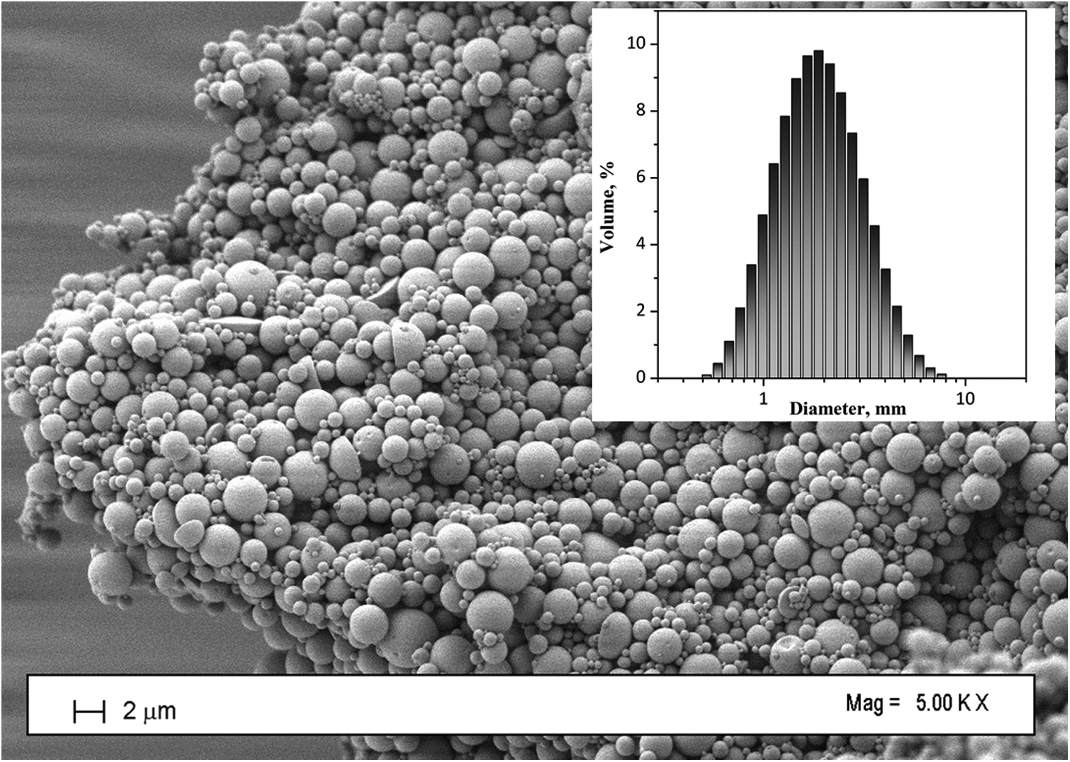
Scanning electron image of PLGA MCs loaded with hGDF-5 fabricated by SEE processing at 8 MPa and 38°C a water/oil/water emulsion (1:19:80); (inset) size distribution data are expressed as volume percentage. SEE, supercritical emulsion extraction.
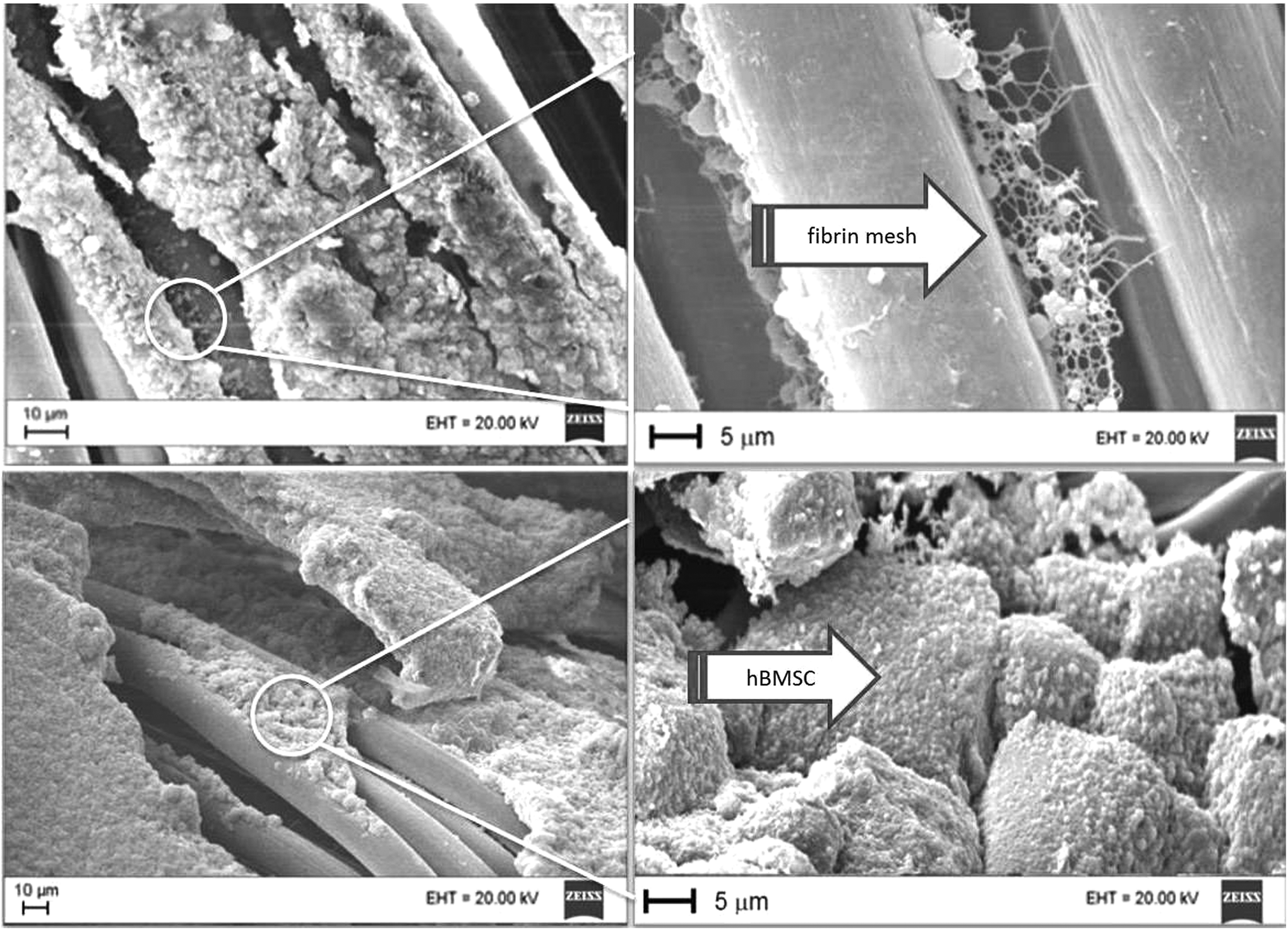
Scanning electron images of the 3D multiphase scaffold formed by hyaluronate fibers (diameter of about 10 μm) assembled with PLGA MCs and hBMSCs by fibrin hydrogel.
Several tests were performed to optimize the hGDF-5 amount delivered each day from the MCs to be inserted into the scaffold, after fixing the number of cells within the 3D structure. Preliminarily, the MC MS and loading were engineered to obtain a rapid release of about 20% of the total protein loaded (56 μg/g hGDF-5/PLGA) during the first 3 days, followed by a lower (3% of the total load per day) and constant rate of hGDF-5 released (Fig. 5) for the following 15 days, when roughly half of the initial load was released. The hGDF-5 diffusion coefficient was calculated as 7.5 × 10⁻¹⁰ cm2/s by fitting the release profiles of free MCs monitored at 37°C in DMEM. Then, following the suggested physiological hGDF-5 values proposed in the litterature, 18 each scaffold (surface 600 mm2, thickness 0.45 mm) was loaded with 100 mg of PLGA MCs (with an overall content of 5.6 μg of hGDF-5) and 500,000 cells to achieve a physiologically relevant level of GF delivered within the scaffold during the first 3 days of cultivation. Using this protocol, an overall amount of 1 μg of hGDF-5 is expected to be available within the scaffold for the cells throughout the 72 h of experiment. In detail, the expected amount released is illustrated in the insert within Figure 5, at day 1 (600 ng) and at day 3 (1000 ng). The measured amount of hGDF-5 in dynamic (120 ± 20 ng at day 1 and 240 ± 50 ng at day 3) and static (60 ± 23 ng at day 1 and 320 ± 48 ng at day 3) media is also illustrated for comparison.
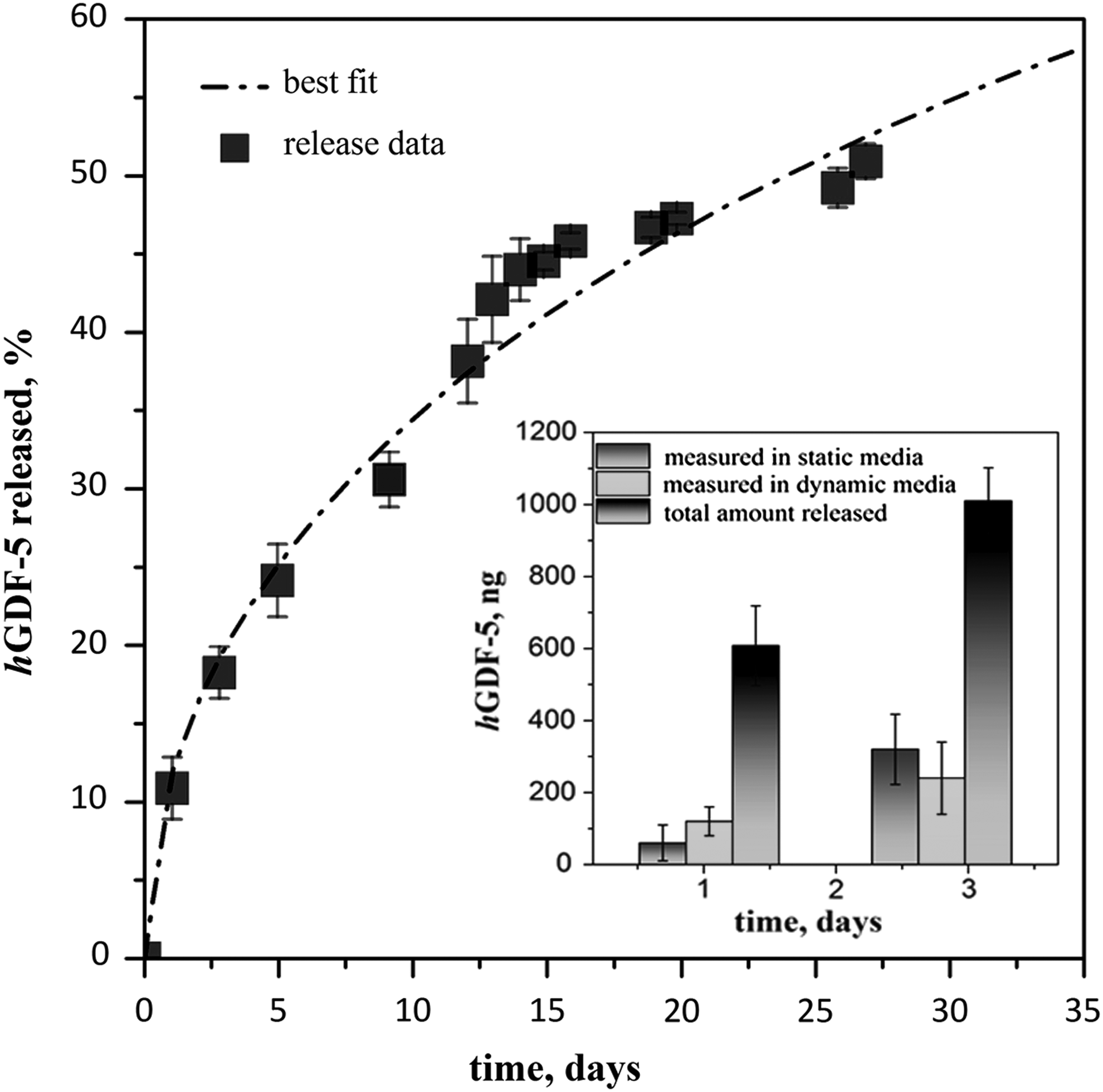
In vitro hGDF-5 release data (square) obtained in DMEM at 37°C with continuous stirring at 50 rpm; fitting profiles are reported as dashed line. The percentage indicates the amount released from the total load contained in the MCs. Values are mean with n = 2. (Inset) Total amount of hGDF-5 released each day from the 3D scaffold (MCs: 100 mg) in DMEM; hGDF-5 measured at day 1 and 3 during static and dynamic cultivations is also reported, for comparison purpose. DMEM, Dulbecco's modified Eagle's medium.
The 3-day lag was the minimal time chosen to monitor the synergistic effect of biochemical (hGDF-5) and physical (cyclic strain) stimuli over ColA1 and SCX gene expression (Fig. 6), which we are referring to as the “early commitment” toward the tenogenic phenotype. The choice of 72 h was undertaken after preliminary experiments, where we monitored the gene expression during culture time. A fragment of our constructs after 24 and 72 h was collected, and cell mRNA expression for Col1A1, Col3A1, DCN, and TCN was analyzed (in this preliminary experiment, mRNA expression of SCX was not analyzed). Although 24 h of stimulation still increased the mRNA expression of tenogenic markers analyzed in DG+, a time range of 72 h was selected to ensure statistically significant data for all genes tested. Only when both cyclic strain and locally released hGDF-5 were supplied to the cells, a significant upregulation of the mRNA levels of the relevant tenogenic markers Col1A1 (p < 0.05), Col3A1 (p < 0.01), DCN (p < 0.01), SCX (p < 0.01), and TNC (p < 0.05) was evidenced in hBMSCs within 3 days of culture (Fig. 6), pointing at a priming of a tenogenic differentiation program within the tissue-engineered construct. hGDF-5 alone determined a significant (p < 0.01) upregulation only for DCN mRNA level. A significant (p < 0.01) upregulation is evidenced only for SCX mRNA level when, on the contrary, the mechanical stimulus was applied alone.
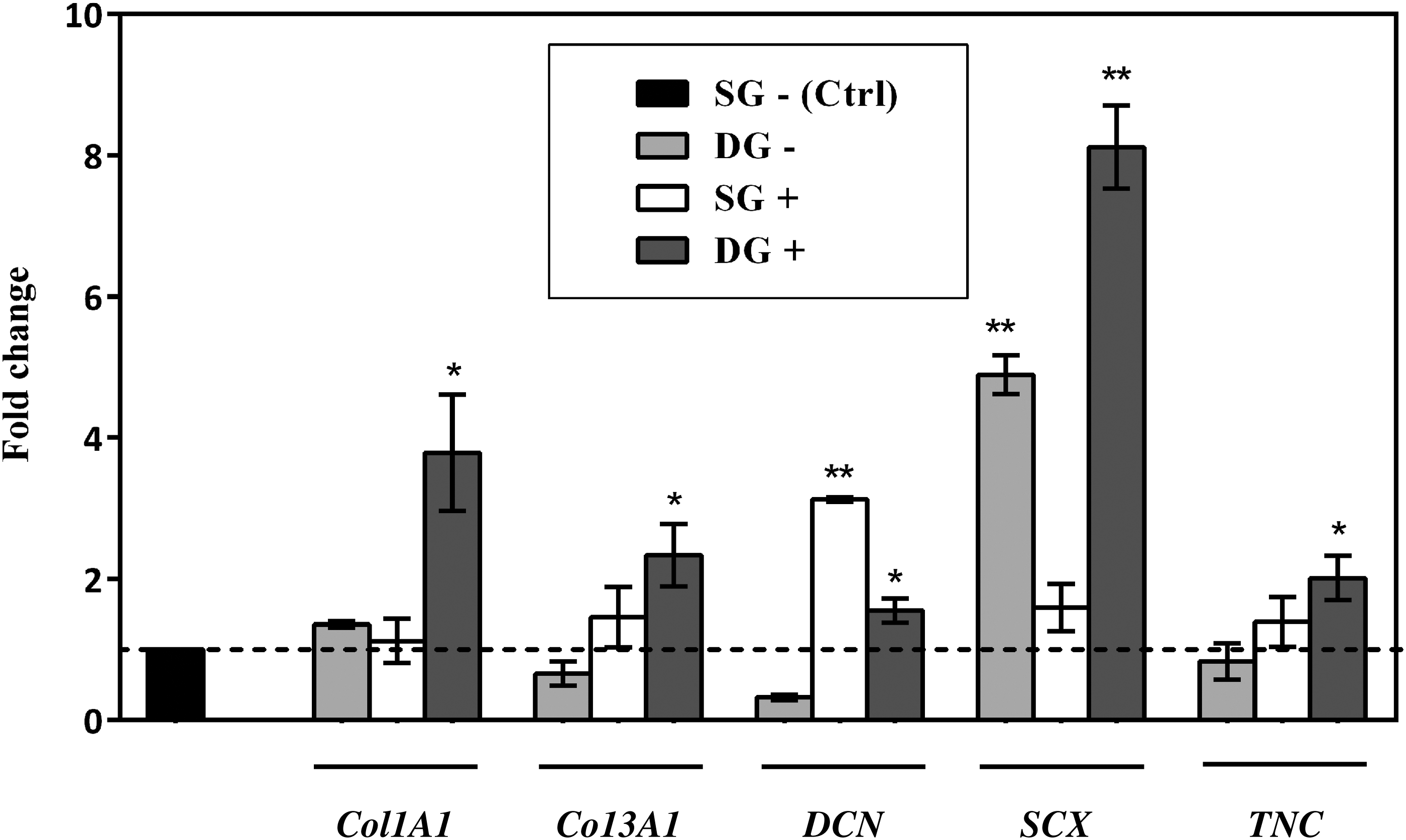
Mean ± SD gene expression data (n = 3) for Col1A1, Col3A1, DCN, SCX, and TNC (GAPDH and HPRT1 used as reference genes). hBMSCs under static conditions in the presence of unloaded MCs were used as control. DG+, dynamic group with hGDF-5-loaded MCs; DG–, dynamic group with empty MCs; SD, standard deviation; SG+, static group with hGDF-5-loaded MCs; SG–, static group with empty MCs. *p < 0.05 versus SG–; **p < 0.01 versus SG–.
Discussion
Even under the best circumstances, surgical repair of tendon injuries or ruptures with auto/allograft or prosthetic devices does not answer full recovery of the mechanical and biological properties of the original tissue. 5 Since these traumas are frequent, better reconstructive options are thus expected. Providing tissue-engineered substitutes with adequate biological and mechanical properties, grown in vitro and intended for subsequent integration with the host tissues is a promising alternative. A combination of appropriate cells, biomaterials/scaffolds, biochemical cues/GFs, and physical stimulation seems a successful approach to this aim.
We thus assembled hBMSCs together with hGDF-5 releasing MCs within a hydrogelling fibrin mesh onboard of an elastic and braided hyaluronate fiber that underwent a cyclic mechanical stimulation. In fact, although each of these relevant signals useful to induce tenogenic lineage commitment has already been individually explored, very few protocols have been proposed to exploit their synergic action. The proposed engineered microenvironment that resulted was able to combine physical and biochemical stimulations and delivered both of them to the cells to promote their fast commitment into the desired phenotype.
The main issue in the proposed approach concerns avoiding the administration of free GFs into the culture medium. hGDF-5 was instead administered locally with a controlled delivery to the cells within the 3D microenvironment where they are cultured. As a consequence, hGDF-5 quantities required to stimulate the cells, that is, the inherent culture costs, were significantly reduced and a concurrent higher stability in the biochemical signal presented to the hBMSCs was obtained. This was allowed by the use of a novel SEE approach to fabricate PLGA MCs loaded with hGDF-5 with a very accurate size tailoring coupled with high encapsulation efficiency (drug encapsulated/drug loaded × 100), thanks to the very fast emulsion processing that prevents any droplet coalescence and allows a rapid polymer hardening. These performances are a consequence of the reduced density and the enhanced diffusivity typical of the supercritical fluid extraction performed by SEE technology compared to a liquid/liquid one.23,37 Moreover, the MC size and composition are tunable by changing the emulsion formulation and droplet size and composition; these factors can affect the sustained delivery of significant molecules. 23
PLGA was selected as a biopolymer to fabricate the MCs, because it offers predictable drug release over prolonged periods thanks to a diffusion-controlled mass transfer of the drug accessible at the solid/media interface and an enhanced release caused by polymer bulk erosion via hydrolysis of the ester bonds.40,46 Moreover, no polymer swelling is reported, and therefore, no tridimensional multiphase structure deformation is expected during cell culture. Molecular release profiles from PLGA MCs have also been extensively studied, and several authors calculated the diffusion coefficient values of different encapsulated drugs/proteins. In our settings, the diffusion coefficient calculated value is of two orders of magnitude higher than the ones reported by Faisant et al. 40 (10−10 cm2/s vs. 10−11/10−12 cm2/s), but it seems consistent with the SEE-fabricated MCs, considering that we used PLGA with lower molecular weight (averaged 46 kDa against 104 kDa reported 40 ), and we fabricated MCs with MS 10 times smaller than the ones reported by the same authors (1 vs. 10 mm, as MS). It is also worth noting that the calculated diffusion coefficient is of several orders of magnitude lower than the ones reported in the literature for the free peptide diffusion through hydrogel scaffolds of thickness similar to the one used in the present work. 22 Hence, (1) the release obtained from PLGA MCs resulted in a more delayed profile when compared to free GF molecules diffusing through hydrogel scaffolds, (2) it can be considered the predominant step, and (3) the diffusion of free protein through the fibrin mesh of the hydrogel can be neglected. Consequently, we suppose that the cells are directly exposed to, and are affected by, released GFs inside the fibrin hydrogel structure, both in static and dynamic experiments.
Looking at the hGDF-5 release profile of SEE-fabricated MCs, it seemed well tailored for a strong hGDF-5 signaling during the first 3 days of stimulation (20% of the load released in the first 3 days) and for a sustained release (3%/day) during a hypothetical subsequent in vivo implant of the bioengineered structure. Moreover, the amount of hGDF-5 measured in the culture media after a 3-day culture (see insert in Fig. 5), in both static and dynamic runs, was a small fraction of the total hGDF-5 released from the loaded microspheres (1000 ng in each scaffold). This suggests that most of the hGDF-5 was retained within the scaffold and available to bind to the cells.
Cyclic tensile strain was also applied using a proprietary bioreactor for the controlled deformation of the overall 3D multiphase scaffold. The braided hyaluronate band, holding the overall 3D structure and transmitting the applied load, tolerates a maximum load of 5 N and shows an elastic modulus of 6.8 MPa (Fig. 2a, b); therefore, when a load of 0.1 N was transferred over 1 Hz cycles, the bioreactor operated within the elastic range of the construct and with an effective strain delivery to the overall components of the 3D structure. The structure also showed a uniform behavior until its breakage, indicating that all the cells dispersed within were uniformly stretched via the applied cyclic strain.
To check for the induction of tenogenesis in hBMSCs, several marker genes, such as Col1A1, Col3A1, DCN, SCX, and TNC, were investigated in this work (Fig. 6). Col1A1 is the major component of tendon tissue (75–85% of the dry mass of tendon) and is responsible for its mechanical strength. Elevated amounts of Col3A1, whose fibers are thinner and more extensile, have been reported at the rupture site of the human tendon. 47 A significant (p < 0.05 vs. SG–) enhancement, up to fourfold, of the mRNA level of Col1A1 compared with control (SG–) was shown when hBMSCs were cocultured for 72 h within the 3D scaffold with hGDF-5—releasing MCs under mechanical stimulation. On the contrary, when mechanical or chemical stimuli were applied alone, no differences from SG– control were observed. Interestingly, when Col3A1 mRNA level reached statistical significance (p < 0.05 vs. SG–) following DG+ protocol application, a favorable ratio of Col3A1 to Col1A1 was maintained, whereas only hGDF-5 stimulation (SG+) resulted in an atypical collagen ratio. 48
In addition to Col1A1, also a small leucine-rich proteoglycan implicated in the regulation of fibrillogenesis, DCN, is a fundamental protein in the ECM of tendon. 49 Our results show a twofold increase of mRNA level (p < 0.05 vs. SG–) following DG+ treatment. The larger DCN upregulation (p < 0.01 vs. SG–) observed upon SG+ treatment is somehow in accordance with Youngstrom et al., who showed a peak in DCN upregulation with a 3%, rather than a 5%, cyclic strain application. This suggests that the 10% elongation level used in our study might exceed a physiological stimulus for this marker, although it appears as the proper one to induce Col1A1 and TNC expression. 48 Indeed, TNC transcript—translated into a six-armed hexabrachion-shaped ECM glycoprotein considered an early marker of tendinous differentiation abundantly expressed in the musculoskeletal tissues during embryogenesis 50 —also reached statistical significance versus SG–, following DG+ treatment in our experiments (p < 0.05).
SCX is a tendon-specific basic helix-loop-helix transcription factor responsible for the transition of MSCs into tendon progenitors, as recently demonstrated by the work of Alberton et al. 51 In addition, more recently, Chen et al. 52 showed how the downregulation of SCX, caused by dexamethasone treatment, inhibited the differentiation of rat tendon MSCs into tenocytes, confirming the key role of this gene in the promotion of a tenocyte-like phenotype in MSCs. Our results showed a fivefold increase in SCX mRNA on DG– treatment (p < 0.01 vs. SG–), in accordance with Mendias et al., 53 which showed how SCX gene was particularly responsive to exercise. This level raised up to eightfold (p < 0.01 vs. SG–) when combined physical and biochemical stimulation (DG+) was applied to the construct.
Taken together, these data suggest that hBMSCs display within 72 h an early commitment toward tenogenesis guided by a mechanical stimulation and a synergetic biochemical signaling, as anticipated by Aspenberg 54 and in agreement with very recent evidence for other tissue phenotypes. 55 Indeed, as a member of the transforming growth factor-β (TGF-β) superfamily of proteins, hGDF-5 (also known as BMP 14) binds to a specific receptor inducing phosphorylation of the downstream substrate proteins Smads. Phosphorylated Smad1/5/8 associate with the co-Smad (Smad4) and the complex translocates to the nucleus, where it further associates with coactivators or corepressors to regulate gene expression. 56 On the contrary, RhoA/ROCK, cytoskeletal dynamics, and focal adhesion kinase are required for mechanical stretch-induced tenogenic differentiation of hMSCs.57,58 The convergence of these different signaling pathways onto common downstream gene promoters appears as a reasonable explanation for the observed synergism of the biochemical and mechanical stimuli.
The proof of concept proposed in this work opens a perspective for developing 3D bioengineered models to understand specific molecular and cellular compositions of damaged systems, as well as for fabricating implantable tissue substitutes. The hydrogel/hyaluronate multiphase structure displays higher elasticity and less tensile strength and passive stiffness (reduced elastic modulus value) when compared to the Hyalonect band alone, without losing resistance (Fig. 2b). The apparently lower tensile strength of multiphase structure (Fig. 2a) might only result from its larger cross section, which is almost double (0.45 mm) for the hydrogel when compared with the simple braided band (0.22 mm). The initial tensile strength of the proposed structure seems within the range of forces reported for passive or unresisted motion in human flexor tendons, 12 because it shows a good mechanical behavior under a load of 5 N. This maximum load is still inadequate to replace most human tendons 59 ; however, a strong improvement of the hyaluronate band by weaving fibers differently and/or in a cylindrical shape to enlarge the support cross-sectional area can increase the strength of the construct, and longer stimulation times might induce a more substantial mechanical improvement of the construct. Moreover, the presence of hGDF-5 loaded within the 3D multiphase structure allows the biochemical signal delivery within the scaffold. This action has to be expected to last also after the scaffold implant by surgery.
Conclusions and Perspectives
Tendon healing is complex and the most important aspects in the context of TE remain to be established. The proposed approach is a potential strategy to address this problem examining the relative importance during a tightly controlled in vitro protocol. Indeed, only few studies assess the use of GF delivery within the scaffold merged with mechanical training of cell differentiation toward tenogenic phenotype. Mechanical and chemical stimuli are traditionally treated separately. To address this issue, we hypothesized that mechanical stimuli coordinated by chemical ones will result in a cooperative effect able to better handle with systems where a combination of both stimuli plays an important role. The encouraging results of the proof of concept proposed in this work will be improved with further experiments exploring on a longer time scale the consistent histological evolution of the engineered tissue construct.
In the future, this will pave the way toward the in vivo test of the functional potential of the substitute, whose macroscopic folding is reasonably expected to engender geometries able to bear suitable loads. Addressing these issues will facilitate effective TE procedures and drive the synthesis of functional engineered tissues for the successful treatment of tendon injuries.
Footnotes
Acknowledgments
Contract grant sponsors: Italian MIUR Grant No. PON01_02512/7 BIAM-EPI; Regione Emilia Romagna grant POR-FESR 2007–2011; PCFF Foundation (Pescara, Italy).
Disclosure Statement
E.G., M.G., and C.M. are listed as inventors in the application with International Publication No. WO2011013067, filed to the World Intellectual Property Organization (WIPO) and entitled “Bioreactor for stem cell stimulation.” G.D.P. and E.R. are listed as inventors of the US Patent n. US Patent US/8628802 B2, January 2014, entitled “Continuous process for microspheres production by using expanded fluids.”