
Abstract
Targeted protein delivery for stimulating tissue repair has been a core focus of the field of tissue engineering for several decades. While many promising protein therapeutics exist, achieving sustained and localized protein delivery to injured tissues remains a challenge. Over the past 25 years, significant breakthroughs have been made in biomaterial-based strategies to improve targeted protein delivery. Protein delivery vehicles that leverage affinity interactions between proteins and materials present an effective approach for modulating the spatiotemporal release of proteins within sites of tissue injury. Stimuli-responsive polymers also enable protein release to be tailored to respond to cell- and tissue-level changes. In this article, we highlight some of the major recent advances in biomaterial strategies for targeted protein delivery with a focus on affinity-based protein delivery systems. We also discuss the future of protein delivery for tissue repair, in which we envision protein delivery strategies that can be tuned in response to the dynamic microenvironment of injured tissues.
Impact Statement
Achieving targeted protein delivery to injured tissues is a core focus of the field of tissue engineering and has enormous clinical potential. This article highlights significant advances made in biomaterial-based protein delivery strategies over the last 25 years and how they will influence research in the next 25 years. These advances will enable protein release rates to be tuned with increased flexibility to deliberately address the challenges of the dynamic injury environment and ultimately lead to better solutions for patients.
Introduction
Protein delivery presents a powerful strategy to stimulate regeneration of damaged tissues. However, the clinical translation of many promising therapeutic proteins in the research pipeline has been hindered by the inability to localize and control their release within the injury site. The only biomaterial-based strategies that have been approved for controlled protein release clinically are the absorbable collagen sponge for bone morphogenetic protein-2 (BMP-2) delivery to repair large bone defects (Medtronic's Infuse Bone Graft) and poly(lactic-co-glycolic) acid (PLGA) microparticles for sustained human growth hormone (hGH) delivery (Genentech's Nutropin Depot). Despite their clinical approval, even these products possess inherent limitations that curb their widespread use: the collagen sponge exhibits rapid BMP-2 release that has been linked with numerous undesirable side effects, 1 while commercialization of the long-acting hGH formulation was eventually halted due to manufacturing concerns over the low protein loading capacity of the PLGA particles. 2 Notwithstanding substantive research over the past 25 years, achieving effective control over protein delivery remains an ongoing challenge.
Despite the lack of clinically translatable protein delivery vehicles, improvements have been made in controlling the in vivo bioactivity, release, and spatiotemporal presentation of therapeutic proteins to more effectively enhance tissue repair and functional recovery in preclinical animal models. Seminal work by Go and Langer, Ritger and Peppas demonstrated the use of both polymeric particles and a variety of hydrogels to control protein release and preserve bioactivity in vitro and in vivo.3–5 Derivation of mathematical models describing drug delivery from polymer systems using chemical engineering principles were essential in understanding the role of network swelling and dissolution in drug diffusion and release.3–5 Several other publications demonstrated the broad utility of polymeric microspheres in preserving protein bioactivity and extending release based on tunable characteristics of the polymer.6,7 These early studies laid the foundation for the design of stimuli-responsive polymers and polymers with targeting ligands, providing intracellular and tissue-specific targeting of molecular cargo.8,9 Moreover, advances continue to be made that improve the loading and preserve the bioactivity of sensitive proteins, including the use of cationic diblock copolymers for encapsulation of proteins in core–shell nanoparticles 10 and nanoparticles for encapsulation-free protein release. 11
Hydrogel platforms are also attractive for controlled protein release because they are typically injectable, biocompatible, and can be engineered to enable degradability. Hydrogels relying on either diffusion-controlled or affinity-controlled protein release have been successfully used in animal models of tissue repair. The development of stimuli-responsive polymers coupled with the discovery of novel affinity interactions have paved the way for the design of more complex protein delivery vehicles that enable protein release to be tailored in response to dynamic injury environments.
The Evolution of Affinity-Controlled Protein Release
Perhaps one of the most promising methods of controlling protein release from a biomaterial, without relying solely on material degradation or crosslinking density, is modulating the interactions between the protein and the material itself. Since a number of potent growth factors involved in repair processes are heparin-binding proteins, heparin-based biomaterials have been used extensively to deliver proteins to stimulate regenerative cell phenotypes in vitro and repair tissue in vivo in preclinical injury models (Fig. 1A). For example, heparin-based materials have been explored for the following: sustained BMP-2 delivery for bone regeneration12,13; nerve growth factor, brain-derived neurotrophic factor (BDNF), and neurotrophin-3 (NT-3) delivery for neurite outgrowth and nerve regeneration14,15; vascular endothelial growth factor (VEGF) delivery for vascularization16,17; stromal-derived factor-1α (SDF-1α) for cell recruitment 18 ; and fibroblast growth factor-2 (FGF-2) delivery for cell proliferation. 19
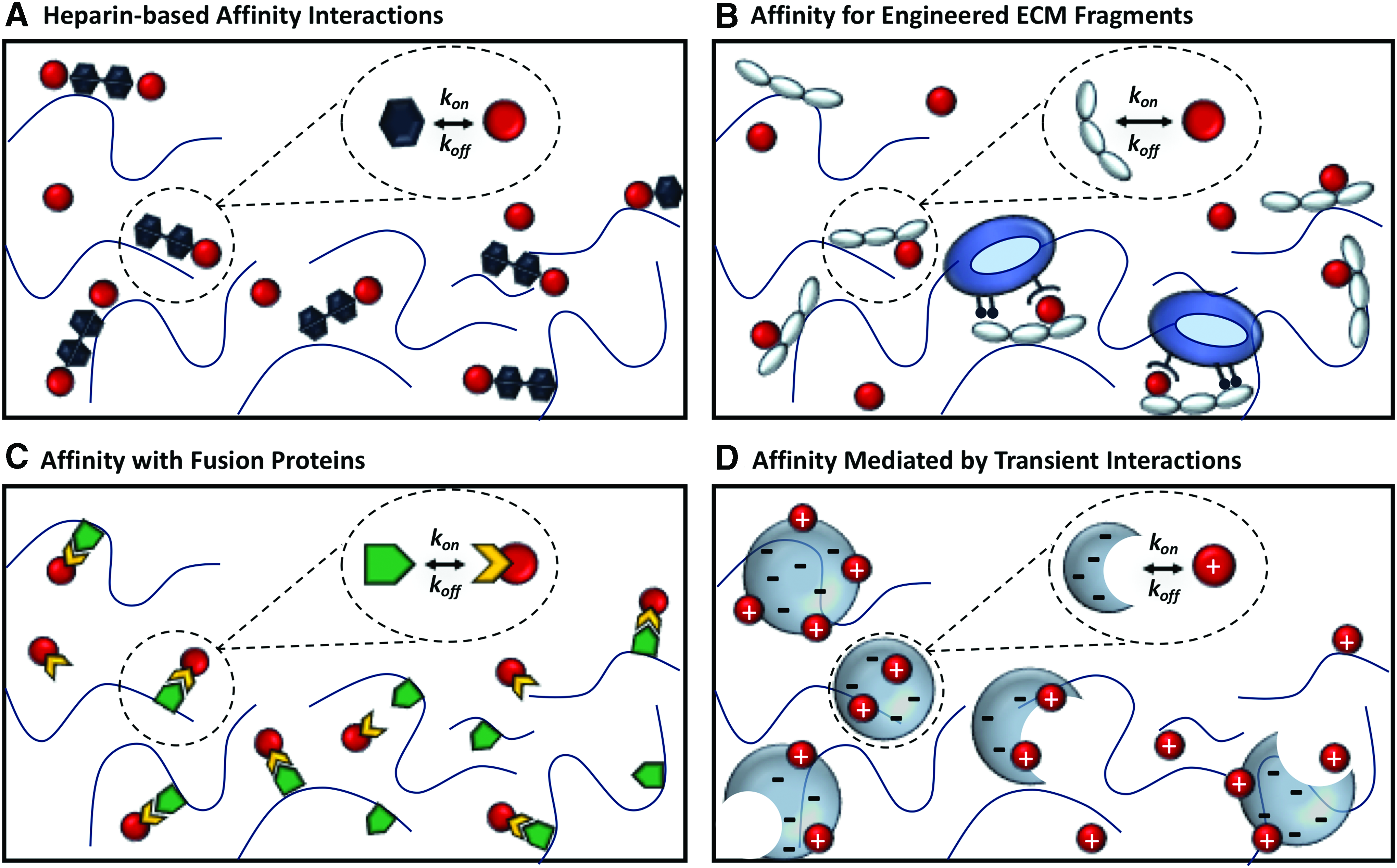
Modulation of protein release through affinity-based interactions between the protein and biomaterial.
Similar promiscuous growth factor binding has also been observed with the heparin-binding domains of fibronectin and fibrin.20,21 Since the high-affinity interactions between heparin and many heparin-binding proteins have been characterized, mathematical models can be used to design heparin-based materials that achieve desired rates of protein release.19,22 In general, such models have demonstrated that growth factor release from heparin-based materials can be tuned by varying the ratio of heparin to growth factor, which can, in turn, modulate both in vitro and in vivo cellular responses.12,17 Affinity-based protein delivery was first modeled by Sakiyama-Elbert and Hubbell, wherein a mathematical model was developed to describe FGF-2 release from a heparin-containing fibrin matrix. 19 This work paved the way for the rational design of affinity-based hydrogels, showcasing how modeling can be used to screen biomaterial properties in silico before experimental evaluation.
Despite the versatility of heparin as a ligand for protein binding, its lack of specificity and ability to bind numerous endogenous serum-borne proteins can interfere with its use as a vehicle for sustained protein release. Competitive binding with large, serum-borne proteins, including extracellular matrix (ECM) proteins (e.g., fibronectin, thrombospondins), has been shown to accelerate the release of protein cargo from heparin-based biomaterials. 23 As such, there is growing interest in creating synthetic affinity-controlled biomaterials that can provide greater tunability and specificity over protein release and subsequently enhance cellular responses. Thus, affinity-controlled protein release has evolved significantly over the past few decades from exploiting natural protein–material interactions, such as those between either heparin and heparin-binding proteins or antibodies and antigens, to engineering synthetic interactions between proteins and a variety of binding ligands (Fig. 1).
One bioinspired approach is based on engineering synthetic protein-binding partners that mimic the natural protein affinity of ECM molecules, such as heparin and fibronectin, but provide better protein localization and activity (Fig. 1B). Efforts to increase the tunability of heparin-based biomaterials include modulating the sulfation pattern of heparin to tune protein release between 5% and 60% 24 and identifying the minimal chain length and sulfation pattern requirements for heparin to control protein release and enhance protein stability/bioactivity. 25 Martino et al. achieved control over the simultaneous delivery of BMP-2, PDGF, and VEGF from a fibrin matrix by engineering a synthetic fibronectin fragment in which the integrin-binding and heparin-binding domains of the protein were fused together (FNIII9-10/12-14). 26 The simultaneous delivery of low doses of multiple growth factors using this synthetic ECM molecule fostered better healing in diabetic skin wounds (with VEGF, PDGF) and critically sized bone defects (with BMP-2, PDGF) compared with the delivery of growth factors alone or engineered fibronectin alone. The efficacy of this strategy was thought to be due to the synergistic effect of highly localized growth factor sequestration and cell adhesion provided by the affinity-based biomaterial.
An alternative strategy modifies the protein itself to enable novel binding capabilities and thereby affinity-controlled release (Fig. 1C). Vulic and Shoichet developed an affinity-based protein delivery strategy in which proteins of interest were expressed as fusion proteins with the well-studied Src homology 3 (SH3) domain. 27 By incorporating binding peptides with a range of known affinities for the SH3 domain (KD = 10−5–10−7 M), release of SH3 fusion proteins was precisely tuned from methylcellulose and hybrid hyaluronic acid/methylcellulose hydrogels over several days, as demonstrated computationally 28 and experimentally with FGF-2, 27 insulin-like growth factor-1 (IGF-1), 29 ciliary neurotrophic factor (CNTF), 30 and chondroitinase ABC (ChABC). 31 This strategy has been used to sustain the delivery of CNTF to the retina 30 and ChABC to the injured brain and spinal cord,32–35 and could be applied to other therapeutic proteins that can be expressed with the SH3 domain. Furthermore, because this strategy does not rely on common heparin-binding domains that exist in many proteins and ECM molecules, the specificity of the protein-binding partner interaction is increased.
Modifying protein affinity has also proved advantageous in preclinical testbeds for cancer immunotherapy with immune checkpoint blockade antibodies. When antibodies for programmed death ligand-1 and cytotoxic T lymphocyte antigen 4 were fused with the high-affinity heparin-binding domain of placental growth factor 2 (PlGF-2123–144) and delivered intratumorally, their retention was mediated by strong binding of the PlGF-2123–144 to the tumor ECM. This approach resulted in high local concentrations within the tumor, increased anticancer efficacy, and decreased systemic side effects. 36 The PlGF123–144 domain has also been used to introduce “super affinity” ECM interactions with BMP-2, VEGF, and PDGF, resulting in improved tissue repair and reduced side effects. 37 These examples demonstrate that fusion proteins are a viable approach for engineering effective affinity-based biomaterials, which may increase both the efficacy and safety profile of protein therapeutics.
Several other unique avenues of engineering protein–material affinity are also being explored for controlled release. For example, Huynh and Wylie demonstrated controlled release of anti-VEGF antibodies using a competitive binding approach in which antibodies conjugated with streptavidin were released from a biotin-functionalized hydrogel containing biotin derivatives with different solubilities. 38 Remarkably, by changing the biotin derivative in the hydrogel, antibody release could be tuned with first-order release kinetics over 100 days.
In another approach, electrostatic interactions, which play a crucial role in mediating many of the natural affinity-based interactions described above, can also be manipulated in synthetic matrices. Building upon a large body of work on polymer-based nanoparticles for drug and protein encapsulation,7,39 recent work led by Shoichet and colleagues 11 and Mooney and colleagues 40 demonstrated that controlled release of proteins from charged nanoparticles could be achieved independent of protein encapsulation when nanoparticles and proteins were mixed into a hydrogel (Fig. 1D). Transient electrostatic interactions between negatively charged PLGA nanoparticles and positively charged proteins within the hydrogel mediate protein–PLGA binding, and the slow dissipation of the negative charge during PLGA degradation initiates protein release. Protein release rate could be further tuned by changing the total available nanoparticle surface area, pH, or salt concentration. Controlled release similar to that of encapsulated proteins was achieved with BDNF, NT-3, and SDF-1, which are all positively charged proteins at physiological pH. This biomaterial strategy enabled local delivery of BDNF and NT-3 in vivo, facilitating tissue repair in animal models of stroke and spinal cord injury, respectively.41,42
Relatedly, the addition of positively charged Laponite XLG nanoparticles in cryogels reduced release of negatively charged granulocyte macrophage colony-stimulating factor and interleukin-2 by twofold and fourfold, respectively, without impacting protein bioactivity. 40 In this system, protein release could also be tuned by changing the amount of Laponite in the cryogel. Since low protein loading in polymeric nanoparticles has been a major hurdle in their clinical success, the discovery that protein encapsulation may not be necessary for sustained delivery using nanoparticles could completely change the future outlook for this protein delivery strategy.
Future Outlook: Tuning Protein Delivery for the Dynamic Injury Environment
Tissue repair typically requires a carefully orchestrated series of events in which numerous cell populations, proteins, and ECM molecules are presented under precise spatiotemporal control. Recent advances in biomaterials that enable better control over protein delivery now provide the opportunity to tune protein release rates with increased flexibility to deliberately address the challenges of the dynamic injury environment. For example, tailoring treatment to the injury environment is a concept that is currently being applied to therapeutic strategies in the central nervous system, where a myriad of inhibitory chemical cues accumulate within the injury site following stroke or spinal cord injury.43,44
Recent work by Anderson et al. aimed to identify and resolve key limitations in the injury environment following spinal cord contusion. 45 A lack of intrinsic capacity for neuronal growth, growth-supportive substrates, and chemoattractants were all hypothesized to contribute to failed regeneration following spinal cord injury and treated with a combination of viral vectors for the endogenous cell expression of osteopontin, IGF-1, and CNTF and hydrogel delivery vehicles containing FGF-2, epidermal growth factor, and glial cell line-derived neurotrophic factor. Interestingly, all of these components were required to stimulate axonal outgrowth across spinal cord lesions. Although cumbersome in terms of approach, sequential placement of hydrogels containing individual growth factors provided both spatial and temporal control over protein delivery.
In other studies, the enzyme ChABC was delivered to the injured spinal cord before the transplantation of cells intended to regenerate damaged tissue, with the rationale being that ChABC can degrade proteoglycans in the glial scar that may inhibit cell integration and axonal regrowth.33,46 Oligodendrocyte precursor cells delivered 1 week following ChABC treatment demonstrated better cell integration and stimulated more robust functional recovery than either treatment alone. Altogether, these studies demonstrate the importance of temporal control over protein delivery and highlight the need to tailor treatment strategies to the injury environment.
Other advancements in material chemistry that render biomaterial delivery vehicles dynamic and stimuli responsive will further enhance our ability to precisely control protein delivery to target tissues. Materials that can be triggered with a variety of external stimuli, such as light, heat, or ultrasound, enable “on-demand” delivery or spatially patterned presentation of bioactive proteins that can mimic protein gradients that emerge during morphogenesis or tumorigenesis.47–49 Alternatively, materials can be designed to respond to stimuli endogenous to the injury environment.
For example, gelatin-based hydrogels or hydrogels crosslinked with matrix metalloprotease (MMP)-cleavable sequences aim to mimic the dynamic injury ECM and can provide protein release that is directly tuned to the rate of cellular activity and protease secretion.50,51 This strategy can render biologically inert hydrogel materials, such as poly(ethylene glycol) (PEG) and poly(N-isopropylacrylamide-co-acrylic acid) cell responsive.51–53 In studies by Lutolf et al., delivery of proteins from an MMP-degradable PEG hydrogel resulted in significantly better material resorption and tissue remodeling, mimicking protein delivery from a natural ECM material (i.e., collagen).52,54 Similarly, Healy and colleagues demonstrated that cardiac progenitor cell delivery using a heparin-containing, MMP-degradable hyaluronic acid hydrogel enhanced cell engraftment and vascular integration through a combination of growth factor sequestration and cell-mediated remodeling. 55
Finally, recent work by DeForest and colleagues 56 has combined the benefits of several individual stimuli-responsive strategies to create sophisticated Boolean logic biomaterials that degrade to release cargo in response to multiple environmental cues. By incorporating crosslinkers that respond to a variety of orthogonal signals (e.g., light, proteases, and reducing conditions), this highly dynamic, modular system can trigger therapeutic release under highly specific spatiotemporal conditions, and has thus far demonstrated utility for both cell and small-molecule delivery applications.56,57
As more is discovered about the roles of various cell populations and cytokines in the healing response, we envision the continued development of biomaterial delivery strategies to provide tunable protein release/presentation. These approaches will be tailored to specific injury environments and overcome instances of dysregulated immune response, poor vascularization, lack of suitable growth substrates, and growth inhibitory cues. The increased availability of novel hydrogel chemistries and engineered protein affinity interactions provide more flexibility and greater tunability for protein delivery to injury sites, 58 and enables rational design of more effective strategies for tissue repair.
Footnotes
Acknowledgments
The authors are grateful to the following for funding this research: Natural Sciences and Engineering Research Council (NSERC Discovery to M.S.S., Postdoctoral Fellowship to M.H.H.), the Canadian Institutes for Health Research (CIHR Foundation to M.S.S.). M.S.S. is also grateful for her Tier 1 Canada Research Chair.
Disclosure Statement
No competing financial interests exist.