
Abstract
We evaluated the applicability of chitosan-g-oligo(L,L-lactide) copolymer (CLC) hydrogel for central nervous system tissue engineering. The biomechanical properties of the CLC hydrogel were characterized and its biocompatibility was assessed with neural progenitor cells obtained from two different sources: H9-derived neural stem cells (H9D-NSCs) and directly reprogrammed neural precursor cells (drNPCs). Our study found that the optically transparent CLC hydrogel possessed biomechanical characteristics suitable for culturing human neural stem/precursor cells and was noncytotoxic. When seeded on films prepared from CLC copolymer hydrogel, both H9D-NSC and drNPC adhered well, expanded and exhibited signs of spontaneous differentiation. While H9D-NSC mainly preserved multipotency as shown by a high proportion of Nestin+ and Sox2+ cells and a comparatively lower expression of the neuronal markers βIII-tubulin and MAP2, drNPCs, obtained by direct reprogramming, differentiated more extensively along the neuronal lineage. Our study indicates that the CLC hydrogel may be considered as a substrate for tissue-engineered constructs, applicable for therapy of neurodegenerative diseases.
Impact statement
We synthetized a chitosan-g-oligo(L,L-lactide) hydrogel that sustained multipotency of embryonic-derived neural stem cells (NSCs) and supported differentiation of directly reprogrammed NSC predominantly along the neuronal lineage. The hydrogel exhibited no cytotoxicity in vitro, both in extraction and contact cytotoxicity tests. When seeded on the hydrogel, both types of NSCs adhered well, expanded, and exhibited signs of spontaneous differentiation. The biomechanical properties of the hydrogel were similar to that of human spinal cord with incised pia mater. These data pave the way for further investigations of the hydrogel toward its applicability in central nervous system tissue engineering.
Introduction
The adult central nervous system (CNS) has only a limited capacity for self-repair, inevitably leading researchers to search for ways to augment this capacity to achieve therapeutic efficacy. The advent of induced pluripotent stem cells technology 1 raised the exciting possibility of generating ethical neural stem cells (NSCs) that could repair the CNS. Importantly, the development of direct reprogramming techniques2–4 introduced a potentially come-at-able and safe source of autologous neural precursor cells (drNPC) that have already been explored in preclinical5–7 studies in models of stroke and spinal cord injury. For direct reprogramming, the following transcription factors are currently being used: Musashi-1 (Msi1), Neurogenin-2 (Ngn2), and methyl-CpG-binding domain protein 2 (MBD2) 2 ; and Ascl1, Brn2, and Ngn2. 8 There are also various techniques for neural transdifferentiation based on microRNA (miR-9/9* and miR-124), 3 various small molecules, 9 and various media compositions 10 (see Samoilova et al. 11 for a review).
Despite the many recent advances in the development of cell-based therapies for CNS repair, important challenges remain that apply to all cell types: (1) ensuring high viability after implantation in an often times very aggressive disease environment; and (2) improving the differentiation capacity of the graft to generate the desired ratio of neurons and glial cells that are needed at the target CNS region. The field has therefore recognized a need to explore cell administration not just in a suspension format, but in a supporting matrix which may provide the required spatial characteristics improving the engraftment outcomes. The search for a fabrication technique for organotypical engineered structures of nervous tissue is proceeding both in the direction of bioprinting 12 or through mimicking cellular microenvironment using three-dimensional (3D) hydrogels, 13 modifications of extracellular matrix proteins, 14 self-assembling peptides, 15 or a combination of a hydrogel and an anisotropic protein scaffold. 16
The main requirements to scaffolds/hydrogels for fabrication of tissue-engineered constructs of nervous tissue are porosity, adhesiveness, biocompatibility, and biodegradability. The applied biomaterials may be either synthetic or natural; poly-ɛ-caprolactone (PCL), polylactide (PLA), polyglycolic acid (PGA), poly-lactide-co-glycolide (PLGA), and polyurethane (PU) belong to the former type of polymers, whereas polysaccharide biopolymers (hyaluronic acid, alginate, etc.)17,18 and protein biopolymers (gelatin, collagen, spidroin, silk proteins, etc.)14,16,19,20 belong to the latter type. Chitosan, the biomaterial used in the current study, entirely meets all the abovementioned requirements.
For applications in neural tissue engineering, it is critical that the composition and mechanical properties of a hydrogel support neuronal differentiation of NSCs. For example, fibrin, 21 hyaluronic acid,17,22,23 alginate, 24 and polyethylene glycol (PEG) 25 -based hydrogels provide better differentiation when their mechanical properties are closer to that of native CNS tissue. Modification of hydrogels with adhesion motifs such as TATVHL 26 or RGD 27 peptides, or laminin28–31 accelerate neuronal differentiation.
Chitosan-based hydrogels are widely used as carriers for various types of cells 23 and growth factors 32 while chemically modified chitosan may also be utilized in the formation of 3D biocompatible structures. 33 Importantly, chitosan implantation into normal connective tissue 34 as well as into damaged brain 35 did not cause any significant immune cell reaction. Chitosan has been shown to enhance adhesion and differentiation of NSCs, enhance electroconductivity, and serve as a substrate for neuronal axonal growth. 36 Recent studies have investigated the application of chitosan and its derivatives in CNS repair.35–40 For instance, chitosan-based hydrogel containing neurotrophin 3 (NT3) has been shown to facilitate regeneration of the damaged brain 35 and spinal cord, 40 highlighting its broad potential application scope in CNS.
In our prior work, we employed a solvent evaporation technique to produce microparticles from copolymers of chitosan and oligo/polyesters. 41 To produce complex-shaped structures, we introduced photocrosslinkers into the polymer mixture that allowed us to tailor the structure and architectonics of the hydrogel by two-photon stereolithography. We also found that chitosan with lower molecular weight (80 kDa as compared with 350 kDa) possesses higher reactivity with oligo(L,L-lactide)/(D,L-lactide) and allows the fabrication of hydrogels with highly reproducible structures and optimized mechanical characteristics using two-photon stereolithography. 42 The resulting hydrogel appeared compatible with neonatal rat cortical cultures. 43 In the current study, we assessed its potential for neural tissue engineering with neural progenitor cells. We started with investigating its mechanical properties and surface morphology followed by a careful evaluation of adhesiveness, cytotoxicity, and applicability as a substrate for proliferating and differentiating neural progenitor cells obtained from embryonic stem cells, as well as by direct reprogramming of somatic cells.
Methods
Hydrogel fabrication
Chitosan-g-oligo(L,L-lactide) copolymer (CLC) was synthesized by mechanochemical treatment of solid powder mixtures of chitosan (with the molecular weight of 80 kDa, degree of acetylation of 0.11) and oligo(L,L-lactide) (with the molecular weight of 5 kDa) at the component ratio of 40/60 w/w. The conditions of the synthesis and the copolymer characteristics have been described previously. 42
To prepare the photosensitive composition, 4.8 wt.% of PEG diacrylate (PEG-DA, M2000; Sigma-Aldrich, Germany) and 0.8 wt.% of Irgacure 2959 photoinitiator (BASF Kaisten AG, Germany) were added to a 4.9 wt.% aqueous CLC solution. Stirring of the mixture was carried out for 24 h at 35°C. Then, the final photosensitive composition (120 μL) was sandwiched between aluminum plates with a nonadhesive coating. The photocuring was conducted for 10 min using a light-emitting diode (Epileds Technologies, Inc.) with the wavelength of 365 nm and total power of 50 W. The resulting hydrogel was placed into a NH4OH (25 vol.%) solution, then washed with distilled water until complete neutralization.
Two-photon stereolithography
The 3D scaffolds were produced using an ytterbium-doped femtosecond solid-state laser “TeMa-100” (Avesta-Project, Troitsk-Moscow, Russia), which delivers 200-fs pulses at 70 MHz repetition rate. The experimental setup is similar to that previously described. 42 The fabricated hydrogels were washed from the uncured material in deionized water for 5 h. The images of the 3D models used for two-photon stereolithography can be found in the Supplementary Figures S1 and S2.
H9-embryonic stem cell-derived neural stem cell cultures
H9-human embryonic stem cell-derived neural stem cells (H9D-NSCs; Fisher Scientific) were cultured in Petri dishes coated with BD Matrigel Basement Membrane Matrix (BD, Canada), according to the manufacturer's instructions, in a medium containing DMEM/F12 + GlutaMAX™ (Gibco) supplemented with 20% KnockOut™ (Gibco), 2% StemPro® Neural Supplement (Gibco), epidermal growth factor (EGF) [20 ng/mL] (Gibco), and fibroblast growth factor-2 (FGF-2) [20 ng/mL] (Gibco). Cells were cultured in a humidified 5% CO2, 5% O2 incubator at 37°C. The culture medium was replaced every 2–3 days.
Before transfer onto CLC hydrogel films, H9D-NSCs were dissociated using Accutase® (StemCell Technologies, Canada) and resuspended to obtain a homogeneous suspension. CLC films (0.5 × 0.5 cm) were seeded with 1.5 × 10⁴ cells/film or 6 × 10⁴/cm2.
Directly reprogrammed neural precursor cell propagation and differentiation
Directly reprogrammed human neural progenitor cells (drNPCs) were kindly provided by New World Laboratories, Inc. (Laval, QC). drNPCs were obtained by direct reprogramming from human bone marrow-derived mononuclear cells, using a cocktail of three factors: Msi1, Ngn2, and MBD2. 2 The cells were cultured in Matrigel-coated (BD, Canada) Petri dishes in NeuroCult-XF Proliferation medium (StemCell Technologies) with addition of B27 (1X; Gibco), EGF [20 ng/mL] (Gibco), and FGF-2 [20 ng/mL] (Gibco)1,5 in a humidified 5% CO2, 5% O2 incubator at 37°C. The culture medium was replaced every 2 days.
Similarly, to the H9D-NSCs, drNPCs were dissociated using Accutase (StemCell Technologies), resuspended to obtain a homogeneous suspension and seeded onto CLC films (0.5 × 0.5 cm) as 1.5 × 10⁴ cells/film or 6 × 10⁴/cm2.
For neuronal differentiation, we used a cocktail of growth factors described in our previous article. 16 Briefly, drNPC cells were replated in NeuroCult-XF Proliferation medium (StemCell Technologies) supplemented with B27 (1X; Gibco), CultureOne (1X; Gibco), brain-derived neurotrophic factor [20 ng/mL] (Miltenyi Biotec, Germany), glial cell-derived neurotrophic factor [20 ng/mL] (Miltenyi Biotec), and Ascorbic Acid (200 μM). The culture medium was replaced every 2 days for 14 days.
Immunocytochemical analysis
The expression of neural stem cell, neuronal and glial markers was detected by immunostaining with appropriate antibodies. The cells were fixed with room-temperature 4% buffered formaldehyde solution containing 0.1% saponin for 30 min. After three washes with Dulbecco's phosphate-buffered saline (DPBS, Gibco), cells were incubated with primary antibodies diluted in DPBS containing 0.2% Tween-80, 0.2% Triton X-100, and 1% goat serum (DPBS-TT) for 1 h at 37°C. The cells were then washed three times in DPBS-TT and incubated with secondary Alexa-conjugated antibodies. Primary antibodies used: Ki-67 (2 μg/mL; Abcam), Nestin (2 μg/mL; R&D), Sox2 (5 μg/mL; BD Biosciences), βIII-tubulin (rabbit polyclonal, R&D, 2 μg/mL or mouse monoclonal, Sigma, 1 μg/mL), microtubule-associated proteins 2b (MAP2b) (5 μg/mL; Sigma-Aldrich), and glial fibrillary acidic protein (GFAP) (5 μg/mL; DAKO). Secondary antibodies used: Alexa Fluor 488 goat anti-mouse IgG (H+L) and Alexa Fluor 633 goat anti-rabbit IgG (H+L), all at 1:400 dilution (Invitrogen). Hoechst 33342 (Thermo Fisher Scientific) (0.1 μg/mL; Gibco) was used for nuclei counterstaining. Immunofluorescence was analyzed using a Nikon A1 scanning laser confocal microscope (Nikon Co., Japan). Quantification of positive cells was conducted using the object counter plug-in in Nis-Elements software (Nikon Co.). All the staining studies were conducted in series, with five repeats in each series.
Flow cytometry analysis
For the flow cytometry analysis, H9-NSC and drNPC were cultured on the CLC hydrogel films and on Matrigel coating separately during 14 days. Before analysis, the cells were dissociated using Accutase (StemCell Technologies) and fixed with 4% paraformaldehyde. Then, the cells were washed trice with PBS containing 1% fetal goat serum. FITC-conjugated anti-human SOX2, βIII-tubulin, MAP2 antibodies, unlabeled primary anti-Nestin, and GFAP antibodies, and Alexa Fluor 488-labeled secondary antibodies were used for staining the cells. All antibodies were purchased from Miltenyi Biotec. The analysis was performed with a CyFlow Space flow cytometer (Sysmex Partec) using the Partec FloMax® flow cytometry Data Acquisition and Analysis Software (Supplementary Figs. S6 and S7).
3-(4,5-dimethylthiazol-2-yl)-2,5-diphenyltetrazolium bromide test
To estimate the cytotoxicity of soluble hydrogel components, we employed the MTT [3-(4,5-dimethylthiazol-2-yl)-2,5-diphenyltetrazolium bromide] test adopted from ISO 10993 Part 5: “Tests for in vitro cytotoxicity,” as described elsewhere. 37 The hydrogel extracts were prepared by 24-h incubation of the ethanol-sterilized hydrogel (total surface area, 7 cm2) in 1 mL of DMEM/F12 culture medium supplemented with 100 U/mL streptomycin, 100 g/mL penicillin, 1% GlutaMAX (Gibco), and 5% fetal bovine serum (FBS; HyClone). The 3T3 murine fibroblasts were seeded in 96-well plates at the concentration of 5 × 10³ cells per well and cultured for 24 h in a humidified 5% CO2 incubator at 37°C in the same culture medium as above, but supplemented with 10% FBS. After that, the culture medium was discarded and the hydrogel extract was added to the wells for 24 h. MTT-reductase activity of the cells was estimated in relation to that of cells cultured without the extracts (max value). A 0.2 mg/mL solution of sodium dodecyl sulfate in the culture medium was used as a positive control (min value) for the assay validation.
Contact cytotoxicity evaluation
Contact cytotoxicity of the hydrogel was evaluated using H9D-NSCs and drNPCs. The cells were seeded on the hydrogel surface in a 24-well plate at a concentration of 1 × 10⁶ cells per well and cultured for 72 h without medium changes. The released lactate dehydrogenase (LDH) activity was measured in the supernatants using the LDH Enzymatic Assay Kit (Thermo Fisher Scientific) according to the manufacturer's instructions. The optical densities at the wavelengths of 490 and 640 nm were measured using a VICTOR Nivo Multimode Microplate Reader (PerkinElmer). The lysis buffer was added to the control wells to estimate total LDH activity of the cells (max value). The results were expressed as percent of the max value. The cells alone were cultured on tissue culture polystyrene (TCPS) to estimate spontaneous LDH release (min value).
Nanoindentation characterization of the hydrogel
The Young's Moduli distribution on the surface of the hydrogel was measured with Chiaro Nanoindenter (Optics11, Amsterdam, Netherlands) on an active vibration isolation table (Halcyonics i4; Accurion GmbH, Germany). For the measurements, we used a probe with the tip radius of 8.5 μm attached to a flexible cantilever (spring constant k = 0.58 N/m). The force/displacement curves were obtained by interferometric detection of the cantilever displacement using an optical fiber. The measurements were performed in distilled water at 20°C. The probing area was 500 × 500 μm, the step size of scanning was 50 μm. The maximal depth of indentation was 2 μm. The Young's modulus of each indentation was calculated based on the force/displacement curves using the Optics11 software and the Hertzian model for the elastic contact between a sphere and an elastic half space.
Scanning electron microscopy
Scanning electron microscopy (SEM) was used to visualize the surface morphology of the hydrogel. The hexamethyldisilazane (HMDS)-drying method was used for sample dehydration. In brief, the hydrogel was washed with PBS and subjected to dehydration with serial grades of ethanol. After the last step in absolute ethanol, samples were dipped in HMDS for 30 min followed by air drying at room temperature for 24–36 h in a fume hood. Dried samples were mounted on aluminum stubs using double-sided conductive adhesive tape and visualized using a Phenom ProX scanning electron microscope (LOT-QuantumDesign, The Netherlands).
Atomic force microscopy
To assess the susceptibility of the hydrogel to proteolytic degradation, we mapped the hydrogel topology using atomic force microscopy (AFM) before and after 4–6 days of incubation with proteinase K (1.5 mg/mL in 20 mM Tris-HCl, pH 7.4 with 0.02% NaN3) at 37°C. AFM images were acquired with Bioscope Resolve AFM (Bruker) in a peak-force tapping mode in PBS buffer. The ScanAsyst-Fluid cantilevers (Bruker) were used with nominal spring constant 0.7 N/m and tip radius 20 nm. Three 10 × 10 μm2 topography images were recorded per each sample. Root mean square (RMS) roughness was calculated as a RMS average of height deviations taken from the mean image data plane using Nanoscope Analysis software.
Water content
CLC hydrogel fragments (1 cm2 each) were soaked in distilled water and sandwiched between two pieces of filter paper to remove the excess of moisture. The weight of the samples was recorded before and after drying at 65°C for 5 h. The water content was expressed as a relative change (in per cent) in the sample weights (wet and dry).
Data processing
All experiments were performed in at least three independent runs to ensure validity of the results. The mean values (M) and standard deviations (σ) were calculated for all the data. Analysis was performed using the one-way analysis of variance (ANOVA) with a Bonferroni post-hoc test. The results with a p-value <0.05 were considered statistically significant.
Results
Characterization of the chitosan-g-oligo(L,L-lactide) copolymer hydrogel film
Biomechanics
We characterized the CLC hydrogel biomechanics by determining the Young's modulus distribution on the hydrogel surface, mapped using a Piuma Chiaro Nanoindenter. The parameter distribution was relatively homogenous with the average value of 51.41 ± 4.82 kPa (Fig. 1A). The shape of the force/displacement curves (Fig. 1B) indicated that the hydrogel was characterized by low adhesion and pronounced viscoelastic behavior.
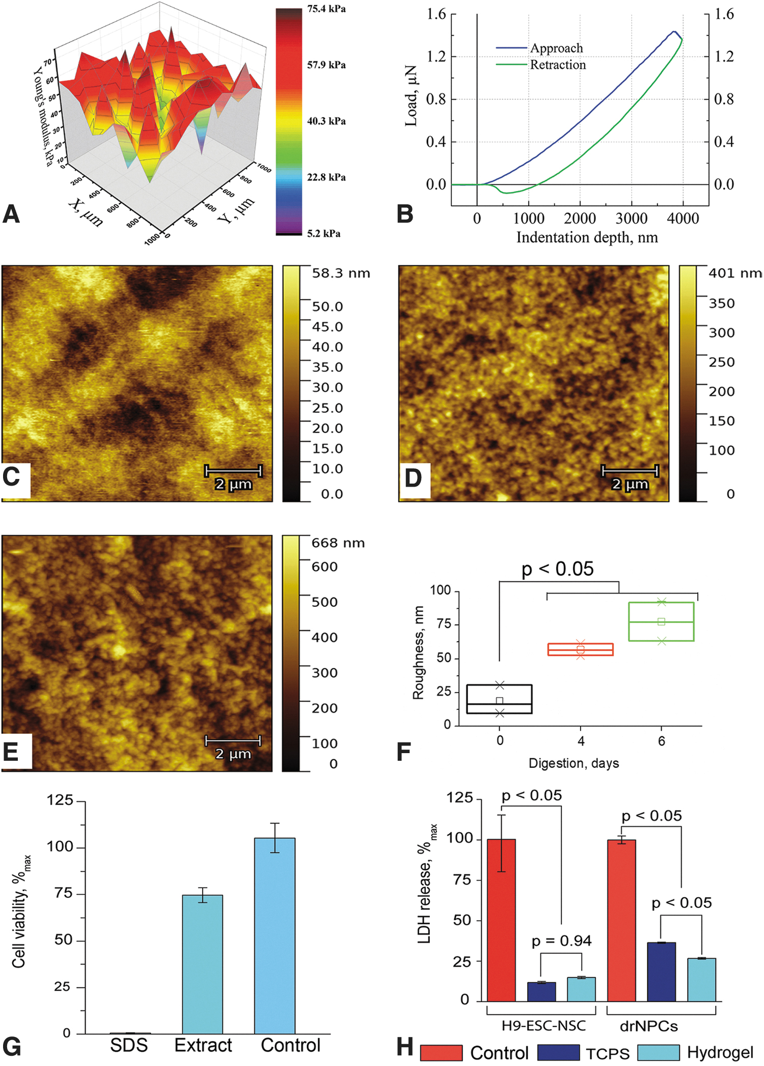
Properties of CLC hydrogel.
Surface morphology, water content, and biodegradation
Under SEM inspection, the hydrogel appeared uniform and nonporous (Supplementary Fig. S1), which is promising for production of the hydrogels with controlled shape and pore size using the techniques of molding and two-photon stereolithography (Supplementary Fig. S2). Water content of CLC hydrogel was found to be 94.0% ± 0.4%.
The susceptibility of the hydrogel to proteolytic degradation was assessed using surface mapping with AFM. The highly active serine protease proteinase K was expected to cleave oligolactide chains in the hydrogel network. We found that 4 days of incubation of the films in a proteinase K solution increased the surface roughness three-fold (57 ± 4 nm vs. 19 ± 9 nm in undigested control) (Fig. 1C—F). Continued incubation for 2 additional days (6 days in total) increased surface roughness to 78 ± 41 nm.
Cytocompatibility evaluation
Cytocompatibility of the CLC hydrogel with mammalian cells was evaluated using two colorimetric tests: MTT and LDH tests. First, we estimated the hydrogel extract cytotoxicity in MTT test using the murine fibroblast 3T3 cell line as a reproducible model. Extracts from about 7 cm2 of the hydrogel in 1 mL of culture medium for 24 h reduced the viability of 3T3 murine fibroblast cells by <30% (Fig. 1G). These data show that the cytotoxicity of the CLC hydrogel conforms to the directives of ISO 10993-5-2011: Medical Devices. Biological Evaluation of Medical Devices, Part 5, where the material is considered noncytotoxic if cell viability is ≥70% of the blank control.
Second, we evaluated the contact cytotoxicity of CLC hydrogel with H9D-NSCs and drNPCs based on LDH release measurement. We revealed slight differences in cell type sensitivity to the material (Fig. 1H). While there was no difference between H9D-NSCs cultured on the hydrogel surface versus TCPS (p = 0.940), the hydrogel improved the survival of drNPCs, as was indicated by the reduced LDH release (p = 0.048), compared with drNPCs plated on TCPS.
The biomechanics and cytocompatibility evaluation of the CLC hydrogel with H9D-NPCs and drNPCs established preliminary applicability as a substrate for culturing neural progenitor cells and warranted continued in vitro evaluation, as described in the following sections.
Immunophenotyping of H9D-NSCs on Matrigel-coated tissue culture polystyrene
It is understood that the biomechanical properties of polymers and gels can influence cellular differentiation.13,39 Therefore, we were interested in investigating how CLC hydrogel films impacted the differentiation potential of neural progenitor cells from different sources.
We started by characterizing the phenotype of Matrigel-seeded H9D-NPCs cultured for 14 days in proliferation medium using immunostaining. We observed abundant expression of the neural stem cell marker Nestin (Fig. 2A), as well as a neural stem cell/glial progenitor marker, GFAP (Fig. 2B). The vast majority (96.7% ± 1.8%) of the H9D-NPC population expressed Sox2 (Fig. 2C), while 37.1% ± 4.4% of the cells expressed Ki-67 (Fig. 2D), indicating that these cells were multipotent and proliferative (Fig. 2 and Supplementary Fig. S3, Supplementary Table S1). The immature neuronal progenitor marker, βIII-tubulin was also expressed, although at lower levels (Fig. 2D). The presence of MAP2+ and GFAP+ cells in the H9D-NSCs cultures after 2 weeks demonstrated spontaneous neuronal and glial differentiation (Fig. 2E), with glial cells being more abundant.

Immunocytochemical analysis of H9D-NSC cultured on Matrigel. (
Under differentiation conditions, H9D-NPCs grown on Matrigel-coated TCPS formed neurosphere-like structures where neurons and glial cells expressed βIII-tubulin and GFAP, respectively (Supplementary Fig. S4).
Immunophenotyping of drNPC on Matrigel-coated TCPS
A similar immunocytochemical analysis was conducted with drNPCs cultured in proliferation medium for 14 days on Matrigel-coated TCPS; high expression of the neural stem cell markers, Sox2 (100%), Ki67 (50.30% ± 15.1%), and Nestin was detected (Fig. 3A and Supplementary Fig. S3A, Supplementary Table S1) as well as the neuronal markers, βIII-tubulin and MAP2 (Fig. 3B, C). The cells coexpressed βIII-tubulin and GFAP (Fig. 3B) as well as Sox2 and βIII-tubulin (Supplementary Fig. S5A), owing to their relatively immature status. Upon switching to differentiation conditions, MAP2-positive neurons emerged (Fig. 3D), which no longer expressed Sox2 (Supplementary Fig. S5B) as well as GFAP-positive astrocytes (Fig. 3D). Thus, the immunocytochemical analysis confirmed that drNPC cells had a multipotent neural progenitor-like phenotype and were capable of differentiating into both neuronal and glial lineages, as shown previously (cited from 16 ).

Immunofluorescence analysis of drNPC cultured on Matrigel. Under growth conditions, drNPC express Sox2 (nuclear staining, green) and Nestin (red)
Proliferation and differentiation of H9D-NSC on CLC hydrogel films
When cultured on CLC films, H9D-NSC adhered well to the substrate. After 2 weeks of culturing in proliferation medium, neurosphere-like structures formed that exhibited high expression of Nestin (close to 100%) and Ki67 (23.5% ± 4.6%), (Fig. 4A), comparable to the Nestin and Ki67 expression levels seen in Matrigel-seeded cultures (Fig. 2C). As expected, Sox2 expression was also abundant (77.7% ± 5.9%) (Fig. 4B) and a large fraction of the Sox2-positive cells also expressed βIII-tubulin, suggesting an early stage of neuronal differentiation. H9D-NSC cells seeded on CLC also exhibited high expression of GFAP and generated agglomerates of MAP2-positive neural progenitors from where a network of neurites emanated (Fig. 4C). A number of cells in these agglomerates were double positive for MAP2 and GFAP (yellow in the merged image), indicating that these cells were likely in the early stages of neuronal differentiation.

Immunocytochemical analysis of H9D-NSC plated on CLC hydrogel films. Panels on the left show individual channels as well as merged image of all three channels while the image of the right is a 3D rendering of the same region showing the relationship between the markers.
Proliferation and differentiation of drNPC on CLC hydrogel films
drNPC cultured on CLC films predominantly spread diffusely over the entire film surface; neurosphere-like structures were detected but they were fewer and smaller when compared with those observed in H9D-NPCs grown on CLC films (Fig. 5). Expression of Nestin and Ki67 (8.64% ± 3.74%, p < 0.01 as compared with drNPCs on Matrigel and H9D-NPCs on CLC films, Supplementary Table S1) was also less abundant than in drNPCs cultured in proliferation media on Matrigel or H9D-NPCs seeded on CLC films, suggesting a more differentiated phenotype of the drNPC cells on CLC (Fig. 5A). In agreement with this hypothesis, Sox2 expression on CLC films (11.9% ± 0.9%) was significantly lower than in drNPCs cultured on Matrigel (p < 0.01) as well as compared with H9D-NPCs on CLC films (Supplementary Table S1 and Fig. 5B). A large proportion of cells were βIII-tubulin and MAP2 positive (Fig. 5B, C). Interestingly, in some areas of the film, oriented growth of neurites from βIII-tubulin-positive neuronal progenitors was seen (Fig. 5B). The coexpression of MAP2 and GFAP was almost absent on CLC films, similarly to what was seen in differentiation media on matrigel. The low expression of Sox2 and Ki67, as well as the decrease of Nestin expression in drNPCs cultured on CLC films were confirmed using flow cytometry (Supplementary Figs. S6 and S7). Taken together, our data shows that culture of drNPCs on CLC film accelerates a spontaneous differentiation program leading to an almost complete loss of neural progenitor cell markers by 2 weeks postplating.

Immunocytochemical analysis of drNPC plated on CLC hydrogel films. Panels on the left show individual channels as well as merged image of all three channels while the image of the right is a 3D rendering of the same region showing the relationship between the markers.
Discussion
Neural stem and progenitor cells are one of the most versatile cell types in neural tissue engineering due to their self-renewal, expansion, and differentiation capacity in vitro. These characteristics are deeply impacted by the culture conditions and also by the microenvironment 14 ; therefore, researchers are actively investigating biomaterials designed to improve the microenvironment surrounding neural stem and progenitor cells so that their unique biology can be explored in therapeutic applications.
Hydrogels have unique characteristics such as flexibility and biocompatibility that are very relevant in CNS tissue engineering: they can serve as carriers of neural stem and progenitor cells, providing oxygen and nutrients, metabolite exchange, and preventing glial scarring.13,35,43 Depending on their biomechanical properties, hydrogels can also induce directed cell differentiation.14,23,32,40 Hydrogels based on chitosan, a naturally derived polymer, are gaining wide attention as candidates for neural tissue engineering due to their attractive physicochemical properties.38,39 The mechanisms of hydrogel curing are diverse and include thermal, photo, and chemical crosslinking approaches; each curing methodology generates hydrogels with different biomechanical characteristics. Photocuring has the advantage of high spatial resolution and offers the possibility to create structures with complex surface architecture using stereolithography. 44 For example, grafting oligo(L, L-lactide) onto chitosan enables photosensitizer-assisted photocuring to produce a hydrogel. 42 This way it is possible to use 3D bioprinting for creating hydrogels of a sophisticated shape, for example, to reproduce the spinal cord cytoarchitecture. 12 The optical transparency of the CLC hydrogel also allows inspection of cell fate using fluorescence bioimaging and immunocytochemistry, making the CLC hydrogel an attractive substrate for studying the behavior of neural stem and progenitor cells as a part of a tissue-engineered construct.
In the current study, we have demonstrated that the CLC hydrogel does not release cytotoxic components into the surrounding culture medium and is noncytotoxic and highly adhesive for neural progenitors and embryonic stem cell-derived neural stem cells cultured on its surface. These results are promising in light of earlier reported charge-dependent cationic cytotoxicity of chitosan derivatives. 45 In that study, Zhou et al. employed grafting of positively charged alkyne-terminated poly(amidoamine) (PAMAM) dendrons to the chitosan chains, which may have led to disturbances of cell membrane and lipid structures, explaining the cytotoxicity reported. In contrast, the oligo(L,L-lactide) employed in our study does not bear a significant charge and has not exhibited the same issues.
The Young's modulus of the hydrogel was around 50 kPa as measured by a nanoindenter. This value is of the same order of magnitude as human spinal cord with incised pia mater, suggesting it can mimic the native microenvironment for transplanted neural cells. Biomimetic mechanical properties of the substrate may play a crucial role in cell fate, by impacting proliferation and differentiation of neural stem and progenitor cells.24,45
SEM and AFM analysis demonstrated that the surface of the molded hydrogel film was quite flat and appeared nonporous at the micron level. These results indicate that two-photon stereolithography enables 3D structuring of the hydrogel with designed pore distribution and architectonics. Incubation in a proteinase K solution resulted in continuous erosion of the hydrogel due to the cleavage of oligolactide chains. This supports the susceptibility of the hydrogel to proteolytic degradation upon implantation. A relatively high water content (94.0% ± 0.4%) is valuable for oxygen and metabolite exchange.
The main objective of our study was to compare the behavior of neural stem and progenitor cells derived from different sources upon culture on CLC hydrogel films. We have shown that H9D-NSC and drNPCs behave differently when cultured for 14 days on CLC films: H9D-NSCs generally preserve their “stemness,” while drNPCs differentiate predominantly along the neuronal lineage.
NSCs obtained from embryonic stem cells of the H9 lineage are well characterized and therefore may be considered as “gold standard” neural stem and progenitor cells. However, their clinical application in neuroregenerative therapy is conditioned by their allogenicity, potential tumorogenicity, and ethical considerations. Direct reprogramming of somatic cells is a promising alternative to the use of pluripotent-derived cells such as H9D-NSCs. 1 Recent studies have investigated the fundamental biology of neural stem and precursor cells derived from reprogrammed somatic cells,2,46 and mature postmitotic neurons derived by transdifferentiation. 3 While the generation of autologous postmitotic neurons has a strong appeal for disorders in which replacement of a specific neuronal cell type is needed, the challenges related to the manufacturing of terminally differentiated neurons are substantial, making the intermediate step that of generating directly reprogrammed NPCs more attainable.
In the current study, we have shown that culture on a CLC film accelerates spontaneous neuronal differentiation of drNPCs but not of H9D-NSC. Differences in the expression of extracellular matrix receptors by the two cell types may underlie these results and are the subject of future studies. Our data also highlights the value of CLC hydrogel as model system in which mechanisms of NSC and NPC self-renewal and differentiation can be studied by modifying various factors in the microenvironment. The drNPCs used in our study have demonstrated regenerative potential in experimental spinal cord injury5,6 and experimental stroke. 7 Investigations into their use in CNS tissue engineering have begun 16 and our current in vitro study suggests that combination of drNPC with hydrogel based on chitosan-g-oligo(L,L-lactide) could improve cell survival upon transplantation into damaged tissue and accelerate neuronal differentiation, supporting improved connectivity with normal tissue. Taken together, the physicochemical properties of CLC hydrogel and its biocompatibility with NSCs indicate that it is a promising biomaterial for 3D bioprinting of nervous tissue constructs, including spinal cord structures for regenerative therapy of the spinal cord injury. 12 Besides, depending on different goals we can use the combinations of each kind of cells and CLC hydrogel. For example, for research of the epigenetic changes of donors and cells signaling in differentiating cells it is better to use drNPC. From the other side, H9D-NSC will be the better object for the disease model. 47
Conclusion
CLC hydrogel is a nontoxic and optically transparent material with biomechanical characteristics suitable for 3D culturing and differentiation of NSCs and NPCs aimed at the creation of tissue-engineered constructs for improving neuroregeneration in vivo by means of two-photon biofabrication. When cultured on films prepared from a CLC hydrogel, embryonic stem cell-derived H9D-NSCs mainly preserved their multipotency, while drNPCs exhibited augmented neuronal differentiation.
Footnotes
Acknowledgments
The authors are grateful to Dr. Jan-Eric Ahlfors, the CEO of New World Laboratories, Inc. for kindly providing drNPCs. The authors are grateful to Drs. Svetlana Kotova and Alexandra Capela for their kind help with the article preparation.
Disclosure Statement
The authors declare that the research was conducted in the absence of any commercial or financial relationships that could be construed as a potential conflict of interest.
Funding Information
This study was supported by the Russian Science Foundation (Project No. 16-15-10432) in the portion pertaining to 2D and 3D H9D-NSC and drNPC culture and analyses. This work was also supported by the Ministry of Science and Higher Education within the State Assignment FSRC “Crystallography and Photonics” RAS in the “Hydrogel Mechanical Analysis,” and by the Russian Foundation for Basic Research (Project No. 18-29-17050) for “Hydrogel Synthesis and Cytotoxicity (MTT, LDH) analysis.” C.Z. was sponsored by the Natural Science Foundation of China (81702161).
References
Supplementary Material
Please find the following supplemental material available below.
For Open Access articles published under a Creative Commons License, all supplemental material carries the same license as the article it is associated with.
For non-Open Access articles published, all supplemental material carries a non-exclusive license, and permission requests for re-use of supplemental material or any part of supplemental material shall be sent directly to the copyright owner as specified in the copyright notice associated with the article.