
Abstract
Electrospun nanofibers is a promising and versatile avenue for building controlled drug release system because of the facile fabrication and the broad range of polymer materials. This research systematically studied the morphological effect of thermosensitive electrospun nanofibers, including porous and coaxial structures, on controllable drug release. Three types of drugs, nicotinamide, paracetamol, and ibuprofen, with different hydrophilicity were applied in this study. The data of drug release were all fitted to the first-order kinetic model regardless of the drug properties, and the release rates paralleled with their hydrophilicity. Sol-gel phase transition of the thermosensitive poly(N-isopropylacrylamide) (PNIPAAm) hydrogel led to slower drug release at 37°C compared with those at 25°C. Regarding morphology, coaxial nanofibers could provide higher loading efficiency and slower drug release rather than porous nanofibers. Our research highlighted the overall effects of compound property, temperature, and the morphological structures of thermosensitive electrospun nanofibers on the controlled drug release. Our results concluded that hydrophobic drug encapsulated in the core-shell PNIPAAm nanofibers could perform excellent sustained release and also controllable release under temperature stumuli.
Impact statement
The behaviors for the controlled release of drugs loaded in the thermosensitive electrospun nanofibers could be affected by various factors including the properties of loaded drug, morphologies of nanofibrous, and lower critical solution temperatures of thermosensitive hydrogels. However, few systematical investigations have been performed in this area. In this article, we designed and fabricated porous and coaxial thermosensitive poly(N-isopropylacrylamide) electrospun nanofibers with different drug loading to study the comprehensive effect. This study suggested when adopting thermosensitive electrospun hydrogel nanofibers as the controllable drug release carrier, the hydrophilicity of loaded compounds and the morphologies of nanofibers are necessary to be optimized.
Introduction
Thermoresponsive hydrogels refer to hydrogels that can respond to external temperature. 1 Some polymers are soluble in a certain solvent in a temperature range but insoluble above a temperature, which is called the lower critical solution temperature (LCST). 2 Poly(N-isopropylacrylamide) (PNIPAAm) is a typical thermosensitive hydrogel that swells at temperature below the LCST (32°C), whereas it distributes into a shrinking network at a temperature beyond the LCST owing to the hydrophilic/hydrophobic balance in the polymer chains. 3 PNIPAAm-based hydrogels have good safety and biocompatibility and thus can be used in the subcutaneous and intradiscal administration. 4 The intelligent thermoresponsiveness makes PNIPAAm hydrogel a promising drug delivery carrier, biosensor, and tissue engineering material.
However, making PNIPAAm into nanofibrous membrane is not easy because of its poor mechanical strength. Till now, PNIPAAm nanofibrous membranes are commonly fabricated by cooperating with other skeletal polymers, such as polycaprolactone (PCL),5–7 polyurethane (PU), 8 chitosan, 9 and so on. Electrospinning recently emerges and acts as alternative technology for PNIPAAm nanofiber formation.
Electrospinning is a simple, low-cost, and versatile method for fabricating electrospun nanofibers.10,11 When the electric field force exceeds the surface tension of electrospun solution, the charged polymer solution is ejected into filaments and then forms into a fibrous web on a metal collector.12,13 To date, many polymers are available for electrospinning including the natural polymers, such as collagen, 14 silk fibroin, 15 cellulose, 16 chitosan, 17 and hyaluronic acid, 18 as well as the synthesized polymers, such as polyvinyl alcohol, 19 poly(lactic-co-glycolic acid), 20 PCL, 21 and nylon 6. 22
Electrospinning is also capable of fabricating nanofibers with a variety of morphologies including multilayer, 23 multichannel, 24 porous,25,26 coaxial,27,28 and hollow structures. 29 Electrospun polymer nanofibers possess many desirable merits such as highly specific surface area, good air permeability, and easy surface functionalization. These excellent properties enabled the application of electrospun polymer nanofibers in tissue engineering, 30 drug release, 31 filtration, 32 and smart wearable device. 33
The release performances of electrospun nanofibers with different morphologies have been studied by many researchers.27,34 Porous nanofibers are effective in hindering burst release. Zhang et al. 35 developed self-sealing porous electrospun nanofibers with carbon nanotubes (CNTs). Polyethylene oxide (PEO) acted as a pore-forming agent in the fabrication of porous polylactide nanofibers. About 0.4 mg/mL CNTs demonstrated photothermal conversion ability for trapping the pores under UV irradiation. Sustainable release of loaded biomacromolecules was achieved as evidenced by the greatly reduced release rate in 24 h and 14 days.
In another study, porous chitosan nanofibers were obtained by leaching PEO in water. 36 Paclitaxel was encapsulated in the pores and hyaluronic acid was coated on the surface of the chitosan nanofibers through electrostatic attraction. It took over 48 h to reach the release equilibrium owing to the attraction of opposite charges.
Apart from porous nanofibers, nanofibers with core-shell structure also played a significant role in sustained drug release. Li et al. 37 explored the release behavior of hydrophobic and hydrophilic drugs in core-sheath nanofibers made by emulsion electrospun. Hydrophobic 10-hydroxycamptothecin (HCPT) was loaded in the shell and hydrophilic diclofenac sodium (DS) was encapsulated in the core. In vitro drug release study demonstrated that the initial burst release of DS and HCPT was extended to 4 and 3 days, respectively.
Although the release performances of electrospun nanofibers have been studied to some extent, there are less sufficient investigations focused on thermosensitive electrospun nanofibers. Lin et al. 8 designed PNIPAAm/PU core-sheath composite nanofibers. The burst release of nifedipine was less in composite nanofibers than that in coaxial nanofibers because composite nanofibers were uniform in blended solution. Shi et al. 38 fabricated core-sheath nanofibers with PCL as the core and PNIPAAm as the shell by single-spinneret electrospinning. The release process of macromolecular substance Nattokinase lasted for >3 h at both 37°C and 20°C.
In addition, it is well known that the controlled release rate of drugs loaded in the thermosensitive nanofibers is affected by the combination effects of compounds' properties, nanofibrous morphologies, and temperature responsiveness. However, few systematical investigations have been performed on the release behavior of different drugs loaded in thermosensitive electrospun nanofibers with different morphologies. For this purpose, we prepared porous and coaxial nanofibrous membranes with PNIPAAm and diverse drug loading by electrospinning. We applied three types of drugs with different hydrophilicity in our study (Fig. 1).
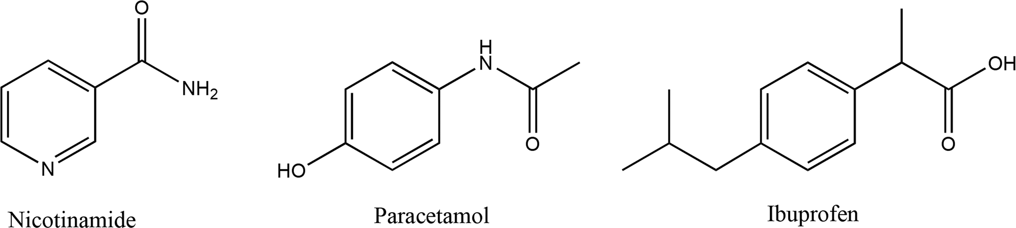
Chemical structures of NIC, PAR, and IBU. IBU, ibuprofen; NIC, nicotinamide; PAR, paracetamol.
Nicotinamide (NIC), a water-soluble drug (log P ≈ −0.4), has the capacity to inhibit the synthesis of proinflammatory cytokines and scavenge oxygen free radicals. 39 Paracetamol (PAR) is a drug slightly soluble in water (log P ≈ 0.55), which can reduce the synthesis and release of prostaglandin E1, bradykinin, and histamine. It is often used for the treatment of headache, fever, arthritis, and other minor pains. 40 Ibuprofen (IBU) is soluble in ethanol, acetone, and chloroform but almost insoluble in water (log P ≈ 3.75). IBU is one of nonsteroidal anti-inflammatory drugs that have been widely used in the relief of mild to moderate pains. 41 Its mechanism of action consists of regulation of the hypothalamic heat-regulating center and inhabitation of cyclooxygenase. 42 These three compounds combined with electrospun nanofibers may attribute to wound repair. In addition, nanofibrous network could realize sustainable drug release and avoid side effects.
Our drug-loaded thermosensitive electrospun porous nanofibers (DTPNs) were fabricated by electrospinning PCL solution using PEO as a pore-forming agent, followed by loading thermosensitive hydrogel PNIPAAm and model drugs. Thermosensitive PNIPAAm hydrogel and drug molecules were diffused into the electrospun porous nanofibrous network by soaking method. 36 Drug-loaded thermosensitive electrospun coaxial nanofibers (DTCNs) were prepared by coaxial electrospinning with PNIPAAm/drug (as core) and PCL (as shell). In vitro drug release behavior of thermosensitive nanofibers at 37°C and 25°C were studied, respectively. The curl of PNIPAAm backbone at high temperature (> LCST) significantly deferred the drug release, especially hydrophobic drug. Moreover, the morphology of electrospun nanofibers could also influence drug release (Fig. 2).
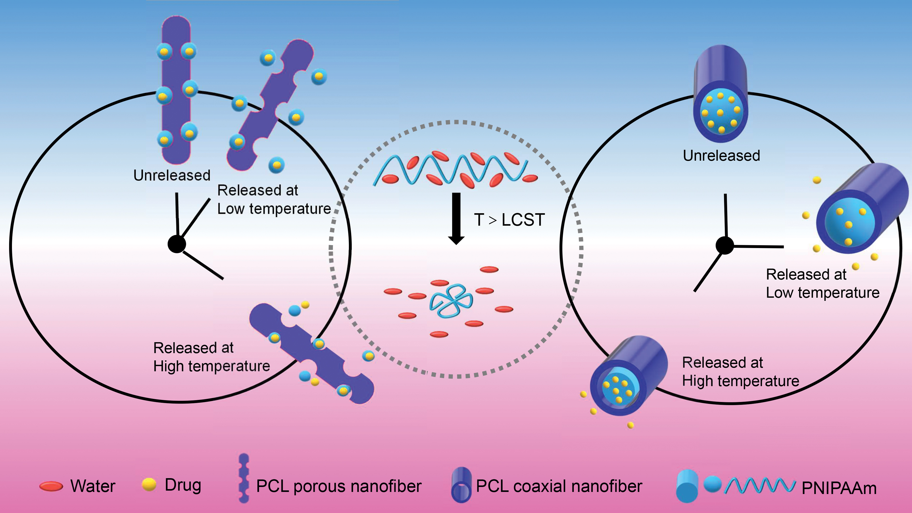
Schematic illustration of drug release behavior of thermosensitive electrospun porous and coaxial nanofibers. LCST, lower critical solution temperature; PCL, polycaprolactone; PNIPAAm, poly(N-isopropylacrylamide). Color images are available online.
Materials and Methods
See Materials and Methods in the Supplementary Data.
Characterization
Characterization of PNIPAAm
The LCST of PNIPAAm was recorded by DSC204 differential scanning calorimeter (NETZSCH Co., Germany). The operation temperature was raised from 20°C to 110°C under the protection of N2 at a heating rate of 2°C/min.
The molecular weight Mν of PNIPAAm was calculated according to the Mark–Houwink–Sakurada formula
43
:
In Eq. 1, [η] is intrinsic viscosity and Mν is viscosity average molecular weight.
The swelling/deswelling characterization: Dry gel was put into a beaker containing deionized water in 25°C water bath. Gel was taken out at regular intervals, wiped by filter paper, and weighed. The above operations were repeated until the hydrogel reached swelling equilibrium. The swelling ratio SR was calculated according to Eq. 2:
In Eq. 2, Wt is the weight of the wet gel at each moment and Wd is the weight of dry gel.
Upon swelling equilibrium, gel was quickly put into a beaker containing deionized water in 40°C water bath. Gel sample was pulled at regular intervals, wiped by filter paper, and weighed. The above operations were repeated until the hydrogel reached deswelling equilibrium. The deswelling ratio, DR, was calculated according to Eq. 3:
In Eq. 3, Ws is the weight of the wet gel at each moment, Wd is the weight of dry gel.
FTIR-8400 infrared spectrometer (Shimadzu Corp., Japan) was applied to determine the chemical structure of PNIPAAm at the wavenumbers range of 400–4000 cm−1.
The stability of PNIPAAm was analyzed by TGA4000 thermo-gravimetric analyzer (PerkinElmer, USA) with temperature setting at 25–700°C.
Characterization of electrospun nanofibers
FTIR-8400 infrared spectrometer (Shimadzu Corp.) was applied to determine the chemical structure of electrospun nanofibers fabricated with different weight ratios of PCL and PEO (Supplementary Table S1) at wavenumbers range of 400–4000 cm−1.
Morphology of porous nanofibers was observed by Regulus810 ultrahigh-resolution field emission scanning electron microscope (FE-SEM; Hitachi, Japan). Samples were precoated with a gold-sputtering layer. Diameters and distribution of over 100 nanofibers were analyzed by Adobe Photoshop CC 2019.
Morphology of thermosensitive coaxial nanofibers was observed by H-7000 transmission electron microscope (TEM; Hitachi) at 100 kV. The coaxial nanofibers for TEM were directly collected with a copper mesh for ∼10 s when the electrospinning process was stable.
A multipoint Brunauer–Emmett–Teller characterization was performed by V-Sorb 2800P specific surface area and pore size analyzer (Gold APP Instrument Corp., China). The sample was preheated in vacuum at 50°C for 10 h and the characterization was performed under high purity N2 (99.9999%) environment at low temperature (−196.15°C).
X-ray diffraction (XRD) was performed to determine the physical structure of drug-loaded nanofibers by BRUKER D8 ADVANCE X-ray diffractometer (Bruker AXS, Germany) with Cu Kα radiation operating at 40 kV and 40 mA over the 2θ range 3–50° by 4°/min.
In vitro drug release study
Two pieces of 10 mg drug-loaded nanofibers were immersed into 10 mL of phosphate-buffered saline (PBS, pH 7.4). The vials were shaken at 100 rpm and incubated at 37°C and 25°C, respectively. At regular intervals, 250 μL of the release solutions were taken from the incubation medium and 250 μL fresh PBS at the same temperatures were added back to the dissolution medium. The released drug, NIC, PAR and IBU, were analyzed by LC-2010 CHT Shimadzu high-performance liquid chromatography (HPLC; Shimadzu, Japan) and the cumulative drug release degree was obtained by the standard curve (r > 0.999). Chromatographic conditions of the three drugs were as follows:
NIC: Column (ZORBAX SB-Aq, 150 × 4.6 mm, 3.5 μm; Agilent); mobile phase: methanol—water (25:75, v/v); detection wavelength: 262 nm; column temperature: 30°C; flow rate: 1.0 mL/min; injection volume: 10 μL.
PAR: Column (ZORBAX Eclipse XDB-C18, 4.6 × 250 mm, 5 μm; Agilent); mobile phase: 0.05 mol/L ammonium acetate solution—methanol (85:15, v/v); detection wavelength: 242 nm; column temperature: 30°C; flow rate: 1.0 mL/min; injection volume: 10 μL.
IBU: Column (Inertsil ODS-SP, 4.6 × 150 mm, 5 μm; Sciences, Inc.); mobile phase: sodium acetate buffer (pH 2.5)—acetonitrile (40:60, v/v); detection wavelength: 263 nm; column temperature: 30°C; flow rate: 1.0 mL/min; injection volume: 10 μL.
Results and Discussion
Preparation and characterization of PNIPAAm hydrogel and thermosensitive electrospun nanofibers
As originally mentioned, PNIPAAm hydrogel has a negative temperature-response characteristic when the ambient temperature rises. The DSC data (Supplementary Fig. S1) showed that the synthesized PNIPAAm had a significant endothermic peak at 31.8°C, which was consistent with previously reported LCST of PNIPAAm. 2 The [η] value of PNIPAAm was determined to be 21.8 (Supplementary Fig. S2). Mv was 2.26 × 10⁴ calculated by the Mark–Houwink–Sakurada formula. The molecular weight of PNIPAAm is closely related to the spinnability of nanofibers.
The SR of PNIPAAm hydrogel increased from 0 to 120 min, and kept unchanged from 120 to 240 min. The SR was 2.63 ± 1.03 g/g at swelling equilibrium (Supplementary Fig. S3a). The DR of PNIPAAm hydrogel decreased sharply in the first 15 min, and kept constant from 15 to 60 min (Supplementary Fig. S3b). When temperature was lower than the LCST, PNIPAAm hydrogels swelled because of hydrogen bonds formation with water molecules. However, when the temperature was higher than the LCST, the PNIPAAm chains are contracted and the water molecules are squeezed out of the hydrogel network. In the Fourier Transform Infrared (FT-IR) spectra of PNIPAAm and NIPAAm (Supplementary Fig. S4), the peak at 3278 cm−1 was caused by the N-H stretching vibration. The C = O stretching vibration and C = C stretching vibration were observed at 1656 and 1620 cm−1, respectively. The peak at 982 cm−1 was attributed to = CH out-of-plane bending vibration.
However, in the spectrum of PNIPAAm, neither C = C stretching vibration peaks nor = CH out-of-plane bending vibration peaks was observed, indicating that NIPAAm had all been polymerized. The 14 wt% weight loss before approaching 200°C was caused by the evaporation of water in the PNIPAAm sample (Supplementary Fig. S5). At ∼300°C, the curve rapidly dropped because the molecular structure of PNIPAAm started to change. At high temperature ranging from 300°C to 450°C, the polymer chain structure was damaged. When temperature was higher than 450°C, a plateau appeared indicating the sample decomposition was basically completed. The purity of PNIPAAm was 86 wt%, which was calculated by the weight loss ratio variation.
FE-SEM images of electrospun nanofibers are given in Figure 3 and a lower magnification version is given in Supplementary Figure S6. The surface of nanofibers was smooth before soaking and then became rough after soaking for 72 h. With the increasing PEO concentration, the number of holes and the average nanofibrous diameter became larger. The leaching of PEO generated pores in the electrospun nanofibers and the pore-forming efficiency increased with the increasing concentration of the pore-forming agent. Electrospun nanofibers held together with adhesive during the soaking process, therefore the fibrous diameters were enlarged. However, as the ratio of PCL/PEO reached 3:5 (w/w), nanofiber holes got partially covered by the folds when soaked for 72 h, which might be caused by the structural deformation or collapse of nanofibers in water.

Field emission scanning electron microscope images of electrospun nanofibers with different ratios of PCL/PEO and soaking states at 80 k magnification.
Figure 4 presents the TEM image of thermosensitive electrospun coaxial nanofiber (TCN). The surface of core-shell nanofibers was smooth with no beads. The light section was PCL shell, whereas the dark section was PNIPAAm core. The core diameter and shell diameter for nanofibers were 169 ± 81 and 282 ± 138 nm, respectively.
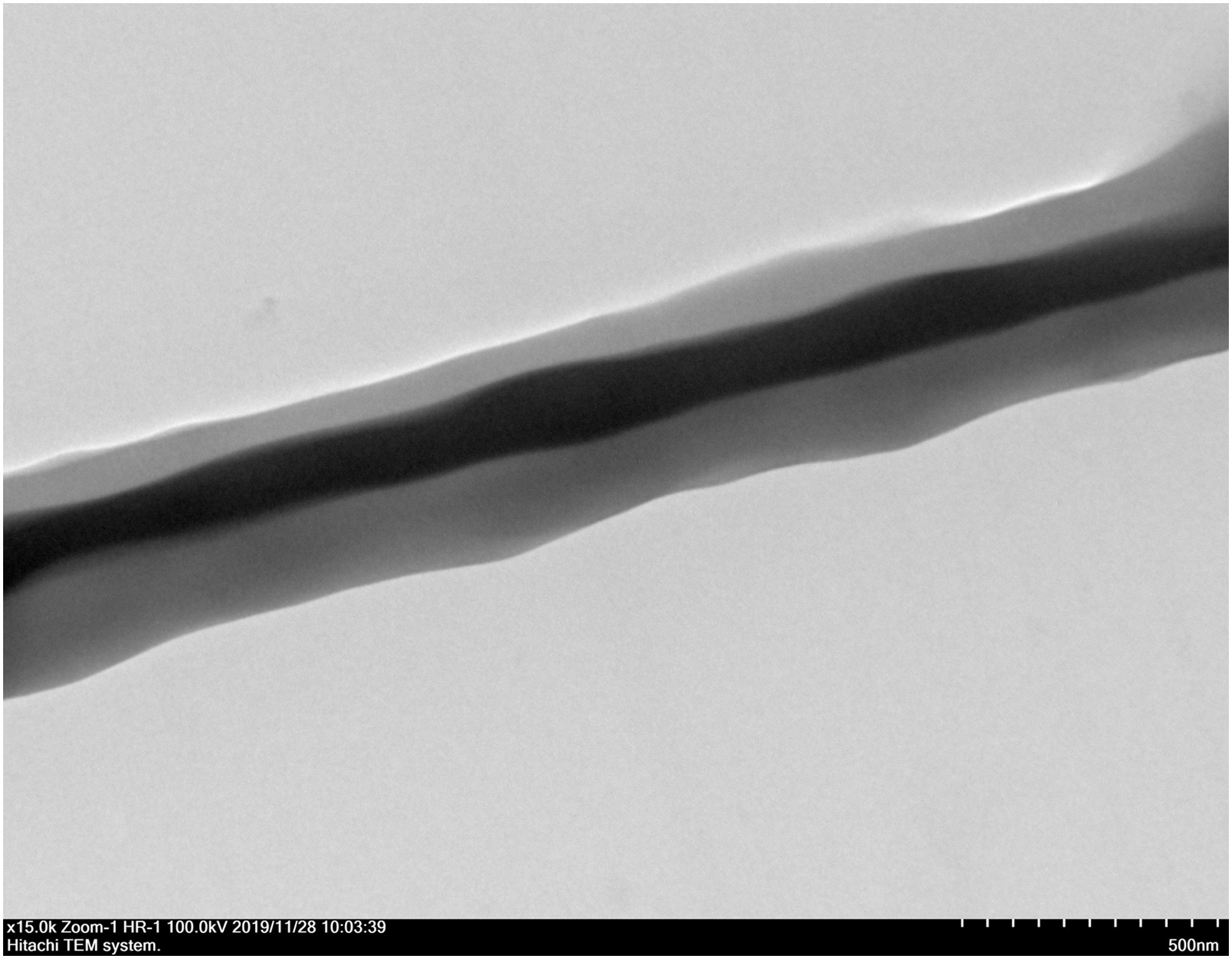
Transmission electron microscope image of TCN. TCN, thermosensitive electrospun coaxial nanofiber.
The N2 adsorption/desorption isotherms and pore size distribution images of electrospun porous nanofiber (PN) prepared at different PCL/PEO composition (designated as PCL/PEO3:2, PCL/PEO3:3, PCL/PEO3:4, and PCL/PEO3:5) were determined (Supplementary Figs. S7 and S8). Table 1 provides the PCL/PEO3:3 sample that had the largest pore volume, whereas PCL/PEO3:4 sample had the largest specific surface area. According to the regulations of the International Union of Pure and Applied Chemistry, the size of the micropore is smaller than 2 nm, the mesopore varies from 2 to 50 nm, and the macropore is bigger than 50 nm. Therefore, the electrospun porous nanofibers prepared in this study have mesoporous structures. Taking SEM images into account, PCL/PEO3:4 was selected as the optimized electrospun composition.
Brunauer–Emmett–Teller Measurements of PN with Different Polymer Ratios
BET, Brunauer–Emmett–Teller; BJH, Barrett-Joyner-Halenda; PCL, polycaprolactone; PEO, polyethylene oxide; PN, electrospun porous nanofiber.
The FT-IR spectra of electrospun nanofibers PCL/PEO3:4 soaked for 0 and 72 h (Supplementary Fig. S9) both displayed characteristic peaks at 1240 and 1293 cm−1, corresponding to C-O-C asymmetrical stretching vibration, C-O and C-C stretching vibration of PCL, respectively. The absorptions of –CH2– twisting vibration and –CH2– wagging vibration of PEO at 1279 and 1340 cm−1 were observed in electrospun nanofibers PCL/PEO3:4 soaked for 0 h, which disappeared when soaked for 72 h. The FT-IR spectra confirmed that PEO was completely removed.
FT-IR spectra of TCN, DTCN, and drugs were characterized as well. The –CH2– stretching vibrations of PCL were observed at 2926 and 2858 cm−1 (Supplementary Fig. S10). The peak at 1722 cm−1 was ascribed to C = O stretching vibration of PCL. Absorption at 1234 and 1286 cm−1 were attributed to C-O-C asymmetrical stretching vibration, C-O and C-C stretching vibration of PCL, respectively. The above absorption peaks also appeared in the FT-IR spectra of DTCN. The FT-IR spectra of NIC-loaded TCN did not show the deformation vibrational absorption peak of pyridine ring at 776 cm−1, the FT-IR spectra of PAR-loaded TCN did not show the p-substituted aromatic ring absorption peak of PAR at 836 cm−1, and the FT-IR spectra of IBU-loaded TCN did not show absorption peak of aromatic stretching bending vibration at 780 cm−1, indicating that drugs were firmly sealed by PCL shells.
XRD diffractograms (Supplementary Fig. S11) demonstrated that NIC was a crystalline compound displaying sharp diffraction peaks appearing at 2θ: 14.868°, 22.266°, 23.425°, 25.452°, 25.909°, and 27.382°. PAR had 2θ (°) peaks of 12.175°, 13.908°, 15.570°, 18.205°, 18.995°, 20.439°, 23.546°, 24.443°, 26.621°, 32.541°, 36.978°, and 46.205°. These diffraction peaks were ascribed to crystalline IBU: 6.143°, 12.238°, 16.767°, 18.790°, 19.079°, 20.198°, and 22.328°. In DTCNs and DTPNs, aforementioned characteristic diffraction peaks of the three drugs disappeared, confirming their formation of fully amorphous solid dispersions instead of crystals. As mentioned in a study, which compared the release performance of drugs in crystalline and noncrystalline form, amorphous drugs were more stable as their solubility were reduced in crystalline form. 44
In vitro drug release behavior
The determination of drug release by HPLC was quick, simple, and specific. The chromatographic peaks of NIC, PAR, and IBU were at 3.9, 4.8, and 3.8 min, respectively (Supplementary Fig. S12).
The drug loading efficiency was evaluated according to the maximum cumulative drug release amount and the weight of electrospun membrane (Table 2). The drug loading efficiency of almost all DTCNs was greater than that of DPNs and DTPNs. The fluctuation of drug loading efficiency in DTCN was small, indicating that DTCN has controllable production stability. The binding force between the drug and the porous fibrous membrane is weak; thus, the drug may fall off the membrane during the preparation. On the contrary, the double-layer structure of the coaxial nanofibrous membrane can well encapsulate drug molecules and result in little drug release.
Drug Loading Efficiency of DPN, DTPN, and DTCN
DPN, drug-loaded electrospun porous nanofiber; DTCN, drug-loaded thermosensitive electrospun coaxial nanofiber; DTPN, drug-loaded thermosensitive electrospun porous nanofiber; IBU, ibuprofen; NIC, nicotinamide; PAR, paracetamol.
The cumulative drug release profiles are given in Figure 5 and percentage statistics in Supplementary Table S2. Generally, a drug with strong hydrophilicity is easy to diffuse in an aqueous medium, so the release rate of a hydrophilic drug is faster than that of a hydrophobic drug. The cumulative release percentages of drug in drug-loaded electrospun porous nanofiber (DPN) in the first 2 min were identical at 37°C or 25°C. However, the cumulative release percentage was lower at 37°C than that of 25°C in DTPNs and DTCNs in the first 2 min.
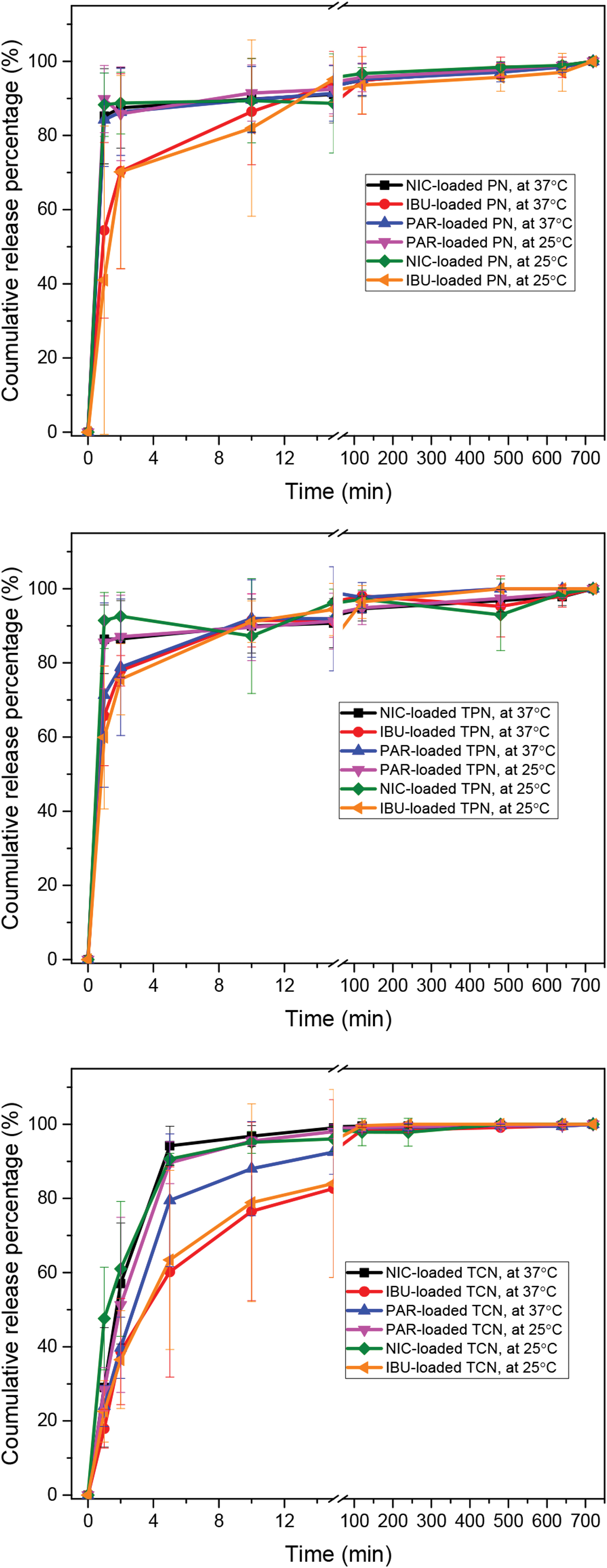
In vitro cumulative release profiles of DPN, DTPN, and DTCN at 25°C and 37°C. DTCN, drug-loaded thermosensitive electrospun coaxial nanofiber; DTPN, drug-loaded thermosensitive electrospun porous nanofiber; PN, electrospun porous nanofiber. Color images are available online.
This could be explained by the thermoresponsiveness of PNIPAAm. When the temperature is higher than PNIPAAm's LCST, the molecular chains entangled and further inhibited drug escape from nanofibrous network. Moreover, the cumulative drug release percentages of NIC-loaded TCN, PAR-loaded TCN, and IBU-loaded TCN at 37°C were, respectively, 57.0% ± 16.3%, 39.7% ± 8.2%, and 38.7% ± 14.3%, which were lower than the DTPN series (86.4% ± 10.3%, 78.8% ± 18.4%, and 77.8% ± 4.1%).
The synergetic effect of thermosensitive hydrogel and electrospun coaxial nanofiber structure could prevent the initial burst release of drugs. The deferred release phenomenon was observed for all the three kinds of drugs, especially the drug with low hydrophilicity. Studies also revealed that the nanofiber with hydrophobic surface could perform better controllable drug release. 27 The porosity of DTPN was not only correlated to the weight ratio of polymers, but also solvent evaporation rate, moisture, and other factors. The loose binding between drug and porous nanofiber might impact the thermoresponsive release effect. However, DTCN with core-shell structure was more stable and fabrication repeatable, resulting in a better sustained release performance.
First-order kinetic model is a common kinetic model of sustained-release preparations, characterized by the release rate and drug concentration. The in vitro release data were introduced to first-order kinetic model (Eq. 4) to explore the release mechanism.
In Eq. 4, M∞ is the cumulative release content at time ∞, Mt is the cumulative release content at time t0, and k is release rate constant.
All the values of correlation coefficient, r, for each equation were greater than 0.98, indicating the first-order kinetic model being a suitable descriptive model of the drug release behavior (Table 3).
The Parameters and Equations of the Drug Release Model from Drug-Loaded Nanofibers
PN, electrospun porous nanofiber; TCN, thermosensitive electrospun coaxial nanofiber; TPN, thermosensitive electrospun porous nanofiber.
The drug loosely attached to the membrane was released rapidly from nanofibers, leading to an initial burst release at the first stage. But the release rate reduced over time at temperature higher than LCST, which was caused by the thermosensitivity of PNIPAAm in the electrospun nanofibers. When temperature rose above LCST, the curling up of PNIPAAm molecular chains lead to the shrinkage of hydrogel, resulting in tighter drug encapsulation in the porous channel of thermosensitive electrospun porous nanofiber (TPN) and less drug escape in TCN. In addition, TCN was more effective for continuous and slow release of drugs, which was a promising sustained-release carrier in clinical application.
Conclusions
Thermosensitive electrospun nanofibers have the potential to be used as a controllable drug release carrier, especially in wound dressings, facial masks, eye patches, and other aspects related to temperature stimuli. In this study, we designed and fabricated thermosensitive electrospun nanofibers with porous or coaxial morphological structures. Three types of drugs with different hydrophobicity were applied to study the controllable drug release behavior of nanofibrous carriers. We systematically investigated the overall effects of drug hydrophilicity, nanofibrous morphology, and thermosensitivity of hydrogel on drug release.
FT-IR and XRD results showed that three model drugs, NIC, PAR, and IBU, were successfully incorporated into electrospun nanofibers. All the loaded drug release results were perfectly fitted to the first-order kinetic model. Hydrophilic drug (IBU) has the slowest release rate, paralleling with its hydrophilicity compared with the other model drugs. Coaxial nanofiber (DTCN) outperformed porous nanofiber (DTPN) as it has less patch-to-patch variation, higher loading efficiency, and tighter drug encapsulation. Thermosensitivity of the hydrogel components enabled much more sustainable drug release when the complex was exposed at physiological temperature 37°C.
This study suggested when adopting thermosensitive electrospun hydrogel nanofibers as the controllable drug release carrier, the hydrophilicity of loaded compounds, and the morphologies of nanofibers are necessary to be optimized.
Footnotes
References
Supplementary Material
Please find the following supplemental material available below.
For Open Access articles published under a Creative Commons License, all supplemental material carries the same license as the article it is associated with.
For non-Open Access articles published, all supplemental material carries a non-exclusive license, and permission requests for re-use of supplemental material or any part of supplemental material shall be sent directly to the copyright owner as specified in the copyright notice associated with the article.