
Abstract
Tissue engineering intends to create functionalized tissues/organs for regenerating the injured parts of the body using cells and scaffolds. A scaffold as a supporting substrate affects the cells' fate and behavior, including growth, proliferation, migration, and differentiation. Hydrogel as a biomimetic scaffold plays an important role in cellular behaviors and tissue repair, providing a microenvironment close to the extracellular matrix with adjustable mechanical and chemical features that can provide sufficient nutrients and oxygen. To enhance the hydrogel performance and compatibility with native niche, the cell-laden hydrogel is an attractive choice to mimic the function of the targeted tissue. Injectable hydrogels, due to the injectability, are ideal options for in vivo minimally invasive treatment. Cell-laden injectable hydrogels can be utilized for tissue regeneration in a noninvasive way. This article reviews the recent advances and future opportunities of cell-laden injectable hydrogels and their functions in tissue engineering. It is expected that this strategy allows medical scientists to develop a minimally invasive method for tissue regeneration in clinical settings.
Impact statement
Cell-laden hydrogels have been vastly utilized in biomedical application, especially tissue engineering. It is expected that this upcoming review article will be a motivation for the community. Although this strategy is still in its early stages, this concept is so alluring that it has attracted all scientists in the community and specialists at academic health centers. Certainly, this approach requires more development, and a bunch of crucial challenges have yet to be solved. In this review, we discuss this various aspects of this approach, the questions that must be answered, the expectations associated with it, and rational restrictions to develop injectable cell-laden hydrogels.
Introduction
Tissue engineering and regenerative medicine are one of the fast-growing branches of knowledge that deals with tissue and organ regeneration. Cells and biomaterials are the inseparable part of tissue engineering.1–3 Appropriate cell delivery to the defected area along with engineered substrate provides a proper condition for tissue repair. Hence, wide ranges of materials have been developed to fulfill this aim.4,5 One of the most promising biomaterials, which have been used widely in this realm, is hydrogels. Hydrogels are a three-dimensional (3D) crosslinked network that can absorb water/biological fluid and swell in a physiological condition.6,7 Hydrogels are composed of hydrophilic segments interacting with water molecules using hydrogen bonding, polar, and ionic interaction to adsorb water.
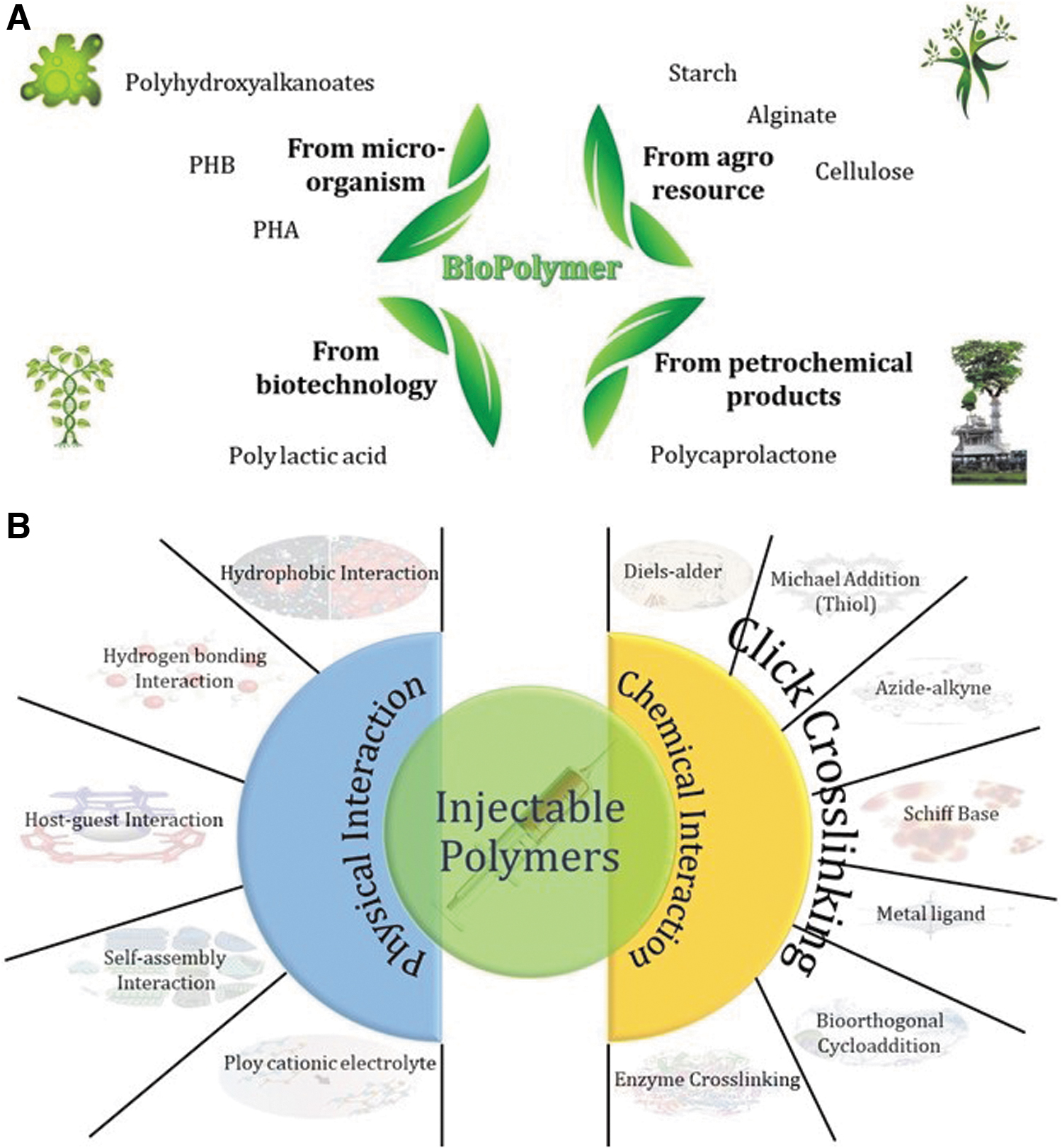
Moreover, the hydrogel's polymer chains are crosslinked to preserve its integrity and prevent the polymer's dissolution in the biological milieu. The utilized hydrogels in tissue engineering and regenerative medicine should show proper biocompatibility and can be classified into four different main groups, including agro-resources-based hydrogel (such as chitosan and cellulose), microorganisms-based hydrogel (polyhydroxyalkanoates), biotechnology-based hydrogel (polylactic acid), and petrochemical-based hydrogel(polycaprolactone) (Fig. 1A).8,9 Thanks to the high-water adsorption capacity, oxygen, nutrients, peptides, and proteins exchange of hydrogel, cells can be loaded in hydrogels and preserve their viability and function. As a result of these benefits, hydrogels have been utilized vastly in biomedical uses.10,11 In the tissue engineering realm, the injectable cell-loaded hydrogel is a minimally invasive technique that attracted significant attention. Compared to traditional surgery, injectable cell-laden hydrogel can be easily applied in targeted areas without large surgical operation in minimally invasiveness, which reduces side effects and pain, increasing the effectiveness of tissue regeneration (Table 1).
Injectable Hydrogel for Biomedical Application
n: power-law index.
ECM, extracellular matrix; LCST, lower critical solution temperature.
Cell loading in hydrogels aims to trap cells within a media resembling the extracellular matrix (ECM). Such matrix should be biocompatible, support cell viability, and permeable to oxygen, nutrients, and extrovert toxic metabolites.12,13 Other features of the platform should be adjustable depending on the target. For instance, encapsulation was utilized for xenotransplantation to cure endocrine disorders such as anemia and diabetes. The injected cells should be protected from the body's immune system to prevent the usage of immunosuppressant treatments, which cause adverse results.14,15 Consequently, the platform should have an adjustable permeability to prevent T cells and antibodies, but allowing the permeation of the signaling ingredients and responses. Moreover, such hydrogel should have adjustable degradation kinetic to protect cells. During degradation, the loaded cells proliferate, differentiate, and generate an ECM. Suitable injectable hydrogel for cell delivery should emulate the ECM and should be fabricated under compatible cell conditions. Hydrogel based on chemical structure, crosslinking degree, and structure shows different swelling behavior and plays a chemical and mechanical obstacle roles toward entering nutrient and outgoing toxic metabolites critical for cell viability. As a mechanical barrier, hydrogel can sieve the molecules based on their size, and as a chemical barrier, hydrogel enables to hinder the smaller molecules.16–18 The suitable hydrogel should allow the easily transport of the oxygen, signaling molecules, nutrients, and metabolites. The cell loading hydrogel properties are illustrated in Figure 2.
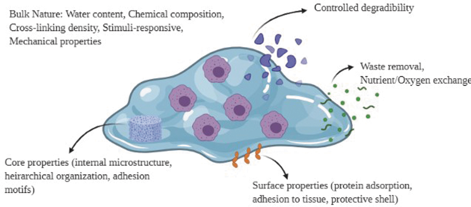
Cell-laden hydrogel properties. The hydrogel embedding the cells should have proper degradation, nutrition/oxygen exchange, porosity, and biocompatibility. Color images are available online.
Mechanism of Injectability
The injectable hydrogel can be crosslinked using physical interaction 19 and chemical reactions. 20 Physically based crosslinking has attracted significant attention because of the easy preparation and higher biocompatibility, while the inferior mechanical properties are the main drawback of the physically crosslinked hydrogels. To overcome such failure, chemically crosslinked hydrogel has been introduced with different chemistry and properties. Limited functional group's stability and toxicity of crosslinkers are the main challenges of the chemically crosslinked hydrogels; however, various ways have been used to decrease the cytotoxicity. Physically crosslinked hydrogels as smart polymers react to different environmental changes such as acidity (pH), 21 light,22,23 electric/magnetic field,24–26 temperature, 27 shear, 28 and ions. 29 Thermo-responsive hydrogels have been widely used because of the simplicity of synthesis and minimum adverse effects on living tissues. 30 The injectable hydrogel's gelation mechanism is based on the physical and chemical bonding, as illustrated in Figure 1B. In this section, the different mechanisms of hydrogel gelation will be discussed.
Hydrophobic bonding interaction
The most popular thermosensitive hydrogels used in tissue engineering are poly(N-isopropyl acrylamide) (PNIPAM), Pluronic chitosan/β-glycerol phosphate. But their gelation mechanism is entirely different. PNIPAM and Pluronic are liquid at room temperature and solidify by increasing the temperature. The mechanism underlying this phenomenon is the coil-to-globule conformational transition with changing the volume by temperature increment which causes the tendency from hydrophobic to hydrophilic state calling lower critical solution temperature (LCST) behavior (Fig. 3). 31 The cloud point as an important factor in thermoresponsivity is affected by the concentration and salt type, which follows the Hofmeister sequences on salt strength. The coil-globule transition of polymeric chains in hydration free energy is affected by hydrophobic interaction. Moreover, entropic factors affect the PNIPAM phase separation. 32 Different force fields affect the PNIPAM thermodynamic characteristics such as solvation and hydration free energy, partition coefficient (temperature function), and density/heat of the segmental unit's vaporization. The reduction of the hydration free energy per unit area with increasing temperature is related to the decrement the molecules in the solvation shell across the NIPAM polar interactions causing conformational changes through the LCST. The force field parameter has been used for the prediction of hydrophobic and hydrophilic interactions. 33
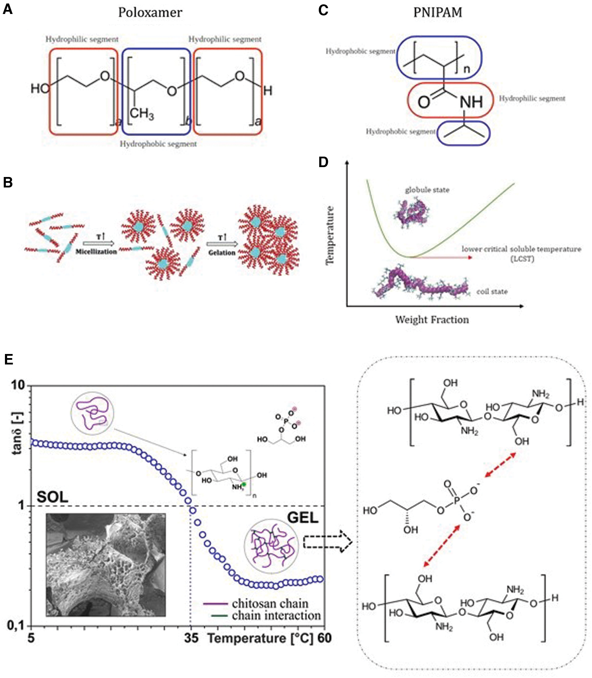
Poloxamer is composed of a triblock copolymer based on polyethylene oxide (hydrophilic segment) and polypropylene oxide (hydrophobic segment), which exhibit the temperature-sensitivity (Fig. 3). Poloxamer solution turns to the solid by increasing the temperature, attributed to the concentration and the segmental ratio. By temperature increment, micelles form due to self-assembly, dehydration, and aggregation of the hydrophobic segments, known as a micellization process. Micelle formation is attributed to the segmental exchange between bulk solution and micelles (on a scale of microseconds), following the Aniansson-Wall model and the forming aggregation of micelles. The micelles' shape can be sphere or worm-like.34,35 Chitosan does not show the thermosensitivity individually. The addition of the glycerol phosphate to the chitosan endows thermosensitivity to the chitosan, which is caused by an ionic-induced mechanism, will be discussed in section Electrostatic gelation/ionic induced gelation (Fig. 3E).
Schiff base linkages
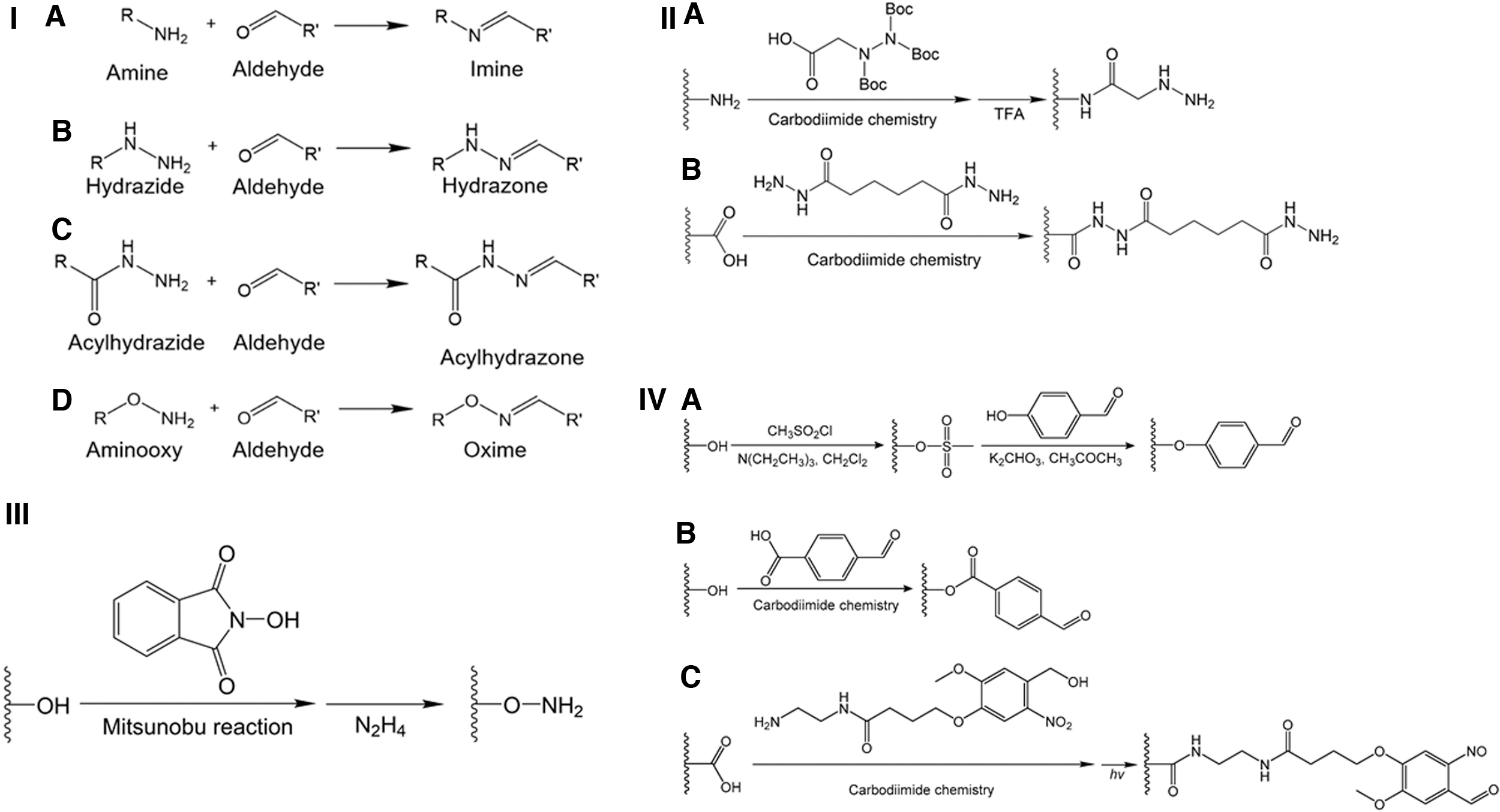
Schiff base linkage uses an aldehyde/ketone as a reversible reaction forms usually under acid or base catalysis condition or temperature increment. The Schiff base interaction is based on amines' nucleophilic attack on the electrophilic carbons of aldehydes and/or ketones. Hydrogels in tissue engineering have been widely fabricated using imines, oximes, and hydrazones synthesized by the reaction of the ketones/aldehydes, aminooxy, and hydrazides, respectively. Oximes and hydrazones show higher durability rather than imines. 36 Moreover, acylhydrazones exhibit better hydrolytic durability rather than them. It was revealed that hydrazones and acylhydrazones had been used to form the hydrogel network with proper stability. Furthermore, benzoic Schiff base linkages have been investigated, and it was revealed that the double bonds of carbon-nitrogen consistency can be enhanced via aromatic replacement with benzene rings linked to the carbon/nitrogen atoms. Usually, benzoic-based Schiff base connections, such as benzoic hydrazides, benzoic imines, and benzoic oximes can be generated using associations of hydrazides, amines, and aminooxy's, respectively, with benzoic aldehydes (Fig. 4).37,38
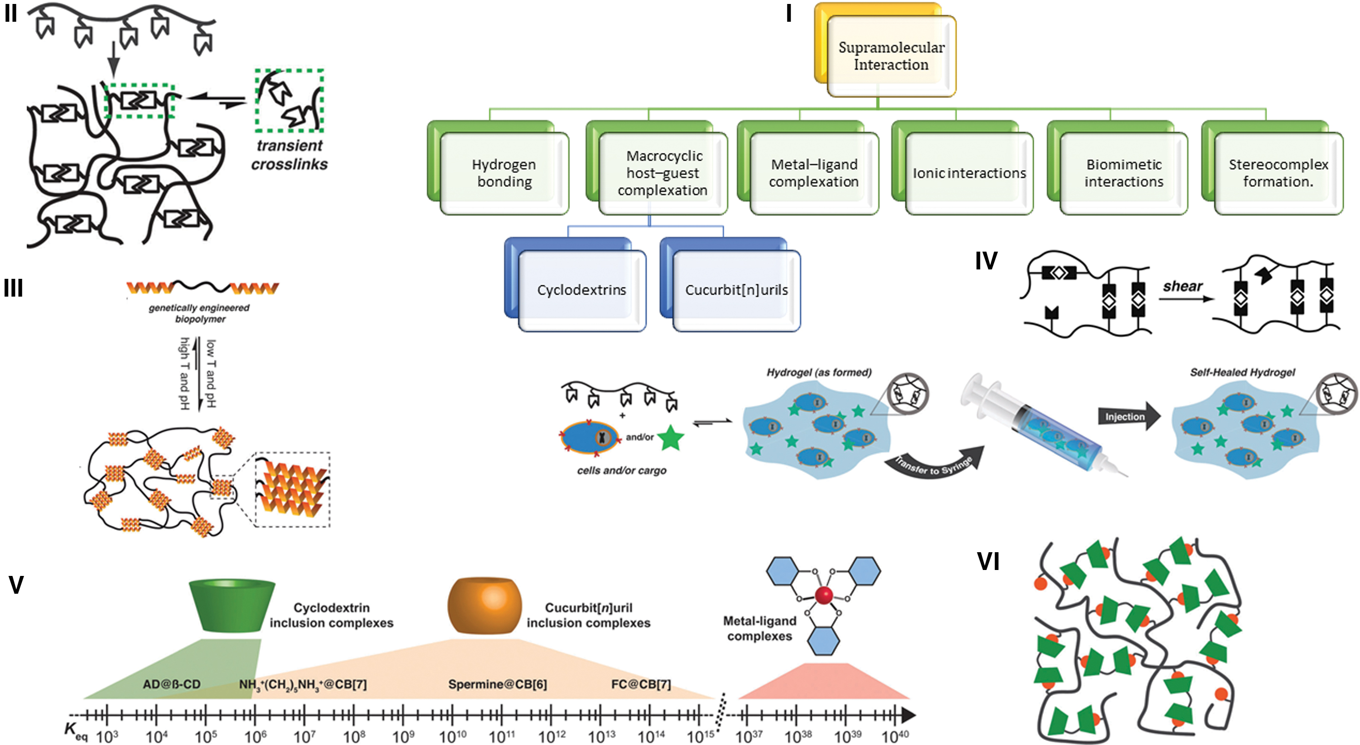
Host–guest interaction
In supramolecular terminology, host–guest interaction refers to the complexes composed of several molecules/ions sticking each other in special structural connections by electrostatic interactions noncovalent bonding rather than covalent bonds. Noncovalent interactions have an important role in preserving the hydrogel integrity and 3D structure. The supramolecular crosslinking of polymer chains in aqueous media using dynamic noncovalent bonds leads to develop novel supramolecular hydrogels such as soft materials with proper injectability, self-healing, and stimuli-responsive properties. Physically crosslinked hydrogels form via the transitory crosslinking among polymer segments. The physically crosslinked hydrogel can be formed with a different mechanism; for example, it can be formed in an aqueous milieu by molecular self-assembly; Hence, a crosslinker is not required, and sol-gel transition happens without considerable volume change. Nevertheless, physically crosslinked gels possess weaker structures and more vulnerable to mechanical forces. However, such a dynamic feature endows the shear-thinning and self-healing features. Physical crosslinking can be formed by entanglements and noncovalent supramolecular connection among polymeric segments. Supramolecular interaction enabling to act in aqueous milieu includes hydrogen bonding, metal-ligand, host–guest biomimetic interactions, multivalent ionic, and stereocomplexes formation. 39 Such unique binding motifs contain variety of dynamics binding with different strength. They are usually orthogonal, letting scientists to reveal a different toolbox to create and manipulate the hydrogels' performance (Fig. 5V). These hydrogels enable scientists to design to synthesize the injectable hydrogel with preserving integrity after injection, which is preferable in medicine as a noninvasive/minimally invasive method by using shear stress during the injection via a syringe (Fig. 6IV). During applying shear stress, a decrement in the gel's modulus facilitates its flow through injection, and after injection, elastic modulus is recovered and reformed the physical network. This implantation technique provides more uniform cell distributions in the injectable cell-laden hydrogel and better control over the material's placement in vivo. 40
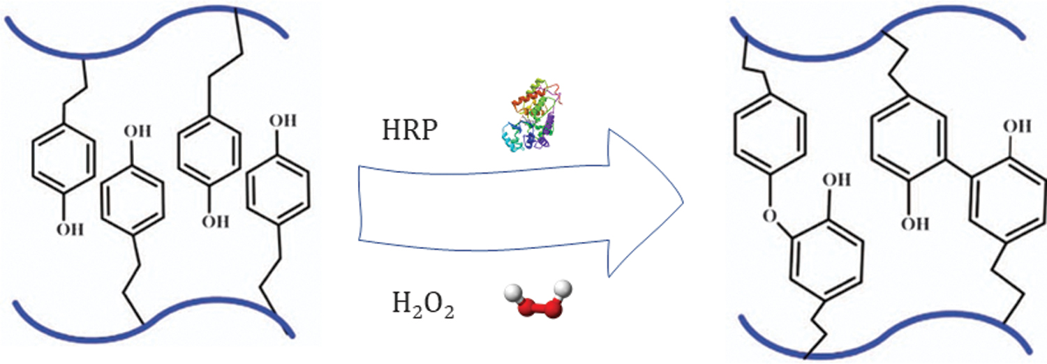
Schematic illustration of injectable hydrogels prepared by the enzymatic crosslinking method with HRP and H2O2. H2O2, hydrogen peroxide; HRP, horseradish peroxidase. Color images are available online.
As discussed, physical interaction between polymer chains such as dynamic entanglement and noncovalent interaction between polymer chains endow hydrogel's injectability. However, few directional noncovalent and specific systems are available for aqueous media and biological environments. Supramolecular motifs based on host–guest interaction have attracted significant attention to synthesizing the injectable hydrogel. In host–guest chemistry, two molecules interact noncovalently and form a complex with reversible properties. Usually, the host molecule is a large one with a specific central cavity like a lock, which can be considered a Lewis base or hydrogen bonding atoms. Guest can be a cationic, anionic, or even neutral atom considering a metallic cationic with Lewis acid properties or hydrogen bond accepting atom. Aqueous supramolecule binding motif such as host–guest has been widely used for synthesizing the injectable hydrogel because of the controllable dynamic and strength. Three fundamental structure has been used in aqueous media, including macrocycles (1) without capping (2) with a flexible/fixed cap at bottom or end, and (3) with a flexible and fixed cap simultaneously at bottom and end.41,42
Electrostatic gelation/ionic-induced gelation
Electrostatic gelation and ionic-induced gelation are the other mechanisms to form the injectable hydrogel. In this system, the ions can be added to the polymer to form a gel, such as adding chloride calcium to the alginate, which can be called ion-induced gelation. In electrostatic gelation, cationic, and anionic polymeric chains are blended and form an electrostatic gel. Classic natural-based anionic hydrogels include alginate, heparin, dextran sulfate, chondroitin sulfate, sodium carboxymethylcellulose, hyaluronate, pectin, and xanthan. Synthetic electrolytic hydrogels are poly-L-glutamates, polyacrylic acids, and poly(methyl vinyl ether-co-maleic anhydride). Chitosan, spermine, spermidine, and polylysine can be classified as a natural cationic polymers and polyvinylamine hydrochloride, poly(allylamine hydrochloride), poly(diallyldimethylammonium chloride), polyethyleneimine, poly(2-dimethylaminoethyl methacrylamide), and poly[2-(trimethylammonium)ethyl methacrylate chloride] can be classified as a synthetic cationic hydrogel. When a cationic/anionic polymer is blended, the hydrogel forms are based on electrostatic interaction. The electrostatic hydrogel performance is related to the opposite charges binding strength, solubility, turbidity, pH, and conductivity. Injecting gel based on electrostatic interaction are homogeneous solutions at room temperature because of the weak electrostatic interaction. The electrostatic interactions were induced by the body temperature to form a gel as an injectable in situ gel.43,44
Alginate (composed of homopolymeric blocks of 1,4-linked β-d-mannuronic acid and α-l-guluronic acid) as the well-known ionic hydrogel can be crosslinked using zinc and calcium cations. The presence of the guluronic acid block causes alginate to interact more selectively with calcium ions. The crosslinking rate is related to the alginate concentration inversely, and the crosslinking rate is affected by the cations concentration and guluronic acid presence. Chitosan as an attractive naturally derived biopolymer has been vastly utilized in regenerative medicine and tissue engineering. Chitosan can be solubilized by reducing the pH to 6.2 because of the amine group protonation, which can be changed to the hydrogel by increasing the pH. Moreover, adding glycerol phosphate endows the thermosensitivity to chitosan because of the ionic interaction between chitosan functional groups and glycerol phosphate.45,46 Chitosan is partially neutralized by introducing the glycerophosphate up to pH 6.8–7.2 without any precipitation because the chitosan chain hydration level is preserved due to the two hydroxyl moieties glycerophosphate. Temperature increment breaks the hydrogen bonds among chitosan, glycerophosphate, and water molecules; then, chitosan forms a 3D hydrogel (Fig. 3E). Temperature increment causes chitosan chains to release proton trapped by glycerophosphate, which pKa is not affected by the variation of temperature.47,48 Using FTIR, it was proved that the phosphate groups are protonated during the chitosan gel formation by temperature increment. 49 It was recommended that the injectable chitosan modified with a low substitution percentage, because high substitution of chitosan groups consume primary amines and prevent the proton exchange and the gelation. For instance, Rossi et al. discovered that trimethylchitosan formed gel using glycerophosphate at the low percentage of substitution (3%) and not at higher ones (78%). 50 Based on this mechanism, it is revealed that, to endow the thermosensitivity behavior to chitosan, a considerable concentration of glycerophosphate (45 wt%) should be added to the solution, but such solution is more susceptible to show inflammatory responses. 51 Nonetheless, such cytotoxicity was not systemic as no renal nor hepatic cytotoxicities were observed. 52 Its intensity is related to the administration route with severe edema by subcutaneous injections and a less obvious one by transdermal infusion in the same condition. 51 The biocompatibility of such hydrogel was further studied as an injectable stem-cell laden hydrogel. Hydrogels with 80 wt% glycerophosphate prevented growth of cell compared to the gels with 40 wt% of glycerophosphate. But such gels cannot form the gel and need a higher temperature of about 46°C. To overcome this problem and form a gel at physiological temperature, hydroxyethyl cellulose (2 wt%), polyethylenoxide, and PEG-4000 (3.6 wt%) can be added to the mixture. 53 Reducing the chitosan solubilizing acid using dialysis and preparing interpenetrating polymer network with other polymers such as collagen can be useful to reduce the glycerophosphate concentration for gel formation. 54
Click reactions
Click chemistry describes a synthetic concept with various reactions, such as Diels–Alder, copper-catalyzed azide-alkyne cycloaddition, thiol-epoxy thiol-ene, tetrazine–norbornene, and thiol-maleimide, which are summarized in Table 2. Such reactions exhibit significant interest in developing injectable hydrogels because of the quick reaction kinetics and low interaction with cellular components. 55 The Michael addition as nucleophilic one of a carbanion and/or nucleophile to an α,β-unsaturated carbonyl complex is the other frequently utilized method to synthesize injectable hydrogels that attract significant attention because of activity in the biological milieu and adjustable reaction time. Poly ethylene glycol (PEG), chitosan, and hyaluronic acid are the most commonly utilized materials to synthesis injectable gel using the Michael addition. 56
Injectable Hydrogel Formation Via Click Crosslinking
PAMAM, polyamidoamine; PEG, poly ethylene glycol.
Enzymatic reaction
Tyramine-conjugated based polymers have been introduced for hydrogel formation in the presence of horseradish peroxidase (HRP) and hydrogen peroxide (H2O2). The hydrogel can be formed using enzymatic crosslinking, by which phenol groups crosslink using H2O2 and HRP. When proteins react with H2O2 and HRP (single-chain β-type hemoprotein), phenol groups of tyrosine oxidize create a di- and tertyrosine conjugated crosslinked protein. 57 The HRP-mediated crosslinking method covalently connects the phenol-conjugated hydrogel to the ECM of the tissue and is thus useful in preserving the injured tissue's structural integrity. 58 Hydrogels containing phenol groups or functionalizing with aminophenol molecules such as tyramine and tyrosine can be enzymatically crosslinked using HRP (Fig. 6). Various enzyme-mediated crosslinking methods have been used to synthesize injectable gels, such as phosphopantetheine transferase, transglutaminase lysyl oxidase, tyrosinase, plasma amine oxidase, thermolysin, β-lactamase, phosphatase, and peroxidase. 59
Kuo et al. synthesized an injectable cell-laden collagen hydrogel that was crosslinked enzymatically. Collagen and tyramine hydrochloride were reacted using carbodiimide reaction to endow the enzymatically crosslinking ability to the collagen. The phenolic hydroxyl (Ph) group bonded to the carboxyl group of collagen, and it was capable of being crosslinked by enzymatic reaction. 60
Applications in Tissue Engineering
The cells loading in a hydrogel is a proper method for many biomedical applications, including the stem cell phenotype maintenance, xenotransplantation, and 3D scaffolds' bioprinting.
Skin tissue engineering
The integumentary system (skin, nails, hair, and sweat glands) is accountable for crucial tasks in the body, including blood clotting, protection of other organs, and transmission of sensory data. Skin as the superficial and the body's biggest organ performs as a practical obstacle against the external environment. Skin injuries can be occurred by environmental stresses and genetic disorders, including acute trauma, thermal, mechanical, chemical, microbial, chronic wounds, radiation effects, and surgical interventions. In the most crucial clinical situations, including third-degree burns, deep dermal, full-thickness wounds, hypodermal adipose tissue, and fascia, internal damages can cause considerable morbidity and dysfunctionality.61,62
One of the critical issues in wound management is moisture, which is crucial for facilitating the healing progression. In this regard, hydrocolloids and hydrogels have been widely applied for the wound healing process to offer a moist milieu and facilitate debridement. Hydrocolloid refers to hydrophilic polymers (colloid particles) forming gels in water. Hydrogels offer several advantages, including the use of bioactive agents and loading cells, because of slight processing situations. The bioactive molecules can be released in an extended period, which signifies vast benefits to topical administration. Moreover, based on the final application, hydrogel features can be adjusted to prevent infection and decrease inflammation. 63 Hydrogels provide significant potential in skin tissue engineering because of several reasons, including (1) hydrogels are the standard therapy for sloughy and necrotic wounds. Hydrogels with high-water content cool the surface of the wound, show a soothing impact, and enhance autolytic debridement. (2) Hydrogel can be designed as a stimuli-responsive platform to release the therapeutic molecules/bioactive agents in the desired manner, which can be applied in the different healing stages. (3) Hydrogels can regulate the inflammatory milieu by carrying antioxidants and anti-inflammatories. (4) Hydrogel can combine with other cells as a cell-laden hydrogel as the prospect of innovative therapies for advanced wound healing to reach the full skin regeneration.64–66
Cell-laden hydrogel with different cells accelerates the wound healing process. For example, endothelial and Schwann cells were used to develop chronic wounds' neoinnervation and neovascularization. Stem cell-laden hydrogels are recommended as alternative therapies that maximize stem cells' immunoregulatory, secretory, regenerative, and homing potential. Such cell-laden hydrogel enables overcoming crucial signs of chronic wounds such as inadequate angiogenesis and constant inflammation. Each nonhealing wound possesses a unique etiology requiring a particular methodology aiming at the wound's specific pathophysiology. 63 Table 3 shows the list of the most utilized cells loaded in hydrogels for skin regeneration.
The Most Utilized Cells Which Have Been Loaded in Hydrogels for Skin Regeneration *
GF, growth factor; IL, interleukin; iPSCs, induced pluripotent stem cells; KGF, keratinocyte growth factor; MSCs, mesenchymal stem cells; TGF-β, transforming growth factor beta; TNF-α, tumour necrosis factor-alpha.
Ke, X., et al. 80
It is proven that the mesenchymal stem cells (MSCs) develop the cutaneous wound healing process. However, cell expansion to achieve adequate cell numbers for regeneration is a limitation in cell therapies. A practical way to solve this problem is to create a method to enhance the immunomodulatory impacts of MSCs. MSCs were loaded within the hydrogel and using electrospray, and the cell-loaded microhydrogel was formed (Fig. 7). After that, such microbeads were loaded in thermosensitive hydrogel and injected into the wounded area. In-vitro results indicated interleukin secretion and transforming growth factor beta 1 (TGF-β1). Full-thickness wounds animal model exhibited granulation and reepithelialization. Furthermore, improved α-smooth muscle actin (α-SMA) expression in the 2 weeks guaranteed wound contraction. Also, decreasing the α-SMA secretion caused reducing the scar formation. Collagen fibrils and the angiogenesis biomarker CD31 expression verified the hydrogel's supporting impact on the wound healing procedure. 67
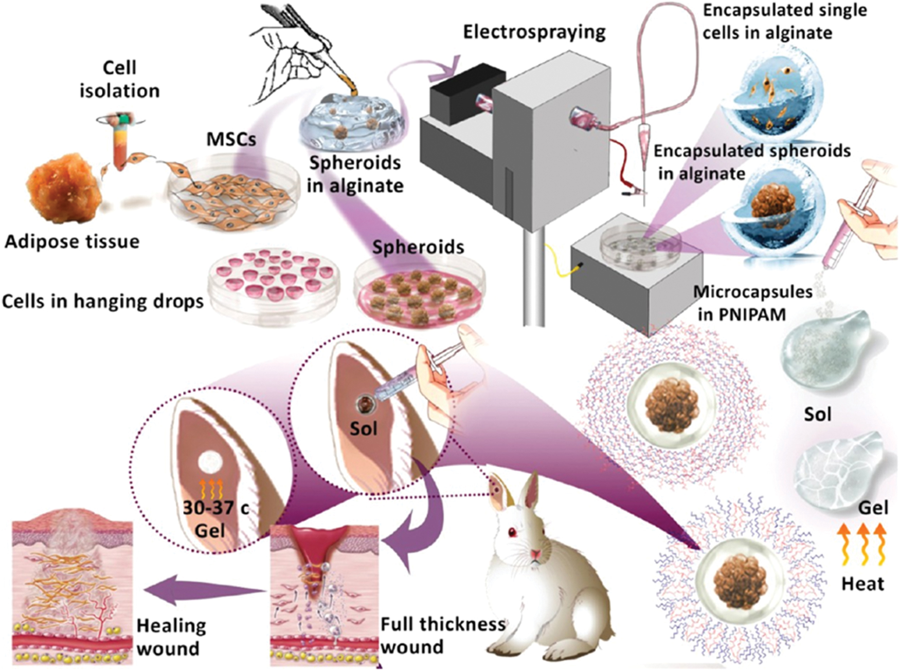
Schematic of the electrospray setup used for cell microencapsulation and cell-laden hydrogel injection. Reprinted with permission. 81 Color images are available online.
Chen et al. synthesized the bone marrow MSCs (BMSCs)-laden hydrogel based on N-isopropyl acrylamide with anti-inflammatory behavior to treat diabetic wounds. In-vitro results indicated that such hydrogel is proper for BMSCs loading, enhancing the TGF-β1 and bFGF secretion. In-vivo results revealed that such hydrogel could hamper the expression of the proinflammatory M1 macrophage. Moreover, cell-laden hydrogel exhibited significant wound contraction and increased angiogenesis, granulation tissue formation, ECM secretion, and reepithelialization. 68
Neural tissue engineering
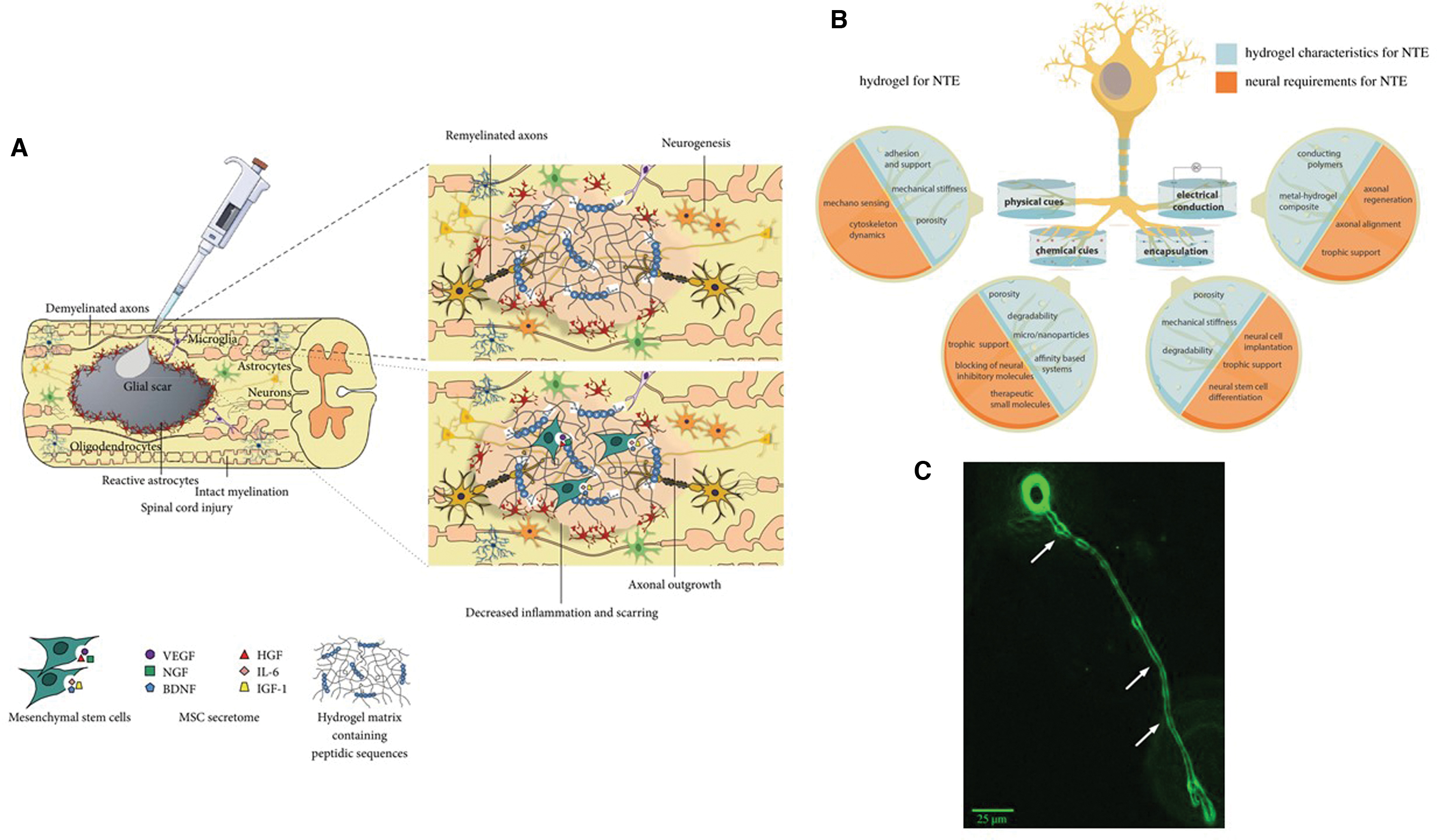
The neural system is separated into the peripheral and central nervous systems. The central nervous system includes the spinal cord, and the brain is affected by various neurological disorders, such as neurodegenerative disorders and demyelinating diseases. Recent therapy methods for neurological disorders can be performed in different approaches. The first action is to restrict the disease pathogenesis, and in the second step, a prolonged strategy needs to inhibit further tissue damage and recurrence. Finally, regenerate and repair the damaged nerve using tissue engineering strategies. These approaches encounter significant challenges, especially in the central system, because it is guarded by the blood–brain barrier. 69 Moreover, the central nervous system persists against regeneration due to the nature of the adult nerves, and cell-secreted molecules, including Nogo, chondroitin sulfate proteoglycans, and myelin-associated glycoprotein, are proven to prevent neural repair. Such inhibitory milieu is mainly available in the central nervous system (CNS), while the peripheral nervous system (PNS) shows relatively improved regenerative capability, mostly due to the lack of neural inhibitory factors. The fundamental assumption of neural tissue regeneration is designing a platform conducive to neural tissue growth. Such platform can be provided by altering the chemical composition, adding biological factors, and using the cells to increase neural regeneration. Neural tissue engineering (NTE) utilizes materials, cells, and neurotrophic factors to accelerate neuronal regeneration. Recently, NTE approaches can be categorized into four groups, including (1) using the guidance signals, (2) components to elevate cell activities, (3) delivery of the drug, growth factor, and cell, and (4) electrical conductivity of the tissue supporting substrate. Hydrogels can easily fulfill the neural regenerative requirements70,71 (Fig. 8). Table 4 shows the most common types of neurodegenerative diseases and the affected cell populations
The Common Types of Neurodegenerative Diseases and the Affected Cell Populations *
Gong, T., et al. 86
Despite the experimental investigation about cell transplantation and material-based therapies for spinal cord injury, their use separately has some limitations. Biomaterials cannot simulate the nerve to regenerate and cannot replace the cells lost during injury. Cell transplantation by itself cannot regenerate spinal cord complicated structural and consistency or even guide axon growth. Therefore, taking benefit of these methods, the injured spinal cord can be regenerated in a proper manner (Fig. 8). 72 Table 5 shows the most essential utilized cells for neural cell therapy.
The Most Important Utilized Cells for Neural Cell Therapy *
CNS, central nervous system; ESCs, embryonic stem cells.
Gong, T., et al. 86
Suri and Schmidt fabricated the cell-laden hydrogel based on collagen, hyaluronic acid, and laminin loaded with Schwann cells to regenerate the damaged nerve. Cell encapsulation did not only show any adverse effect but also at high cell concentration. Cells underwent migration, growth, and secretion of brain-derived neurotrophic factors and growth factors. In some cases, cells aligned and created constructions reminiscent of Bands of Büngner. Adding laminin to the collagen/hydroxyapatite (HA) enhances the nerve growth factor and brain-derived neurotrophic factor. 73 Hsieh et al. synthesized a thermosensitive polyurethane and loaded the hydrogel with neural stem cells (NSCs). NSCs-laden hydrogel exhibited excellent proliferation and differentiation. Moreover, such cell-laden hydrogel is administered into the neural embryo model of zebrafish and rescues the damaged neural system. 74 Tseng et al. fabricated self-healing, injectable cell-laden hydrogel based on chitosan loading with neurosphere-like progenitors. Such cells differentiated to the neuron-like cell and rescued the CNS function. 75
Cardiac tissue engineering
Heart diseases are among the main death reasons worldwide. The significant problems in cardiac injury treatment are the inadequate ability of cardiac tissue to repair. The golden standard of cardiac damage treatment is heart transplantation. Nevertheless, a considerable disparity between the certified persons for heart transplants and the accessible donors causes a severe problem. The low efficacy of this method is another problem. Myocardial infarction (MI) causes myocyte slippage, heart-wall tightening, and ventricular dilation, along with advanced injury to the heart-wall muscle. MI happens after the oxygen and nutrients of the cardiac muscle are reduced because of obstructed coronary arteries. Muscle damage in the left ventricle results in advanced dilation and the myocardium's fundamental changes. Consequently, the contractile efficiency decreases. 76 The MI treatments such as pharmaceutical therapy, implants, and organ transplants possess massive restrictions, including high-level invasiveness, lack of donor organs, immune rejection, thrombosis/stenosis of devices, and extended hospitalization. Injectable gels are developed as an attractive method for in situ cardiac tissue regeneration in infarcted hearts, which such hydrogels can carry therapeutic and cells to the injured section of the to repair damaged tissue. Cell-laden injectable hydrogel offers a minimally invasive delivery method that can overcome the disadvantages of the recent therapies. 77
The myocardium cells can be categorized into four fundamental cell kinds: cardiomyocytes (CMs) (20–40%), cardiac fibroblasts (60–80%), endothelial cells (ECs), and smooth muscle cells. It is known that less than 1% of such cells can be replaced by progenitor cells annually. Entirely, less than 50% of all CMs can be renewed during their lifespan. A high amount of cell slippage happens through MI; hence, recruiting the progenitor cells to compensate for such loss is essential. Different sources have been utilized to achieve CMs for the regeneration of MI. These sources include embryonic stem cells (ESCs), BMSCs, induced pluripotent stem cells, cardiac stem cells (CSCs), bone marrow-derived mononuclear cells (BMMNCs), cardiac progenitor stem cells (CPSCs), endothelial progenitor cells, and adipose-derived mesenchymal stem cells (ADMSCs). Stem cells (SCs) are the ideal source for myocardial repair. Generally, the contribution of SCs to myocardial repair is assumed to happen by two separate processes: paracrine effects and direct differentiation. Restoration is mainly facilitated by the paracrine impact, while direct differentiation has a slight effect (Fig. 9).76,78
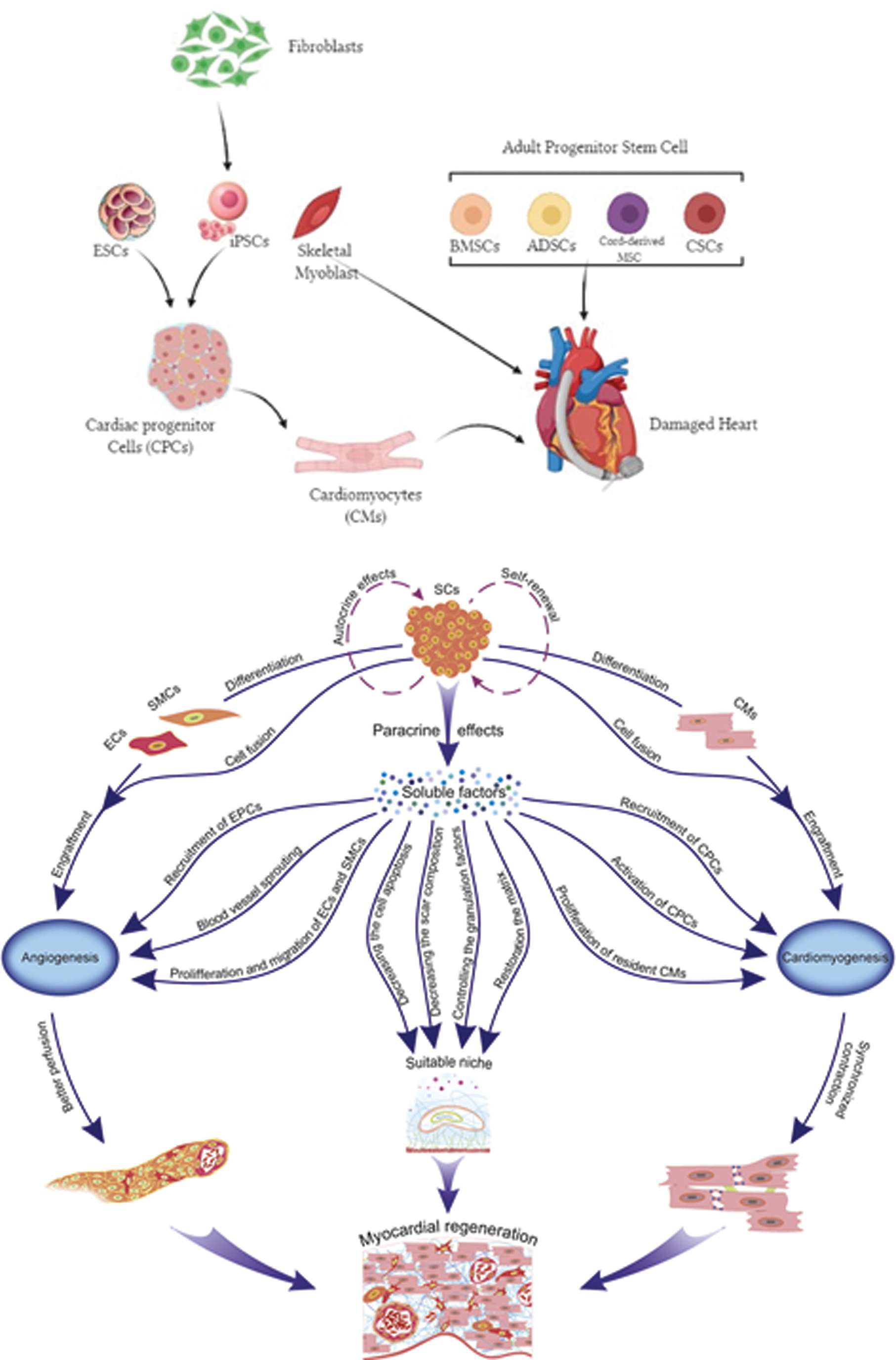
(Top) Cell-based therapies for cardiac regeneration. Several populations of SCs were used for cardiac regeneration. ESCs and iPSCs were first differentiated to CPCs and/or to CMs before transplantation, whereas skeletal myoblasts and adult SCs—such as BMSCs, ADSCs, cord-derived MSCs, and CSCs—were directly employed to regenerate the heart. (Down) Mechanisms involved in myocardial SC therapy. SCs differentiate into ECs, SMCs, and CMs. Cell fusion between transplanted and host cells. Paracrine effects include recruitment and activation of resident EPCs and cardiac progenitor cells (CPCs) (and/or CSCs), along with proliferation of CMs, ECs, and SMCs. Also, they impact the contractility of the CMs; stimulate ECs sprouting from preexisting blood vessels; reduce cell apoptosis; prevent ECM degradation, and inhibit granulation factors and scar composition in the matrix. Reprinted with permission. 92 ADSCs, adipose-derived stem cells; BMSCs, bone marrow MSCs; CMs, cardiomyocytes; CPCs, cardiac progenitor cells; CSCs, cardiac stem cells; ECM, extracellular matrix; ECs, endothelial cells; EPCs, endothelial progenitor cells; ESCs, embryonic stem cells; iPSCs, induced pluripotent stem cells; SCs, stem cells; SMCs, smooth muscle cells. Color images are available online.
Direct cell transplantation to MI in the clinical stage has not been successful, as well as in-vitro studies. The infarcted milieu is acidic, and the ECM strand is inadequate. Moreover, decrement of the ruptured ECM mechanical performance results in ventricular tension be higher than the threshold of damaged tissue, which results in the myocardium perforation. In this situation, a large number of cells experience apoptosis as soon as transplantation, although it was revealed that even a tiny amount of transplanted SCs could restore the infarcted myocardium. In this progress, it seems that the paracrine effects have a significant role, especially in the CMs restoration. It is believed that transplanted SCs cause the CSCs recruitment from the host tissue. A considerable quantity of restored CMs in the damaged area results from the host CSCs differentiation. While cell recruitment, neovascularization, differentiation, and proliferation are stated in cell therapies with poor outcomes, the cell-laden hydrogel can play an essential role in infarcted myocardium regeneration, thanks to the cells and biomaterials' synergic effect (Fig. 10). 76 Dong et al. fabricated a conductive, self-healing, injectable cell-laden hydrogel for cardiac cell therapy based on chitosan-oligoaniline. The conductivity of hydrogel was approximately 10–3 S/cm, similar to the cardiac tissue conductivity. Such hydrogel exhibited proper injectability, thanks to the Schiff-base reaction along with appropriate self-healing properties. Moreover, different cells showed suitable biocompatibility with such hydrogel, and the in-vivo test revealed that such hydrogel possesses adjustable biodegradability. 79 Ke et al. synthesized injectable hydrogel based on chitosan and dextran as an injectable cell delivery system for MI. Injectable gel exhibited the thermosensitive feature thanks to the glycerolphosphate. Umbilical cord mesenchymal stem cells were loaded in a hydrogel, improving the cardiac markers of cTnI and Cx43 expression and p-Akt and p-ERK1/2 signaling pathways. 80
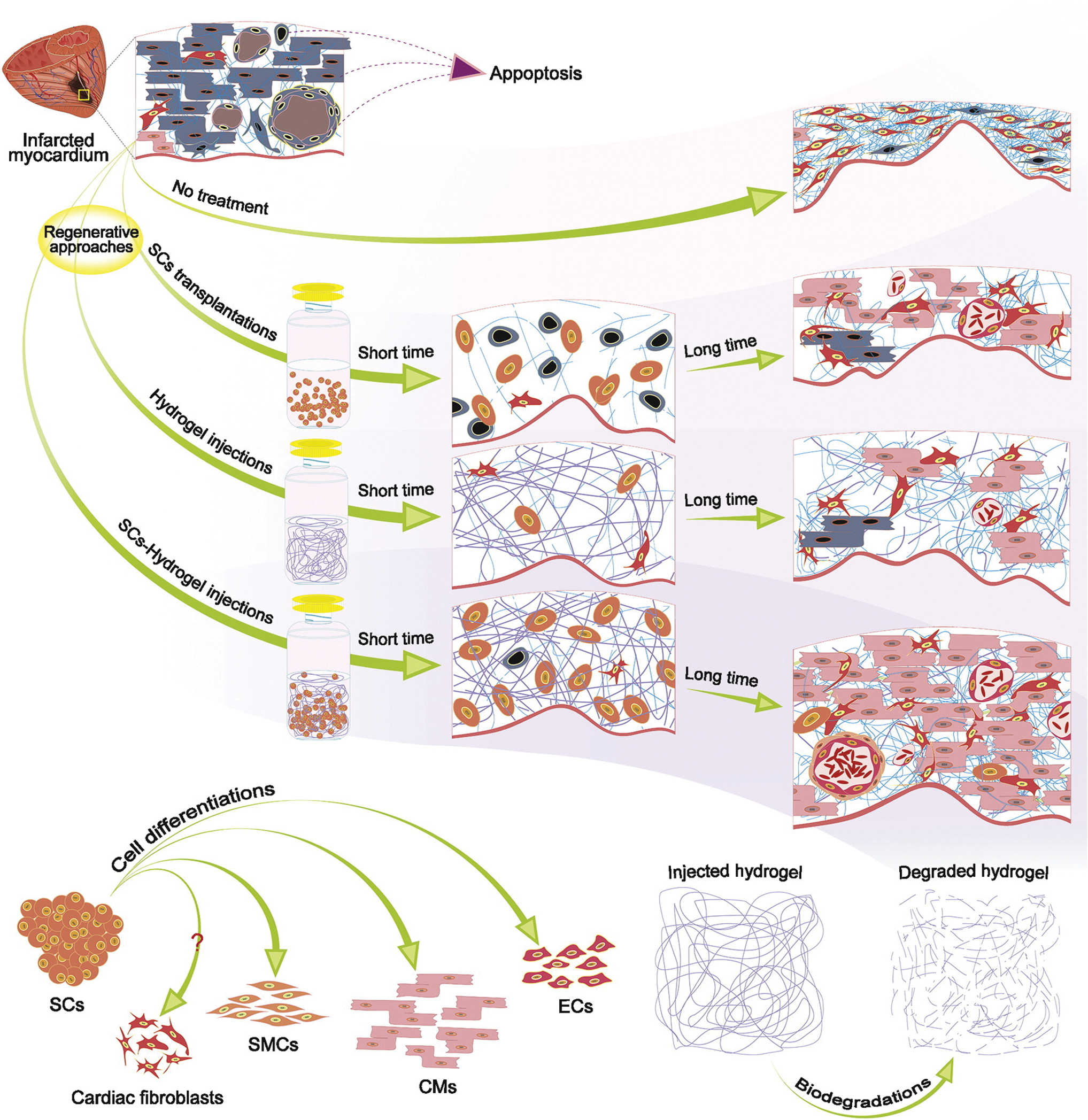
Regeneration methods. After initial cell transplantation, the high volume of cells failed because of an unsuitable milieu. Nevertheless, the remaining cells start to secrete soluble factors that cause autocrine and, more importantly, paracrine effects. In this method, the present initial tension killed a broad volume of cells and decreased myocardial wall thickness. Hydrogel injection hindered myocardial wall thickness reduction, retained cardiac function, prevented fibrous tissue formation, and offered an appropriate milieu for cell survival. Hydrogel performance can be improved by loading SCs and GFs. SC-laden hydrogels prevent decreases in wall thickness by damping the physical tensions, offering a proper condition, and substantially increasing SC therapies' efficacy. The natural and grafted SCs can supply all necessary cell lines for the myocardium—CMs, ECs, and SMCs. There is doubt regarding fibroblasts' resources, as they are frequently regarded as recruited cells from the endocardium. Hydrogels degrade with different processes (hydrolysis, enzymatic, pH, etc.) in the reverse pathway of myocardial ECM restoration. Reprinted with permission. 92 Color images are available online.
Christman et al. used skeletal myoblasts-laden cells to treat the MI using direct and cell-laden hydrogel injection. Fibrin glue was used as a delivery hydrogel, which showed a promising effect than direct cell injection. It was revealed that fibrin glue protected cardiac performance and infarct wall after a MI. Also, it preserved the cells from harsh conditions and increased cell performance during regeneration. 81 Ryu et al. compared the direct injection of the bone marrow mononuclear cells (BMM) and cell-laden hydrogel injection using a fibrin matrix. Loading the BMM within the fibrin matrix and its implantation caused more tissue regeneration in MI compared to direct cell transplantation. Microvessel density and the average internal diameter of microvessels were higher in cell-laden fibrin compared to direct implantation of BMM. 82 Kofidis et al. loaded ESCs within the Matrigel for myocardial restoration. The results indicated that the cell-laden Matrigel increased the fractional shortening and wall thickness.83,84 Liu et al. loaded adipose-derived mesenchymal stem cells (ADSCs) within the chitosan. The cell-loaded chitosan shows a better result than the direct injection of the cell. Cell-laden chitosan enhances cell viability, improved heart performance, increased the wall thickness/angiogenesis, and caused a smaller infarct size. 85 Table 6 shows the essential injectable cell-laden hydrogels for cardiac tissue engineering applications and indicated the cell-laden hydrogel performance compared to the direct cell implantation.
Injectable Cell-Laden Hydrogels for Cardiac Tissue Engineering
ADSCs, adipose-derived stem cells; CSCs, cardiac stem cells; LV, left ventricle.
Bone tissue engineering
Bone with complicated structure with distinct hierarchical levels is depicted in Figure 11. Bone tissue contains two major sections, a dense outer part known as a cortical bone and a porous inner part known as a spongiosa or trabecular bone (Fig. 11a). Cortical and cancellous bone are constituted of repeating osteon units and an interconnecting trabeculae structure, respectively. Osteon units and Trabeculae are constituted of calcium phosphate crystals and collagen fibers (Fig. 11b). Such fibers stiffness is increased by embedded HA crystals in the collagen molecules' spaces and results in increasing the bone stiffness (Fig. 11c). The bone features depend on the ECM and cells, where the ECM organization is hierarchical and spans from nanometer to centimeter. Hence, bone regeneration necessitates novel approaches that represent the nanoscale to macroscale hierarchical construction. Bone repair and regeneration need various factors, including a morphogenetic signal, responsive host cells, signal carrier, and proper vascularized host bed.86–88
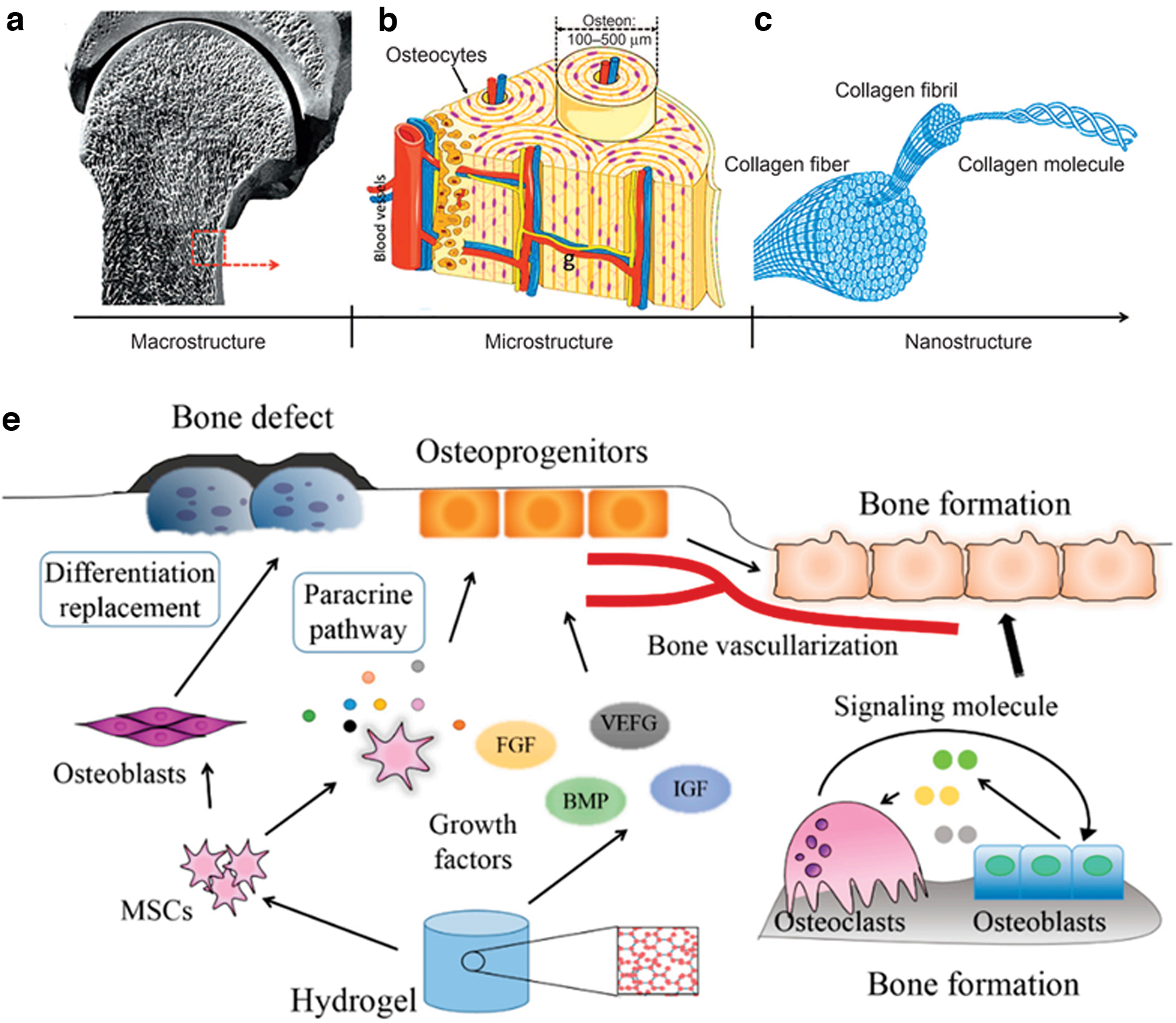
MSCs and GFs performance in tissue regeneration: differentiation replacement and paracrine pathway. The structure of bone
Bone tissue usually can repair spontaneously, although some fractures and diseases prevent the spontaneous healing process and necessitate intervention to help the bone to repair. It was revealed that bone injury causes apparent chondrocyte death and metabolic malfunction and also the surrounding cartilage function is influenced by the injury, thus hampering the spontaneous healing of bone tissue.89,90 Table 7 summarizes the most essential utilized cell-laden injectable gels for bone regeneration.
Most Utilized Cell-Laden Injectable Hydrogel for Bone Tissue Engineering
RGD, arginine-glycine-aspartate.
Biomaterials in bone regeneration should be osteoconductive, osteogenesis, and osteoinductive to repair the bone defect completely. Osteoconduction as the capability to develop new bone on the scaffold's surface requires the platform to attain efficient bone regeneration. Bone conduction biomaterials help the bone progenitor cells migrate, proliferate, differentiate, and facilitate calcification and deposition of bone matrix and vascular growth. Osteoconductive scaffolds provide proper environment to develop prevascular tissue, capillaries, osteoblasts, and the bone's porous structure. 91
Osteogenesis is established by osteoprogenitor cell maturation and proliferation followed by differentiating to osteoblasts. Porous hydrogels facilitate bone conduction. Generally, osteoconductive scaffolds are provided the proper environment for growing the fibrous vascular tissue. Different materials have been added to hydrogel to mimic the bone properties and increase the osteoconductive properties, such as tertiary calcium phosphate and HA. Type I collagen accelerates depositing the minerals by binding to noncollagen matrix proteins, which can initiate and control the mineralization process as an osteoconductive substrate.92,93
The osteoinductive hydrogel can adsorb endogenous growth factors such as bone morphogenetic proteins to stimulate mesenchymal cells to chemotaxis and migration into the hydrogel to generate new bone. To evaluate the osteoinduction, usually, a hydrogel is examined by introducing it to the ectopic soft tissue such as muscle and monitoring the development of new bone in/on the scaffold. The osteoinductive feature of the hydrogel is mostly influenced by two ways: paracrine pathway and differentiation substitution. The accumulated morphogenetic proteins can induce chain-level reactions, stimulate differentiation of MSCs into chondrocytes, enhance cartilage matrix secretion, and mineralize to generate bone. The biomaterials' osteoinductive feature can be related to the development of blood vessels. Angiogenic factors such as TGF and insulin-like growth factor releasing from hematomas, fractured ends, and inflammatory cells recruit osteoblasts and stimulate differentiation to generate bone. MSCs proliferate in an agglomerated form on the HA surface, which is widely utilized to synthesize the osteoconductive hydrogel. HA can help cell proliferation, differentiation, and mineralization through MEK1/2-ERK1/2 and JNK MAPK pathways94,95 (Fig. 11e).
Vo et al. synthesized an injectable dual-gelling cell-laden hydrogel for bone tissue engineering. MSCs were loaded in thermoresponsive PNIPAM/gelatin composite. Gelatin microparticles incorporation enhanced the cellular viability, hydrogel mineralization, and bony bridging. Simultaneous incorporation of gelatin microparticle and cells resulted in forming osteoid and tissue infiltration. 96 Wu et al. synthesized injectable cell-laden microspheres hydrogel for bone regeneration. The microfluidic synchronous crosslinked method was used to fabricate the homogenous and porous microparticle. The freeze-dried microparticle easily adsorbs SCs and enhances the viability and osteogenic performance. Using this method to fabricate the cell-laden hydrogel, preserve the cell from harsh crosslinking conditions. 97 Zhao et al. fabricated the gelatin methacrylate (GelMA) photocrosslinkable microsphere to encapsulate the SC for bone regeneration. It was revealed that such microspheres enhanced cell proliferation, viability, osteogenesis, and mineralization. 98 Ma et al. used injectable hydrogel loaded with periodontal ligament SCs to treat the alveolar bone defect. The hydrogel was based on GelMA and poly(ethylene glycol) (PEGDA) dimethacrylate as a photocrosslinkable hydrogel. The GelMA and PEGDA ratio (4/1) was optimized to enhance the SC activity to achieve better repair. 99
Cartilage tissue engineering
Cartilage as a fiber-supported complex is constituted of chondrocytes surrounded by ECM composing of glycoproteins and glycosaminoglycans. The cartilage structure can be stratified into different parts based on aligning the collagen fiber and proteoglycan composition. Proteoglycan increases gradually from the outer layer to the inner layer. In the outer layer, the collagen fibers are located horizontally. Collagen fibers in the central part are random and tangential to the cartilage surface. Collagen fibers in the calcified section tend to be organized and mineralized.56,100
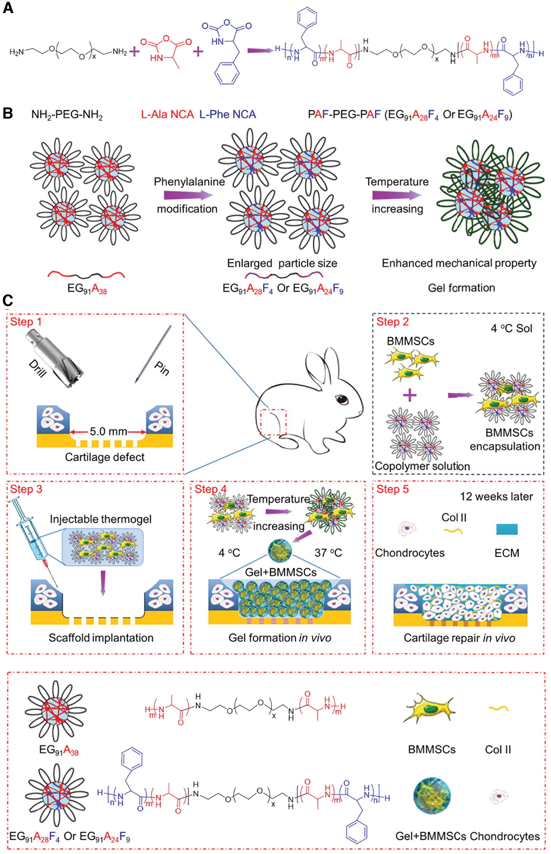
Articular cartilage spontaneous regeneration is restricted due to the limitation of innervation and blood vessels. Cartilage damage is the main reason for temporary disability of the knee, leading to osteoarthritis. Several surgical methods have been used for treating the damaged cartilage, such as microfracture and autologous chondrocyte implantation. However, it was reported that the regenerating damaged cartilage using such methods causes inferior mechanical feature, degenerate gradually, and without long-term rehabilitation. 101 In this regard, various tissue engineering methods have been utilized to form hyaline-like cartilage using cell-laden hydrogels. Effective cartilage repair needs a platform with good mechanical properties, adjustable biodegradation, and high porosity to offer a suitable milieu for adequate cell–cell interaction and cellular activity such as proliferation and differentiation. It has been revealed that scaffolds with large pores exhibit higher permeability and improve cell–cell interaction, nutrient transport, cellular activity, and ECM deposition. For in situ cartilage regeneration, the scaffold plays an important role in supporting regenerative tissue, in which it should tolerate the physiological loads, particularly in large defects. However, it is challenging to increase and preserve the mechanical strength due to the unstable framework during aperture expansion. 102 It has been reported that several thermoresponsive polypeptides and polyalanine-based hydrogel, as a 3D matrix of chondrocytes, improved the cell proliferation and biomarker expression of articular cartilage, such as collagen type II and sulfated glycosaminoglycans. Polyalanine sol-gel transition, mechanical properties, and porosity can be adjusted by introducing the hydrophobic amino acid.103,104 Liu et al. synthesized the injectable cell-laden hydrogel for cartilage regeneration. Poly(l-alanine-co-l-phenylalanine)-block-poly(ethylene glycol)-block-poly(l-alanine-co-l-phenylalanine) as an injectable hydrogel was used to encapsulate the BMSCs to regenerate the cartilage. Such hydrogel possesses different ratios of alanine to phenylalanine. The introduction of hydrophobic phenylalanine into polyalanine-based thermosensitive hydrogel caused an increase in gelation behaviors and mechanical properties. The sol-to-gel transition temperature and the critical gelation concentration were reduced by increasing the phenylalanine units, which caused to grow the pore size and enhance mechanical strength. These characteristics improve hyaline-like cartilage regeneration with lower fibrous tissue formation (Fig. 12). 105 Table 8 summarizes the information about the incorporation of cells into injectable scaffolds for cartilage regeneration.
Incorporation of Cells into Injectable Scaffolds for Cartilage Regeneration *
GO-PEI, graphene oxide-polyethylenimine.
Li, J., et al. 113
Future Perspective and Concluding Remarks
Hydrogels are considered an attractive platform that supports cell proliferation, adhesion, and differentiation by providing requisite physical, mechanical, and chemical cues to grow cells and their subsequent development into tissues and organs for regenerative medicine applications. Hydrogel design and synthesis approaches play a substantial role in imparting the microenvironment of native tissue, which is essential to attain the eventual goal of the development of biological replacements for injured tissues/organs. Injectable hydrogel attracts significant attention because it minimizes the surgical process and can be used in a minimally invasive method. Nevertheless, the shear stress during an injection can harm the cells and decrease the therapy's efficiency. This obstacle can be addressed by encapsulating the cells into microspheres and loading the microsphere in the injectable gel. Microfluidic technology can produce microspheres wherein the cells are suspended in the prepolymer and crosslinked to form microspheres. The encapsulated cells within the microsphere can survive during injection and immune rejection, and the hydrogels' porousness permits nutrient/waste exchange between the cells and milieu. 97
Cell delivery using the injectable hydrogel has offered excellent chances for tissue regeneration. Direct cell transplantation has some challenges, such as correct delivery of cells to the desired site, cell viability during injection, immune response of the body against injected cells. Injectable cell-laden hydrogels offer an adjustable platform to deliver the cells for tissue engineering. Despite limited proofs from clinical trials and unsolved hurdles of the cell-laden injectable hydrogel, many instances of commercial and clinical usage of the injectable hydrogel are available. 106 However, such systems encounter different problems, including mechanical properties, nutrient/waste exchange, compatibility, and degradability. Using cell-laden injectable hydrogels for regeneration is an exciting realm for researchers and technology owners, which can be used as a leading therapeutic method for tissue regeneration.
Footnotes
Acknowledgment
The authors thank Prof. Moztarzadeh for helpful comments and insights.
Disclosure Statement
No competing financial interests exist.
Funding Information
Seeram Ramakrishna wishes to acknowledge the funding support from the UParis-NUS 2020 grant with IdEX code: ANR-18-IDEX-00001 and project number: 2020-06-R/UP-NUS.