
Abstract
Periodontium is the rally of soft and hard tissues, which will be devastated continuously by the compromise of periodontitis. Current periodontal therapeutic methods cannot effectively reconstruct periodontal ligament (PDL), which is oriented at an angle with tooth root and combined hard tissues to form cementum-PDL-alveolar bone complex. Hence, it is urgent to find new techniques for PDL reconstruction to achieve functional regeneration of periodontium. Herein, we developed a novel method to manipulate the distribution and growth of periodontal ligament stem cells (PDLSCs) by utilizing highly paralleled static magnetic field (SMF) and magnetic nanoparticles (MNPs). PDLSCs were incubated with MNPs in vitro to label with them. Meanwhile, CCK8 and live/dead cell staining assay were used to detect the impact of SMF and MNPs on cell viability. The directional migration and growth of PDLSCs were visualized under microscope. Furthermore, real-time quantitative PCR and western blot were utilized to calculate the expression level of PDL-related genes. The results showed that PDLSCs could rapidly take up MNPs without compromising cell proliferation and viability, consequently endowed with the ability to respond via magnetic force. The cell migration analysis indicated that PDLSCs could move along the magnetic induction line, testifying that SMF exerted forces on PDLSCs that labeled with MNPs. It was demonstrated that collective application of SMF and MNPs not only induced PDLSCs organized and grew directionally, but also initiated elongation of cells and nucleus. Furthermore, the morphological alteration of the nucleus could also effectively enhance the gene and protein expression of Collagen Iα2, Collagen III, and Periostin, suggesting the capability of PDLSCs to differentiate into PDL. In conclusion, this study exhibits a new approach for directional reconstruction of PDL to obtain physiological and functional regeneration of periodontium. The Clinical Trial Registration number: WCHSIRB-D-2022-458.
Impact statement
Periodontitis is a common oral disease and characterized by compromising the integrity of periodontium without ideal regenerative method. Magnetic manipulation based on static magnetic field and magnetic nanoparticles has potential in regulating cellular morphology and function, which are crucial for tissue regeneration. However, it is unclear about the role of magnetic manipulation in periodontal ligament (PDL) reconstruction. In this study, we established a novel device to explore the effects of magnetic manipulation in PDL reconstruction. The results provided theoretical basis of magnetic manipulation as a potential strategy to achieve physiological and functional reconstruction of PDL.
Introduction
Periodontitis is the primary factor that gradually compromise the integrity of periodontium, which will eventually result in tooth loss. 1 The unique feature of periodontium is that the oriented collagen fibers of periodontal ligament (PDL) are embedded with cementum and alveolar bone. PDL serves as a dense connective tissue that supports teeth in sockets and meanwhile withstand considerable forces of mastication. 2 Besides, PDL also plays a crucial role in maintaining the stability of tooth structure. Loss of PDL usually leads to ankylosis, which may subsequently initiate tooth resorption. 3 However, current therapeutic methods for periodontitis by initial therapy and flap surgery often result in fragile long junctional epithelium on the exposed root surface of periodontal pocket, and cannot effectively restore the original anatomies of PDL. 4
Guided tissue regeneration (GTR) is capable of reconstructing PDL in the newly formed cementum and alveolar bone to a certain extent, yet, the arrangement of PDL fibers remains unpredictable and uncontrollable. 5 Furthermore, the indication of GTR is rather strict and it is impractical to apply GTR to all kinds of periodontal defect. 6 It's still unclear how to reconstruct highly ordered PDL fibers through the combination of stem cells and scaffolds in terms of tissue engineering. Hence, it is in urgent need to explore a new approach to guide the directional and regular growth of PDL fibers, and organically connect cementum and alveolar bone to achieve effective reconstruction of PDL.
Mechanical force is considered to be a potential strategy to manipulate cell growth and distribution. Mesenchymal stem cells (MSCs) can perceive external mechanical stimuli, thus initiating transformation of cellular structure and function, and ultimately determine the fate of MSCs. 7 To exert forces on MSCs and manipulate their differentiation behavior is a practical way to achieve targeted tissue regeneration. 8 In recent years, various techniques involved in cell manipulation have emerged, for instance, contact guidance by designed substrate patterns, or chemical stimulation through utilizing immobilized biomolecules.9–11 Technically, magnetic manipulation based on magnetic field (MF) and magnetic nanoparticles (MNPs) has been explored as an alternative approach as mechanical stimulation. 12 MF has a bunch of categories such as static magnetic field (SMF) and pulsed electromagnetic fields and so on. 12
Compared with the others, SMF is characterized by easy-access and long-term application without maintenance of any source of energy, which makes it an ideal candidate for noninvasive and remote magnetic manipulation. 13 MNPs refer to superparamagnetic iron oxide nanoparticles with a diameter of 1–100 nm, which can be actuated and mediated by outer MF. 14 By exerting magnetic forces on cells, magnetic manipulation is capable to control cell assembling, migration, and directional growth. 15 In recent years, many researchers have focused on the application values of magnetic manipulation in cells and biomolecules homing, neuron regeneration, and microtissue construction, yet, few reports have been involved in periodontal regeneration.16–19
There's a growing body of evidence indicating that cell morphology and orientation play essential roles in tissue regeneration. For one thing, magnetic manipulation is specialized in determining the direction of cell polarization. For another, stable magnetic forces can deform cytoskeleton and nucleus, which will in turn modulate cellular function. Hence, one of the potential methods for reconstructing PDL is to enable periodontal ligament stem cells (PDLSCs) to respond to the outer magnetic forces by regulating cellular distribution, morphology, and function. 20 To clarify the role of magnetic manipulation in PDL reconstruction will provide theoretical basis for periodontal regeneration therapy, which is dedicated to obtain functional regeneration of periodontium.
In this study, a novel technique is established to demonstrate the potential role of magnetic manipulation in regulating cellular structure and function of PDLSCs, especially with regard to differentiation into PDL.
Materials and Methods
All the experiments were sanctioned by the Ethics Committee of West China Hospital of Stomatology, Sichuan University (Approval No. WCHSIRB-D-2022-458). Besides, all of the teeth were collected with informed consent of the patients or their legal guardian.
Cell culture
Human PDLSCs were derived from the middle third root surface of premolars extracted from patients (13–22 years old) because of orthodontia. All of the PDL tissues were digested by 1% collagenase (Sigma) for 1 h, and they were incubated in minimum essential α-medium (α-MEM; GIBCO), including 20% fetal bovine serum (GIBCO) and 1% antibiotic−antimycotic solution (Solarbio, China) at a stable and moist atmosphere of 37°C and 5% CO2. When the cells grew to 80% of the culture dish, cell passage is performed. PDLSCs utilized in this study were third to sixth passage.
Detection of cell labeling with MNPs
MNPs solution (Macklin, China) was deliquated with α-MEM. The PDLSCs were incubated in this medium for 2 h. To confirm PDLSCs internalized MNPs, PDLSCs were immersed into Fast red and Prussian Blue staining solution (Leagene, China) according to the instruction. The specimens were examined under microscope (Olympus, Japan).
Cell proliferation and viability assay
Both CCK8 and live/dead cell staining were used to measure cell proliferation and viability. PDLSCs were seeded in 96-well plate (1 × 10³ cells per well) and cultured overnight, and incubated with MNPs at 50, 100, and 200 μg/mL, SMF at 200, 300, and 400 mT, SMF+MNPs at 300 mT +100 μg/mL. The SMF was generated by rubidium iron boron magnet (Nd2Fe14B) and the strength and the direction of the SMF is measured by Gauss meter (Laisida, China). Both SMF and MNPs were combined or separately applied to PDLSCs for 24 h/day and 1–7 days. After 1–7 days, the CCK8 reagent (KeyGEN, China) was added to each well and coincubated with cells for 30 min at 37°C. The OD (450 nm) of each well were examined.
Live/dead cell staining assay was used for examining cell viability when PDLSCs were exposed to SMF (300 mT) and MNPs (100 μg/mL). In brief, 5 × 10⁵ cells were seeded in 9.5 cm2 plastic dish and stained with Live & Dead Viability Assay Kit (KeyGEN) for 30 min. The live/dead cells were observed by optical microscope (Olympus).
Cell migration assay
The cell migration assay was evaluated by the 8 μm pores transwell system (Corning®), modified wound healing assay, and observation under optical microscope. For transwell assay, 5 × 10⁴ PDLSCs were inoculated in the top chamber with a cylindrical magnet (300 mT) placing at the bottom of the transwell system. After 24 h culture, PDLSCs on the top chamber were removed, while PDLSCs on the bottom chamber were dyed with purple crystal (KeyGEN) based on the instruction. The cells were examined by optical microscope and calculated in five stochastic fields.
For modified wound healing assay, PDLSCs were incubated with 100 μg/mL MNPs and seeded in 55 cm2 plastic dish to form cell monolayers. A scratch wound on one side was made by 1 mL pipette tip. A cuboid magnet (300 mT) was placed at the one side of the Petri dish. Images were captured under the optical microscope at 24 and 48 h after scratching. The area of wound closure was calculated from five independent views by ImageJ.
In addition, the migration path of PDLSCs were directly observed under the confocal microscope (Olympus).
Preparation of electrospun fibers
Ten percent (wt.%) poly (
PLLA/gelatin was crosslinked to forestall gelatin from dissolution in medium. 0.28 g NHS and 0.47 g EDC were separately dissolved in 95% 100 mL ethanol solution and were agitated for 12 h.
Physicochemical characterization of eletrospinning
Scanning electron microscopy (FEI) analysis were applied to observe the surface morphology of PLLA/gelatin. Water contact angle measurement was used to evaluate the hydrophilicity of PLLA and PLLA/gelatin.
PDLSCs growth, morphology, and orientation using confocal microscope
PDLSCs were separately seeded in confocal dish and PLLA/gelatin. The cells were cocultured with 100 μg/mL MNPs for 2 h. Then the cells were treated with two cubic magnets at both sides to create parallel SMF. The MF intensity (300 mT) in the center of two magnets is measured by Gauss meter. After 7 days, the distribution and morphology of PDLSCs were observed by confocal microscope. The length and the arrangement of PDLSCs were calculated from five random views. The orientation of PDLSCs was quantified by orientation index (OI) defined as OI = cos(θ) (0 < θ < π/2). Nuclear Aspect Ratio = max/min diameter of nucleus.
Real-time quantitative PCR
Total RNA was taken from PDLSCs that seeded in PLLA/gelatin and treated with SMF and MNPs for 7 and 14 days in control and experimental groups by FastPure® Cell RNA Isolation Kit V2 (Vazyme, China). RNA was transcribed to cDNA using Hiscript®III RT SuperMix (Vazyme). The obtained cDNA was amplified with Taq Pro Universal SYBR (Vazyme) and performed by Real-Time PCR System (Illumina, Japan). The relative RNA expression was calculated by the 2−ΔΔCt method and normalized to GAPDH as internal reference. The primer sequences applied in the study are presented in Table 1.
The Primers Sequences Used in Real-Time Quantitative Polymerase Chain Reaction
Western blot
The total cellular proteins of the PDLSCs that seeded in PLLA/gelatin and treated with SMF and MNPs for 7 and 14 days were isolated by RIPA buffer (KeyGEN). The extracted specimens (20 μg) were transferred to the polyvinylidene fluoride membranes (Millipore). The membranes were cocultured with primary antibodies, including β-ACTIN (ZenBio, China), COL Iα2 (ZenBio), COL III (Abcam, United Kingdom), and Periositn (Santa Cruz), at 4°C overnight. ImageJ was applied to visualize and calculate the stripe areas, and β-ACTIN was utilized as internal reference.
Statistical analysis
All data are expressed as mean ± standard error of the mean of three independent experiments and were analyzed using GraphPad Prism 9.0.0. Comparisons between two groups were calculated by nonparametric unpaired two-tailed Student's t-test. *p < 0.05, **p < 0.01, ***p < 0.001, and ****p < 0.0001 were regarded statistically significant.
Results
PDLSCs labeling and viability assay
MNPs were shaped with granular, and the sizes are at nanometer levels (Fig. 1A). The Fast Red and Prussian Blue staining verified the internalization and distribution of MNPs in PDLSCs. Also, it showed that the MNPs were accumulated and distributed in the perinuclear region (Fig. 1B, C). Then the effects of SMF and MNPs on the proliferation of PDLSCs were tested by CCK8 with respect to the incubation time from 1 to 7 days, and there was no obvious proliferation inhibition of SMF (200 and 300 mT) and MNPs (50 and 100 μg/mL). However, SMF (400 mT) and MNPs (200 μg/mL) inhibited the cell proliferation from 1 to 7 days (Fig. 1D, E). Hence, SMF (300 mT) and MNPs (100 μg/mL) were separately selected as SMF strength and MNP concentration, and they did not compromise cell proliferation from 1 to 7 days (Fig. 1F).

The effects of SMF and MNPs on cell uptake, proliferation, and viability.
Furthermore, the live/dead cell staining assay also used to explore the impact of SMF and MNPs on cell viability. It was displayed that solitary or concomitant application of SMF (300 mT) and MNPs (100 μg/mL) did not induce cell death compared with the control group (Fig. 1G). It showed excellent biosafety and biocompatibility of magnetic manipulation system in this study.
The effect of SMF and MNPs on PDLSCs migration behavior
The modified wound healing assay revealed that SMF+MNPs, SMF, or MNPs did not have any obvious impact on PDLSCs migration at 24 h. However, at 48 h, SMF and MNPs obviously promote PDLSCs migration compared with the control group. At the same time, PDLSCs migration ability was apparently inhibited when labeled with MNPs after 48 h. When SMF exerted on PDLSCs alone, the cell migration capability was not compromised (Fig. 2A, B).

SMF (300 mT) and MNPs (100 μg/mL) regulate migration of PDLSCs.
PDLSCs exerted with SMF and MNPs had the highest number of migrated cells in transwell system, significantly more than the other groups (Fig. 2C, D). Furthermore, PDLSCs moved directly along the direction of magnetic induction line (Supplementary Figure S1 and Supplementary Video S1 in Supplementary Data). The results suggest that the magnetic manipulation system apply forces on PDLSCs in the direction of SMF, which induced PDLSCs movement toward the magnetic pole.
Characterization of electrospinning membrane
The surface structure of the PLLA/gelatin membrane and PLLA/gelatin crosslinked membrane was observed to be directionally arranged by scanning electron microscopy (Fig. 3A, B). Water contact angle of the PLLA/gelatin group was significantly lower than that of the PLLA group, which indicates that the adhesion of cells can be significantly increased by adding gelatin into the electrospun membrane (Fig. 3C).

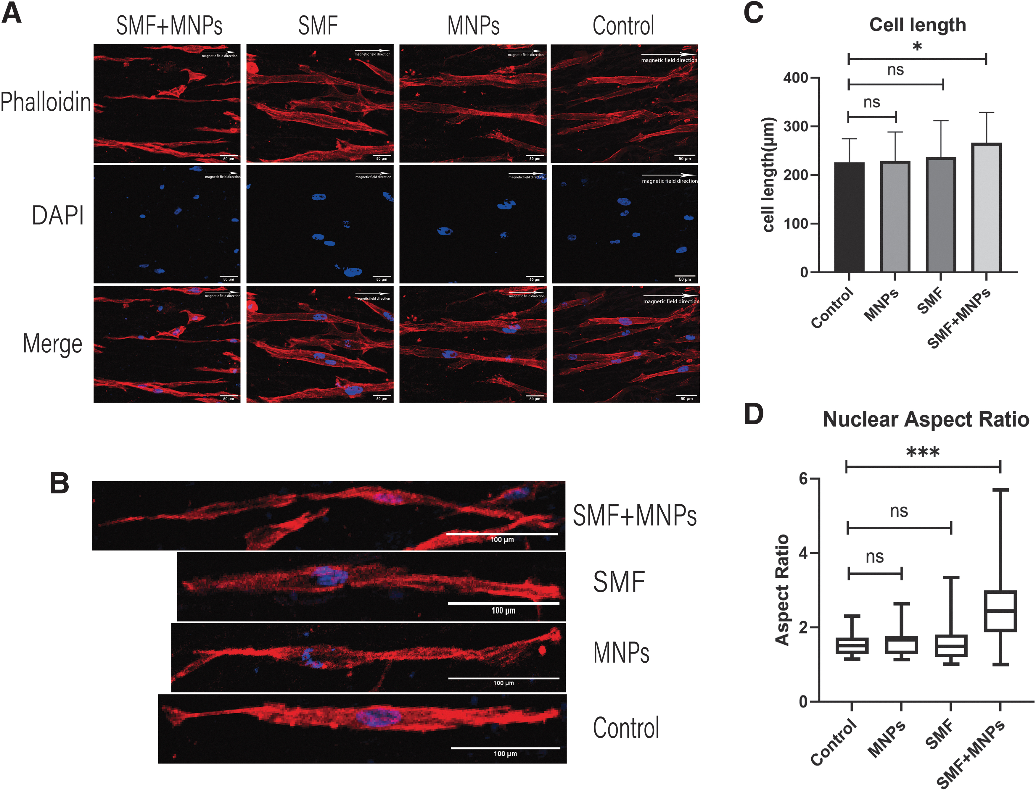
The effects of SMF and MNPs on PDLSCs cell arrangement, cell length, and nuclei.
PDLSCs morphology, growth manipulated by SMF and MNPs
The impact of SMF and MNPs on cell length and nuclear deformation were evaluated under confocal microscope. Cytoskeleton was stained with the actin filaments marker phalloidin, while nucleus was stained with DAPI. When exposed to the SMF and MNPs, the arrangement of PDLSCs was highly consistent with SMF, as was analyzed by OI (Fig. 3D, F, J). The average length of PDLSCs and nuclear aspect ratio are higher compared with the control group when treated with SMF and MNPs (Figs. 3E, H, I, and 4B, C, D). In brief, the results indicated that SMF and MNPs could significantly promote cell arrangement, enhance cell length, and elongate nucleus.
The expression of PDL-related genes
The PDLSCs treated with SMF and MNPs in PLLA/gelatin were collected to measure the gene and protein expression of COL Iα2, COL III, and Periostin. Real-time quantitative PCR showed that col 1α2, col 3, and periostin significantly increased in SMF+MNPs group at 7 and 14 days (Fig. 5A, B). In addition, the protein expression of COL Iα2, COL III, and Periostin enhanced remarkably at 7 and 14 days (Fig. 5C, D). For PDL fibroblast-lineage genes, col 1α2, col 3, and periostin were significantly upgraded in SMF and MNPs group, suggesting that the magnetic manipulation system can promote PDLSCs to differentiate into PDL fibers.
The effects of SMF and MNPs on cell length and nuclei of PDLSCs seeded on PLLA/gelatin.
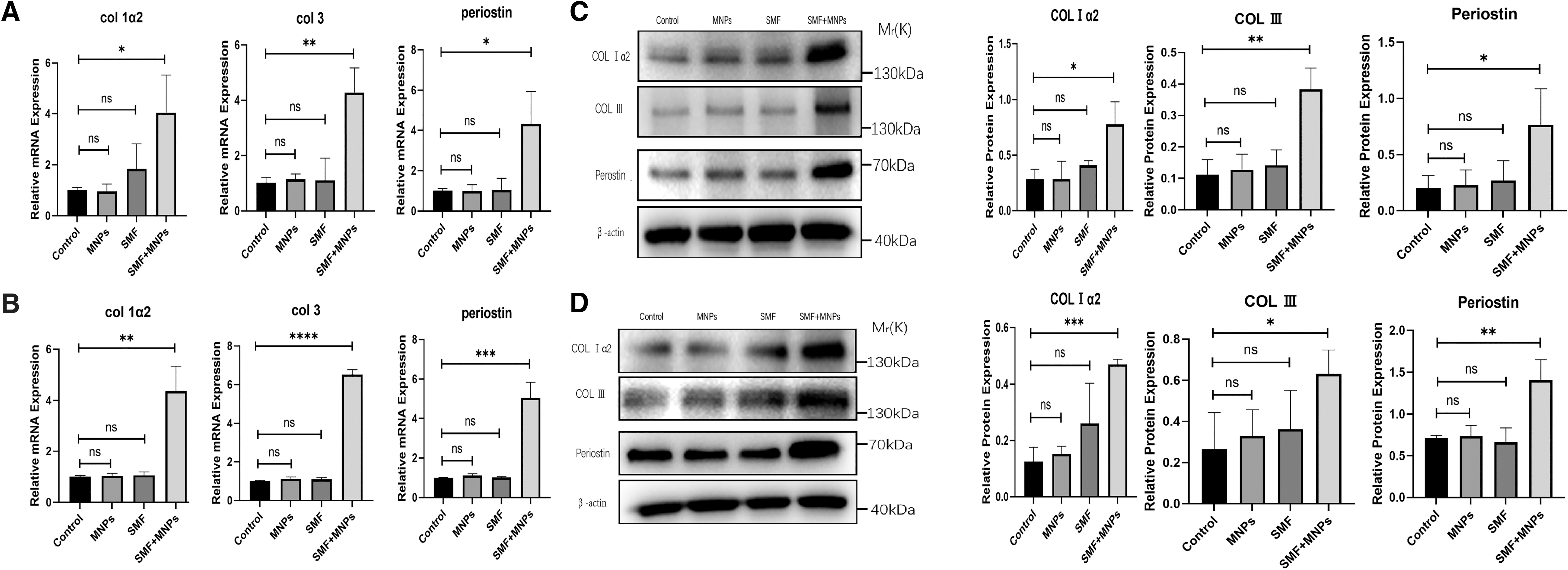
The expression of PDL-related genes.
Discussion
In this study, it was demonstrated that SMF and MNPs were capable to regulate PDLSCs movement and polarization, to elongate the shape of PDLSCs cytoskeleton and nucleus, and to promote PDLSCs fibroblast-lineage differentiation.
To apply SMF and MNPs on cells and tissues, systematic and standardized biosafety evaluation is the prerequisite and fundamental step. In vitro experiments, cytotoxicity test, are calculated by the effects of SMF and MNPs on cell proliferation and viability. 21 Generally speaking, the toxicological profile of MNPs is related to doses, coatings, exposure time, cell types, and diameters, of which doses is the most critical one.22–25 Wu et al. analyzing MNPs cytotoxicity concluded that the biosafety of MNPs was dose-dependent. 26 Hu et al. reported that 500 μg/mL MNPs decreased cell viability by 20%, while Wang et al. observed that 300 μg/mL compromised cell viability by 8.64%.15,27 Most studies reached a consensus that MNPs (<100 μg/mL) induce none or faint cell viability inhibition, which can be defined as noncytotoxic and biocompatible.22,27,28
The toxicological safety of SMF is closely associated with magnetic strength. SMF is a constant vector MF that can be classified as weak (1 mT), moderate (1 mT–1 T), strong (1–5 T), and ultrastrong (>5 T) according to magnetic flux intensity. 29 Various studies have conducted to explore the toxic impacts of SMF on human tissues and cells, yet, no definite consensus have been reached and elucidated.30–32 Moderate intensity of SMF has been widely utilized in biomedicine, which are generally considered to be biocompatible.33,34 Besides, it is worth noting that concomitant application of SMF and MNPs are preferred to enhance cellular uptake and intracellular aggregation of MNPs, thus may trigger unsuspected cytotoxic effects. 35
It is imperative to determine the optimal working concentration of MNPs and magnetic strength of SMF, respectively, not only for the purpose of biosafety but also for seeking a magnetic manipulation system with sufficient strength, which is positively correlated with MNPs concentration and SMF intensity. Hence, we selected SMF (300 mT) and MNPs (100 μg/mL) within the scope of biocompatibility, and they did not compromise cell proliferation and viability, which presented excellent biosafety of the magnetic manipulation system.
To efficiently manipulate PDLSCs with SMF, labeling cells with sufficient MNPs is compulsory. It has been elucidated by synchrotron radiation-based techniques that MNPs can remain chemical stability for up to 14 days. 36 Therefore, it is practical to manipulate cells with SMF and MNPs in a relative long-term. MNPs were observed to be transported to the perinuclear region of PDLSCs, which are basically identical to the previous reports. 37 The entire process of cellular uptake MNPs is termed magnetic labeling, thus making it attainable for cells responding to magnetic forces. 38
It aims to establish a magnetically guiding methodology that enables PDLSCs to grow and differentiate directionally. Hence, it is necessary to verify the operating mechanism of magnetic forces, which can be elucidated by observing the migration pattern of labeled cells. 39 MNPs can be actuated and exerted forces by external SMF through assembling, migration, and orientation, and cells that have taken up MNPs named “magnetic cells” have the capacity to be activated by SMF as well. 37 The results presented that labeled PDLSCs migrated toward the magnetic pole. Meanwhile, the trajectory of migration is straight to the magnet and coincide with magnetic induction line within the restricted region. The motility of labeled PDLSCs conjugates is principally determined by the external SMF profile and intensity, the hydrodynamic characteristics of the cells in its surroundings, and the amount of MNPs uptake by the cells. 40
Compared with the control group, an obvious inhibition of migration occurred in labeling PDLSCs with MNPs. Interestingly, the phenomenon of reduced migration capacity could be reversed by additional applying external SMF. In brief, the migration-promoting effect of SMF and MNPs precisely clarified that magnetic forces followed the direction of magnetic induction line. Meanwhile, the magnetic manipulation system has a relative strong force on labeled cells, which is sufficient to transform the morphology and function of PDLSCs.
In recent years, mounting evidence has focused on the potential of mechanical stimuli to guide the arrangement and growth of cells for tissue regeneration.19,41 In vitro, parallel PDLSCs induced by near-field electrospinning are preferential to upgrade PDL-related genes, while random PDLSCs tend to exceedingly express osteogenic genes. 20 In vivo, highly oriented MSCs can effectively regenerate mature PDL collagen fibers in an orderly and regular manner perpendicular to the surface of tooth root. 11 Magnetic force, serving as the mechanical stimulation, is a promising approach for cell arrangement and polarization. The results presented that SMF+MNPs induced parallel distribution of PDLSCs by placing two parallel cubic magnets on both sides of Petri dishes. By measuring OI, it is worth noting that the parallel pattern of cellular arrangement is highly consistent with the magnetic induction line.
SMF is capable of precisely organizing arrays of “magnetic cells” to form localized patterns in diverse shapes, thus making it attainable to construct desired biological structures by manipulating the position and shape of the magnetic poles. 37 In addition to regulating polarization of cells, magnetic forces also have the ability to reconstruct cell morphology and structure by rearrangement cytoskeleton. 15 Gahl et al. demonstrated that cells could perceive forces ranging from 1 fN to 25 nN, and magnetic forces from this section are capable to stimulate intracellular and cellular reactions. 8 It has been reported that 3–6 pNs could be sufficient for neuronal filopodia elongation and induce alteration of cell morphology, while other research noticed that up to 100 pN cell morphology starts to transform.7,42 By treating PDLSCs with MNPs and external aligned SMF, it was observed that cytoskeleton and cell length were significantly enhanced, which were identical to some prior studies.7,15
Notably, labeled PDLSCs are preferential to grow and extend parallel to the SMF, which is attributed to the effect of external magnetic forces acting on the intracellular MNPs. In brief, magnetic manipulation provides a promising strategy to achieve the desired orientation and growth, which will be conducive to the targeted differentiation of stem cells.
To comprehensively assess the functional modality of magnetic manipulation in vivo is critical. However, due to the limitations of the experimental apparatus, we utilized electrospinning to mimic extracellular matrix (ECM) environment. The high surface area-to-volume ratio of electrospinning is beneficial for cell attachment, proliferation, and differentiation. 43 Furthermore, micro-/nano-sized fibers form either aligned or interlaced patterns, resembling ECM, which plays a crucial role in regulating cell function and arrangement. 20 Both PLLA and gelatin are of high biocompatibility and are FDA-approved biomaterials.44,45 It is believed that gelatin enhance the hydrophilicity of PLLA electrospun membrane, which is profit to cell adhesion and proliferation. Furthermore, the microstructure of relative oriented electrospun fibers is similar to that of directionally arranged PDL fibers. The morphological changes in cytoskeleton as is described above will impose forces to the nucleus through actin-intermediate filament system, thus is likely to initiate transformation of nuclear function and structure. 46
After being subjected to external force, cytoskeleton is something more acting like a mechanosensory, which will induce nuclear deformation while rearranging itself. 45 The significant elongation of nucleus is correlated with the length enhancement of the PDLSCs. A previous study demonstrated that nuclei elongation played a critical role in gene expression profile and stem cell differentiation. 47 Yim et al. showed that extension of the human bone marrow MSCs'nucleus resulted in upregulation of neuronal associated marker SOX2, MAP2, neurofilament light peptide, and tyrosine hydroxylase. 48 Ankam et al. presented that temporal changes in nuclear morphology caused alteration of lamin A/C expression through high-throughput multi-architecture chip analysis. 47 When the PDLSCs were inoculated on the electrospun membrane and treated with SMF (300 mT) and MNPs (100 μg/mL), the gene and protein expression of COL Iα2, COL III, and Periostin were upgraded compared with the three other groups at 7 and 14 days.
These three fibrogenic-related genes are exceedingly expressed in highly ordered fibrous tissues and are indicators for PDL reconstruction.20,49,50 For one thing, Periostin is a matricellular protein, which is increasingly expressed in fibrous connective tissues that are rich in collagen and continuously undergo mechanical strain. 51 For another, COL Iα2 was exceedingly expressed in PDL fibroblasts and was closely associated with collagen fabrication.52,53 Besides, col 3 is one of the marker genes of fibrogenic lineage. 54 Therefore, it is reasonable to believe that the magnetic manipulation system has potential in PDL fibrous tissue reconstruction in vivo. However, it is still noteworthy that we did not find high expression of these PDL-related genes when PDLSCs seeded on Petri dish.
Many researchers have reached a consensus that contact guidance induced by aligned fibers is a practical method to promote differentiation of stem cells by extension of nuclear morphology.55,56 Yet, the mechanism of the role in contact guidance remains unclear. The results render a postulation that the ordered nanotopography of the eletrospun membrane acts like an initiator to boost expression of these three PDL-related genes. After that, the magnetic manipulation system perpetually exerted forces on the labeled PDLSCs, which make it possible to upregulate PDL-related genes overwhelmingly.
This study not only demonstrates a potential method for the directional reconstruction of PDL but also provides a theoretical basis of magnetic manipulation on periodontal regeneration. Given the advantages and the increasing application of magnetic materials in stomatology, it is reasonable to believe that the method we apply will become a promising strategy for PDL reconstruction. However, there are still several unsolved issues in this study. For instance, the mechanism of the magnetic manipulation system promoting PDL-related genes expression remains unclear. Besides, it is uncertain whether the function of “magnetic PDLSCs” in vivo is as good as that in vitro, and how to increase the number of PDLSCs that attached to the electrospun membrane and apply this model to the periodontal defect in situ are still required to be explored.
Conclusion
This study demonstrates that SMF and MNPs are utilized to manipulate the migration, directional growth, and differentiation of PDLSCs. The integrated application of SMF and MNPs has the capacity to promote migration and length enhancement of PDLSCs. Furthermore, the extension of nucleus induced by SMF and MNPs can upregulate PDL-related genes col 1α2, col 3 and periostin. Hence, magnetic manipulation provides a new clinical strategy for PDL reconstruction, which is crucial for physiological and functional regeneration of periodontium.
Footnotes
Authors' Contributions
W.L.: Conceptualization, Data curation, Formal analysis, Investigation, Methodology, Software, Validation, Visualization, and Writing; W.T.: Conceptualization, Funding acquisition, Resources, Supervision, and Writing—review and editing (supporting); Y.W.: Conceptualization, Funding acquisition, Resources, Supervision, and Writing—review and editing (supporting); S.G.: Conceptualization, Funding acquisition, Resources, Supervision, Project administration, Methodology, Validation, and Writing—review and editing (equal).
Disclosure Statement
No competing financial interests exist.
Funding Information
National Key Research and Development Program of China (No. 2022YFA1104400), the National Natural Science Foundation of China (No. U21A20369).
References
Supplementary Material
Please find the following supplemental material available below.
For Open Access articles published under a Creative Commons License, all supplemental material carries the same license as the article it is associated with.
For non-Open Access articles published, all supplemental material carries a non-exclusive license, and permission requests for re-use of supplemental material or any part of supplemental material shall be sent directly to the copyright owner as specified in the copyright notice associated with the article.