
Abstract
Adipose tissue engineering requires effective strategies for regenerating adipose tissue, with adipose-derived stem cells (ASCs) being favored due to their robust self-renewal capacity and multipotent differentiation potential. In this study, the efficacy of poly-L-lactic acid (PLLA) mesh containing collagen sponge (CS), seeded with ASCs to promote adipose tissue formation, was investigated. PLLA-CS implants seeded with GFP-positive ASCs were inserted at high concentration (1 × 10⁶ cells/implant, H-ASC) and low concentration (1 × 10⁵ cells/implant, L-ASC), as were unseeded controls. Adipogenesis was evaluated at 3, 6, and 12 months using a rat inguinal model. At 3 months, the weight and volume of newly formed tissues in the H-ASC group were significantly higher than those in the control group. Histological assessment revealed that the area of all newly formed tissue, including the adipose tissue inside the implants in the H-ASC group, was larger at 6 and 12 months compared with that of the control and L-ASC groups, with the adipose percentage at 12 months being higher in the H-ASC group than in the control group. GFP-positive ASCs in both the L-ASC and H-ASC groups adhered to the CS scaffolds and survived for up to 12 months postimplantation, with spontaneous differentiation into adipocytes observed exclusively in the H-ASC group. Double immunofluorescence confirmed the presence of GFP-positive adipocytes. In summary, this study demonstrated that ASCs coimplanted with PLLA-CS implants could enhance adipose tissue formation within the implants. Uninduced ASCs were capable of spontaneously differentiating into adipocytes within the PLLA-CS implants, with differentiation correlating with the number of implanted cells.
Impact Statement
The bioabsorbable implants developed by our research group, consisting of poly-L-lactic acid (PLLA) mesh containing collagen sponge (CS), exhibited adipogenesis limited to areas directly adjacent to native adipose tissue, with minimal adipose regeneration within the implants. To enhance adipogenesis in regions of slow adipogenesis, PLLA-CS implants were investigated in combination with ASCs, and the underlying mechanisms were elucidated. The transplanted ASCs survived and promoted adipogenesis within the implant by partially spontaneous differentiation into adipocytes, without prior induction or added exogenous growth factors. These findings are expected to advance adipose tissue engineering, offering more effective regenerative medical options for clinical applications.
Introduction
The rising incidence of breast cancer in young women has made it one of the most prevalent malignancies. 1 Postmastectomy reconstruction plays a critical role in improving patients’ psychological health and quality of life. 2 However, the prevailing techniques for breast reconstruction, including silicone implants, autologous flaps, and autologous fat injections, remain limitations. Reconstruction by autologous flap causes abdominal hernia, wound dehiscence, infection, and flap necrosis, and approximately one-third of patients require revision surgery.3,4 Autologous fat injections often require multiple procedures due to low fat retention rates and are associated with the risks of nodule formations, necrosis, and the development of oil cysts. 5 Silicone implants lead to rupture, capsular contracture, and infection. In addition, the long-term implantation of silicone implants causes the development of breast implant-associated anaplastic large cell lymphoma (BIA-ALCL).6,7 Therefore, the development of absorbable breast implants able to promote fat regeneration without long-term implantation is required.
We developed a bioabsorbable implant composed of poly-L-lactic acid (PLLA) mesh as an external scaffold and collagen sponge (CS) as an internal filler. 8 Long-term maintenance of the internal space in vivo is crucial for adipogenesis.8,9 Our implant promotes the regeneration of autologous adipose tissue by maintaining the internal space and avoiding tissue pressure, with the PLLA as an external frame, without requiring additional adipose-derived stem cells (ASCs) or growth factors.8,10,11 It poses no risk of promoting tumor metastasis and is considered safe for use. In previous studies, we confirmed the adipogenesis capability of PLLA-CS implants using rats and rabbits.8,10–12 To achieve substantial adipose formation, multiple PLLA-CS implants were transplanted as implant aggregates in a minipig model.13,14 Although adipose tissue formation was confirmed, it was predominantly limited to areas directly adjacent to native adipose tissue, with minimal adipose tissue regeneration within the implants.
ASCs possess extensive regenerative potential and hold great promise in tissue engineering and regenerative medicine. 15 They exhibit multipotent differentiation and secrete various growth factors and cytokines. It is reported that ASCs seeded onto collagen-based biodegradable scaffolds can enhance adipose tissue formation in vivo.16–18 However, mechanical stress from surrounding tissues and limited vascularization can adversely affect the survival and integration of transplanted ASCs, thereby diminishing their effectiveness in promoting fat formation.19,20
It is crucial to promote adipogenesis even in regions where fat formation is slow, especially in areas not directly adjacent to normal adipose tissue. In this study, we hypothesized that the incorporation of ASCs into PLLA-CS implants would enhance adipogenesis, leading to increased adipose formation within the implant. Adipogenesis using PLLA-CS implants was investigated in combination with ASCs and the underlying mechanisms were elucidated.
Materials and Methods
Ethical statement
The animals were maintained at the Laboratory Animal Research Institute, Graduate School of Medicine, Kyoto University, Japan. The number of animals used in this study was kept to a minimum and every effort was made to reduce animal suffering according to the protocol established by the Animal Research Committee of Kyoto University. The experimental protocol was approved by the University Animal Research Committee (license number: Med Kyo 23110). A schematic overview of the experimental procedures is provided in Figure 1.
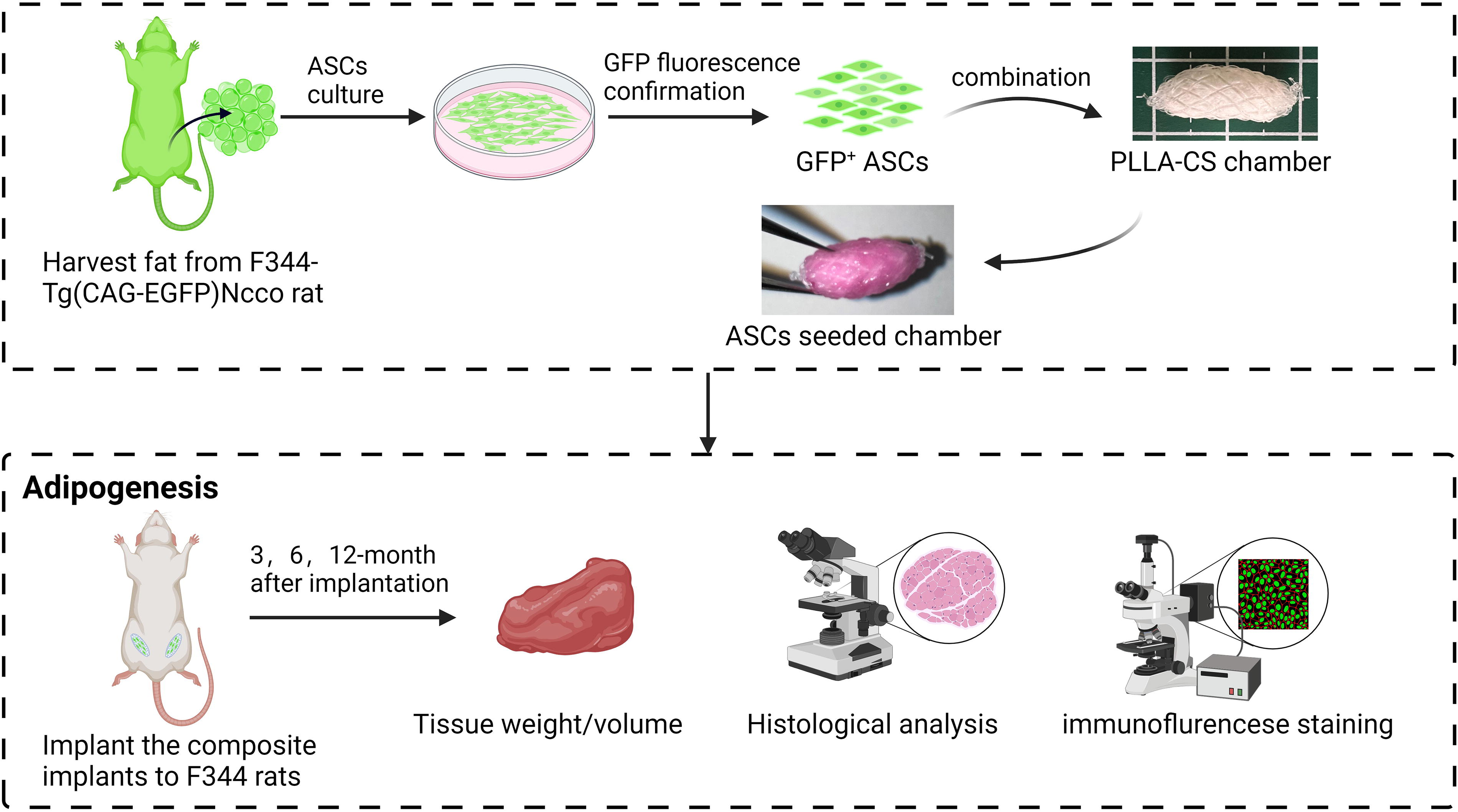
Schematic drawing of experimental procedure (created with biorender.com). ASCs, adipose stem cells; CS, collagen sponge; GFP, Green Fluorescent Protein; PLLA, poly-L-lactic acid.
Isolation and cultivation of GFP-positive ASCs
ASCs were isolated from a 5-week-old male F344-Tg(CAG-EGFP)Ncco rat (Kyoto University Animal Research Center) using a method described previously. 21 The rat was sacrificed by carbon dioxide inhalation and its bilateral inguinal adipose tissues were aseptically harvested. After the adipose tissues were washed extensively with phosphate-buffered saline (PBS), they were cut into small pieces and incubated with 3 volumes of 0.1% collagenase (Sigma-Aldrich, St. Louis, MO, USA) solution with constant shaking at 40°C for 40 min. After adding high glucose Dulbecco's Modified Eagle Medium (DMEM) (Fujifilm Wako Pure Chemical Corporation, Osaka, Japan) containing 10% fetal bovine serum (FBS, Gibco-BRL, NY, USA) and 1% antibiotics (Gibco-BRL) (complete medium), the tissue was centrifuged at 400 g for 3 min. The cellular debris was then removed through a 100-μm nylon mesh (BD Falcon, Franklin Lakes, NJ, USA), and the cells were incubated in a dish with the complete medium. The primary ASCs were cultured for 4–5 days until they reached confluence. These cells were defined as passage “0.” Cells were cultured at 37°C with 5% carbon dioxide (CO2) until reaching 70–80 confluence and passage.
Flow cytometry analysis of ASCs surface markers
Flow cytometry was performed to identify the expression of surface antigens characteristic of ASCs. ASCs at passage 3 were harvested and resuspended in FACS buffer (PBS with 2% FBS) at a concentration of 1 × 10⁶ cells/500 μL. To block Fc receptors, cells were incubated with purified mouse anti-rat CD32 (550270, BD Biosciences, San Jose, CA, USA) for 5 min at 4°C. After blocking, the cell suspension was aliquoted into 100 μL per tube and incubated with fluorochrome-conjugated antibodies, including CD90 (202519, BioLegend, San Jose, CA, USA) and CD29 (102215, BioLegend) as positive markers, and CD45 (202313, BioLegend) as a negative marker, for 40 min on ice, protected from light. Following incubation, the cells were washed with FACS buffer and filtered through a 40 μm cell strainer (Falcon, Corning, NY, USA) to remove cell clumps. Flow cytometric analysis was performed using a BD FACS Canto II flow cytometer (BD Biosciences). Data were analyzed using FlowJo software (Tree Star, Ashland, OR, USA) to quantify the percentage of cells expressing each surface marker.
Adipocyte differentiation of ASCs
Adipogenic differentiation of ASCs was induced in vitro. Cells were cultured in 10 cm dishes in DMEM supplemented with 10% FBS until reaching 90% confluence. Subsequently, an adipogenic induction medium (AdipoInducer Reagent for animal cells, Takara Bio Inc., Shiga, Japan) containing 10 μg/mL insulin, 2.5 μM dexamethasone, and 0.5 mM 3-isobutyl-1-methylxanthine was added and cells were cultured for 48 h. The medium was then replaced with the maintenance medium containing 10 μg/mL insulin, and the cells were cultured for an additional 10 days, with medium changes every 5 days.
At the end of the adipogenic differentiation, the cells were washed with PBS, fixed in 4% formaldehyde (PFA, Fujifilm Wako Pure Chemical Corporation) at room temperature for 10 min, treated with 60% isopropanol for 1 min, and stained with Oil Red O (Sigma-Aldrich) solution (3 g/L) dissolved in isopropanol for 20 min at room temperature. After washing with PBS, the cells were stained with 4',6-Diamidino-2-Phenylindole (DAPI) (1:1000, 62248, Invitrogen) for 5 min and washed with PBS. Representative micrographs were captured using Keyence BZ-X800 (KEYENCE Corp., Osaka, Japan).
For the perilipin staining, following fixation, the cells were washed with PBS and blocked for 60 min at room temperature using immunoblock (CTKN001, KAC Co., Ltd.). The cells were then incubated overnight at 4°C with a rabbit monoclonal antibody against Perilipin-1 (9349S, CST) at a 1:200 dilution. On the following day, the cells were washed and incubated with a secondary antibody, Donkey anti-rabbit AF594 (1:1000, A21203, Invitrogen), for 1 h at room temperature. After washing off the secondary antibody, the cells were stained with DAPI (1:1000, 62248, Invitrogen) for 5 min and washed with PBS. Representative images were captured using a Keyence BZ-X800 microscope (KEYENCE Corp.).
Osteogenic differentiation of ASCs
Osteogenic differentiation of ASCs was performed using the Osteoblast-Inducer Reagent (Takara Bio Inc.), which contains 10 μM l-ascorbic acid, 100 nM hydrocortisone, and 10 mM β-glycerophosphate. On day 14, the cells were washed with PBS and fixed with 4% PFA at room temperature for 30 min. After fixation, the cells were stained using the TRACP & ALP double-stain kit (Takara Bio Inc.) to detect alkaline phosphatase activity as a marker of early osteogenic differentiation. On day 21, Alizarin Red S staining was performed to detect calcium deposits, indicating late-stage osteogenic differentiation.
For ALP staining, after washing and fixation, the cells were permeabilized by adding a 1:1 ratio of acetone and ethanol, incubating for 1 min at −20°C. Following washing with PBS, the ALP substrate solution (premixed) was added, and the cells were incubated at 37°C for 15 min. The ALP-positive areas were visualized after incubation. For Alizarin Red S staining, 1 g of Alizarin Red S (011-0192, Wako) was dissolved in 100 mL of Milli-Q water, and the pH was adjusted to 6.3–6.4 using 28% ammonia solution. After the cells were washed and fixed, the Alizarin Red S staining solution was added to the osteogenic differentiation wells, and the cells were stained for 10 min. After staining, the cells were washed thoroughly with Milli-Q water. The representative images were captured using a Keyence BZ-X800 microscope (KEYENCE Corp.).
Chondrogenic differentiation of ASCs
Chondrogenic differentiation of ASCs was performed using the MesenCult™-ACF Chondrogenic Differentiation Kit (STEMCELL Technologies, Canada), following the manufacturer’s protocol. Briefly, ASCs were resuspended at a concentration of 2 × 10⁶ cells/mL in a complete MesenCult-ACF Chondrogenic Differentiation Medium. The cell suspension (0.5 mL) was transferred into 15 mL polypropylene tubes and centrifuged at 300 g for 5–10 min to form cell pellets. The caps were then gently loosened, and the tubes were incubated at 37°C in a humidified atmosphere with 5% CO2 for 3 days. On day 3, the medium was replaced, and the cultures were maintained until the end of the 21-day differentiation period. At the end of the induction, the cell pellets were washed with PBS and fixed with 4% paraformaldehyde at room temperature for 30 min. Alcian blue solution (Alcian Blue Solution, pH2.5 Fujifilm Wako Pure Chemical Corporation) was added, and the samples were incubated overnight in the dark. After washing with 1% acetic acid, representative micrographs were captured using a Keyence BZ-X800 microscope (KEYENCE Corp.).
Preparation of bioabsorbable implants
PLLA was prepared as the external frame and supplied by Gunze Ltd. (Tokyo, Japan). Next, CS was prepared with a porosity of 80–95% (PELNAC®, Gunze Ltd.) as the internal filling material. The 54 implants were prepared as described previously. 10 Each columnar mesh (10 mm in diameter and 10 mm in height) was knitted using 2–0 PLLA threads. Then the top and bottom of the meshes were closed by purse string sutures of the respective thread after tight packing with 40 × 20 × 3 mm CS. The largest diameter of the short axis was approximately 7.5 mm, while the longest length of the long axis was approximately 18 mm. The gaps between the meshes were squares measuring approximately 1.5 × 1.5 mm.
Cell seeding
Green Fluorescent Protein (GFP)-positive ASCs at passages 3–5 were harvested and resuspended in DMEM to achieve a final concentration of 5.0 × 10⁵ cells/mL and 5.0 × 10⁶ cells/mL. Subsequently, aliquots of the cell suspension were evenly seeded onto CS inside the PLLA-CS implants. For the control group, 200 µL of DMEM was added to each CS. For the L-ASC group, 200 µL of cell suspension containing a concentration of 5 × 10⁵ ASCs was evenly added to each CS (Supplementary Video S1). For the H-ASC group, 200 µL of cell suspension containing a concentration of 5 × 10⁶ ASCs was evenly added to each CS. The seeded PLLA-CS implants were placed in a 6-well plate and allowed to incubate statically in a CO2 incubator at 37°C for 30 min to facilitate cell attachment to the CS. The attachment of ASCs to the surface of CS inside the PLLA-CS implant-seeded ASCs was confirmed under a fluorescence microscope.
Experimental design and operating procedures
Twenty-seven male, 10-weeks-old F344/Jcl rats were purchased from CLEA Japan (Osaka, Japan). According to standard practice, the rats were anesthetized and maintained by an inhalation of isoflurane (Pfizer Inc., Tokyo, Japan). Antibiotics were not administered during the perioperative period. The following procedures were performed on both sides of the inguinal region, and three groups were randomly assigned to each side of each rat. After shaving and depilation, a 2 cm long skin incision was made 5 mm cranially from the inguinal ligament. An incision was made in the inguinal fat pad and a pocket was created to insert the implants. PLLA-CS implants were placed in the pocket above the femoral vessels and fixed to the fat pad using 4-0 nylon sutures (Diadem; Alice Morks Inc., Tokyo, Japan). The fat pad and skin were closed using 4-0 nylon sutures. The number of each group was 18.
Evaluation of weight and volume of inguinal area tissue
Three, six, and twelve months after implantation, the rats were euthanized by CO2 inhalation. All the newly formed tissue was harvested from the iliac crest and midline above the muscle layer in the abdominal region, and above the muscle layer in the femoral region. The weight of the excised specimen was determined using an electronic balance (PM460; Mettler-Toledo International Inc., Japan), and its volume was determined using a common water displacement approach. 22
Histological evaluation of the formed tissue and adipose tissue inside implants
Harvested specimens were fixed in a 10% formalin neutral buffer solution (Fujifilm Wako Pure Chemical Corporation). Each specimen was equally divided into four pieces along the long axis, resulting in three cross-sections. The blocks were embedded in paraffin for hematoxylin and eosin (HE), immunohistochemical, and immunofluorescence staining. A total of 3-μm-thick HE-stained sections were prepared at the respective three aspects in accordance with standard procedures. The images were captured using Keyence BZ-X800 (KEYENCE Corp.) at ×10 magnification. The area of formed tissue and formed adipose tissue inside the implants were manually measured using the BZ-X800 Analyzer software (KEYENCE Corp.), red lines were used to outline the formed tissue area, and yellow lines were used to demarcate the area of formed adipose tissue inside implants. And the average area of the three cross-sections of each specimen was used for statistical analysis.
Immunohistochemical staining evaluation of GFP-positive cells and adipocyte
Immunohistochemical staining of anti-GFP was performed to evaluate GFP-positive cells and adipocytes. At the middle aspect, 3-μm-thick paraffin sections were prepared. After deparaffinization and dehydration, the sections were immersed in antigen inactivation solution (code: 415211; Nichirei Biosciences Inc., Tokyo, Japan) for 20 min at 98°C in a water bath. After cooling to room temperature, the sections were rinsed with distilled water and soaked in 3% hydrogen peroxide for 10 min at room temperature. The sections were then rinsed twice for 5 min in distilled water and Tris–HCl buffer containing 0.05% Tween-20 and 0.15 M NaCl (TBST). To block nonspecific protein binding, sections were immersed in 3% bovine serum albumin diluted with PBS for 60 min at room temperature. A rabbit monoclonal antibody (GFP (D5.1) Rabbit mAb #2956s; Cell Signaling Technology, Danvers, Massachusetts) at a 1:200 dilution was applied to the sections and incubated overnight at 4°C. Sections were rinsed in TBST three times for 5 min each. Next, a rabbit anti-goat simple stain MAX-PO (Histofine 724142; Nichirei Biosciences Inc., Tokyo, Japan) was applied at room temperature for 30 min. The sections were rinsed again with TBST, exposed to DAB (3-3′-diaminobenzidine–4HCl) (Signal Stain® DAB Substrate Kit 725191; Nichirei Biosciences Inc., Tokyo, Japan), and counterstained with hematoxylin. The images were captured using a Keyence BZ-X800 (KEYENCE Corp.) at ×10 to ×20 magnification. The number of GFP-positive cells inside the implants was manually measured using the BZ-X800 Analyzer software (KEYENCE Corp.).
Double immunofluorescence staining evaluation of implanted cell differentiation
Double immunofluorescent staining of perilipin and GFP was performed to evaluate the transplanted cell differentiation. After antigen retrieval and blocking, as mentioned above, sections were incubated with chicken monoclonal antibody (anti-GFP ab13970, abcam) at a 1:500 dilution, and rabbit monoclonal antibody (Perilipin-1, 9349S, CST) at a 1:500 dilution, overnight at 4°C. After washing, sections were then incubated at room temperature in the dark for 60 min with secondary antibodies: Goat Anti-Chicken AF488 (ab150173, abcam) and Donkey anti-Rabbit AF594 (A21203, Invitrogen). Following another wash, slides were mounted using Mountant containing DAPI (P36962 ProLong™ Diamond Antifade Mountant with DAPI). The images were captured using a KEYENCE BZ-X800 (KEYENCE Corp.) at ×10 to ×40 magnification.
Statistical analysis
All data are expressed in terms of the mean ± standard deviation. A one-way analysis of variance was used, with a Bonferroni post hoc analysis between multiple groups. Statistical significance was set at p < 0.05. All statistical analyses were performed using IBM SPSS Statistics for Windows version 28 (IBM Corp., Armonk, NY, USA).
Results
Flow cytometry analysis of ASC surface marker expression
The analysis of CD90, CD29, and CD45 is shown in Figure 2A–C. The ASCs exhibited high levels of the mesenchymal stem cell (MSC) markers CD90 and CD29, with both markers being expressed in 99% of the cell population. In contrast, the negative marker CD45 was expressed in only 0.8% of the cells (Fig. 2C). These findings confirm the MSC phenotype of the isolated ASCs.
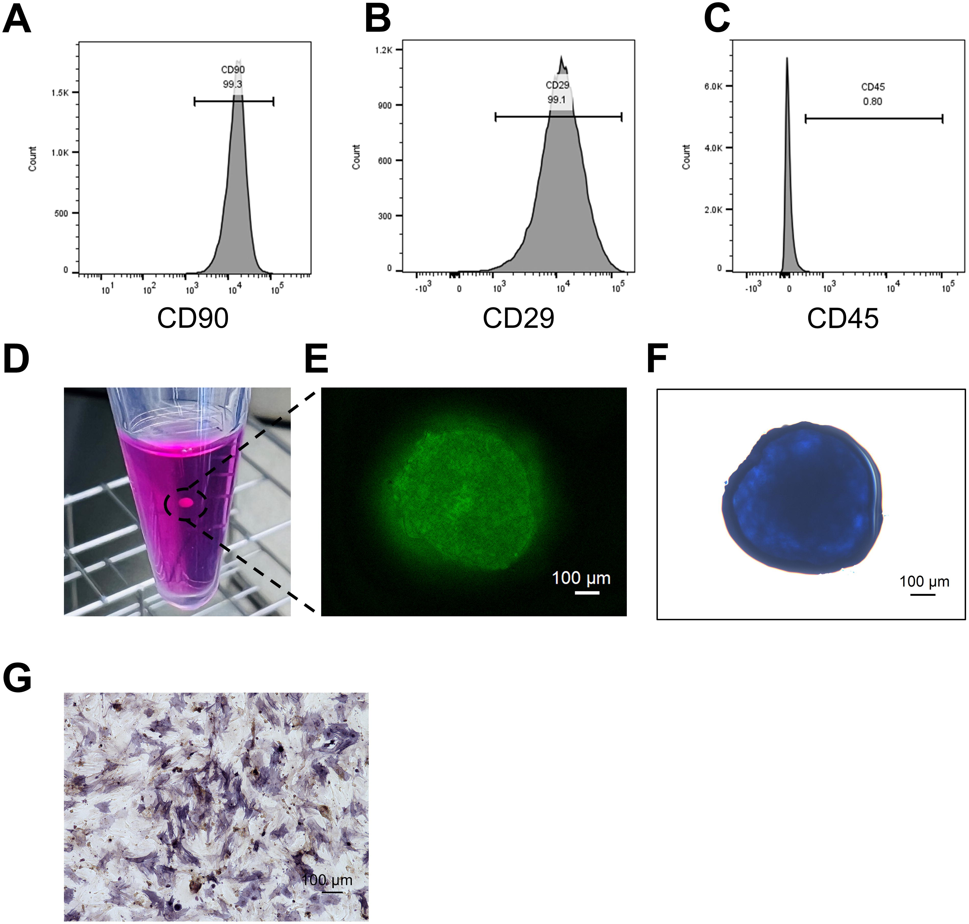
Characterization and differentiation of ASCs.
Chondrogenic differentiation of ASCs
The formation of ASC-derived chondrogenic pellets was observed, as shown in Figure 2D. Strong GFP expression within the chondrogenic pellets was revealed by fluorescent imaging (Fig. 2E). Additionally, Alcian blue staining demonstrated the successful deposition of an extracellular matrix characteristic of chondrogenesis (Fig. 2F), confirming the differentiation of ASCs into chondrocytes.
Osteogenic differentiation of ASCs
The analysis of ALP staining is shown in Figure 2G. ALP expression was observed on day 14, indicating early-stage osteogenic differentiation. This finding confirms the initiation of osteoblast activity.
Adipogenic differentiation of ASCs
The analysis of the adipogenic differentiation potential of ASCs is shown in Figure 3A and B. Oil Red O staining was used to visualize lipid accumulation within the differentiated ASCs, with clear staining observed in vitro (Fig. 3A). In addition, perilipin staining demonstrated the presence of perilipin-positive lipid droplets, confirming perilipin expression on the lipid droplet membrane (Fig. 3B). These findings validate the successful adipogenic differentiation of the ASCs.

Adipogenic differentiation of ASCs.
Identification of ASCs attachment on PLLA-CS implants
The attachment of ASCs on the scaffolds was confirmed using fluorescence microscopy. Both the L-ASC and H-ASC groups showed a uniform distribution of ASCs on the surface of the PLLA-CS implant (Fig. 4).
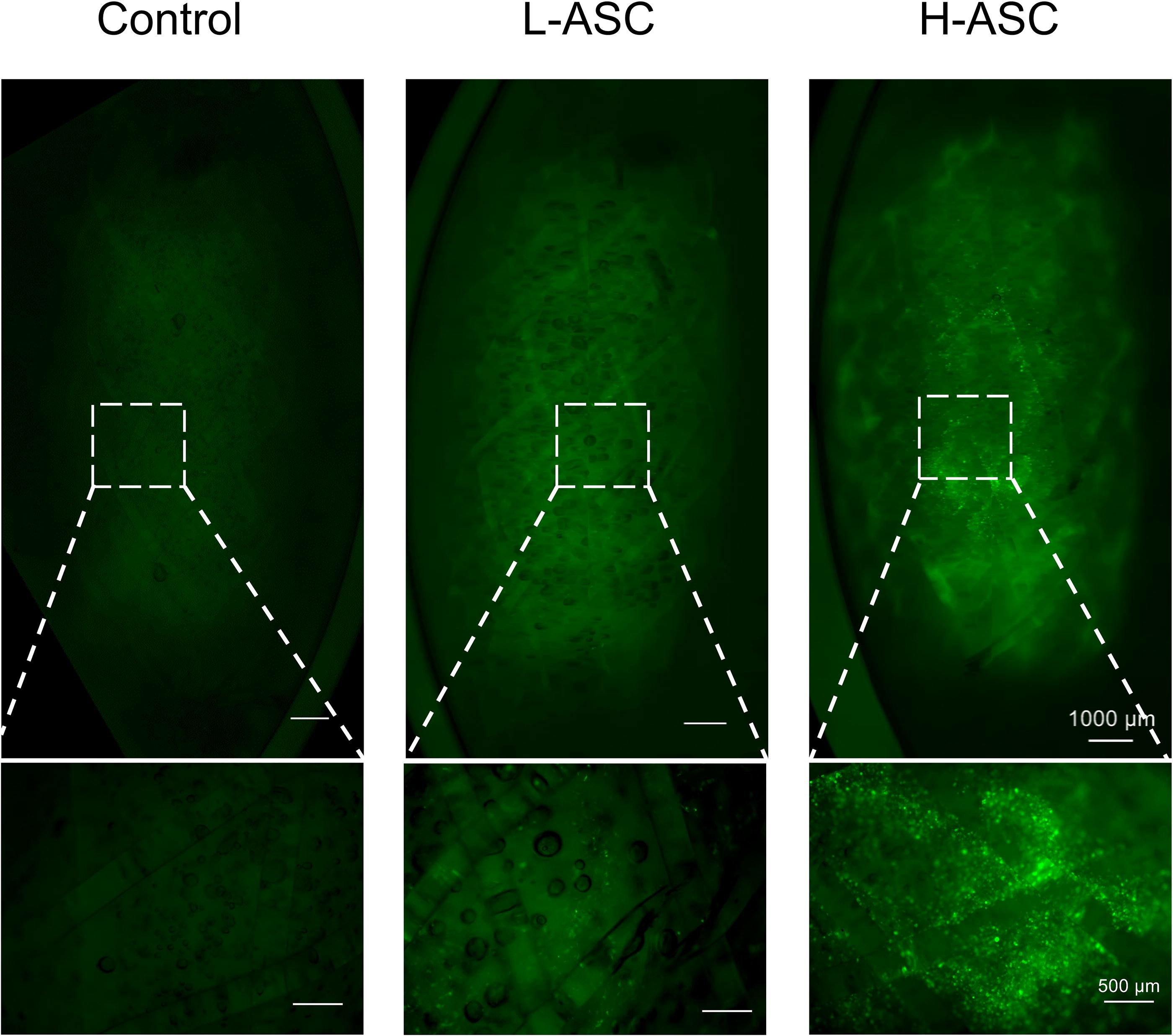
Confirmation of ASCs attachment on PLLA-CS implants under a fluorescence microscope. After seeding, ASCs were evenly distributed on the surface of the PLLA-CS implants. Scale bar: 1000 μm. The dotted squares highlight regions shown at higher magnification. Higher magnification scale bar: 500 μm.
Evaluation of the weight and volume of all the newly formed tissues
No infection or tumors were observed during postoperative follow-up. The gross appearance of all newly formed tissue is shown in Figure 5A. All the newly formed tissue increased in size over time. The time courses of the weight and volume of all newly formed tissue are shown in Figure 5B and C. At 3 months, the weight and volume in the H-ASC group was greater than that of the control group. At 6 and 12 months, the weight and volume were not significantly different between the groups. The weight and volume of the control, L-ASC, and H-ASC groups at 6 and 12 months were greater than those at 3 months, and those at 12 months were greater than those at 6 months.

Weight and volume of all the newly formed tissues.
Histological assessment of the area of formed tissue and adipose tissue inside implants
Micrographs of the HE-stained sections are shown in Figure 6A. Gradual adipose regeneration was observed in areas in contact with native adipose tissue at all time periods. The time course of tissue formation inside the implants, adipose tissue area inside the implants, and percentage of adipose tissue in the tissue formed inside the implants are shown in Figure 6B–D. At 3, 6, and 12 months, the area of the formed tissue in the H-ASC group was significantly larger than that in the control and L-ASC groups. At 6 and 12 months, the area of adipose tissue in the H-ASC group was significantly larger than that in the control and L-ASC groups. Furthermore, the adipose tissue area at 6 and 12 months in the L-ASC and H-ASC groups was significantly larger than those at 3 months in the corresponding group. For the adipose percentage, at 12 months, the H-ASC group was significantly larger than the control group. At 6 months, the adipose percentage in the L-ASC and H-ASC groups was significantly larger than that at 3 months. At 12 months, all the groups had better adipose percentages compared with those at 3 months, while the adipose percentage of the H-ASC group was better compared with that at 6 months.

Micrographs and the time course of the formed tissues, adipose tissue, and the percentage of adipose tissue inside the implants.
Assessment of GFP-positive cells inside implants
Micrographs of anti-GFP-stained sections at 3, 6, and 12 months are shown in Figure 7A. In both the L-ASC and H-ASC implants, spindle-shaped GFP-positive cells were observed across all time points inside the PLLA-CS implants. Circular GFP-positive adipocytes were confirmed inside PLLA-CS implants in the H-ASC group both at 6 and 12 months. These GFP-positive adipocytes exhibited a similar morphology to GFP-negative adipocytes inside PLLA-CS implants. GFP-positive cells and GFP-positive adipocytes were predominantly distributed within the regions of collagen fibers. The number of GFP-positive cells inside the implants is shown in Figure 7B. The H-ASC group had significantly more GFP-positive cells compared with the L-ASC group at 3, 6, and 12 months.

Micrographs of the GFP-positive cell counts and adipocytes inside implants.
Transplanted cell morphology and differentiation
Immunofluorescent micrographs of GFP (green), perilipin (red), and DAPI (blue) stained sections at 6 months are shown in Figure 8. In the L-ASC and H-ASC groups, GFP-positive cells with a spindle shape were observed, indicating potential undifferentiated ASCs or other cell types.

Morphology and differentiation of transplanted cells in the scaffold at 6 months. Representative images of sections stained for GFP (green), perilipin (red), and DAPI (blue) are shown. In the H-ASC group, GFP-positive adipocytes coexpressing perilipin are indicated by yellow due to the overlap of green and red fluorescence. Scale bar: 200 μm. The dotted squares highlight regions shown at higher magnification. Magnified images show cells with overlapping GFP and perilipin, spindle-shaped GFP-positive cells, and GFP-negative adipocytes. Higher magnification scale bar: 100 μm. Arrows indicate GFP-positive cells, asterisks indicate GFP-positive adipocytes, and sharps indicate GFP-negative adipocytes.
Discussion
Herein, we investigated the potential of PLLA-CS implants combined with ASCs to promote de novo adipose regeneration. Our observation revealed that ASCs transplanted with PLLA-CS implants significantly enhanced adipose tissue formation. The transplanted ASCs persisted within the implants for up to 1 year and spontaneously differentiated into adipocytes without prior induction.
The flow cytometry analysis of the isolated ASCs in this study exhibited the typical immunophenotype of MSC, consistent with previous reports. 23 Furthermore, the differentiation ability of GFP-positive ASCs into adipocytes, chondrocytes, and osteoblasts was confirmed. (Figs. 2D–G and 3A and B). However, the in vitro Oil Red O staining revealed that only a portion of ASCs differentiated into lipid droplets (Fig. 3A). This limited differentiation efficiency may be attributed to the inherent ASCs, where only a fraction possess multipotent differentiation potential.24–26 Despite this, a higher concentration of ASCs significantly promoted adipose tissue formation in vivo, suggesting that even with reduced differentiation potential in vitro, ASCs may enhance adipose regeneration through paracrine effects. 27
Long-term maintenance of the internal space within an implant facilitates adipogenesis, with the external frame material playing a crucial role, while the internal filling material provides a supportive function.11,13 PLLA is a biocompatible and biodegradable synthetic polymer that has been safely utilized in various clinical applications for over 30 years.28–30 In this study, PLLA maintained internal space stability for up to 1 year in all groups (Fig. 4), consistent with our previous study. 8 On the other hand, stem cell attachment and maintenance on scaffolds are fundamental in tissue engineering. 31 PELNAC® is a lyophilized CS known for its biodegradability, biocompatibility, and safety, which has been widely used in tissue engineering.32–34 Its porous structure facilitates cell infiltration, supports cell attachment and growth, and promotes extracellular matrix synthesis, thereby inducing neo-adipogenesis.35,36 In in vitro studies, MSCs adhere firmly, proliferate, and maintain typical fibroblast-like morphology and surface markers after seeding on a CS. 37 ASCs seeded onto a CS secrete various growth factors, including Platelet-Derived Growth Factor-αβ, Transforming Growth Factor-β1, and Vascular Endothelial Growth Factor. 38 In this study, ASCs were observed to successfully adhere to CS (Fig. 3). Therefore, CS is the preferred scaffold for adipose regeneration. Furthermore, the PLLA-CS implants degraded gradually in vivo. During this study, no infections or hematomas were observed in either ASC-seeded or non-ASC-seeded implants. Our implants did not require secondary surgical removal, thereby mitigating complications associated with permanent materials. Overall, our PLLA-CS implants represented safe, tissue-engineered composites capable to support ASCs.
The exploration of in situ adipogenesis utilizing ASCs has garnered considerable attention. Prior research demonstrated that preadipogenically induced ASCs can successfully differentiate into adipocytes upon implantation. 39 Nonetheless, predifferentiated cells exhibit the differentiation process in vitro and may lose self-renewal and pluripotency, which can adversely affect their integration in the host. 40 ASCs combined with controlled release of bFGF within type I collagen scaffolds facilitate long-term de novo adipogenesis.16,18 However, it should be noticed that bFGF is associated with tumor growth, angiogenesis, and metastasis in breast cancer. 41 Our findings indicate that the combination of ASCs in PLLA-CS implants results in superior outcomes in the formation of adipose tissue. The efficacy of adipose tissue formation was elevated in accordance with the dose dependency of ASCs (Fig. 5). Therefore, the incorporation of ASCs into PLLA-CS implants has proven to be a safe and effective strategy for enhancing adipogenesis.
Regarding the mechanism of adipose tissue regeneration using PLLA-CS implants, previous studies have elucidated several aspects of the adipose tissue regeneration process. A notable observation is the proliferation of surrounding adipose tissue and the contribution of cells from the adjacent tissues, which often leads to adipose tissue formation. Additionally, the formation of blood vessels within the implant likely plays a role in supplying cells to the area, further supporting adipogenesis around the newly formed vascular structures.42,43 In this study, GFP-positive cells and GFP-positive adipocytes were identified in the stromal region. This finding suggests that the mechanism of adipose regeneration might involve both the proliferation and differentiation of transplanted ASCs into adipocytes, making them a significant source of adipocyte precursors. Additionally, the growth factors released by ASCs may have promoted angiogenesis within the stromal area, as well as the proliferation of surrounding tissues, further enhancing adipose tissue formation. Another significant factor contributing to this enhanced adipogenesis is likely to be the lack of tissue pressure within the PLLA, thus enabling the survival and function of the ASCs.
This study has several limitations. Firstly, the number of ASCs that underwent spontaneous differentiation was relatively low. Secondly, in the H-ASC group, the weight and volume increased at 3 months, possibly as a result of the accelerated vascularization and stimulation of adipose tissue growth by ASCs. However, this advantage diminished by 6 months, so that there was not an increase in all adipose tissue. Thirdly, while our results demonstrate the long-term survival and differentiation of ASCs, we were unable to fully elucidate the specific molecular mechanisms involved in this process. Finally, the H-ASC group had a high concentration of ASCs, indicating that a large amount is required for substantial adipose tissue formation. In future research, the characteristics of ASCs during long-term implantation will be investigated, and the superior ASCs concentration for more efficient adipose tissue formation will be determined, with the aim of increasing the total amount of adipose tissue.
In summary, this study highlights the innovative application of ASCs seeded onto PLLA-CS implants to enhance adipose tissue proliferation and facilitate spontaneous differentiation into adipocytes in vivo, without the requirement for predifferentiation or exogenous growth factors. Particularly, the group seeded with a high dose ASCs of 1.0 × 10⁶ cells per implant demonstrated a superior outcome. These findings are expected to advance adipose tissue engineering, offering more effective regenerative medical options for clinical applications.
Footnotes
Acknowledgment
This work was supported by “Startup business support program IPG-Advance” of Kyoto University.
Authors’ Contributions
Q.Z. conducted the experiments, collected and analyzed the data, and drafted the article. S.O. contributed to the study design and participated in drafting and revising the article. Y.S. conceived the idea and designed the experiments. Y.K., Y.L., S.L., Y.T., and T.N. assisted with the experiments and contributed to data collection. H.Y., M.S., and E.S. collected, analyzed, and interpreted the data. N.M. conceived the ideas, designed and approved the study, collected and interpreted the data, and critically revised the article. All authors gave final approval and agreed to be accountable for all aspects of the work.
Ethics Statement
The authors confirm that the ethical policies of the journal, as noted on the journal’s author guidelines page, have been adhered to, and the appropriate ethical review committee approval has been received.
Disclosure Statement
The authors report no proprietary or commercial interests in any product mentioned or concepts discussed in this article.