
Abstract
The development of molecularly imprinted polymers (MIPs) using biocompatible production methods enables the possibility to further exploit this technology for biomedical applications. Tissue engineering (TE) approaches use the knowledge of the wound healing process to design scaffolds capable of modulating cell behavior and promote tissue regeneration. Biomacromolecules bear great interest for TE, together with the established recognition of the extracellular matrix, as an important source of signals to cells, both promoting cell–cell and cell–matrix interactions during the healing process. This review focuses on exploring the potential of protein molecular imprinting to create bioactive scaffolds with molecular recognition for TE applications based on the most recent approaches in the field of molecular imprinting of macromolecules. Considerations regarding essential components of molecular imprinting technology will be addressed for TE purposes. Molecular imprinting of biocompatible hydrogels, namely based on natural polymers, is also reviewed here. Hydrogel scaffolds with molecular memory show great promise for regenerative therapies. The first molecular imprinting studies analyzing cell adhesion report promising results with potential applications for cell culture systems, or biomaterials for implantation with the capability for cell recruitment by selectively adsorbing desired molecules.
Introduction
M
Intelligent polymers are macromolecular systems that exhibit strong thermodynamic interaction with the surrounding environment and with associated components. Therefore, an intelligent polymer material has strong interactions with the environment based on pH-, or temperature sensitivity, but most importantly based on thermodynamic interactions with a recognitive compound. This “intelligence” is rendered to the polymer either by external decoration with required functional groups, or (better) by internal molecular imprinting of micro- and nanocavities that provide recognitive characteristics.
Therefore, the fundamental idea for these new technology platforms is to combine a template molecule with a functional monomer giving rise to a polymer network (after cross-linking and removal of the template) with template-specific cavities in size, shape, and chemical functionality 5 (Fig. 1). The ability of these molecular imprinted polymers to then recognize template molecules through thermodynamic interacts present the intelligent capabilities of these polymers.
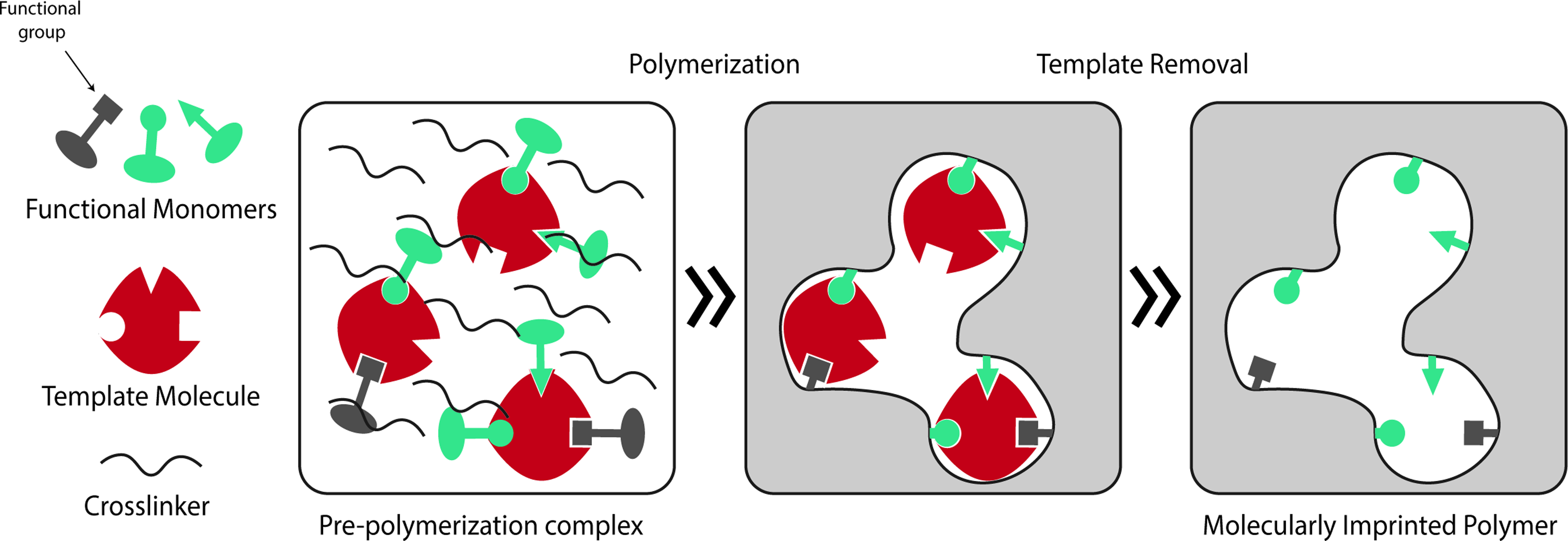
Molecular imprinting process. Once the appropriate template molecule, functional monomer(s) and cross-linker(s) are selected, all components are mixed together in a proper solvent (e.g., deionized water, phosphate buffer) forming the prepolymerization complex (template-functional monomer). Polymerization is then allowed to take place and specific cavities and binding sites are stabilized. Finally, removal of the template and other washing steps eliminate unreacted monomer and cross-linker molecules, resulting in a MIP. The use of heteropolymeric systems (or a functional monomer with more than one functional groups) may improve imprinting features as the different polymer functional groups can interact with various protein domains. In addition, these functional groups can modify the chemical and/or mechanical properties of the final MIP. MIP, molecular-imprinted polymer. Color images available online at www.liebertpub.com/teb
Molecular imprinting is well characterized for low-molecular weight molecules although, despite the promising results, products based on this technology are currently not available on the market. 2 In contrast, molecular imprinting of high-molecular weight molecules exhibit several obstacles, such as, size, structure, functionality, and solubility, which are not relevant when imprinting small molecules. 6 The large size of these molecules restricts their transfer within highly cross-linked polymer networks during both template removal and rebinding studies. The complex structure of biomacromolecules (e.g., proteins) plays an important role in their functionality, leading to high sensitivity to pH, ionic strength, temperature, and organic solvents.6,7 Despite these challenges, studies have reported results of the molecular imprinting of proteins supporting its application, namely in the fields of biosensing and chromatography.8–15
Due to the specificity and recognition capabilities of molecular imprinting, this has the potential to become a tool utilized for the production of scaffolds with high bioactivity and recognition capacity toward specific molecules or cells for tissue engineering (TE) applications. TE is an alternative clinical approach to conventional therapies, which resort to the use of organ grafts and transplantation (xeno, allo, or auto). Organs and tissues are difficult to obtain and have short storage time. The methods used for organ and tissue transplantation are highly invasive, usually comprising tremendous burden to the patient, and are associated with high risk of rejection and permanent need for immunosuppressive therapy. 16 TE aims to restore, replace, and/or regenerate tissues or organs to their normal function by combining acquired knowledge on the wound healing process with cell- and/or scaffold-based approaches.
The design of biomimetic structures can be accomplished by either top-down or bottom-up strategies. In top-down TE strategies, scaffolds are seeded with cells so that they can populate the structure to eventually form a tissue. Bottom-up approaches create small modular parts, which are then assembled to form the final tissue structure. 17 The latter approach in particular has been of recent interest due to its potential to better mimic the microarchitectural features of native tissues, therefore, providing additional guidance at the cellular- and molecular-levels. 17
Despite the potential of using molecular imprinting as a tool for tissue regeneration applications, this topic is currently far from being fully explored. Biomacromolecules (i.e., high molecular weight molecules present in living organisms) are ones that bear great importance for TE applications due to their involvement in the wound healing processes. However, biomacromolecules present major challenges when used for molecular imprinting. Nonetheless, molecular imprinting could specifically be used in bottom-up techniques to create complex cues at a microstructural level to improve cell- and scaffold-based approaches. This review aims to provide an overview of utilizing molecular imprinting as a tool for various TE applications by exploring the fundamentals and challenges, specifically, regarding the use of imprinting macromolecules. Additionally, potential strategies of molecular imprinting in TE based on recent approaches in the field of protein molecular recognition will be surveyed.
Natural Body Response to Injury
The design of effective TE approaches requires the understanding of the body's natural recovery from an injury. Upon tissue injury, the wound healing process is divided into three stages: inflammation, tissue formation, and tissue remodeling. 18 This is a highly regulated and complex process of paracrine, autocrine, and juxtacrine signaling events. 19 In summary, immediately after injury, platelets and polymorphonuclear leukocytes (neutrophils) aggregate and become entrapped within a fibrin mesh forming a thrombus. 18 Initial release of growth factors [e.g., platelet-derived growth factor (PDGF) or vascular endothelial growth factor (VEGF)] by these cells give rise to the inflammatory process, where monocytes-macrophages, lymphocytes, fibroblasts, and endothelial cells are chemoattracted to the site, and activated to scavenge the damaged tissue.18,19 These cells then produce various growth factors and cytokines, which promote the formation of granulation tissue. Angiogenesis (i.e., the formation of new blood vessels) then promotes and accelerates the wound healing process. 20
Two major outcomes can result from this body response to injury: scarring or regeneration of tissue. In mammals, the most frequent body response is to quickly produce a fibrous tissue, usually referred to as scar, to immediately stop bleeding and avoid microbial infection at the injury site. In this typical response, the overall function of the tissue or organ is maintained, even though not fully recovered since composition, aesthetic and structure is not preserved. Harty et al. 21 proposed this lack of regenerative capacity as a possible result of the evolution of the mammalian immune system, which rather promoted wound microenvironments to improve tissue defense and facilitate tissue repair. The second, and less frequent response to injury, is regeneration. Upon injury, the tissue or organ is able to fully recover structure, composition, and function at a functional-unity level. In mammals, this process occurs namely at embryonic stages, 22 and below a critical size injury.
Duration and intensity of each wound healing stage, namely inflammation, dictate the final outcome of tissue repair. In fact, inflammation has been proposed to play an important role in the success of the wound healing process. Unbalanced inflammatory reactions and/or cytokine profiles frequently result in differences in scarring. 19 Such an example is observed in spinal cord injuries, where the occurrence of inflammatory events with subsequent formation of a glial scar is the primary barrier to achieving neuroregeneration. 23 Successful tissue repair after injury requires resolution of the inflammatory response, with inflammation being a prerequisite to scarring. 19 The topics of wound healing and repair processes have been extensively reviewed and revised over the past few decades.18,19,24
The wound healing process is characterized by the existence of direct cell–cell and cell–matrix interactions, along with indirect crosstalk between different cell populations through soluble mediators. 18 These interactions are highly complex and tissue-specific, comprising of a series of biochemical and mechanical signals. These signals then modulate subsequent cell behavior, such as, migration, proliferation, and differentiation. The extracellular matrix (ECM) plays a pivotal role in this process. The natural ECM not only provides mechanical support to cells, but is also a reservoir of growth factors (matrix-embedded). In addition, the ECM serves as platform for cell–cell interaction by allowing dispersion of secreted factors and mechano-transduction signals. 25 Therefore, the ECM is largely capable of modulating cell migration, cytoskeletal organization, proliferation, and differentiation. 26 Tissue vascularization is also affected by the ECM structure, namely by its microporosity, which will promote or incapacitate angiogenesis. 19 Thus, effective design of biomimetic structures for TE applications should involve a complex combination of topographical, architectural, and biochemical features mimicking the natural ECM.
TE and the Relevance of Cell-Instructing Scaffolds
An important goal of TE is to replace tissues or organs by mimicking and/or modulating the natural events of wound healing to produce a fully regenerated and functional tissue. Significant understanding of the wound healing process has led to the development of several TE strategies, which can be divided into either cell- or scaffold-based approaches. Cell-based approaches use both adult and embryonic stem or progenitor cells to induce the formation of new tissue. These approaches are centered on stimulating tissue progenitor cells in situ, or promoting their expansion and differentiation in vitro, for further implantation to the site where regeneration is desired. 27 Limitations using cell-based approaches include difficulties regarding cell obtainment from scarce sources, and in vitro cell expansion and differentiation.
Scaffold-based approaches rely on the understanding of the ECM function, maintaining homeostasis (a dynamic process achieved through a balance between degradation of the old and formation of the new ECM) and providing signals (viz., chemical, physical, and mechanical) to cells. TE uses this understanding to create 2D or 3D matrices, which can provide mechanical support and physical-chemical cues to promote cell seeding. This can be accomplished by either using man-made scaffolds, or natural-based scaffolds, such as decellularized ECM from allogenic or xenogenic tissues. 25
Developing scaffolds with degradable biomaterials is pivotal in TE research. It has been established that mimicking the natural cell environment produces favorable outcomes with successful cases reported in a wide variety of tissues (e.g., heart valves or skin) when decellularized extracellular matrices were used. 25 However, these matrices do present drawbacks including the methods in which they are processed, and possibilities of contamination and immunogenicity. A significant portion of these efforts is followed to create a bridge between man-made and natural structures. This requires development of new, intelligent biomaterials and production technologies, which better resemble the native structural, mechanical, and biological cues.
TE scaffolds can be made from many types of biomaterials, although polymeric scaffolds are widely investigated due to their chemical and mechanical versatility associated with biodegradability. Natural polymer biomaterials present high biocompatibility and biodegradability, but low mechanical properties, limiting their applications. Commonly studied natural polymer biomaterials used for TE applications include collagen, 28 alginate,29,30 chitosan, 31 and silk. 32 Extensively explored scaffolds based on synthetic polymeric biomaterials produced with poly-L-lactic acid (PLLA), polyglycolic acid (PGA), or poly(lactic-co-glycolic) acid (PLGA) are easily tailored in their architecture, mechanical properties, and degradation characteristics. However, these materials have been associated with risks of rejection due to reduced bioactivity, and may cause cell and tissue necrosis upon degradation. 33 Bioceramic-based TE scaffolds, primarily made of calcium phosphates, such as hydroxyapatite 34 and tricalcium-phosphate, 35 are biocompatible and characterized by high mechanical stiffness and low elasticity, limiting their use to hard tissues (e.g., for orthopedic applications).
Several characteristics should be considered when developing a biomaterial/scaffold for TE applications. Ideally, a scaffold for such applications should be designed considering the following: (1) biocompatibility, (i.e., promote cell adhesion, support native cell activity, such as proliferation or migration, without causing an immune reaction that could lead to severe inflammation or rejection); (2) biodegradability (i.e., scaffolds should be replaced by cells and their ECM, in addition to having degradation byproducts be nontoxic and be excreted by the body without harming other tissues or organs); (3) bioactivity, which can be provided by including biochemical, biophysical, and mechanical cues into the scaffold (e.g., biological relevant molecular features, such as proteins, or functionalizing with chemical functional groups able to interact with these molecules); (4) architecture, by designing scaffolds with adequate porosity and preferentially interconnected pore allowing cell migration and diffusion of oxygen, nutrients, and waste products, while promoting vascularization; (5) mechanical profile, by designing scaffolds as a temporary mechanical stabilizer with mechanical properties similar to that of the tissue/organ of interest; (6) manufacturing technology, as cost effective and scalable as possible. 33
Molecularly Imprinted Polymers
Molecular imprinting characteristics
Molecular imprinting is a technology based on naturally occurring molecular events, which aim to insert molecular memory into a material, usually polymeric, 3 increasing specificity, loading capacity, and release control of biomaterials. 5 A molecularly imprinted polymer (MIP) is produced by combining a functional monomer (or mix of monomers) with a template molecule. These monomers interact with the template molecule either by reversible covalent interactions, or noncovalent interactions, forming a prepolymerization complex. 36
Macromolecular MIPs usually rely on noncovalent interactions (e.g., H-bonding, electrostatic and hydrophobic interactions) for recognition.
3
Polymerization is allowed to take place, and the addition of a cross-linker promotes the fixation of polymer positions to help memorize the geometry of the cavities once the template is removed. Template removal is achieved by performing a series of washing steps using organic or inorganic solvents, or by enzymatic digestion.
37
Once the imprinting process is completed, imprinting success can be evaluated by two main parameters: adsorption capacity (Q, mg g−1) (1), which determines the amount of adsorbed molecules per weight of polymer; and imprinting efficiency (2), which corresponds to the ratio between both MIP and correspondent nonimprinted polymer (NIP) adsorption capacities.
Ci and Cf–initial and final concentration of template solution, respectively (mg mL−1)
Molecular imprinting considerations
To create a material for TE applications, one should carefully mind several aspects of MIP production. After choosing an appropriate template molecule, the choice of the functional monomer(s) (and their ratios) bears great importance. The selected monomer(s) should be biocompatible, nontoxic, and have high affinity to the template so that template-monomer complexes are thermodynamically favorable and stable under reaction conditions. 3 Since proteins contain many different binding sites and heterogeneous regions, the choice of the polymer matrix will influence MIP selectivity by enabling or preventing cross-reactivity with proteins similar to the template molecule. 38 For instance, it is not desirable for the polymer to reactively polymerize with the template, a phenomenon frequently observed while imprinting biomolecules into polyurethanes, where one of the monomeric units is an isothiocyanate, capable of reacting with alcohol or amine groups present in most biomolecules. 2 On the other hand, heteropolymer systems appear to favor macromolecular memory.5,12,39 This can be explained by the conjugation of their different functional groups, thereby increasing the number of available interaction sites with the different regions of the template and improving their physico-chemical properties.12,13
Another possibility is the use of natural ligand derivatives as functional monomers for the polymer matrix. Chou et al. 40 produced a C-reactive protein imprinted polymer using a phosphorylcholine derivative (4NPPC) as the functional monomer and the micro-contact approach. This approach uses a monolayer of a minimal protein mass to which the recognition substrate will form an imprint. 40 The resulting MIP showed rebinding capacity and selectivity toward the C-reactive protein, leading the authors to conclude that the proposed micro-contact approach is a suitable method to produce protein MIPs. 40
The MIP should be biodegradable to promote its replacement by the natural ECM, but stable to withstand the harsh environmental body conditions. Biodegradability is also important to allow exposure of new binding sites, especially in bulk imprinted materials. MIP biodegradability, and flexibility, can be controlled by the degree and type of cross-linking used. Higher degrees of cross-linking can promote higher selectivity by reducing movement (e.g., swelling) and fixating binding sites. 1 However, an excessive degree of cross-linking interactions can result in difficulties during template removal, and reduce rebinding capacity due to impaired diffusion. 41 Despite existing covalent and noncovalent cross-linking techniques used for MIP production, biological applications of MIPs prefer noncovalent techniques due to more mild template removal methods. 1
Molecular imprinting conditions should be mild to avoid protein denaturation or polymer distortion. Biomacromolecules have complex structures that are highly sensitive to pH, ionic strength, temperature, and solvents. 6 If imprinting conditions are nonphysiological, changes in the natural conformation of the protein may result in imprinting cavities specific for this altered form. 3 Kryscio et al. 42 performed an elaborate study on how different functional monomers (acrylamide, methacrylic acid, acrylic acid, 3-aminophenylboronic acid, and N-isopropyl acrylamide) and cross-linkers (N,N’-methylenebisacrylamide and ethylene glycol dimethacrylate) widely used in MI influence protein template conformational stability. The authors used circular dichroism studies to analyze conformational changes in bovine serum albumin (BSA), and concluded that these reactants induced significant changes in the secondary structure of the template protein. This is a severe matter of concern as it can explain the low rate of success of macromolecular imprinting. 42
The choice of the polymerization solvent brings challenges when it comes to protein solubility. 2 While proteins are usually more soluble and stable in aqueous solutions, these types of solvents are far from ideal as they affect the prepolymerization complex by interfering with hydrogen bonding (an extremely important type of interaction for many template-monomer complexes). 3 The solvent is responsible for mixing all of the components (i.e., template, functional monomer(s), cross-linkers, and polymerization initiators) into one phase, in addition to, creating pores in macroporous polymers. 43 Some studies on protein molecular imprinting use organic solvents, such as chloroform, 44 as they better stabilize the polymerization process. However, for biomedical applications involving cell contact (i.e., drug delivery and TE applications), these solvents must be avoided or replaced during MIP production processes due to their high toxicity.45,46
During the imprinting of macromolecules, template removal is one of the critical phases and consists of several washing steps using solvents, acids, bases, or detergents. 2 Other possibilities include template removal by enzymatic digestion.37,47 Template removal is a particularly delicate step during the imprinting process due to the large sizes of macromolecules. Efficient template removal is often hard to obtain and still presents a major challenge when imprinting large molecules.
For each developed system, it is important to work on the optimization of the template removal phase since it influences the final rebinding capacity and success of the MIP. Hawkins et al. 47 optimized a template removal strategy for a bovine hemoglobin (Hb) molecularly imprinted polyacrylamide gel using sodium dodecylsulfate (SDS) and acetic acid (AcOH). Even though a SDS:AcOH ratio of 10%(w/v):10%(v/v) did not present the highest template removal percentage (only 47.6%), the highest imprinting efficiency (>90%) was obtained. Higher template removal percentages can result in lower imprinting efficiencies if the agents begin to promote conformational changes on the imprinting matrix. 47 The authors also tested an enzymatic digestion-based template removal system with trypsin. This showed an even higher template removal capacity (87.4%), compared to the SDS:AcOH maximum (71.5%), but a significantly lower imprinting efficiency (20.4%). This can be a result of remaining protein fragments inside the polymer matrix that may block potential binding sites. 47
On the other hand, SDS can damage the cell membrane and lead to cell death. In BSA-imprinted hydrogel films, even after several washing steps with deionized water, Zhao et al. 9 reported that SDS remains a possible cause for total cell death of L929 cells after 5 days of culture. Optimization of molecular imprinting systems should also focus on improving the wash steps needed for template removal as the reagents can be toxic to cells.
Finally, depending on the final application of a MIP system, various polymerization methods can also be applied. These methods include bulk suspension, two-step swelling, precipitation, emulsion core-shell polymerization, film synthesis, aerosol polymerization, and polymerization on silica-beads. 48 Bulk polymerization is the most widely used method by molecular imprinting groups, however, it is used exclusively with organic solvents, and it has the disadvantage of wasting a significant percentage of polymer produced in the process of grinding to obtain smaller irregular particles. 48 On the other hand, two-step polymerization requires an aqueous polymerization medium and produces monodispersed particles with control of the final size and number of particles. 48 Ideal molecular imprinting production methods should be controllable and reproducible. For instance, Ying et al. 49 developed a gas jetting-dropping method to produce beads of a controllable diameter in aqueous medium at a constant rate of production (600 mL/h). This possibility is very important when it comes to industrial applications for MIP materials.
Molecularly imprinted intelligent scaffolds
Molecular imprinting can be a powerful tool used to create scaffolds with high bioactivity and recognition capacity to specific molecules. Recognition, neutralization, and clearance of target peptides, such as toxin melittin, in the bloodstream has been achieved in vivo using molecularly imprinted polymeric nanoparticles with binding affinity and selectivity comparable to that of natural antibodies. 50 Similarly, molecular imprinting can be applied during the construction of TE scaffolds with the ability to recognize and adsorb a biomolecule transported through the bloodstream, or present in situ, for ultimately promoting cell recruitment to the site of injury or to trigger specific cell behaviors.
For TE applications, biomacromolecules bear great relevance due to their involvement in the healing process. Increasing interest on the imprinting of macromolecules to exploit its potential for clinical and pharmaceutical applications has driven many research groups to develop protein MIP systems. The majority of protein MIP studies are usually developed for model macromolecules like lysozyme (Lyz), BSA and Hb. However, few studies use biomolecules of interest for TE technologies such as fibronectin (Fn). Fn, a high molecular weight protein (composed of two polypeptides of molecular ∼220 kDa each) with adhesive activity, is present in the ECM and involved in several biological events, including wound healing and tissue repair. 51 Recent advancements in the protein molecular imprinting field can strongly impact regenerative medicine technologies through the use of molecular imprinted intelligent scaffolds. 52
Many imprinting strategies exist, although majority are successful when imprinting small molecules, but present several challenges for use with larger molecules such as proteins. Figure 2 represents the main molecular imprinting strategies for macromolecules divided into two categories: namely, according to either the substrate, or template type used.
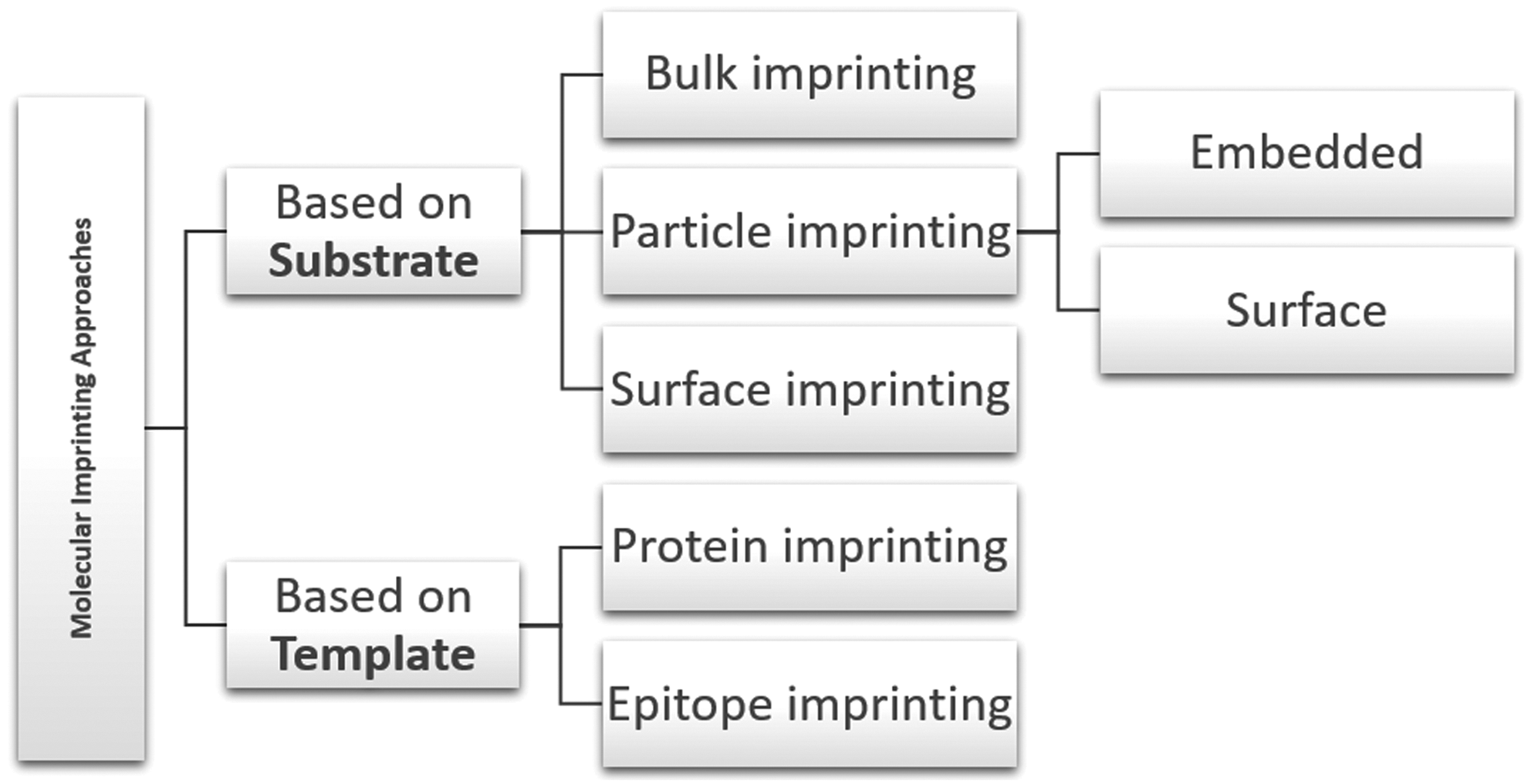
Molecular imprinting strategies for macromolecules. Molecular imprinting approaches can be divided according to the imprinted substrate they produce: bulk imprinting involves creating imprinting cavities in a material, namely in its inner layers (bulk); particle imprinting is based on the direct production of embedded- or surface-imprinted nano/microparticles; surface imprinting creates scaffolds that are only imprinted at the surface, or at the layers near it. Additionally, molecular imprinting approaches can be divided according to the type of template used, namely, in protein imprinting when the whole protein is used as a template, or in epitope imprinting when only relevant portions or domains of the molecule of interest are used.
Molecular imprinting mechanisms
Molecular imprinting approaches can be classified according to the substrate imprinted and further subdivided into bulk, particle, or surface imprinting. Bulk imprinting is a rather successful strategy used for small molecules, which involves imprinting cavities in the inner layers of a polymeric material. However, this strategy presents some challenges when addressing mass transfer, associated with the size of macromolecules. Usually, macromolecularly imprinted materials produced by this approach strongly retain their templates and require extensive grinding and sieving. This process produces irregularly shaped polydisperse particles and shorter lengths of diffusional pathways.6,53
For protein MIP strategies, particle-based imprinting and surface imprinting are more commonly used. These are some of the most successful because they reduce the diffusion pathway length, promoting protein movement inside-out and outside-in. Not only does this lead to successful removal of the template and increase the number of available binding sites, but also promote protein rebinding by facilitating diffusion of proteins through the imprinted material. Particle-based strategies use polymerization methods, such as emulsion or suspension, to directly synthetize micro-/nanoparticles with the advantage of using lower quantities of monomers and template molecules. 3 However, this presents the drawback of using stabilizers and surfactants (which may cause the disruption of monomer-template complexes), since residual levels of stabilizers remain even after an extensive washing process. 3
Surface imprinting is the most common approach for protein MIPs, and consists of imprinting binding sites only at the surface or on the superficial layers. This approach is similar to bulk imprinting, with the difference being that surface imprinting is applied on thin films, or by attaching the protein on the surface of a substrate. 3 In addition to alleviating macromolecule transfer, surface imprinting enables the imprinting performance to be improved through the introduction of surface functional ligands. These ligands allow interaction with the template protein by reversible covalent binding or affinity interactions, and by physical adsorption on the surface. 54
An alternative way to address the classification of molecular imprinting of macromolecules is based on whether the whole protein is used during the imprinting process, or if only relevant domains (epitopes) are used (Table 1). Epitopes are small domains or sequences exposed in macromolecules that can be recognized by their receptors. Epitope imprinting uses small protein domains or sequences, as opposed to the whole protein, to create materials with molecular recognition toward proteins. This approach aims to overcome obstacles inherent to protein imprinting caused by size complexity, and conformational characteristics of proteins. 3 In the same way, a MIP can recognize a whole protein if previously imprinted with its epitope. Since these sequences can be synthetically produced, this approach also overcomes difficulties associated with obtaining pure template proteins. 53 Additionally, epitope imprinting can create highly selective scaffolds as reported by Zhao et al. 55 who developed Fe3O4 silica-coated nanoparticles with high adsorption rates and high selectivity toward BSA by using a BSA-specific nonapeptide C-terminal amino-acid sequence as a template. The authors reported these MIPs to have low tolerance for single amino acid mismatch since a significant reduction of imprinting factor values were observed in MIPs imprinted with mismatched sequences of BSA, compared to MIPs imprinted with the original sequence. 55 The authors also reported better recognition features on epitope imprinted particles when compared to BSA imprinted particles. 55 This supports the idea that molecular imprinting can be used to create highly specific and selective systems at a sequence level.
AAm, acrylamide; AcOH, acetic acid; ANP, atrial natriuretic peptide; APTES, 3-aminopropyl triethoxysilicane; BA, benzoic acid; BHb, bovine hemoglobin; BSA, bovine serum albumin; Cyt, cytochrome c; DPP, diphenyl phosphate; DW, deionized water; EGDMA, ethylene glycol dimethacrylate; EGMPA, ethylene glycol methacrylate phosphate; HEMA, hydroxyethyl methacrylate; HRP, horseradish peroxidase; HSA, human serum albumin; IF, imprinting factor; Lyz, lysozyme; MAA, methacrylic acid; MAsp, N-methacryolyl-(
Papaioannou et al. 56 reported promising results regarding epitope imprinting by producing a MIP for RGD (Arg-Gly-Asp tripeptide) recognition, based on noncovalent interactions. The RGD domain is known to be responsible for Fn adhesive activity. 51 RGD and Fn are frequently used to functionalize different types of substrates (e.g., alginate) to promote desirable cellular responses, either alone, or in combination with other bioactive molecules or sequences.57,58 This approach could be applied to several other biologically relevant domains or sequences. For instance, different domains of neural cell adhesion molecules (e.g., fibronectin type III motifs FGL and BCL) have been proven to interact with fibroblast growth factor (FGF) receptors, inducing neurite outgrowth in primary cerebellar granule neurons, promoting synaptogenesis and enhancing presynaptic function. 59 Wang et al. 60 produced a self-assembling RADA16 peptide nanofiber hydrogel functionalized with FGL enabling it to promote spinal cord-derived neural stem cell proliferation and migration into the three-dimensional scaffold. These, and other biologically relevant motifs, could be imprinted in a biocompatible polymer scaffold to promote molecular recognition and desired cell functions.
High protein adsorption rates can occur in a biomaterial without any significant outcomes if proteins undergo conformational changes, or hide relevant functional groups upon adsorption, preventing cell adhesion to the scaffold. Using anti-BMP-2, a monoclonal antibody against bone morphogenetic protein 2 (a protein known to promote the osteodifferentiation of progenitor cells), Ansari et al. 61 reported the influence of orientation during protein immobilization (without imprinting) in scaffolds for bone repair. While using Protein G as a linker to immobilize the antibody through its Fc fragment on a collagen scaffold, the authors were able to achieve higher antibody binding to cells concomitant with increased bone formation in vivo, compared to passively adsorbing anti-BMP-2 antibodies to the same scaffolds. 61 This enhanced performance can be explained by the increase in the number of available binding sites for the anti-BMP-2 antibody to interact with its ligand.
Protein orientation control has been achieved by epitope imprinting of L-lysine residues, typical of C-terminus of immunoglobulin G Fc fragment in poly(hydroxyethyl methacrylate-N-methacryolyl-(
One methodology that could further amplify potential applications of molecular imprinting are postimprinting modifications (PIMs), by allowing the modulation of on/off switching of the molecular recognition, or the introduction of other desirable functions (such as transduction of binding events into fluorescence events).63,64 PIMs can be used to improve imprinting features by the transformation of binding sites as reported by Taguchi et al. 37 who produced thin films with the ability to molecularly recognize cytochrome-c (Cyt) by generating peptide-fragment binding sites inside imprinted cavities. This was possible by using enzymatic digestion as the template removal strategy, where pepsin would digest Cyt at specific sites, leaving behind lysine residues grafted to the polymer backbone. 37 The authors reported high selectivity of these scaffolds toward Cyt as only enzyme-accessible regions of MIPs can be transformed into protein-binding cavities, reducing the possibility of nonspecific binding sites commonly left behind by other conventional removal processes. 37
Hydrogels for macromolecular imprinting
Hydrogel structures
The use of hydrogels constitutes another promising approach to overcome molecular imprinting problems related to mass transfer of high molecular weight templates, and provide the additional possibility of creating sensitive systems, which respond to external stimuli.5,6 Hydrogels have been widely explored for biomedical applications, including TE, due to their natural similarity to the ECM. Hydrogels are commonly used as drug carriers in pharmaceutical applications, and they are useful tools to create controlled release systems. Hydrogels are polymeric networks with high hydrophilicity, that is, they can absorb considerably high amounts of water, expanding their volume, and promoting diffusion within their bulk. 65 They can be produced from natural or synthetic polymers, are chemically stable, have biodegradable capabilities, and perform interactions that are reversible, depending on the external stimuli applied (e.g., pH or temperature). 65 Additionally, they can be processed to acquire different forms (e.g., coatings, capsules, microspheres, tubes, or sheets) and functionalizations,58,65 to improve their bioactivity.
Molecular imprinting within hydrogels may be more challenging than in rigid structures, owing to their inherent movement capacity. 5 Hydrogels can either expand or collapse easily, deeply affecting the imprinting efficiency by distortion of binding sites. So far, most studies using molecular imprinting in hydrogels are intended to develop drug delivery systems with high loading capacity and to improve controlled release.1,5 Nevertheless, several considerations are taken into account for these systems and can be applied for TE applications. This section will be focused on molecular imprinting using natural functional monomers, namely alginate and chitosan (Fig. 3), due to their common use and great potential in TE applications.
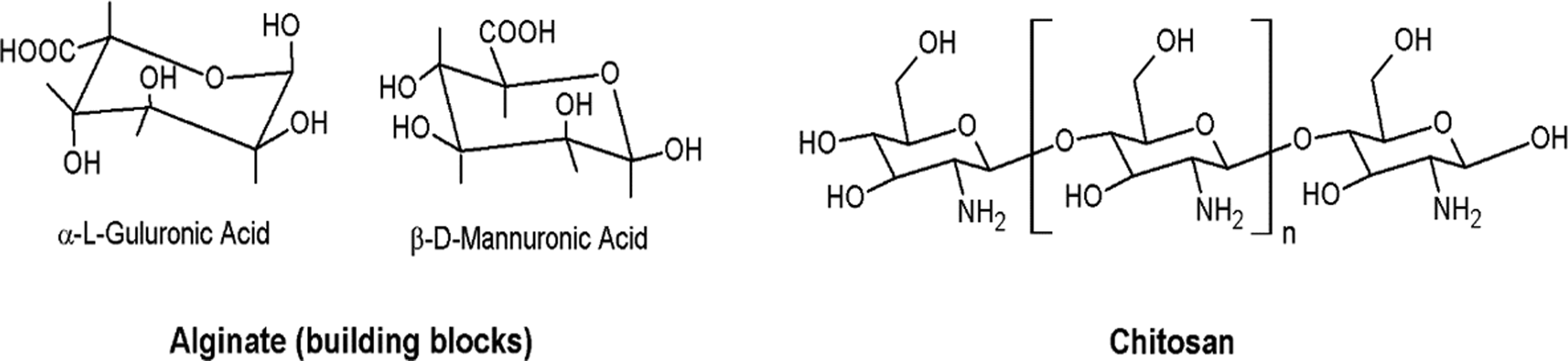
Molecular structure of alginate and chitosan building blocks.
Alginates and alginate composite polymeric hydrogels
Molecular imprinting studies in alginate hydrogels have been performed using Fn and the model protein BSA (Table 2). Alginates are water soluble linear polysaccharides derived from brown seaweed, composed of alternating blocks of 1–4 linked α-L-guluronic and β-D-mannuronic acid residues. 66 Along with their great biocompatibility and biodegradability, alginates have the ability to form gels with good mechanical properties by reacting with divalent cations. 66 Alginates enable the production of various platforms for biomedical applications, 67 including the development of alginate microspheres for growth factor delivery, such as bioactive VEGF to promote osteogenic differentiation. 68
AA, acrylic acid; AMPS, 2-acrylamido-2-methyl-propanosulfonic acid; CaCl2, calcium chloride; CS, chitosan; DAP, diammonium phosphate (NH4)2HPO4; EAB, egg albumin; ECH, ephichlorohydrin; EMIPMs, embedded molecularly imprinted polymer microspheres; Fn, fibronectin; Glo, γ-globulin; HAc, anhydrous acetic acid; Hb, hemoglobin; g-MIP, grafted-molecularly imprinted polymer; HEC, hydroxyethyl cellulose; Lac, α-lactalbumin; Lyz, lysozyme; MAPS, 3-methacryloxypropyl trimethoxysilane; MIP, molecularly imprinted polymer; Myo, myoglobulin; NIP, nonmolecularly imprinted polymer; NIPA, N-isopropylacrylamide; Ova, ovalbumin; PAAm, poly(acrylamide); Pap, papain; Pep, pepsin; PP, polypropylene; PU, polyurethane; SA, sodium algiante; SMIPMs, surface molecularly imprinted polymer microspheres; SPA, sodium polyacrylate; Try, trypsin; [VAF- MIM]Cl, 1-vinyl-3-aminoformylmethyl imidazolium chloride.
Signnificant work using sodium alginate as a functional monomer to create BSA-MIP has been performed by Zhao and co-workers.9,44,69–72 This group uses an inverse suspension method and ionic gelation to develop MIPs under mild conditions to avoid protein denaturation. Furthermore, Zhao and co-workers frequently conjugate alginate with phosphate groups by adding diammonium phosphate [(NH4)2HPO4, DAP], creating composite hydrogels with improved imprinting features.44,69,71,72 The addition of phosphates into alginate matrices combines the functionality of organic compounds with the stability of inorganic compounds. 69
In addition to developing heteropolymeric systems based on both natural and synthetic polymers (where alginate is combined with acrylamide (AAm) and sodium polyacrylate9,70–72) to improve mechanical and imprinting properties of the final MIP, the group has developed MIPs fully based on synthetic polymers. 73 Throughout some of these studies,44,69,71,72 the authors compared imprinting parameters (adsorption capacity and imprinting effect) of alginate-embedded MIP microspheres (EMIPMs) and surface MIP microspheres (SMIPMs) using BSA as the template molecule. Preparation methods for these approaches differ on the timing and the amount of template added. Despite SMIPMs requiring lower template concentrations during the molecular imprinting process, the microspheres usually present increased imprinting performance compared to EMIPMs.44,69,71,72 This is easily explained since EMIPMs face mass transfer problems inherent to bulk imprinted approaches. A practical application of these natural polymer-based microspheres is as building blocks for TE scaffolds, specifically in bottom-up approaches, to improve protein loading within the scaffold. 74 As the majority of template molecules of interest for TE applications are expensive and difficult to obtain in high rates of purification, it is important to mind cost/efficiency ratio, which will dictate production viability and market diffusion of any biomedical product.
A problem encountered by many of the previously cited works is the use of cytotoxic compounds as solvents or template removal agents (e.g., SDS), which may impair the application of these systems for TE. However, the development of more biocompatible systems for biomedical and food industry applications is the subject of continuing research. Peppas and co-workers75,76 have developed molecular imprinting methodologies for biomedical purposes using alginate and calcium chloride. Herrero et al. 76 developed a biocompatible system with the ability to achieve 3.0 mg of BSA per gram of capsules, compared to 0.7 mg achieved via inverse suspension by Zhao et al., 69 without using any chemicals besides sodium alginate and calcium chloride. These results are promising and are the first steps toward optimization of protein molecular imprinting systems for delivery and implantation in the human body.
The potential of alginate imprinted systems for generating platforms for cell culture studies has been previously investigated. Zhu et al. 77 recently created an Fn-imprinted alginate/polyacrylamide (PAAm) hydrogel, supported by a polypropylene nonwoven scaffold, which promoted Fn adsorption in addition to adhesion and spreading of mouse fibroblast cells (L929). The combination of more than one functional monomer is a widely used methodology to improve imprinting efficiency. This increases the number of potential binding sites available for the template, and improves physical and chemical characteristics (e.g., swelling behavior or mechanical stability) of molecularly imprinted hydrogels.
In this particular case of alginate/PAAm, an improvement in mechanical and elastic properties of the combined polymer systems was reported. 78 Alginate/PAAm scaffolds have also proven to be suitable platforms to support human stem cell proliferation and chondrogenesis. 78 An additional study, also based on the imprinting of Fn, was developed by Fukazawa et al. 79 who created a Fn-MIP system based on a water soluble biocompatible phospholipid polymer constituted by 2-methacryloyloxyethyl phosphorylcholine (MPC). MPC is a biocompatible polymer unfavorable for protein adsorption, thus frequently used as a coating to achieve antithrombogenicity for medical devices. 79 Using silica beads with adsorbed Fn to create protein stamps, cavities with high selectivity to Fn were imprinted on the polymer, creating a cell capturing system with low rates of nonspecific adsorption. 79 Cell adhesion tests performed with L929 cells, the authors proved that cells only adhered in the bead cavities where Fn had been imprinted, and not on the surface of the NIP. This approach has the potential to be applied as a cell patterning technique by designing scaffolds with precise control of local cell adhesion. 80
Chitosan and chitosan composite hydrogels
Another widely used polymer is chitosan, a natural polysaccharide obtained from the deacetylation of chitin from the exoskeleton of crustaceans. It has been widely explored for biomedical applications due to its abundant source, biocompatibility, and biodegradability. 81 Chitosan amino and hydroxyl groups make it possible to interact with different protein regions through van der Waals interactions, hydrogen bonding, and hydrophobic interactions. 81 Chitosan has already been used as a functional monomer for molecular imprinting of low molecular weight molecules such as dye adsorption, enantioselective separation of amino acids, metal ions removal in decontamination activities, and some chemical/pharmaceutical applications.82–87
A MIP sensitive system was reported by Singh et al. 83 who created a hydrogel system sensitive to pH, temperature, and ionic strength with molecular recognition to carnosine, a dipeptide highly concentrated in muscle and brain tissues with reported antioxidant properties. 83 However, for macromolecular imprinting, most studies performed with chitosan combine this polymer with synthetic monomers (e.g., PAAm or 2-hydroxyethyl methacrylate) or use modified chitosan (Table 2).
Chitosan is commonly used to improve mechanical properties of other polymeric materials during molecular imprinting processes. Guo and co-workers12–14,39,88 developed several studies focusing on chitosan-based matrices for Hb recognition for biosensing applications. In most of these studies, chitosan is combined with epichlorhydrin, a water soluble cross-linker that preserves the cationic amine function of chitosan and improves its wet strength, to produce beads that are used as a matrix for acrylamide monomers during the molecular imprinting process.12–14 Guo et al. 13 reported a chemical modification of porous chitosan beads by adding maleic anhydride groups to improve chitosan bonding to AAm. This modification increased vertical growth of PAAm chains from the surface of the beads, leading to higher protein adsorption rates, but lower selectivity toward the template molecule since the geometry of cavities could not be successfully preserved. 13
Later, Xia et al. 39 developed an Hb-imprinted semi-interpenetrating polymer network in an aqueous medium by combining chitosan and AAm. The MIP produced revealed high capacity to retain water, and higher adsorption and selectivity when compared to molecular imprinting hydrogels made only of PAAm. 39 Nevertheless, there is evidence of significant nonspecific binding of imprinted chitosan/PAAm polymers. 89 Fu et al. 89 analyzed different conditions where BSA and Hb-imprinted polymers showed increased recognition when compared to NIPs, but in much higher quantities than those used as a template during MIP synthesis. This behavior was hardly explained, since the yield in binding sites relative to the amount of imprinting molecule should be low. 89 When washing NIPs the same way as MIPs (i.e., including template removal solutions), the authors found that NIPs presented an increase in protein binding, which was comparable to MIPs. 89 Therefore, all procedures, with the exception of template addition, should be equally performed on the controls as they are for imprinted polymers to assure chemical changes promoted by solvents are taken into account when both materials are compared. Very few studies are performed solely on AAm as a functional monomer47,90 despite its great biocompatibility. Combining AAm with other functional monomers has been shown to improve mechanical properties and imprinting features of the matrix when comparing separate functional monomers.9,12–14,39
Dan et al. 81 studied different copolymerization systems for chitosan and other synthetic polymers at different pH values and temperatures for the recognition of ovalbumin. Copolymers composed of chitosan and methacrylic acid showed increased imprinting features and selectivity at different pH and temperature levels. 81 Gao et al. 15 recently reported a MIP system conjugated with alginate and chitosan to improve imprinting features of a magnetite (Fe3O4) nanoparticles-based system for molecular recognition of ovalbumin. With a two-step method, the authors modified Fe3O4 nanoparticles with sodium alginate to attract chitosan onto the surface (since these polysaccharides are oppositely charged) and promoted polymerization to create a molecularly imprinted shell around the particles. The imprinting effect was reported to be dependent on the alginate to chitosan mass ratio, with 1:2 exhibiting the best adsorption capacity and imprinting efficiency values toward ovalbumin. 15 The authors also showed that this system was highly selective toward different template molecules, including ovalbumin, BSA, BHb, and Lyz. 15 The existence of oppositely charged functional groups, carboxyl and hydroxyl groups from alginates, and amine groups from chitosan, may have promoted the imprinting effect as they support interactions between the polymer matrix and the differently charged domains of these proteins.
Cell Imprinting
Molecular imprinting principles for macromolecules can be applied for the imprinting of larger templates such as cells. Cell imprinting has been vaguely explored, but is based on imprinting of morphological and topographic features of cells to improve cell adhesion in/on a substrate. To program mammalian cell adhesion and growth, DePorter et al. 91 proposed a cell-imprinting approach to produce a culture system as an alternative to the conventional high cost, multi-step fabrication processes. The authors successfully imprinted substrates by casting a PAAm gel on prefixed (4% formaldehyde/PBS) monolayers of HeLa, HEK-293T (epithelial-like cells) and MRC-9 (fibroblast-like cells) cells. Although cell viability was maintained, there is room for improvement. Adhesion experiments revealed that HEK-293T and MRC-9 cells preferentially adhered to epithelial-like imprinted surfaces, with MRC-9 cells losing their fibroblast-like morphology features, and HeLa cells adhering to all imprinted substrates. 91
Jeon et al. 92 developed a PDMS imprinted surface using MG63 osteoblast-like cells (as a template) fixed with glutaraldehyde at the proliferation stage. The group used an electric field-assisted casting method during different culturing times and showed that rough surfaces promoted cell viability and increased alkaline phosphatase activity and mineralization. 92
Currently, cell imprinting is at its early stages, and more studies need to be performed to further explore its applications and influences on various cell functions. Further investigation of this topic would lead to an understanding if this technology is feasible and reproducible compared to existing technologies for cell patterning and culturing.
Final Remarks and Future Prospects
Molecular imprinting is a technology with the potential to be applied for biomedical applications, such as TE. Even though little work has been developed on biologically relevant macromolecules for TE applications, studies performed on model proteins are a helpful platform to optimize molecular imprinting systems. One can expect molecular imprinting technologies to evolve for more biocompatible production approaches, which will enable cell growth and survival. Mild conditions to avoid protein denaturation are required during molecular imprinting, and all cytotoxic components should be avoided without compromising imprinting features, something already achieved. Additionally, more systematic and reproducible technologies with greater control of imprinting parameters and MIP structure (e.g., beads diameter or porosity) will present greater industrial viability. Once solved, these key aspects will undoubtedly catapult the use of molecular imprinting with living organisms and biomedical applications. Some additional aspects of biopolymer polymeric design have been recently discussed by Peppas and Clegg. 100
Molecular imprinting will especially be relevant for scaffold-based strategies (Fig. 4) as they depend on scaffold bioactivity to modulate cell activity in situ. As previously mentioned, scaffold-based approaches rely on the knowledge of the ECM as an important source of signals to cells and the maintenance of ECM homeostasis. Developing a scaffold with molecular recognition capabilities, enables MIP technologies to not only promote loading capacity of MIPs, but also promote recognition of a specific molecule in a complex mixture of biological compounds (a real scenario all implants face when introduced in the organism) (Fig. 4A). The latter option brings a new set of possibilities for scaffold-based strategies, since the majority of conventional methods rely on the delivery of previously immobilized factors.
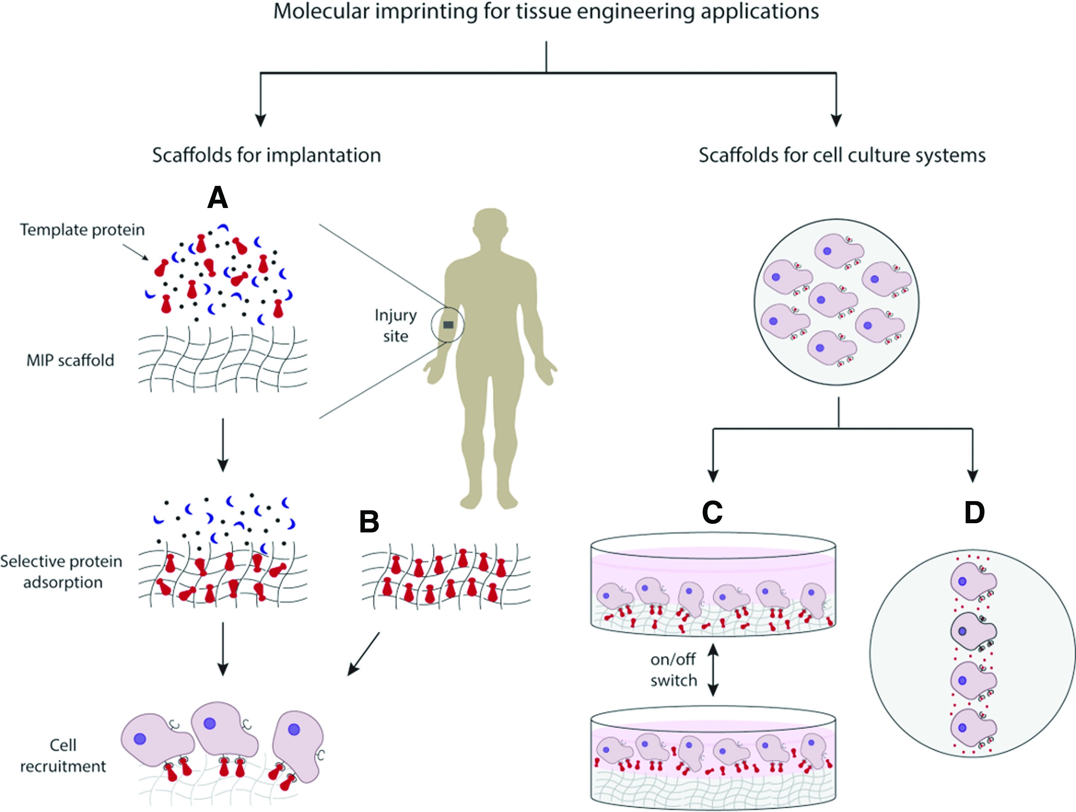
Applications of molecular imprinting in TE. Molecular imprinting of proteins can support TE approaches, especially scaffold-based bottom-up strategies on the development of cell instructive scaffolds for implantation, or for cell culture systems. Regarding scaffolds for implantation, scaffolds produced by molecular imprinting are capable of selectively recognizing and adsorbing a macromolecule within a complex mixture of biological compounds present at the injury site. As a result of the molecular memory imprinting process
Besides poor loading capacity, these methods have therapeutic efficacy impaired as protein conformation changes due to immobilization methods. Molecular imprinting systems can be optimized to overcome these problems as research performed in the protein template area proceeds. As adsorption capacity and selectivity are essential to dictate the success of a MIP-based technology, if any major conformational changes were to occur during the production process, basic imprinting features would be compromised, and the MIP would present no major differences from a NIP. Epitope imprinting approaches can also be an alternative to minimize these effects, as they are less prone to conformational changes by their significantly lower complexity and size, with the additional advantage of promoting correct protein orientation, thereby increasing scaffold bioactivity as more protein-binding sites are available for interaction with cell receptors (Fig. 4B).
Several biomolecules can be used as a template for developing scaffolds for regenerative medicine applications. Growth inducing cytokines, peptides, and proteins, including bone morphogenetic proteins (BMPs), transforming growth factor beta (TGF-β), vascular growth factor (VEGF), FGF, PDGF, insulin-like growth factor (IGF), and stromal-derived growth factor (SDF1) have generated interest by their roles in the wound healing processes. Additionally, ECM molecules such as Fn can be successfully imprinted to promote cell adhesion on/in scaffolds. Once molecularly imprinted with these molecules, scaffolds can be implanted at the site of injury and adsorb the template molecule at higher rates than other compounds, promoting cell binding to the adsorbed proteins, which will consequently lead to a specific cellular response. MIPs have been shown to selectively adsorb template molecules when in a complex solution and promote cell adhesion, prospecting the potential application for scaffolds capable to attract the template molecule in situ and trigger a specific cell behavior.
An application of molecular imprinting for the near future will definitely go through cell culture systems in vitro. PIMs allow further versatility of MIP scaffolds enabling an on/off switch of the binding recognition, a feature that can be suitable for cell sheet technologies (Fig. 4C). MIPs can also be considered for cell patterning techniques, as cell adhesion can be controlled by the imprinted and nonimprinted regions (Fig. 4D).
Hydrogels present themselves as suitable solutions to overcome current drawbacks in molecular imprinting of bio-macromolecules due to the inherent characteristics and tunable mechanical and chemical properties of these biomaterials. Similar to other functionalization techniques, several natural and synthetic biomaterials, or their combinations, have been characterized in the literature for the production of MIPs with promising results. The possibility of utilizing different polymerization and production methods in mild and biocompatible conditions (without compromising imprinting features) gives rise to the potential of developing molecularly imprinted hydrogels on the design of highly precise bottom-up strategies for the production of scaffolds with improved bioactivity.
Another imprinting strategy is cell imprinting, a technology currently at its early stages, but presents itself as a dauntless new perspective. Nevertheless, the imprinting of cells is an untapped molecular imprinting approach, which is the topic of continuing research.
Footnotes
Acknowledgments
The authors wish to thank Dr. Julia Vela-Ramirez, Ms. Heidi Culver, and Mr. John Clegg for important discussions and suggestions. This work was supported in part by the University of Texas-Portugal Collaborative Research Program, and the Grant UTAP-ICDT/CTM-BIO/0023/2014. M.E.W. is supported by a National Science Foundation Graduate Research Fellowship.
Disclosure Statement
No competing financial interests exist.