
Abstract
This review focuses on the recent strategy in the preparation of thiolated polymers and fabrication of their hydrogel matrices. The mechanism involved in the synthesis of thiolated polymers and fabrication of thiolated polymer hydrogels is exemplified with suitable schematic representations reported in the recent literature. The 2-iminothiolane namely “Traut's reagent” has been widely used for effectively thiolating the natural polymers such as collagen and gelatin, which contain free amino group in their backbone. The free carboxylic acid group containing polymers such as hyaluronic acid and heparin have been thiolated by using the bifunctional molecules such as cysteamine and L-cysteine via N-(3-dimethylaminopropyl)-N′-ethylcarbodiimide/N-hydroxysuccinimide (EDC/NHS) coupling reaction. The degree of thiolation in the polymer chain has been widely determined by using Ellman's assay method. The thiolated polymer hydrogels are prepared by disulfide bond formation (or) thiol–ene reaction (or) Michael-type addition reaction. The thiolated polymers such as thiolated gelatin are reacted with polyethylene glycol diacrylate for obtaining interpenetrating polymer network hydrogel scaffolds. Several in vitro cell culture experiments indicate that the developed thiolated polymer hydrogels exhibited biocompatibility and cellular mimicking properties. The developed hydrogel scaffolds efficiently support proliferation and differentiation of various cell types. In the present review article, the thiol-functionalized protein-based biopolymers, carbohydrate-based polymers, and some synthetic polymers have been covered with recently published research articles. In addition, the usage of new thiolated nanomaterials as a crosslinking agent for the preparation of three-dimensional tissue-engineered hydrogels is highlighted.
Introduction
T
In recent years, the development of an interpenetrating polymer network (IPN) has attracted considerable interest because of its versatility in tissue engineering. The polyethylene glycol diacrylate (PEGDA) is used widely with other thiolated polymers, such as thiolated hyaluronic acid (HA-SH) or thiolated collagen (COL-SH) to prepare the IPN hydrogel. 4 The physicochemical properties of the IPN hydrogel can be modulated by adjusting the degree of thiolation and changing the weight ratio of the polymers.
Chemical oxidation and photochemical reactions are used widely in crosslinking or disulfide formation reactions. In the chemical oxidation reaction, the thiolated polymers are oxidized using an oxidizing agent, such as hydrogen peroxide, at 37°C under atmospheric oxygen at the physiological pH 7.4. 5 During this process, the disulfide bond was formed by the oxidation of a sulfhydryl group at pH 7.4. The ultraviolet (UV) light-triggered thiol–disulfide exchange reaction was used successfully in the development of tailored biodegradable hydrogels. 6 The photochemical method provided spatiotemporal control to obtain the specific structure and physical properties of disulfide-crosslinked hydrogels. The photochemical method could be followed to fabricate macro-/microcustomized patterned hydrogels. 6 On the contrary, the photochemical method involves the use of photoinitiators, such as Irgacure-2959, which are harmful to human cells. Therefore, it is essential to introduce biocompatible photoinitiators.
Although enormous efforts have been made in development of 3D hydrogel scaffolds, the methodologies used for the controlled fabrication of in situ injectable thiolated polymer hydrogels are inadequate. The fabrication of a stem cell-loaded injectable thiolated polymer hydrogel matrix involves four steps: (1) solubilization of the thiolated polymers in an aqueous medium, (2) mixing the polymer solution with stem cells and initiators, (3) injection to the desired part of the animal, and (4) formation of gel by crosslinking under the physiological conditions. The solubility of the thiolated polymers in aqueous media is generally poor because of the formation of a disulfide bond during the preparation of thiolated polymers. 7
Recently, reducing agents, such as dithiothreitol (DTT) and mercaptoethanol were used to break down the disulfide bond. 8 On the contrary, rigorous purification by dialysis followed by freeze drying for several days have limited the wider use of this method. Therefore, a simplified and efficient method for the preparation of thiolated polymers is needed. As the time taken for the in situ formation of injectable hydrogels is either very fast (or) slow, the precise experimental conditions need to be optimized to achieve the desired time duration. Recently, the rate of disulfide formation was delayed by the introduction of a sterically hindered bulky group to the hyaluronic acid polymer backbone. 8
The present review article outlines the thiolated protein-based biopolymers (e.g., collagen [COL] and gelatin [GEL]), thiolated carbohydrate-based biopolymers (e.g., thiolated heparin [HEP-SH], HA, pectin [PECT], and polygalacturonic acid [PGA]), and various thiolated synthetic polymers developed to fabricate hydrogel matrices for a series of tissue engineering applications. The following sections are covered in the present review article: (1) synthesis of thiolated polymers, (2) thiolated gelatin or collagen-based hydrogels, (3) thiolated carbohydrate polymer-based hydrogels, and (4) thiolated synthetic polymer-based hydrogels.
Synthesis of Thiolated Polymers
Thiolated polymers are synthesized by several synthetic approaches using various thiolation reagents. Chemical reagents, such as L-cysteine (L-CYS), cysteamine (CYS), thioglycolic acid (TGA), and thiolactone-based reagents are used widely for the preparation of thiolated polymers.9,10 Two major synthetic approaches can be used depending on the various types of functional groups present in the polymer chain: (1) N-(3-dimethylaminopropyl)-N′-ethylcarbodiimide/N-hydroxysuccinimide (EDC/NHS) coupling reaction, and (2) ring-opening addition of thiolactone-based reagents. As the 2-iminothiolane hydrochloride (Traut's reagent) and thiolactones, such as N-acetyl homocysteine thiolactone and 4-butyro thiolactone, are highly reactive toward the amine group, they were attached effectively to the amine group of the polymers.11,12
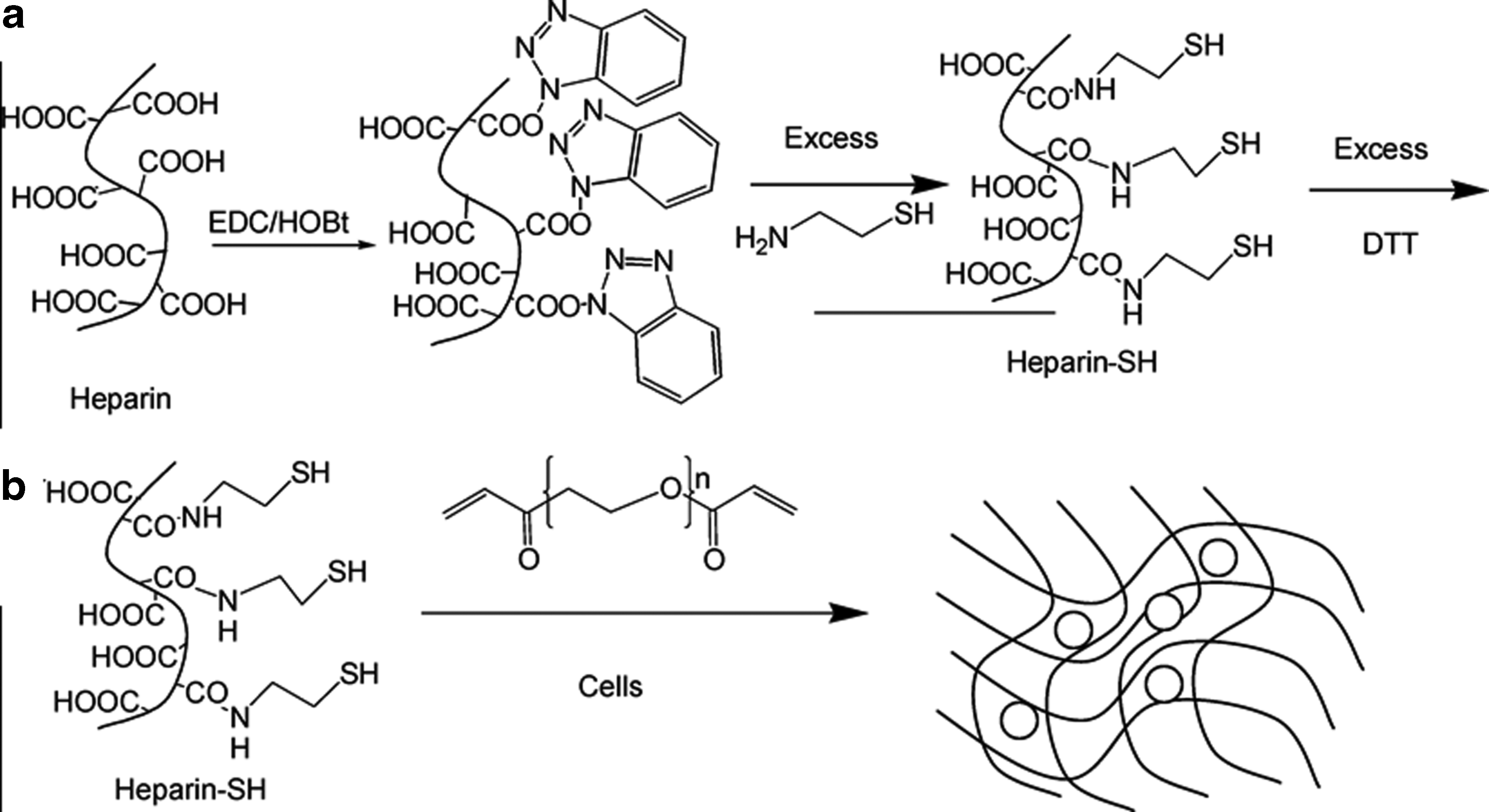
The EDC/NHS coupling reaction was followed to attach the carboxylic acid group of the thiolating agents with the amine group of the polymer or amine group of thiolating agents with the carboxylic acid group of the polymer (Fig. 1). 13 For example, CYS was attached to the carboxylic acid group of HEP, whereas the carboxylic acid group of TGA was attached to the amine group of chitosan via the formation of an amide bond via the EDC/NHS coupling reaction.13,14 In another case, in the first step, the carboxylic acid group of GEL was reacted with ethylenediamine via an EDC coupling reaction to obtain the amine group-grafted biopolymers. In the second step, it was reacted with Traut's reagent to obtain the thiolated gelatin (GEL-SH). 11
Reaction scheme for
The Traut's reagent-based synthetic pathway was used widely to prepare GEL-SH because of the higher effectiveness of the reaction, 15 but it cannot be used to prepare HEP-SH (or) HA due to the absence of a free amine group in these polymer chains. The EDC/NHS coupling reaction was used to synthesize the thiolated carbohydrate-based polymers due to the presence of free carboxylic acid (or) hydroxyl groups in these polymers. 13 The degree of thiolation in the thiolated polymers has been determined widely using the reagent (5,5′-dithio-bis-(2-nitrobenzoicacid) (Ellman's assay). 16 Recently, the development of S-protected thiolated polymers have attracted considerable interest to protect the free sulfhydryl group, and the S-protected thiomers could form a hydrogel in the presence of dithiols via a disulfide exchange reaction. 17 Table 1 lists the various chemical reagents used for the thiolation of different polymers.
COL-SH, thiolated collagen; DTBH, dithiobis(butyric dihydrazide); DTP, 3,3′-dithiobis-propanoic dihydrazide; DTPH, dithiopropionic dihydrazide; DTT, dithiothreitol; EDC, N-(3-dimethylaminopropyl)-N′-ethylcarbodiimide; GEL-SHP, thiolated gelatin; HA-SH, thiolated hyaluronic acid; HEP-SH, thiolated heparin; MPTS NPs, 3-mercaptopropyltrimethoxysilane nanoparticles; PAA-SH, thiolated poly(aspartic acid); PECT-SH, thiolated pectin; PGA-SH, thiolated polygalacturonic acid; TGA, thioglycolic acid.
Thiolated gelatin (or) collagen-based hydrogels
The protein-based hydrogel fabrication has been applied extensively in the field of tissue engineering. Protein molecules, such as GEL and COL, have found enormous applications in tissue engineering because of the ease of functionalization on their polymer backbone and the ease of further crosslinking in a physiological environment. The physicochemical properties of the hydrogel material could be tuned by adjusting the degree of crosslinking and the degree of functionalization. Recently, the thiolation of GEL and COL fibers have attracted significant attention for the formation of in situ gelation and cell encapsulation into the hydrogel matrix.
GEL is a protein molecule prepared from COL by the partial hydrolysis of COL fibers. The material is soluble in hot water but insoluble in most organic solvents. GEL forms a thermally reversible hydrogel with a minimum concentration of 0.5% (w/v) over the pH range of 5–8 at low temperatures (<35°C). GEL has potential applications in the biomedical field, particularly in tissue engineering, because of its biocompatibility and hydrogel properties. The development of COL-SH has attracted vast attention for the development of new types of 3D hydrogel matrices and IPN.
Recently, Dong et al. developed an injectable GEL-SH/PEGDA hydrogel with tunable mechanical properties. 18 PEGDA is generally used for the fabrication of GEL-SH/PEGDA hydrogels, but in their research, they used 2% (w/v) multifunctional hyperbranched PEG acrylate for crosslinking with 2.5% (w/v) of GEL-SH via the Michael addition pathway. The multifunctional PEG acrylate enhanced the crosslinking efficiency, and gelation was achieved spontaneously within 2 min. The advantage of using the acrylate group-enriched hyperbranched PEG polymer was to flexibly adjust the crosslinking ratio and simultaneously maintain the mechanical properties of the hydrogel structure. The murine adipose-derived stem cells (ADSCs) were loaded effectively into the injectable hydrogel matrix. 18
Vlierberghe et al. developed reversible GEL hydrogels via a crosslinking of GEL-SH. 15 GEL was thiolated using N-acetyl homocysteine thiolactone (or) Traut's reagent. The degree of thiolation, which was controlled by adjusting the concentration of the thiolating reagent, was determined using Ellman's assay method. The thiolation of GEL was also confirmed by size exclusion chromatography. Thiolation was more effective when Traut's reagent was used as a thiolation agent, which was inferred by the higher molecular weight (330,000 Da) of the Traut's reagent-modified GEL than that (280,000 Da) of N-acetyl-homocysteine thiolactone modified GEL. The GEL-SH was crosslinked through the formation of a disulfide bond by reacting with a 0.5 equivalent of hydrogen peroxide on silanized glass plates. 15
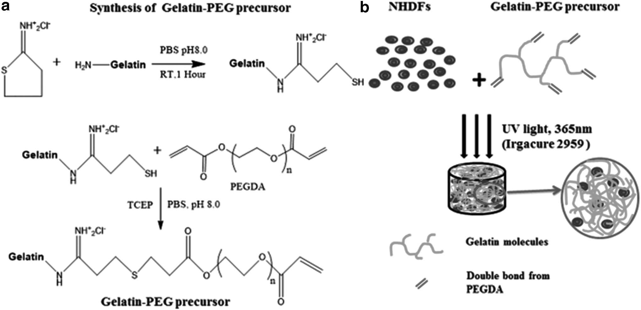
Cao et al. synthesized GEL-SH using the Traut's reagent and then conjugated it to PEGDA via a Michael addition reaction to obtain the precursor (Fig. 2A). 19 Finally, neonatal human dermal fibroblast (NHDF)-encapsulated GEL-SH/PEGDA hydrogels were fabricated by the photopolymerization of the precursor in the presence of NHDFs under UV light irradiation at 365 nm using Irgacure-2959 as an initiator (Fig. 2B). They suggested that the mechanical properties of the GEL-SH/PEGDA hydrogels could be modulated by varying the amount of GEL-SH/PEGDA precursor and by changing the ratio of GEL-SH and PEGDA. The GEL-SH/PEGDA 3D hydrogel with a storage modulus less than ∼100 Pa and mesh size >150 nm improved the viability and proliferation of the NHDFs cells. 19
Schematic representation for
Thiol–ene click chemistry is used widely for synthesizing GEL hydrogels. Russo et al. synthesized the GEL-SH by reacting with γ-thiobutyrolactone and then crosslinking it with pentenoyl GEL via the thiol–ene reaction pathway under UV light irradiation at 365 nm using Irgacure-2959 as a photoinitiator. 20 Human bone marrow stromal cells (hBMSCs) were suspended in a prepolymer solution of GEL-SH and pentenoyl GEL, and photopolymerized at 365 nm to obtain the hBMSC-embedded hydrogel matrix. The hydrogel derived from the lowest concentration of total GEL (5% w/v) showed better cytocompatibility compared with the hydrogel derived from a higher total GEL concentration (10% w/v). 20
Fu et al. also developed a GEL-PEGDA 3D hydrogel via the thiol–ene click reaction. 21 In another report, GEL-SH was synthesized by conjugating with L-CYS via bis-N-hydroxysuccinimide-functionalized PEG (bis-NHS-PEG). A NHDF-loaded covalently crosslinked GEL-PEG hydrogel was produced by a UV light-induced thiol–ene reaction in the presence of NHDFs. The 3D hydrogel supported the cell attachment, adhesion, and proliferation of NHDFs. 21
The thiolation of GEL was also achieved by reacting GEL with thiol group containing various bifunctional reagents via the EDC coupling reaction. Heffernan et al. reacted the amino group of GEL with dithiopropionic dihydrazide (DTPH) via an EDC coupling reaction to obtain GEL-SH. 22 The resulting GEL-SH was crosslinked chemically with PEGDA via a Michael-type addition reaction to obtain the hydrogel scaffold. Cell invasion into the scaffold was examined by fabricating spheroids on the surface of the hydrogels. The effect of growth factor on cell invasion was assessed by adding the human hepatocyte growth factor (HGF) to the samples at a 20 ng/mL concentration. HGF greatly enhanced both the invasion and proliferation of U87R cells in the hydrogel. 22
Recently, the COL-SH-based IPN hydrogels have attracted considerable interest because of their wide spectrum of properties and ease of controlling the elastic modulus over a broad range. For example, Xu et al. prepared COL-SH and oligo(acryloyl carbonate)-b-poly(ethylene glycol)-b-oligo(acryloyl carbonate) (OAC-PEG-OAC) copolymer-based injectable hybrid hydrogel through a Michael addition reaction. 3 COL-SH/OAC-PEG-OAC hydrogels formed with the gelation time ranged from 0.4 to 8.1 min at 37°C and pH 7.4. The presence of COL in the hybrid hydrogel facilitated its enzymatic degradation against collagenase. An in vivo experiment on a rat model indicated that the BMSC-encapsulated hybrid hydrogel showed improved cardiac function within 28 days. 3
Yamauchi et al. synthesized COL-SH by reacting COL with 4-butyrothiolactone. The crosslinked COL-SH polymer film was synthesized via the formation of a disulfide bond between the COL-SH polymer chains. 23 The collagenase enzymatic degradation was slow in the case of the crosslinked COL-SH films compared with noncrosslinked control COL. The disulfide crosslinked COL-SH film supported the growth of L929 fibroblasts similar to the control COL film. 23 Singh et al. synthesized COL-SH by reacting COL with N-succinimidyl S-acetylthioacetate and followed by a reaction with hydroxylamine followed by reduction with tris (2-carboxyethyl) phosphine. The COL (type I)-PEGDA hydrogel was then synthesized by the photopolymerization of COL-SH and PEGDA. The endothelial and fibroblast cells were encapsulated within the hydrogel scaffold and their successive coculture resulted in the formation of capillary vessel-like networks. 24
Thiolated Carbohydrate Polymer-Based Hydrogels
Carbohydrates are abundant in all prokaryotic and eukaryotic organisms. They are connected through glycosylated proteins attached directly to the cell membrane. Carbohydrates present in the outer shell of the membrane play a vital role in cell adhesion, communication, and anti-inflammatory activity. Because of their potential functionality and biocompatibility, carbohydrate molecules, such as HEP, HA, PECT, and alginate, are used extensively in tissue engineering. In recent years, the thiolation of carbohydrate molecules has become important for fabricating 3D hydrogel matrices and regulating their physicochemical properties for tissue regeneration.
HEP-SH-based 3D hydrogels have been developed. The carboxylic acid group present in the HEP was used for further functionalization of the thiol group containing bifunctional molecules.13,25 For example, Tae et al. synthesized HEP-SH through an EDC-based coupling reaction at pH 6.8 between the carboxylic acid of HEP (1% [w/v]) and the amine group of excess CYS. 13 The degree of thiolation was controlled by varying the molar feed ratio of HEP and CYS. The possibly oxidized disulfide bonds were reduced using a 10 times higher molar ratio of DTT (moles per COOH group of HEP) to convert to free thiol groups.
The HEP-SH was crosslinked with PEGDA via the in situ gelation of a thiol group in the presence of fibroblast cells to obtain a fibroblast cell-encapsulated HEP-SH/PEGDA hydrogel matrix. The fibroblasts exhibited cell proliferation inside the hydrogel matrix and the proliferation of fibroblasts was increased five-fold in the presence of fibrinogen in the hydrogel matrix. 13
Recently, Gwon et al. followed the EDC coupling reaction of HEP with CYS to obtain HEP-SH, and fabricated the HEP-SH/HA hydrogel via a photocrosslinkable thiol–ene reaction between the thiol group of HEP and the methacrylate group of methacrylated HA. 25 Gelation was controlled by the light intensity, concentration of the initiator, and pH of the medium. Fu et al. reported the visible light-initiated photopolymerization of HEP-SH with acrylated PEG. 26 HEP was thiolated using CYS via an EDC coupling reaction, and the degree of thiolation was determined by Elman's assay. 16
The photopolymerization reaction between HEP-SH and acrylated PEG (1:1–1:0.25 molar ratio of thiol/acrylate) in phosphate-buffered saline (pH 7–8) was carried out using eosin-Y as an initiator and triethanolamine as an electron donor under the irradiation of green LED light at 525 nm to obtain the transparent hydrogel (Fig. 3). The prepared hydrogel exhibited biocompatibility toward the fibroblast cells and acted as a good carrier for epidermal growth factor. 26
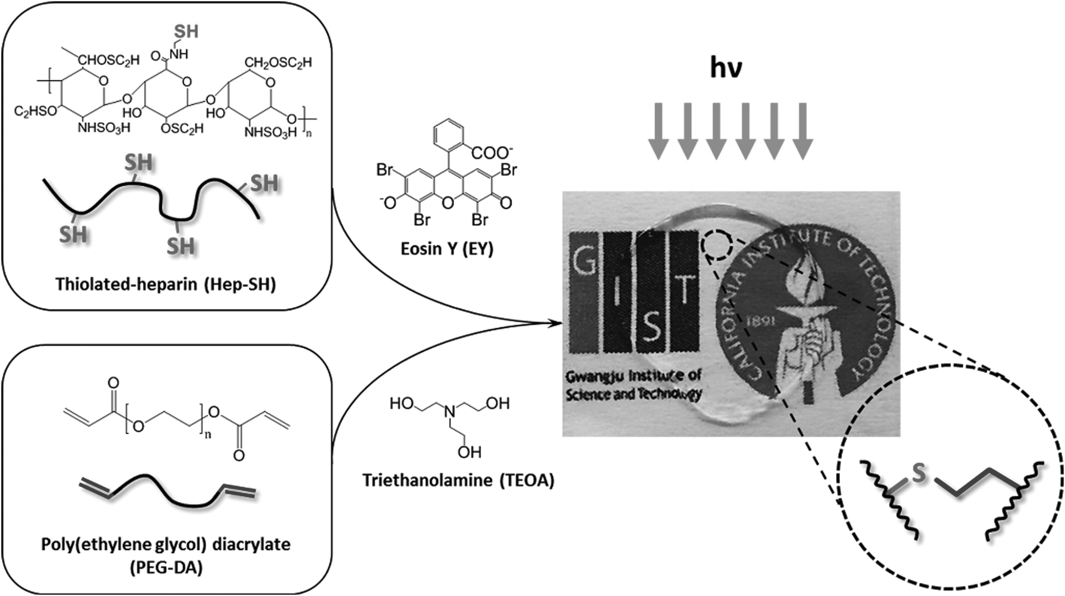
Schematic representation of visible-light-activated HEP-SH/PEGDA-based hydrogel formation. Reproduced from Fu et al., 26 with permission from American Chemical Society Copyright 2015.
The HEP-SH-based hydrogels were used to cultivate the primary hepatocytes. For example, Kim et al. prepared a HEP-SH/PEGDA-based 3D hydrogel scaffold for hepatocyte culture via a Michael addition reaction at 37°C. 27 The HEP-SH/PEGDA hydrogel was nontoxic and stimulated the secretion of albumin by the primary hepatocytes. HEP-SH was also used to fabricate micropatterned HEP-SH/PEGDA hydrogels via a photocrosslinking reaction. These hydrogels were formed faster and stronger than those prepared by the Michael addition reaction. 28
The HGF was fabricated with a micropatterned hydrogel, and immunostaining showed that the HGF was retained in the hybrid HEP-SH/PEGDA hydrogel, while it was released rapidly from the pure PEG hydrogel microstructures. The rat hepatocytes residing at the HEP-SH-based hydrogel produced four times more albumin on day 7 than the hepatocytes residing in the pure PEG hydrogels. 28
HA-SH-based hydrogels have been developed for various tissue engineering applications. Recently, the self-crosslinked, single-component HA-SH was investigated as an injectable 3D hydrogel scaffold for a fibroblast and chondrocyte cell culture platform. 29 The HA was reacted with CYS via an EDC/NHS coupling reaction at pH 4.75 to obtain the HA-SH, and the HA-SH injectable hydrogel was fabricated by in situ self-crosslinking via the formation of a disulfide bond at pH 7.4 with exposure to atmospheric oxygen. The degree of thiolation played a vital role in the stability of the hydrogel matrix. The hydrogel matrix was stable for up to 28 days when the degree of thiolation was more than 50%, while the stability period was decreased to 14 days when the degree of thiolation was less than 15%.
The gelation time was governed by three factors: concentration of HA-SH, degree of thiolation, and temperature. The gelation time period decreased with increasing concentration and degree of thiolation of HA-SH. Interestingly, the gelation process could be delayed for up to 2 h at 4°C, which provided sufficient time to conveniently mix the cells with a HA-SH solution and the 3D hydrogel matrix could be achieved via a subcutaneous injection within a few minutes at the physiological temperature (37°C). In vitro and in vivo analyses indicated that the self-crosslinked 3D HA-SH hydrogels showed good biocompatibility and induced cell proliferation. 29
The thiolation of HA has been achieved widely using 3,3′-dithiobis-propanoic dihydrazide (DTP) via carbodiimide chemistry.30–32 For example, Hao et al. synthesized HA-SH by reacting with DTP via a carbodiimide coupling reaction with 33% thiol incorporation. 30 The HA-SH was crosslinked with an acrylated copolymer carrying multiple copies of the cell adhesive peptide (PolyRGD-AC) at pH 7.8 to obtain the hybrid biomimetic hydrogel matrix with a mean pore size of 50–100 nm and an elastic modulus of 630 Pa (Fig. 4). The hybrid hydrogel acted as a microenvironment for LNCaP prostate cancer cells with high cell viability, and the hydrogel facilitated the 3D cell expansion to produce a physiologically relevant tumor model. 30
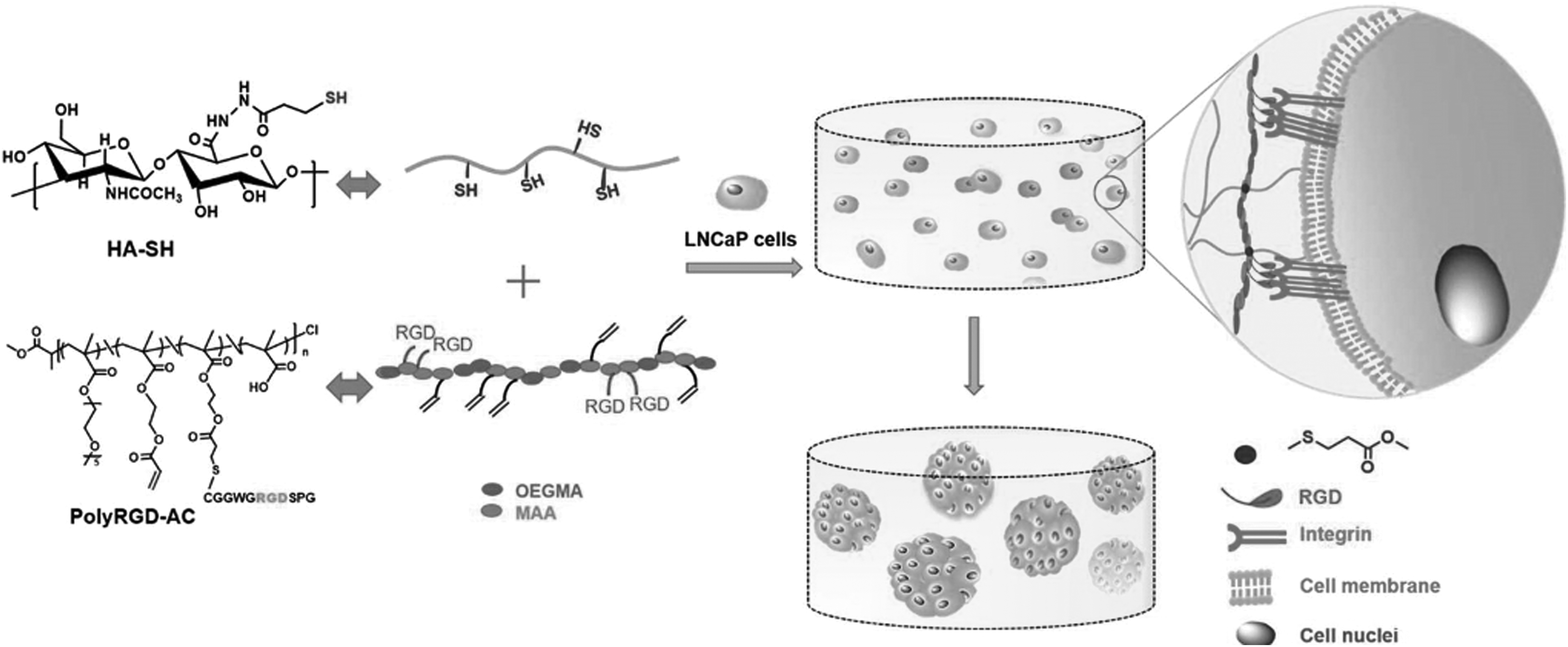
Fabrication of HA-PolyRGD hydrogels for the assembly of LNCaP prostate tumoroids. Reproduced from Hao et al., 30 with permission from American Chemical Society Copyright 2016. HA-SH, thiolated hyaluronic acid.
Shu et al. used DTP and dithiobis(butyric dihydrazide) (DTBH) as a thiolation agent via carbodiimide chemistry to obtain the thiolated derivatives of HA, that is, HA-DTPH (pKa = 8.87) and HA-DTBH (pKa = 9.01), respectively. 31 The L-929 murine fibroblasts cells were fabricated in a HA-DTPH hydrogel using the in situ gelation and cell encapsulation method under physiological conditions. The in vitro cytotoxicity study indicated that the L-929 murine cells were viable and proliferated after 3 days incubation.
Horn et al. synthesized HA-SH via the DTP via carbodiimide reaction. The HA-SH was crosslinked with PEGDA via a Michael-type addition reaction to yield the crosslinked hydrogel. 32 Chick dorsal root ganglia were cultured on a 3D crosslinked HA-PEGDA hydrogel; the crosslinked HA-SH hydrogel exhibited more than 50% extension of neurite growth compared with that of crosslinked fibrin hydrogels. On the contrary, it was indicated that the crosslinked HA-SH hydrogel did not overcome the strong inflammation caused by the spinal cord transection. 32
Bi et al. attempted the reaction of HA-SH with the polyamidoamine (PMAM) dendrimer via a thiol–ene reaction to obtain the HA-PMAM hydrogel matrix. 33 The hydrogel matrix showed biocompatibility toward human umbilical vein endothelial cells (HUVEC) and the incorporation of RGD peptide enhanced the viability of the HUVECs. Zarembinski et al. obtained the HA-Gel hydrogel by crosslinking the carboxymethylated thiolated HA (CMHA-S) with GEL-SH via a disulfide exchange reaction. 34 The CMHA-S was reacted with oxidized glutathione (GSSG) and then with GEL-SH to form the HA-Gel hydrogel. The hydrogel showed in vitro biocompatibility to the ADSC line and in vivo biocompatibility in white adult rabbits. 34
HA-SH was also used to synthesize a prodrug hydrogel for drug release specific to cancer cells. For instance, Fu et al. conjugated the doxorubicin hydrochloride (DOX-HCl) with HA-SH through acid-liable hydrazone linkage. 35 The HA-SH was synthesized by the serial conjugation of adipic acid and 3, 3-dithiodipropionic acid (DTDPA) to HA through the EDC coupling reaction. DOX-HCl was attached to HA-SH via the formation of a hydrazone bond and finally exposed to air for self-gelation. The DOX-conjugated HA hydrogel exhibited pH responsive drug release at acidic pH as well as reduction-responsive drug release in the presence of glutathione (GSH). 35
The thiolated PECT (PECT-SH)-based carbohydrate polymers have also been developed for biomedical applications. 36 The PECT-SH showed better mucoadhesive properties over PECT. 37 Sharma and Ahuja synthesized the thiolated PECT with 0.6 mmol of thiol groups/g of polymer via esterification with TGA. 37 An evaluation of the mucoadhesive properties of the polymers on the goat intestinal mucosa showed that PECT-SH exhibited a higher ex vivo bioadhesion time than PECT. The better mucoadhesive properties of PECT-SH could be attributed to the formation of a disulfide bond between the mucus and PECT-SH.
Recently, PECT-SH nanoparticles (NPs) were prepared using an ionotropic gelation technique for various biomedical applications.37,38 Magnesium chloride was used as an ionic crosslinker and the particle size was increased with increasing magnesium chloride concentration. The optimal entrapment efficiency (94.6%) of a model drug timolol maleate was achieved when PECT-SH (0.01% [w/v]) and magnesium chloride (0.01% [w/v]) were used. The PECT-SH nanoparticulates showed considerably higher ex vivo corneal permeation of timolol maleate through the goat cornea than a PECT solution. 38
Peng et al. synthesized the thiolated PGA (PGA-SH) using L-CYS as the thiolation agent with a 16.24% of degree of modification. 39 The thiolated PGA hydrogel film was prepared to act as a drug carrier for rosmarinic acid and to prevent postsurgical adhesion. An in vivo implantation to Sprague–Dawley rats showed that the PGA-SH hydrogel films reduced 90% of the adhesion. 39 Hence, PGA-SH can be used to prepare the 3D hydrogel matrix and might have potential applications in tissue engineering. The thiolated pullulan polysaccharide was also prepared for mucosal drug targeting. 40 Thiolated pullulan was synthesized by the oxidation of pullulan by periodate and then coupling with CYS. The thiolated pullulan exhibited 46 times higher mucoadhesive properties. 40
Thiolated Synthetic Polymer-Based Hydrogels
Various types of thiolated synthetic polymers were developed in addition to thiolated proteins and carbohydrate polymers for the fabrication of a 3D hydrogel scaffold matrix. For example, the synthesis of thiolated poly(aspartic acid) (PAA) (PAA-SH) and its mucoadhesive properties have been studied extensively. CYS was used effectively as a thiolation agent for PAA.41,42 Horvát et al. synthesized PAA-SH using CYS as a thiolation agent and fabricated the hydrogel in the presence of mucin. 41 Mucin played a vital role in crosslink formation, and the PAA-SH gels showed strong mucoadhesion properties that were more effective at lower polymer concentrations (3% and 5% [w/w]). The anti-inflammatory drug, diclofenac sodium (DS), was encapsulated in PAA-SH, and the PAA-SH gel exhibited a burst release within 1 h and sustained DS release for up to 23 h. 41
Gyarmati et al. synthesized a pH-responsive CYS-modified PAA and examined its reversible sol–gel transition behavior. The CYS-modified PAA exhibited dual responsive characteristics: (1) reversible pH and (2) redox sensitivity in aqueous solutions. 42 Juriga et al. attached the CYS to the PAA, which was then reduced using DTT to obtain PAA-SH. The PAA-SH hydrogel supported the survival and proliferation of MG-63 osteoblast-like cells. 43 In addition, Montero-Rama et al. synthesized thiolated poly[2-(2-methoxyethoxy)ethyl methacrylate (MEO2MA)]-co-[2-thioethyl methacrylate] P(MEO2MA-co-SEMA) hydrogel. 44 They reported that the free thiol group was utilized to modulate the transition temperature of the thermo responsive P(MEO2MA-co-SEMA) hydrogel by changing the pH of the solution. 44
NPs have also been utilized to enhance the intrinsic properties of thiolated polymeric hydrogels. NP-mediated fabrication of 3D composite materials could also be used for the development of various potential tissue engineering applications. For example, the gold nanostructures were used to synthesize spatially confined chitosan nanogel. 45 When the cationic polymer chitosan was introduced to the glutathione functionalized gold nanoclusters (GSH@AuC), the carboxylate anions in the GSH@AuC were neutralized by the cations present in chitosan, leading to the formation of a nanogel. Owing to their higher stability in water and good biocompatibility, the nanogel may have potential in the biomedical field.
Silver nanoparticles (AgNPs) have recently been reported to act as a crosslinker for the fabrication of AgNPs/thiolated poly(N-isopropyl acrylamide) (PNIPAM-SH) nanocomposite hydrogels. The AgNPs acted as both a crosslinker and antialgal agent. The mechanical properties of the hydrogel could be tuned by varying the molar feed ratio of silver/sulfur (Ag/S), and highest mechanical properties of the hydrogel were achieved with a 0.9 molar feed ratio of Ag/S. 46 Because metal NPs, such as Au and AgNPs, exhibit active surfaces due to the quantum confinement effect, they are highly reactive toward the thiol group. Therefore, the biocompatible metal NPs could be used as a potential crosslinker for the formation of thiolated polymer hydrogels.
In addition, Irmukhametova et al. synthesized the thiol group-functionalized organosilica NPs by the self-condensation of 3-mercaptopropyltrimethoxysilane (MPTS) in the presence of atmospheric oxygen. The formation of a disulfide bridge between the thiol group containing the MPTS NPs by the partial oxidation of a thiol group resulted in the necklace-shaped self-assembled NPs and exhibited mucoadhesive properties (Fig. 5). 47 Therefore, the thiol group-enriched MPTS NPs could act as efficient crosslinkers via the formation of a disulfide bond with the thiol group of various thiolated polymers.
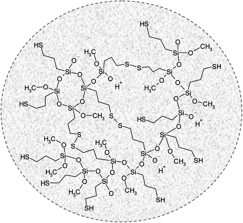
Schematic structure of thiol functionalized 3-mercaptopropyltrimethoxysilane NPs. Reproduced from Irmukhametova et al., 47 with permission from American Chemical Society Copyright 2011.
Summary and Future Perspectives
The thiolated polymers could be synthesized using various multifunctional chemical reagents with a sulfhydryl group, such as L-CYS, CYS, and thiolactones. Although bi-(or) multifunctional reagents such as L-CYS, CYS, and DTPH were functionalized with the carboxyl group of the polymers via an EDC coupling reaction, the Traut's reagent was prominent for adding to the amine group of the polymers. Irgacure-2959 has been used widely as an initiator in photocrosslinking reactions. The GEL-SH (or) COL-SH could be crosslinked successfully with PEGDA via thiol–ene click (or) Michael addition reactions to obtain the GEL-PEGDA (or) COL-PEGDA hydrogel. The addition of growth factors, such as HGF to the hydrogel greatly improved both the invasion and proliferation in the U87R cells in the GEL-PEGDA hydrogel.
The thiolated carbohydrate polymers, such as HEP-SH, HA-SH, PECT-SH, and PGA-SH were synthesized, and their corresponding hydrogels were prepared by the formation of a disulfide bond (or) via a crosslinking reaction with other polymers, such as GEL or PEGDA. The thiolated carbohydrate-based hydrogels showed better mucoadhesive properties than that of the thiol free polymers. Some thiolated synthetic polymer hydrogels, such as PAA-SH and P(MEO2MA-co-SEMA), were also fabricated and the PAA-SH hydrogels exhibited thermally reversible sol–gel transition behavior.
Extensive in vitro cytotoxicity experiments were conducted to examine the viability and proliferation of various stem cells, such as NHDFs, hBMSCs, L-929 murine fibroblasts, ADSC, and MG-63 osteoblast-like cells, encapsulated in various thiolated polymer hydrogels. These results indicated that the thiolated polymer hydrogels showed cell viability and improved cell proliferation. Some in vivo experiments were also conducted to examine the potential of the thiolated polymer hydrogels. These recent studies clearly showed that the biocompatible thiolated bio-(or) synthetic polymers could play important roles in the tissue engineering field.
Because the initiator used in the crosslinking reactions, such as Irgacure-2959 and hydrogen peroxide, are harmful to animal cells, serious consideration should be taken when selecting biocompatible and efficient initiators (or) crosslinking agents. Because the thiolated nanomaterials have attracted as much interest as crosslinking agents, extensive studies should be undertaken to determine the biocompatibility and effectiveness of the various thiolated nanomaterials. Extensive in vivo studies will be needed to highlight the potential of the developed thiolated polymer hydrogels.
Footnotes
Acknowledgments
The research was supported by the International Cooperative Research Grant (2014) of Incheon National University and Basic Science Research Program through the National Research Foundation of Korea (NRF-2017R1C1B1003665).
Disclosure Statement
No competing financial interests exist.