
Abstract
With the increasing prevalence of bone tissue diseases, three-dimensional (3D) bioprinting applied to bone tissue engineering for treatment has received a lot of interests in recent years. The research and popularization of 3D bioprinting in bone tissue engineering require bioinks with good performance, which is closely related to ideal material and appropriate construction form. Hydroxyapatite (HAp) is the inorganic component of natural bone and has been widely used in bone tissue engineering and other fields due to its good biological and physicochemical properties. Previous studies have prepared different bioinks containing HAp and evaluated their properties in various aspects. Most bioinks showed significant improvement in terms of rheology and biocompatibility; however, not all of them had sufficiently favorable mechanical properties and antimicrobial activity. The deficiencies in properties of bioink and 3D bioprinting technology limited the applications of bioinks containing HAp in clinical trials. This review article summarizes the construction forms of bioinks containing HAp and its modifications in previous studies, as well as the 3D bioprinting techniques adopted to print bioink containing HAp. In addition, this article summarizes the advantages and underlying mechanisms of bioink containing HAp, as well as its limitations, and suggests possible improvement to facilitate the development of bone tissue engineering bioinks containing HAp in the future.
Impact statement
This review summarizes the construction form and application in bone tissue engineering of three-dimensional (3D) bioprinting ink containing hydroxyapatite (HAp). Currently, the use of HAp as a 3D bioprinting ink material in clinical applications is rare. We suggested possible improvements for this kind of ink to facilitate its research and popularization.
Introduction
Bone tissue engineering refers to the implantation of autologous osteoblasts, bone marrow mesenchymal stem cells (BMMSCs), or chondrocytes onto a three-dimensional (3D) cell scaffold that is biocompatible and can be gradually degraded and absorbed by the human body after in vitro expansion. This allows cells to grow on the preformed 3D scaffold. Subsequently, this hybrid cell material is implanted into the bone defect area, where the scaffold material degrades, while cells continue to proliferate, thereby achieving the goal of repairing bone tissue defects.1,2 Bone tissue engineering is of increasing interest due to the high prevalence of bone disease that cannot heal itself by the regenerative capacity, and the limitations of other treatment strategies include artificial bone transplantation and autologous bone transplantation.3–7
3D bioprinting is a method applied in bone tissue engineering, which involves using a custom-designed 3D model as a basis to create tissue structures using biocompatible materials, biomolecules, and live cells. 3D bioprinting technologies can be categorized into three distinct groups according to the American Society of Testing and Materials standards—jetting-based, extrusion-based, and vat polymerization-based bioprinting. This technology has been extensively researched in the fields of organ regeneration, pharmaceutical manufacturing, and food industry. 8
The combination of different cells and scaffold materials has been proposed in bone tissue engineering using 3D bioprinting, and the mechanical strength and biocompatibility of resulting products are being further researched to achieve the desired goals in bone repair. 9 Currently, some issues have been exposed in bone tissue engineering using 3D bioprinting, including limited mechanical performance of the products resulting in poor structural stability, suboptimal biocompatibility, and the need for further improvement in biomimetic properties.10,11
The ideal bioink serves as the foundation for improving the mechanical performance and biocompatibility of printed products. Bioink refers to a mixture of biological materials and living cells, possessing characteristics similar to the extracellular matrix (ECM) environment. It is capable of supporting cell adhesion, proliferation, and differentiation. To achieve good structural and functional outcomes in printed results, bioink must meet different criteria in terms of biointeractions, structural fidelity, viscosity, bioprintability, biodegradability, and biocompatibility, depending on its intended use. Currently, most bioinks are deposited with high precision layer-by-layer, undergoing a phase transition from fluid to semisolid or gelatinous states through cross-linking or gelation processes. 12
To prepare an ideal bioink, researchers have been searching for materials with excellent biological and physicochemical properties, among which hydroxyapatite (HAp) has been widely studied due to its inherent characteristics. HAp is an inorganic compound composed of calcium and phosphate salts, with the main component of calcium hydroxyapatite. It is the main inorganic component of bone tissue and has bone-conducting properties, which can induce the osteogenic differentiation of stem cells. The human bone tissue predominantly comprises 65% (w/w) HAp crystals and 35% (w/w) collagen matrices.
The HAp crystals are of hexagonal crystal structure and belong to the P63/m space group (Fig. 1).13–15 In clinical practice, HAp is a common material for tissue engineering and is used in orthopedics, dentistry, otorhinolaryngology, wound repair, and soft tissue filling.16–21 Besides, HAp is often used as drug carriers for targeted therapy.14,22 Some studies have confirmed the potential application of HAp in the treatment of neurodegenerative diseases and cancer.23,24
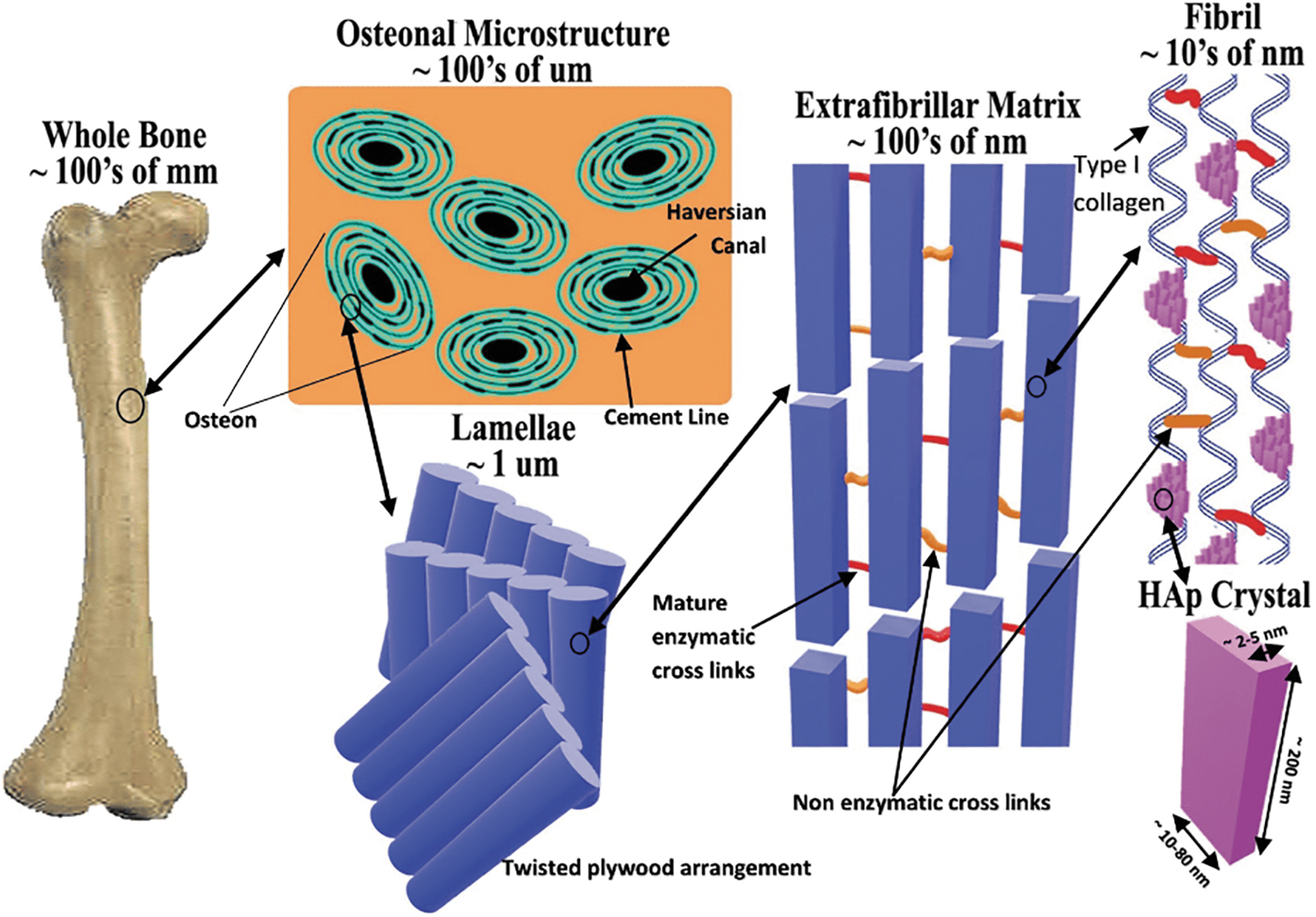
A microstructural representation of bone with size scales: Schematic illustrating the microstructure of and the distribution of HAp of nature bone. 15 Copyright © 2018 by the authors. Licensee MDPI, Basel, Switzerland (http://creativecommons.org/licenses/by/4.0/). HAp, hydroxyapatite.
Previous studies have shown that the addition of HAp to bioinks improves the compressive resistance of printed products to a certain extent, and exhibits excellent biocompatibility, which can enhance cell viability and osteogenic differentiation. However, HAp bioink has limited mechanical properties and poor antimicrobial activity, which restrict its application and clinical applications in bone tissue engineering. This review differs in focus from previous reviews by taking the new perspective of combining HAp with 3D bioprinting, summarizing more comprehensively in terms of different aspects of 3D bioprinting, such as printing technique and properties of bioink. And it covers new materials, in addition to the traditional ones in terms of hydrogel materials, such as decellularized matrices and plasma, which were not discussed in previous reviews. To facilitate the development of bone tissue engineering bioinks containing HAp in the future, this review also provides suggestions for possible improvements.
Construction Forms and Properties of Bioinks Containing HAp
Combined materials
Previous studies have combined HAp with various biomaterials such as natural polymers and synthetic polymers to prepare bioink (Fig. 2). These bioinks have been evaluated for their printability, mechanical properties, biocompatibility, and degradation properties. Table 1 presents the incorporate materials, form of HAp, composition of bioink, cells, and printing techniques used in different studies.
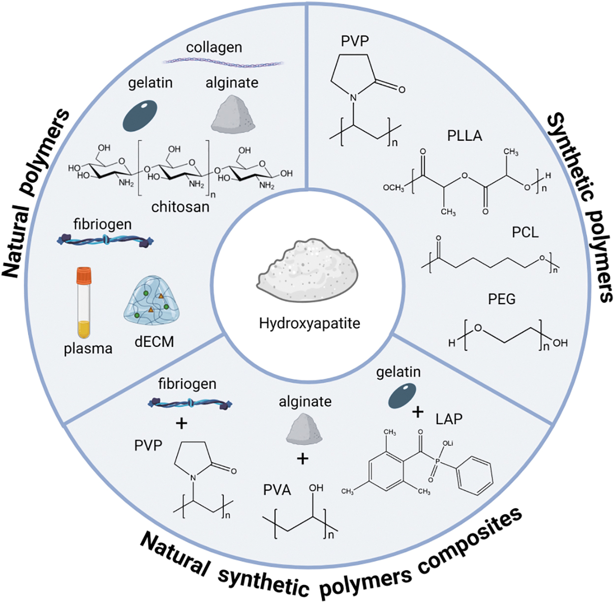
Combined materials of bioink containing HAp: Schematic illustrating three types of materials combined with HAp in three-dimensional bioprinting inks, including natural polymers, synthetic polymers, and natural synthetic polymer composites.
Summary of Bioink Containing Hydroxyapatite
ADA, alginate dialdehyde; CaSO4, calcium sulfate; CPC, calcium phosphate bone cement; CSP, core/sheath plotting; dECM, decellularized extracellular matrix; D1-MSCs EPO, D1 MSCs modified to produce erythropoietin hormone; ECs, endothelial cells; FS, fish scale; GelMA, Gelatin methacryloyl; GP, glycerol phosphate; HAM, methacrylated hyaluronicacid; HAp, hydroxyapatite; hASCs, human adipose-derived stem cells; hBMSCs, human bone marrow stromal cells; HBO, human-patient-derived osteoblast-like; hDPSCs, human dental pulp stem cells; hMSCs, human mesenchymal stem cells; HPDLSCs, human periodontal ligament stem cells; hOBs, human preosteoblasts; HUVECs, human umbilical vein endothelial cells; LAP, lithiumphenly-2,4,6-trimethylbenzoylphosphite; Na2HPO4, disodium phosphate; nHAp, nano-hydroxyapatite; pADSCs, Porcine adipose tissue-derived stromal cells; PEGDA, polyethylene glycol diacrylate; PG, Prionace glauca (a kind of blue shark); PLLA, poly(L-lactic acid); PTH, parathyroid hormone; PVA, polyvinyl alcohol; PVP, polyvinylpyrrolidone; QCS, quaterinized chitosan; SPIONs, superparamagnetic iron oxide nanoparticles; β-TCP, β-tricalcium phosphate; VP, vat polymerization-based.
Natural and synthetic polymers are common materials used in HAp bioinks. Common natural polymers include alginate, collagen, gelatin, cellulose, and chitosan (CS). Common synthetic polymers include polyvinylpyrrolidone (PVP), poly(L-lactic acid) (PLLA), polycaprolactone (PCL), polyethylene glycol, and polylactic acid. 12 Natural polymers are usually more biocompatible; however, the degradability, mechanical properties, and structural fidelity of their printed products are often limited.49,50 Synthetic polymers have better mechanical properties, but are difficult to stimulate cell proliferation and differentiation due to the lack of mediators inherent in the natural ECM. 51 To improve the performance of bioinks and ensure that the mechanical and biological properties match the target tissue, researchers often use hybrid materials to prepare bioinks so that multiple components complement each other and work together to form complex tissue structures. 50
The decellularized extracellular matrix (dECM) of natural polymers has received increasing attention. dECM refers to a natural biomaterial composed of ECM proteins, cytokines, polysaccharides, and others made from animal or human tissues and organs by removing all the cellular components and antigens in a certain way to preserve their 3D structure.52,53 dECM has good biocompatibility, tissue specificity, and biodegradability and is difficult to be mimicked by synthetic materials. It has been widely used in the fields of tissue engineering, tissue filling, and trauma repair.54,55
However, dECM has poor mechanical properties and needs to be combined with other materials such as PCL to enhance scaffold stability, 50 and the use of dECM as a scaffold coating is also a strategy. Kang et al. decellularized fresh porcine skins and mixed the dECM with gelatin, quaternized CS and nano HAp to make a bioink, and printed a 3D scaffold with a rich network of microchannels for craniofacial bone regeneration.
The results showed that the printed products had high porosity, degradation rate, antimicrobial activity, and biocompatibility. 38 Kumar et al. decellularized the human osteoblast cell culture medium and used the matrix as a coating material for the HAp scaffolds. The results of the study showed that the decellularized matrix on the surface of scaffolds increased cell adhesion and proliferation, and enhanced the expression level of myosin and albumin. 40 Similar results were reported in a study by Hwangbo et al. where porcine thigh bone was decellularized and used as a coating material for nano HAp-PLLA scaffolds, and the results of the study showed that the dECM-coated scaffolds exhibited more pronounced cell adhesion, cell proliferation, and osteogenic differentiation. 39
Plasma extracted from patients is highly specific and biologically active and has also been considered a substrate material for 3D bioprinting inks.56,57 Ahlfeld et al. prepared bioinks by mixing plasma with alginate, methylcellulose, and calcium phosphate bone cement, and the results of their study showed that the bioink print products containing plasma had good shape fidelity, cellular viability, and intercellular interactions, and bone progenitor cells containing plasma had significantly increased alkaline phosphatase (ALP) activity in the plasma-containing scaffolds compared to plasma free, 41 suggesting that the addition of plasma promoted osteogenic differentiation.
Growth factor-rich plasma (PRGF) contains a rich library of biomolecules, including platelet growth factor (platelet growth factor 4), platelet-derived growth factor (PDGF), epidermal growth factor, vascular endothelial growth factor (VEGF), and transforming growth factor (transforming growth factor beta), which are commonly used for accelerated tissue repair and regeneration. Anitua et al. prepared alginate-gelatin-conformant hydrogels bioink incorporated with HAp-PRGF, and compared the print products of three inks, gelatin and alginate (GA), gelatin and alginate enriched in HAp (GAHA) and gelatin and alginate enriched in HAp and PRGF (GAHAP). The results showed that the addition of PRGF significantly increased cell adhesion and chemotaxis, and the scaffolds containing HAp had a higher Young's modulus and significant cell proliferation and osteogenic differentiation, indicating better mechanical properties and cytocompatibility. 42
Bioink viscosity and HAp concentration
The viscosity of the bioink has an essential effect on printability and biocompatibility during the printing process, and it is important to note that these two properties are often contradictory in terms of viscosity (Fig. 3).
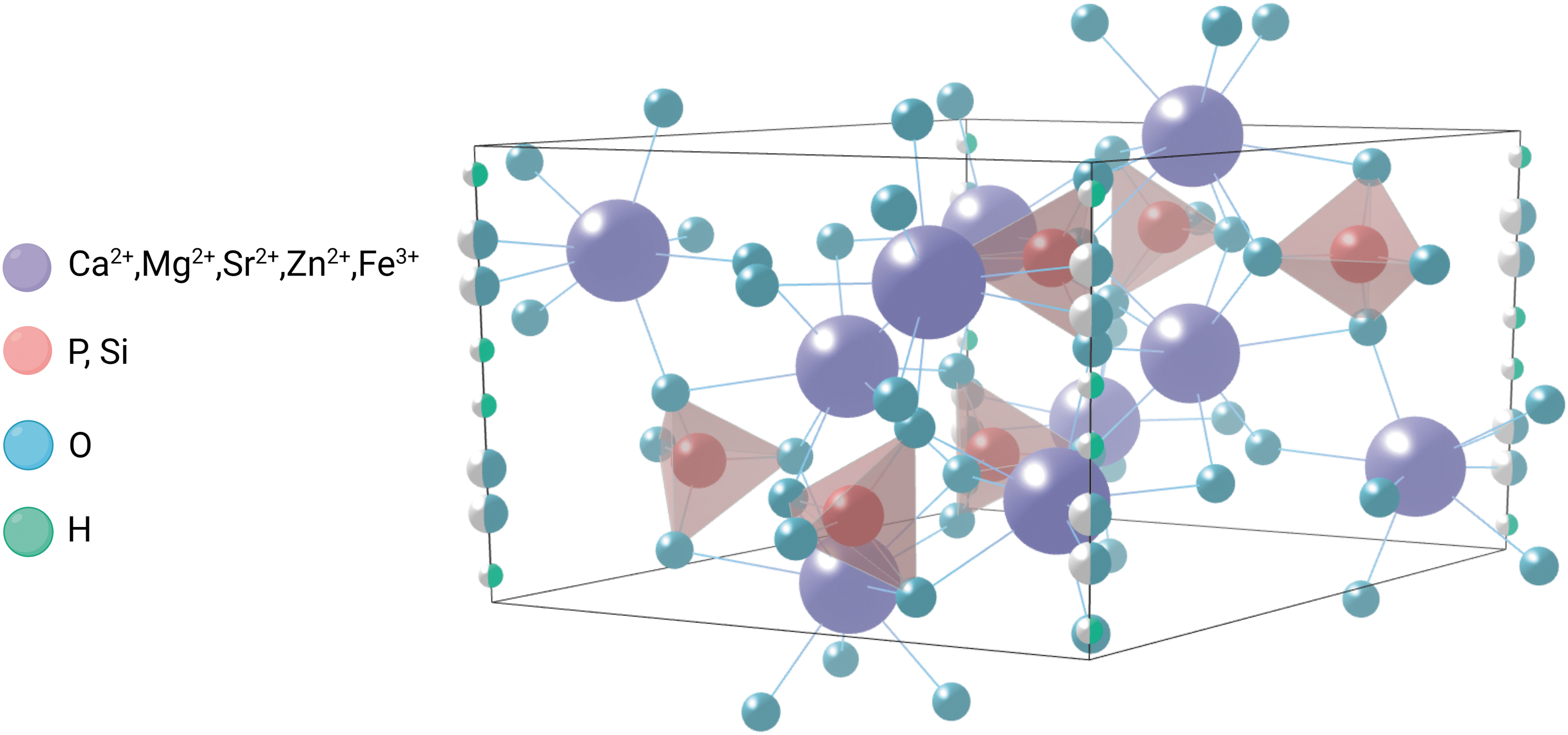
HAp crystal ion substitution: Schematic illustrating the crystal structure of HAp and ions can be used for substitution. Mg2+, Sr2+, Zn2+, and Fe3+ can substitute Ca2+, and Si can substitute P in PO43−.
Most of the current research with shear rate and viscosity testing adopted hydrogels composed by alginate or hybrid materials formed by alginate and other polymers. The addition of HAp increases the viscosity of bioinks, and we summarized the concentration of HAp and the corresponding viscosity of bioinks with best printability and optimal cell survival rates in different research. All the studies where viscosity testing was performed used extrusion-based bioprinting techniques, so printability needs to be considered in terms of keeping the viscosity within a range that prevents clogging of the nozzles, while ensuring structural integrity and accuracy.
Seok et al. used alginate at the concentration of 5 wt% to fabricate bioinks and tested the rheological properties at HAp concentrations of 0 wt%, 10 wt%, and 20 wt%, resulting in viscosity of 3486, 5712, and 6988 Pa∙s, respectively, with a shear rate of 0.1/s. The bioink with 20 wt% had best printability and cell survival. 34 Researchers fabricated hybrid bioink with alginate and Gel.
Wüst et al. developed hybrid materials with 2 w/v% alginate and 10 w/v% Gel and tested the rheological properties at HAp concentrations of 0 w/v%, 4 w/v%, and 8 w/v%. With a shear rate of 0.0001/s, the viscosity was ∼5.6 Pa∙s for 8 w/v% HAp, 3.4 Pa∙s for 4 w/v% Hap, and ∼2.3 Pa∙s for 0 w/v% HAp. The 8 w/v% HAp performance was best in biocompatibility with 15.2 ± 9.7% dead cell. 31 Tian et al. developed hybrid hydrogel of 3 w/v% sodium alginate, 7 w/v% Gel, and 5 w/v% Nano-hydroxyapatite (nHAp). The maximum viscosity is 248.3 Pa∙s when the shear rate was near 0/s, and the viscosity decreased as the shear rate increased. The fluorescence microscopy showed bioink with the addition of HAp had better cell survival than without. 32
Bendtsen et al. fabricated hybrid bioinks with alginate and PVP. The study showed that the addition of HAp increased viscosity, while still maintaining excellent biocompatibility with cell viability of ∼95%. 48
Modification of HAp
Isomorphic substitution of cations and anions can be performed without disrupting the crystal structure of the HAp unit, and substituted ions have similarities in size and charge with the substituting (Fig. 4). As a result of substitution, HAp has low crystallinity, small lattice, and increased solubility, which further affects the bone resorption process. 59 In addition, modification of HAp using oxides or reduced oxides and polymers has also been used to better properties of bioink. Table 2 summarizes the modifications of HAp and their effects on the properties of HAp bioinks.
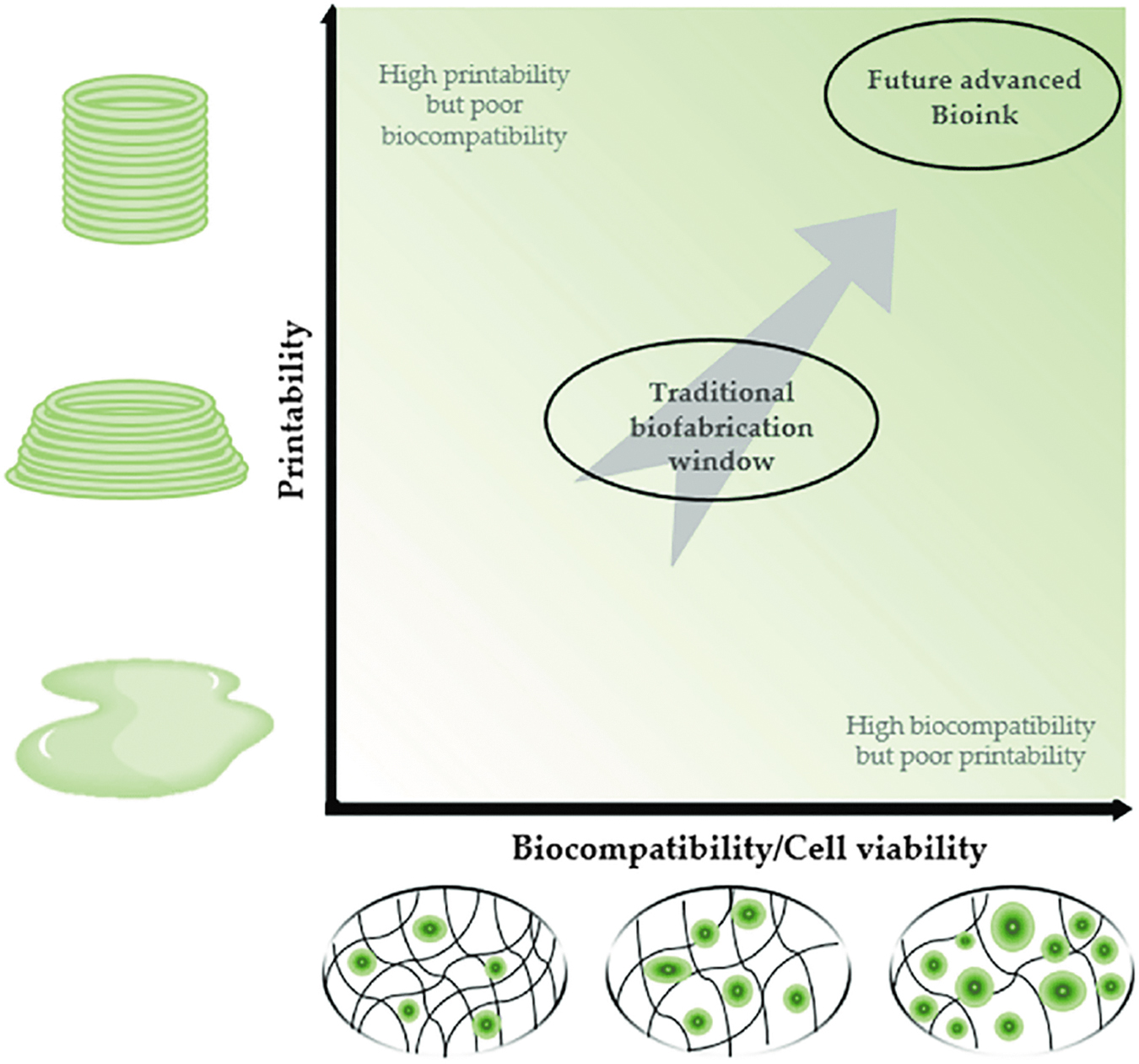
Printability and biocompatibility of bioink: Schematic illustrating the contradiction between bioink printability and biocompatibility/cell vatility. 58 Copyright © 2021 by the authors. Licensee MDPI, Basel, Switzerland (http://creativecommons.org/licenses/by/4.0/).
Modifications of Hydroxyapatite to Better Properties of Bioink
3D, Three dimensional; rGO, reductive graphene oxide.
3D bioprinting technique used to print bioink containing HAp in different applications
Jetting-based bioprinting uses piezoelectricity or heat to drive nozzle and divides bioinks into a series of droplets and manipulates the number of printed cell-laden droplets at specific spots/regions, including microvalve bioprinting, acoustic bioprinting, and laser-assisted bioprinting. The low cost and adjustable concentration of cells and biomaterials make jetting-based bioprinting an attractive technique; however, the inevitable sputtering of nozzle spraying causes the droplets to stray from the predetermined position, and this in turn lowers the precision of printing products. In addition, it is often necessary to use a large number of nozzles spontaneously because of the formation tiny droplets, which will form discontinuous areas between the droplets and weaken the mechanical properties. Therefore, the ink-jet bioprinting scaffolds are generally small in size. 84
Extrusion-based bioprinting uses a pneumatic or mechanic nozzle to extrude bioink in microfibers, the microfibers are deposited layer by layer to form a 3D structure. Researchers tend to adapt this technique in their studies due to its simplicity, affordability, and scalability. Compared to jetting-based bioprinting, extrusion-based bioprinting can achieve higher precision and print bioink containing higher concentration of cells and materials. One of the main limitations of this technique lies in the mechanical forces during bioprinting process; among these mechanical forces, shear stress is of a special significance and concern as it is considered the main cause of cell damage/death. 85
Vat polymerization printing uses different photoinitiators to promote the cross-linking of bioresin in the printing process to construct complex tissues with high separation rate. It includes stereolithography, digital light processing, and two-photon polymerization (Fig. 5). vat polymerization-based (VP) bioprinting can achieve nanoscale printing resolution with greater precision and accuracy than other bioprinting techniques, making it attractive for manufacturing release environments close to the complex ECM found in natural tissues. The viscosity of bioresin is the major difficulty of this technique. Bioresin with low viscosity is preferred because of its high speed in construction and simplicity in cleaning; however, low viscosity leads to unfavorable cell sedimentation and poor structure uniformity in printing products. 86
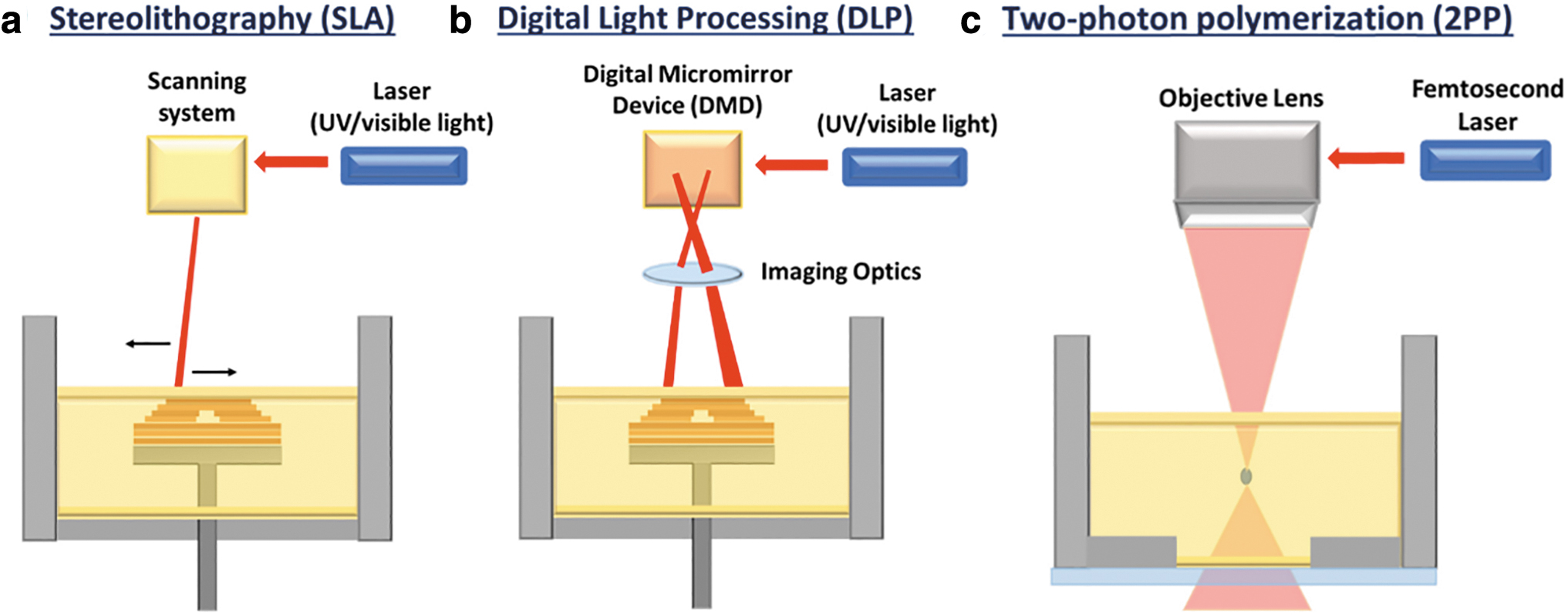
SLA, DLP, and 2PP bioprinting.
As we summarized in Table 1, extrusion-based bioprinting is the most frequently used technique of bioink containing HAp. Compared to jetting-based printing, extrusion-based printing can achieve higher accuracy as well as larger product sizes, and in addition, since HAp must be combined with other materials to prepare bioinks, extrusion-based printing's acceptance of multiple materials can better serve this point. Considering the high technical requirements of VP, further experimental studies on bioresins with high biosafety and appropriate viscosity are still needed before they can be widely published and disseminated.
Different 3D bioprinting techniques have different applications in combination with HAp. Extrusion-based bioprinting can be used for regeneration of bone and cartilage tissue, including compensating defects, osteoporosis, and arthritis. Animal experiments have demonstrated that HAp can promote osteoblast differentiation after lumbar spine surgery in mice,25,87 and mimic the heterogeneous structure of endochondral bone in rabbit humerus joints for the treatment of arthritis, 30 compensate for femoral midshaft bone defects in rats, 45 and promote bone regeneration in cranial defects in rats, 38 and that PLLA-HAp porous screws can immobilize grafted tendons in rabbits and promote osteointimal fusion at the tendon–bone interface. 88
Jetting-based bioprinting enables the variations of HAp concentration in different spots, and its precise control of components at specific locations allows for more bionic and natural variations than with extrusion-based core-sheath plotting, which is suitable for the fabrication of bone and cartilage at the interface. Christensen et al. developed collagen structures with a HAp content gradient by jetting-based bioprinting. 29 VP-based bioprinting enables the preparation of excellent porous scaffolds due to the nanoscale structural control, which is important for tissues and organs with demanding vascularization requirements.
Properties of bioink containing HAp
Advantages and mechanisms of bioink containing HAp
HAp improves the structure of printed products. The viscosity of bioink is the main factor affecting the printing process; if the viscosity is too high the hydrogel will be clogged, and if the viscosity is too low, the hydrogel cannot form the desired shape. 89 The addition of appropriate amount of HAp can keep the viscosity of the hydrogel within a reasonable range, so that the printed product has a complete and high-fidelity structure.
HAp can improve the mechanical properties of bioinks, thus compensating for the disadvantages of hydrogels in terms of mechanical properties. Most studies have shown that the addition of HAp leads to an increase in the compression modulus of the scaffolds compared to the control25,26,31,32,34; some experiments have confirmed an elevated maximum force for scaffold deformation, 31 suggesting that the scaffolds have better mechanical properties to withstand compressive forces and maintain the stability of the structure. The possible mechanism lies in the formation of new chemical bonds, such as hydrogen bonds. The fracture energy increases after HAp binds to the hydrogel compared to pure hydrogels. 17
The incorporation of HAp resulted in a high degree of swelling of the printed product,26,37,48 which was related to the hydrophilicity of HAp, and the higher degree of swelling indicated that the scaffolds had good water absorption and provided good nutrient transport for the subsequent proliferation and differentiation of cells. Some studies have combined HAp with sodium alginate to prepare bioinks, and the results show that the appropriate amount of HAp can keep the degradation rate of the scaffolds within a reasonable range, which is compatible with the rate of tissue regeneration in vivo. Some researchers pointed out that HAp may replace the sodium ion in sodium alginate and avoid the degradation of sodium alginate. 26
HAp improves the biocompatibility of bioinks, enabling the use in animal experiments. HAp improved osteogenic differentiation and resulted in high expression of osteogenic genes such as osteocalcin and osteopontin and high ALP activity,28,32,34,45 providing strong evidence that HAp promotes osteogenic differentiation. The inherent osteoinductive effect of bioceramics, such as HAp, has been widely accepted; however, the intrinsic mechanism still needs to be further investigated. Studies in the field of osteoimmunology have shown that immune cells play an ample role in maintaining bone homeostasis and regulating bone reconstruction, 90 and the immune and skeletal systems share many regulatory molecules, including cytokines, chemokines, receptors, and transcription factors. 91
Macrophages and T cells are important cells of the immune system that secrete a variety of cytokines to regulate the inflammatory response, and it has been demonstrated that HAp can cause macrophages to elongate in a spindle-like shape and promote macrophage polarization, 92 and macrophage polarization has been shown to affect the osteogenic differentiation of bone marrow stromal cells, which in turn affects the outcome of bone regeneration.93,94
Furthermore, it has also been demonstrated that HAp increases the number of T cells and induces IL-22 production, 95 and not only do T cells have a recruiting effect on MSCs but T cell-derived cytokines have also been shown to play an active role in bone regeneration.96–98 The possible mechanism by which HAp promotes bone differentiation is to promote the proliferation, maturation, and activation of immune cells and secretion of cytokines, through which osteogenic differentiation is induced. Bioinks containing HAp in studies have not only shown higher osteogenic differentiation and cell survival but also more significant biological behaviors such as metabolism, proliferation, and migration.25,26,31,32,34–37,45,46 These biological effects are closely related to the fact that HAp increases the roughness and surface contact area of the scaffold, both of which are altered to promote cell adhesion as well as cell-cell interactions.36,37,43,45
Limitations and challenges of bioink containing HAp
HAp bioink faces constraints between biocompatibility and structural precision and stability. The increase in viscosity caused higher shear stress during the printing process,31,32 which is detrimental to the survival of the cells. To overcome this problem, bioinks must bear strong shear thinning behavior to free cells from the high stresses, 99 and can quickly recover viscosity after shear removal to make sure the structure fidelity. 100
Since HAp is an inorganic material, the combination with organic polymers often results in poor mechanical properties and structural stability due to the difference in polarity; therefore, the products cannot be used for the fabrication of mechanically safe load-bearing bulk implants or prostheses and are limited to biofunctionalized coatings for metal implants and porous scaffolds for low- or no-load bone defect restoration.101,102
Antimicrobial activity of HAp bioink needs to be improved. Bacterial infections of HAp implants are frequent in recent years and can lead to serious postoperative complications. Due to the inflammatory response after infection, the pH value of the peri-implant microenvironment decreases, and the interface between the implant and human tissues is loosely bound, which reduces the long-term stability of the implant. 103 Besides, the acidic degradation products of polymers may lead to local inflammation and infection, which may ultimately lead to implant failure. 104
The mimicry of the in vivo tissue using HAp bioink still needs to be further improved. Bone growth is closely related to angiogenesis, and HAp does not directly stimulate angiogenesis, which is often achieved by adding different growth factors such as VEGF and PDGF; however, these growth factors may lead to ectopic or unintended bone formation, so HAp composites need to be further investigated in promoting bone angiogenesis. 105 The imitation of arterial, venous, and capillary networks and functions compatible with organism such as homeostasis regulation has not yet been achieved.11,104
There are issues with immune response and cellular biology at the postprocessing node; however, these cannot be insightfully recognized and resolved until the print product has been implanted in the patient, and the long-term effects of the print product in the patient remain to be further investigated.104,106 To make the results more instructive, it is important to conduct a large number of toxicity and safety tests using nonhuman primates that are highly similar in structure and function to humans 107 ; however, ethical controversies in primate experimental animals have limited many animal tests.106,107
Prospect and Outlook
To prepare ideal 3D bioprinting inks containing HAp for bone tissue engineering, the following improvements can be considered.
Combined materials
The performance of HAp bioinks can be improved by innovating combined material forms.
Commonly used natural polymers have different properties and can be selected or synthesized as hybrid hydrogels to meet the criteria of different target scaffolds. Silk fibronin (SF) have excellent rheological properties (shear thinning and fast thixotropic recovery) and good mechanical properties, which can significantly improve the brittleness of HAp crystals, as well as high biocompatibility and low immunogenicity, which can be an alternative material to other hydrogels. 46 CS has good antibacterial and antifungal activity, and its chemical structure is similar to that of ECM from components, which has been widely used in dentistry and bone tissue engineering. 108
To further improve the cell proliferation and differentiation of HAp ink scaffolds, collagen material is a suitable choice, collagen is extracted from various animal tissues and contains arginine-glycine-aspartic acid sequences, which can provide cellular recognition sites and facilitate cell attachment. 109 Alginate bioinks tend to have poor mechanical properties, and to compensate for this, a study prepared hybridized hydrogels by adding gelatin to alginate, achieving transient stability, maintaining the ability of the printed hydrogel to form filaments, allowing greater structural fidelity, and improving the degradation rate of the hydrogel. 31
Synthetic polymers can be chemically modified to selectively synthesize target scaffold materials with better mechanical properties, but poor biocompatibility. Some studies have combined natural and synthetic polymers to make full use of the advantages of both materials and compensate for the disadvantages to design innovative composite bioinks.46–48 Composite bioinks generally exhibit good printability, mechanical properties, and biocompatibility.
Plasma and decellularized matrices have also received widespread attention due to their higher degree of mimicry of the in vivo microenvironment due to their richness in biofactors and other substances. Bioinks combining plasma and decellularized matrices exhibit higher osteogenic differentiation activity, angiogenic activity, and hemocompatibility.38–42
In addition, Hwangbo et al. have used thermal annealing and in situ plasma processes to improve the stability of the HAp and bonding material combination and enhance the mechanical properties of the scaffolds. 39
Modification of HAp
Modification of HAp has been widely studied and some of them have led to sufficient improvements in the properties of HAp; among them, certain modifications have not been applied in 3D bioprinting. It has been reported that loaded reductive graphene oxide can improve the adhesion of BMMSCs and promote the proliferation and spontaneous osteogenic differentiation of BMMSCs, and the degradation rate of scaffolds is matched with the rate of new bone generation. 76 Silver cation substituted-HAp enhances the antimicrobial activity in a manner that is not dependent on antibiotics to prevent Acinetobacter baumannii infections in orthopedic or dental implants. 70 Lanthanide substitution can provide HAp with higher stiffness and electrical conductivity, and promote osteoconductivity and angiogenesis, reduce osteoclastogenesis, decrease reactive oxygen species production, promote anti-inflammatory macrophage phenotypes, and confer antimicrobial properties to HAp. 102
Bioactive factors
The addition of bioactive factors can enhance osteogenic and angiogenic activity. The most commonly used bioactive factors that have been studied include bone morphogenetic protein-2 (BMP-2) and VEGF, and some studies have shown that nerve growth factor (NGF) has a potential role in bone repair.110–114 Fitzpatrick et al. prepared HAp bioinks with the incorporation of BMP-2, VEGF, and NGF, and the results of their study showed that genes related to osteoblast differentiation were upregulated after the addition of the growth factors, resulting in the upregulation of osteoblast differentiation-related genes. 115 In one study, parathyroid hormone (PTH) was combined with HAp bioink, and the results showed that PTH combined with different biochemical cues could promote chondrogenesis or osteogenesis, and rabbit synovial joint scaffolds with heterogeneous structure of exochondral endosteum were successfully fabricated. 30
Bioprinting techniques
The advantages of jetting-based printing in terms of precise distribution of components should not be overlooked. Christensen et al. used jetting-based printing in their study, which enabled control of HAp density in different layers for gradient printing. 29 However, this study did not perform in vivo experiments, so it is not yet sufficient to illustrate its applicability in bone tissue engineering.
Highly accurate VP bioprinting benefits the fabrication of porous scaffolds to promote biocompatibility. Zhou et al. fabricated scaffolds with VP bioprinting and the resultant scaffolds provide a highly porous and interconnected 3D environment to support cell proliferation 43 ; however, the aggregation of nHAp was observed. Further studies are still needed to investigate the relationship of nHAp aggregation and effects of bioresin viscosity and pi in VP bioprinting.
Conclusion
Bioinks containing HAp with excellent printability and biocompatibility are potential materials for tissue engineering and regenerative medicine. Due to the unsatisfactory mechanical properties and poor antimicrobial activity of printed products, as well as the limitations in the experimental process, bioinks containing HAp have not been widely used in clinical trial. The use of hybrid binding materials with improved properties, enrichment of HAp modifications, incorporation of bioactive factors into bioinks can significantly improve the product properties. Future research could focus on improving the properties of Hap bioink, as well as enhancing 3D bioprinting technology to promote vascularization of printed products to increase the degree of biomimicry and achieve other physiological functions.
Footnotes
Disclosure Statement
No competing financial interests exist.
Funding Information
This study was funded by Beijing Municipal Natural Science Foundation (Grant No. 7232201) and National Natural Science Foundation of China (Grant No. 81873939).