
Abstract
Recent studies have shown potential ways for improving stem cell cryopreservation. The major need for autologous stem cell use is a long-term storage: this arises from the humans' hope of future use of their own cells. Therefore, it is important to evaluate the cell potential of vitality and differentiation before and after cryopreservation. Although several studies have shown a long-term preservation of adipose tissue, a few of them focused their attention to stem cells. The aim of this study was to evaluate the fate of cryopreserved stem cells collected from adipose tissue and stored at low a temperature in liquid nitrogen through an optimal cryopreservation solution (using slowly cooling in 6% threalose, 4% dimethyl sulfoxide, and 10% fetal bovine serum) and to develop a novel approach to efficiently preserve adipose-derived stem cells (ASCs) for future clinical applications. Results showed that stem cells, after being thawed, are still capable of differentiation and express all surface antigens detected before storage, confirming the integrity of their biology. In particular, ASCs differentiated into adipocytes, showed diffuse positivity for PPARγ and adiponectin, and were also able to differentiate into endothelial cells without addition of angiogenic factors. Therefore, ASCs can be long-term cryopreserved, and this, due to their great numbers, is an attractive tool for clinical applications as well as of impact for the derived market.
Introduction
Cryopreservation of cells and tissues has been improved, 14 but, up to now, mainly hematopoietic stem cells have been extensively cryopreserved15,16 and then successfully used for transplantation.17,18 Dental pulp stem cells have also been reported to be successfully cryopreserved for long term. 19 Regarding the adipose tissue, previous studies have described long-term preservation of adipose aspirates after lipoplastic surgery 20 for autologous fat transplantation and proliferative capacity of human ASCs after long-term cryopreservation using standard protocols.21,22 Fat grafting is a common cosmetic procedure performed by plastic surgeons because it is still considered an ideal soft tissue filler23,24 to rejuvenate the face, hands, or other parts of the body secondary to aging. Fat grafting has also been used to correct irregularities or various soft tissue deficiencies of congenital, posttraumatic, or iatrogenic defects. 25
The modern technique of cryopreservation of adipose cells or tissue 26 allows to a long-term storage of living cells and tissue that may have many potential clinical applications. Prefreeze processing, cryosubstrate variation of temperature and shelf-life, and postfreezing procedure associated with reliable and automated cooling methods must be taken into consideration to establish banking of MSCs.
The greatest challenge during cellular cryopreservation is the lethality of the cooling and thawing processes. Optimization of cryopreservation protocols to maintain the quality of stem cells is a critical task for their banking.
For cryopreservation, dimethyl sulfoxide (DMSO) is used as the most common cryopreservant for cells. Usually, the cell freezing requires the use of a solution composed of 10% DMSO and 90% fetal bovine serum (FBS). DMSO prevents the formation of intracellular ice, but it is known to be toxic at room temperature. Therefore, DMSO has to be removed after thawing by several washing steps. It might be important to develop DMSO-free preservation methods or at low concentration of DMSO, and choose the best alternative for a standard protocol. Trehalose is a nontoxic disaccharide of glucose that possesses a good ability to stabilize and preserve cells and cellular structures during freezing.27,28 Both threalose and DMSO are used in solution for organ protection before transplants. There are two main theories regarding how trehalose works within the organism in the state of cryptobiosis: the vitrification theory, a state that prevents ice formation, and the water displacement theory, whereby water is replaced by trehalose, although it is possible that a combination of the two theories is at work. 29
The purpose of this study was to test different combinations of DMSO and threalose to lower the DMSO concentration, obtain the better vitality of ASCs without injury of cell membrane and intracellular ice formation, and maintain their stemness and differentiation characteristics. ASCs were cryopreserved and recovered up to 1-year period. Our results have demonstrated that a better freezing solution is composed of 4% DMSO, 6% threalose, and 90% FBS.
Materials and Methods
Adipose tissue extraction, digestion, and cell culture
Subcutaneous adipose tissue of abdominal and mammary origin has been obtained with informed consent from 25 women patients (mean age, 31 ± 0.8 years; mean BMI, 26 ±1.1 kg/m2) who have endured elective procedures of plastic surgery. The adipose tissue has been obtained by lipectomy or liposuction in the Plastic and Reconstructive Surgery of the Second University of Naples. The adipose tissue has been placed in a physiological solution (NaCl 0.9%). It has been washed two times in phosphate buffered saline (PBS: NaCl 137 mM, KCl 2.7 mM, Na2HPO4 10 mM, and KH2PO4 1.8 mM), chopped into pieces, and digested by type I collagenase (Invitrogen, San Giuliano Milanese, Milan, Italy), 3 mg/mL and dispase (Invitrogen) 4 mg/mL in PBS for 60 min, under permanent shaking in a water bath at 37°C. Digestion was stopped by adding Dulbecco's modified Eagle's medium (DMEM) containing 10% FBS followed by an incubation in erythrocyte lysis buffer (NH4Cl 155 mM, KHCO3 10 mM, and EDTA 0.1 mM, pH 7.3) for 10 min at room temperature.
Cell suspension was centrifuged (for 7 min at 1300 rpm), and the pellet was immersed in DMEM, with 10% FBS, L-glutamine 2 mM, penicillin 100 U/mL, and streptomycin 100 μg/mL, and placed in 25 cm2 flasks with filtered valves. Flasks were incubated at 37°C in a 5% of CO2, and the medium was changed twice a week. Just before cells become confluent, they were subdivided in new flasks.
Cryopreservation technique
In this study, DMSO, a permeable cryoprotective agent that could reduce cell injury due to the intracellular ice formation, and trehalose, a nonpermeable cryoprotective agent that could protect the membrane, were used. Cells (stocks of 500,000 cells, as described by Goh et al. 30 ) were resuspended in 1%, 4%, 8%, and 10% DMSO (Sigma, Milan, Italy) and in 9%, 6%, and 2% trehalose (Sigma) plus 90% FBS into standard 1.8 mL cryotubes (Barloworld Scientific Italia, Riozzo di Cerro al Lambro, Milan, Italy) and frozen in liquid nitrogen. After extraction and enzymatic digestion from adipose tissue, cells were frozen for 1, 6, and 12 months without passage in culture.
Freezing and thawing protocol
Cells were paced in 1.8 mL cryotubes and mixed with 1 mL of four combined freezing solutions: (i) 1% DMSO + 9% trehalose + 90% FBS; (ii) 4% DMSO + 6% trehalose + 90% FBS; (iii) 8% DMSO + 2% trehalose + 90% FBS; (iv) 10% DMSO +90% FBS. After adding cryoprotective agents, the cryotube was placed at −20°C for 30 min, then at −80°C for 1 h, and transferred into liquid nitrogen (−196°C) for long-term cryopreservation. This procedure prevents the formation of ice crystals inside the cell because freezing occurs slowly.
The cells were thawed in culture medium at 10% FBS without the use of the water bath, and immediately centrifuged. Then, they were washed two times in culture medium, placed in flasks, and incubated at 37°C with 5% CO2 in humidified atmosphere, and the medium was changed twice per week. This thawing protocol prevents the toxicity due to the DMSO, as the thawing occurs quickly in culture medium at room temperature.
Fluorescence-activated cell sorting
Cells were detached using a solution of trypsin–EDTA (EDTA 200 mg/L and trypsin 500 mg/L; Lonza, Milan, Italy) and pelleted (7 min at 1300 rpm), washed in PBS at 4°C, and then incubated with primary antibody at the concentration of 5 μg/μL in PBS for 30 min at 4°C in the dark. After washing twice in PBS, cells were subsequently incubated with a secondary antibody at the concentration of 8 μg/μL in PBS, or, alternatively, they were incubated directly with fluorescent antibody already conjugated at the concentration of 2.5 μg/μL in PBS for 30 min at 4°C. Phenotypic analysis of the stem cell population was performed at day 7, and after day 30 of culture by a FACS Vantage cell sorter (Becton & Dickinson, Mountain View, CA), using anti-CD34 FITC and PE (Miltenyi–Biotech, Calderara di Reno, Bologna, Italy), anti-CD90 FITC (Chemicon, Prodotti Gianni, Milan, Italy), anti-CD29 Cy (BD Pharmingen, Buccinasco, Milano, Italy), anti-VEGF (Santa Cruz Biotechnology, Santa Cruz, CA), anti-VEGFR-2 (Santa Cruz), anti-CD54 PE (Miltenyi–Biotec), anti-CD44 FITC (Miltenyi–Biotec), anti-CD45 Cy and PE (BD Pharmingen), and anti-CD14 PE (Miltenyi–Biotec). Cells were sorted using CD34 and CD90 markers. Isotype-matched antibodies and nonprobed cells were used as control.
Proliferation assay and growth curves
After thawing, cells were plated at a density of 6.0 × 10³ cells/cm2 in six-well plates. Every 12 h, cells were harvested and re-suspended in PBS. An aliquot of cell suspension was counted under a microscope at 200 × magnification. The number of viable cells for each experimental condition was counted and represented on a linear graph. The doubling time (DT) was determined from the growth curves or using the formula:
where t and t0 are the times at which the cells were counted, and N and N0 are the cell numbers at times t and t0, respectively. Fresh cells were used as controls.
Apoptosis assay
To analyze the apoptosis, thawed cells were processed using the Annexin V- FITC Apoptosis Detection KIT (BD Pharmingen). Fresh cells were used as controls.
Adipocyte differentiation
To induce adipogenesis, fresh and thawed ASCs were cultured in DMEM with 10% FBS, 1% penicillin 100 U/mL and streptomycin 100 μg/mL, dexamethasone (1 μM; Sigma), human recombinant insulin (10 μM; Sigma), indomethacin (200 μM; Fluka, Milan, Italy), and 3-isobutyl-1-methyl-xantina (0.5 mM; Sigma) for 21 days. The medium was changed twice a week. ASCs cultured in basal medium have been used as controls.
Immunohistochemistry
Cells were fixed in 4% paraformaldehyde for 15 min at room temperature. They were then washed twice with PBS and covered with kit Dako Cytomation (En Vision + System-HRP—AEC) according to the use protocol. Antibodies were the following: adiponectin (diluted 1:200 in PBS), PPARγ (diluted 1:50 in PBS), and VEGF (diluted 1:100 in PBS) (all purchased from AbCam, Cambridge, UK).
RNA isolation and polymerase chain reaction
RNA was extracted with TRI reagent (Sigma). cDNA synthesis was lead on total RNA by SuperScript II reverse transcriptase (Invitrogen). Following are the used primer sequences:
GADPH: fw AGCCGCATCTTCTTTTGCGTC; rw TCATATTTGGCAGGTTTTTCT VEGF: fw TGACAGGGAAGAGGAGGAGA; rw CGTCTGACCTGGGGTAGAGA PPARγ: fw ACAGCAAACCCCTATTCCATGC; rw ATTACGGAGAGATCCACGGAGC Adiponectin: fw CAACATTCCTGGGCTGTACT; rw CCTGTGAAGGTGGAGTCATT
Statistical analysis
Student's t-test (two-tailed) was used for statistical evaluation. Level of significance was set at p < 0.05.
For each donor, all the experiments described were performed in triplicate.
Results
Freezing and thawing protocol
To assess which protocol for freezing could be better to be used, we made four types of combined freezing solutions with DMSO–trehalose–FBS, as described in Materials and Methods. Better freezing solution was composed of 4% DMSO, 6% threalose, and 90% FBS compared to all the other protocols. In fact, thawed cells showed a greater vitality and increased efficiency of differentiation and levels of antigen expression close to those obtained from freshly isolated cells. The concentration of 1% DMSO and 9% threalose induced a high cell death, most probably due to intracellular ice crystal formation. The concentration of 8% DMSO and 2% threalose led to viable cells but with an initial delay in proliferation (10–11 days). The same results were obtained using 10% DMSO.
Isolation and morphological analysis of ASCs after thawing
ASC cryostorage length was performed for 1, 6, and 12 months. When thawed, stem cells were recovered, and we found a level of viability >80% for cells frozen for 1–6 months, while cells frozen for a year showed a viability of 70%. Cells were plated in basal medium (DMEM), and we observed that they adhered and re-started to proliferate in 48–72 h after plating. After the first passage of culture, cells proliferated slowly with respect to freshly isolated ones (p < 0.05) and gave rise to confluence in 7–9 days. The ASCs formed adherent heterogeneous cell population, which consisted of polygonal and spindle-shaped cells. After two passages of culture, the heterogeneous population acquired a fibroblast-like shape, similar to fresh-derived stem cells, at this time (Fig. 1A, B).
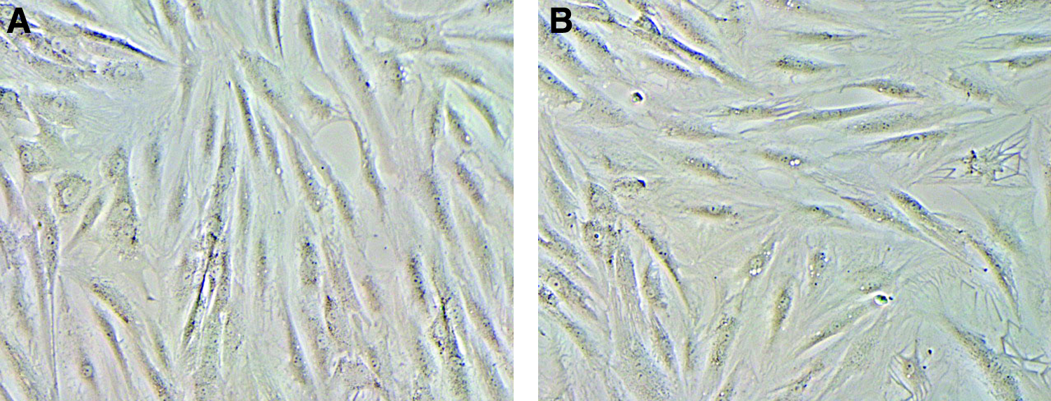
Representative image of adipose-derived stem cells (ASCs) in culture at day 21: (
Immunophenotypes and differentiation of ASCs before and after cryostorage
To evaluate surface antigens expressed by these cells, before and after the freezing process, we performed flow cytometric analysis. For this purpose, the cell-surface antigen profile of cryopreserved ASCs was analyzed and compared with that of the freshly cultured ASCs. We used the following antibodies: anti-CD34, marker of stromal progenitors, and anti-CD90 and anti-CD105, markers of MSCs. Moreover, we also analyzed anti-CD44, anti-CD54, anti-VEGF, and anti-VEGFR2 (Flk-1), which are endothelial markers. Cytometric analyses (Table 1) showed that the mean positivity levels for CD34, CD90, CD105, CD29, CD44, CD54, VEGF, and VEGFR2 were comparable to those found in our previous experiments performed with fresh cells (Fig. 2A, B). Cells were negative for CD14 (monocyte antigen) and CD45 (leukocyte common antigen).
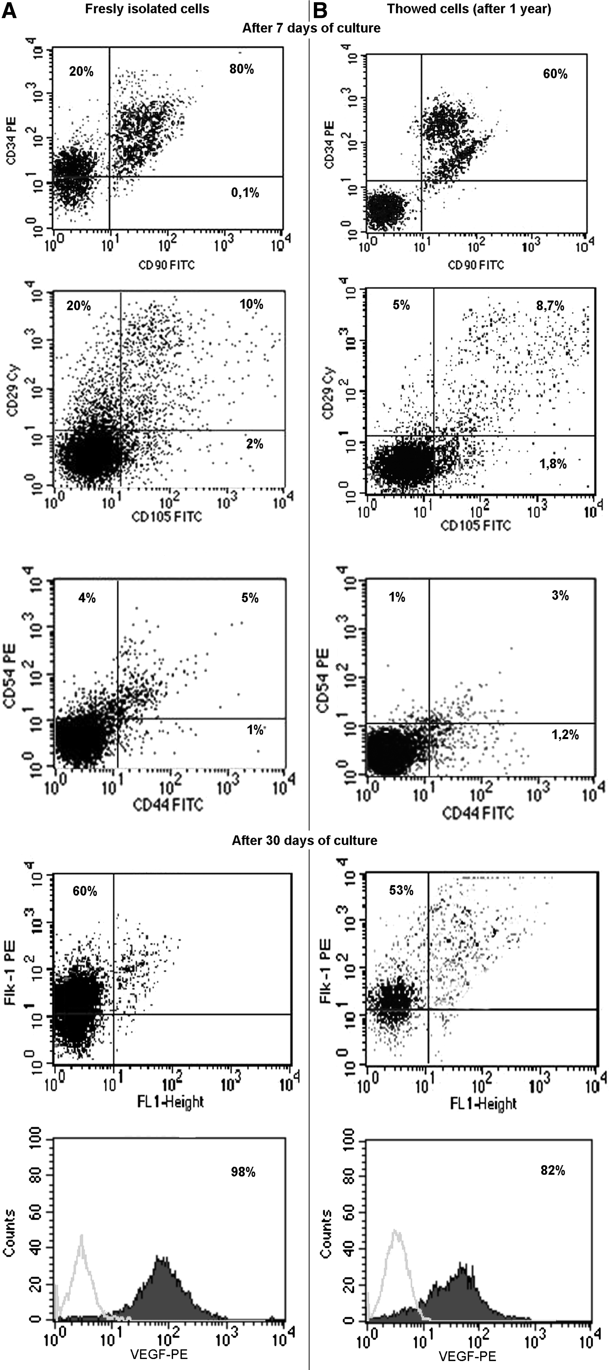
Representative fluorescence-activated cell sorting of ASCs for CD34, CD90, CD105, CD29, CD44, and CD54 after 7 days of culture, and VEGFR2 (Flk1) and VEGF after 30 days of culture (
Markers levels are mean percentage of positive cells ± SD. CD34, CD90, CD105, CD29, CD44, and CD54 were analyzed after 7 days from thawing. VEGF and VEGFR2 were analyzed after 30 days from thawing.
Proliferation and apoptotic assays
The time needed for recovery and proliferation was of about 48 h. We performed apoptosis assays at 1, 6, and 12 months from freezing, and compared it to fresh stem cells (Fig. 3A) using the Annexin/PI kit. Cells positive only for PI are considered to be necrotic, cells positive only for annexin are considered to be in early apoptosis, cells positive for both annexin and PI are considered to be in late apoptosis, while cells negative for both annexin and PI are alive. Results showed that the cells thawed after 1 month (Fig. 3B) from freezing were positive for PI (7%), while cells positive for annexin were 0.12% and cells positive for both PI and annexin were 0.34%. Cells negative for PI and annexin were 92.5%. At the sixth month (Fig. 3C) from freezing, thawed cells positive for PI were 14%, while cells positive for annexin were 1.01% and cells positive both for PI and annexin were 0.34%. Cells negative for PI and annexin were 84.6%. Twelve months (Fig. 3D) from freezing, thawed cells positive for PI were 20%, cells positive for annexin were 1.11%, and cells positive for both PI and annexin were 10%. Cells negative for PI and annexin were 70% (Table 2). Thawed ASCs showed high proliferation rate starting from the third passage of culture. In fact, growth curves were similar when fresh cells were compared with thawed cells, and there were no significant differences in doubling time of fresh cell samples with respect to thawed cell samples after three passages of culture (Fig. 4).
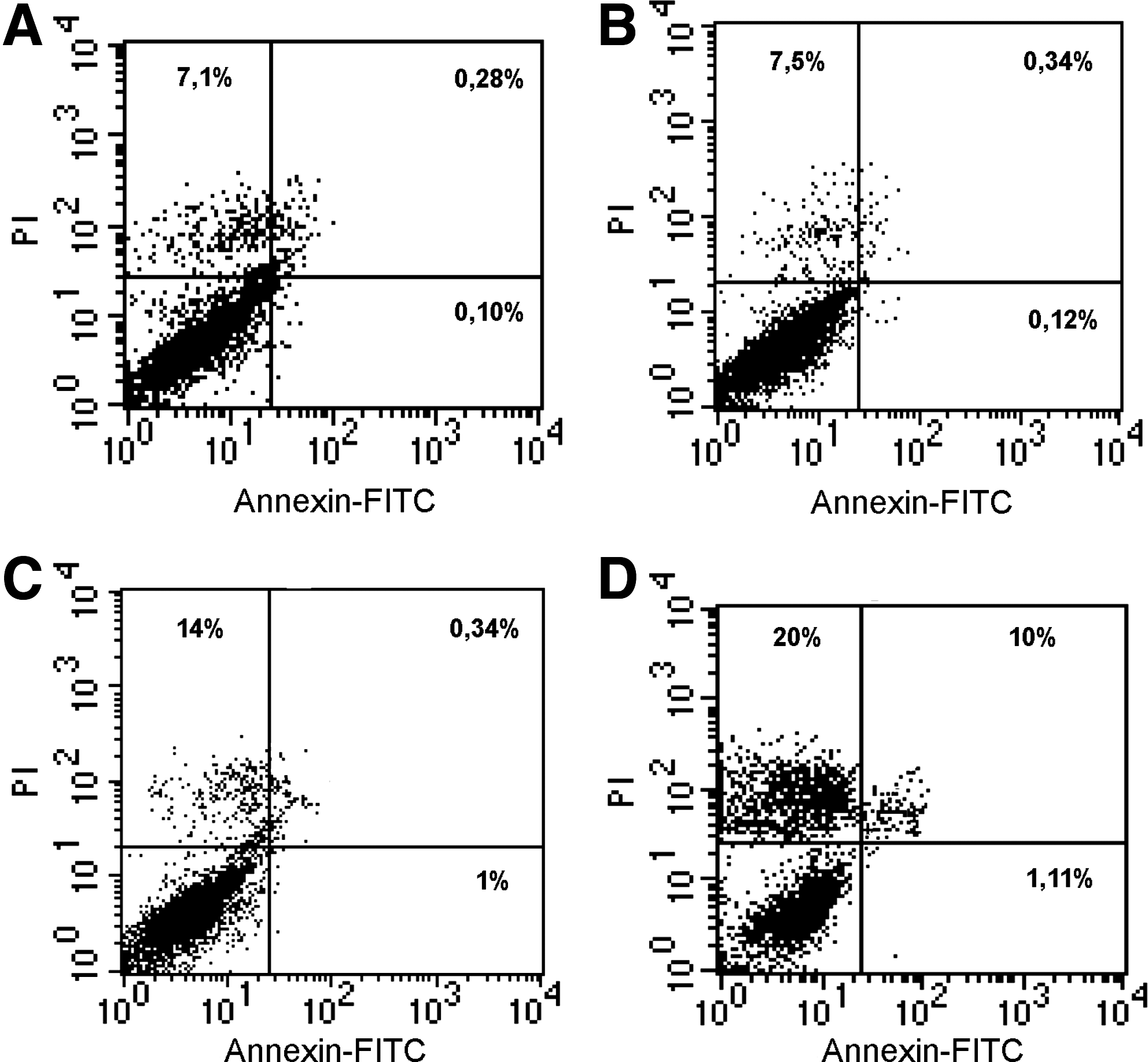
Cytometric analysis of ASCs apoptosis, performed using PI and annexin. The image shows the level of viability of cells frozen for (
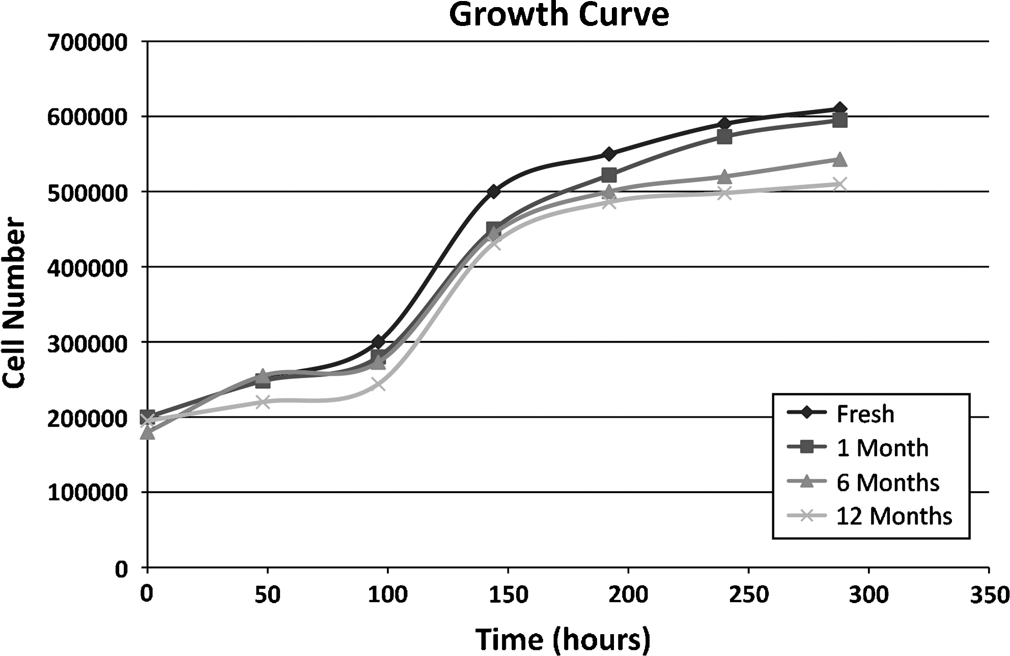
Representative image of growth curves of ASCs thawed at 1, 6, and 12 months with respect to fresh cells, showing that doubling time does not change.
Values shown are mean percentage of positive cells ± SD.
Differentiation of cryopreserved ASCs in adipocyte
To evaluate the differentiation potential of cryopreserved adipose tissue–derived cells into mesenchymal lineages, we grew these cells in adipogenic induction media and examined the formation of multilipid vacuoles. ASCs were positive for specific markers, including PPARγ and adiponectin, at the immunohistochemistry staining and mRNA expression pattern (Fig. 5D, E) compared to freshly isolated ASCs grown in adipogenic medium (Fig. 5A, B).
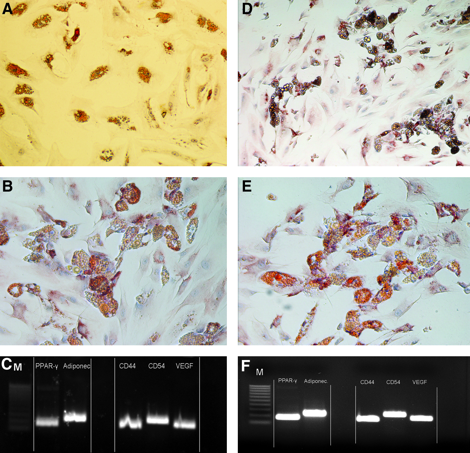
Representative image of immunohistochemistry performed on freshly isolated stem cells under adipogenic induction for (
Differentiation of cryopreserved ASCs in endothelial cells
To assess the differentiation of ASCs into endothelial cells, we grew the ASCs in standard medium, and, after 40 days, thawed cells were able to differentiate into the endothelial lineage as confirmed by cytometry and by m-RNA transcript levels for CD54, CD44, and VEGF and (Fig. 5F) compared to freshly isolated ASCs grown in standard medium (Fig. 5C).
Discussion
Human adult stem cells have an extraordinary plasticity. These cells retain their multilineage potential.11,31 Thus, studies on the long-term storage are of critical importance, mainly when related to ASCs due to the considerable amount of these cells after collection. In addition, we have observed that, up to now, studies focusing on ASCs' long-term preservation using DMSO and trehalose have not been performed, except those regarding cryopreservation of adipose aspirates20,26 from conventional lipoplasty.
Robust, secure and monitored process of isolated stem cells will prevent possible change of their behavior after cryostorage or cell death by intracellular ice formation during the freeze–thaw process. Optimal customized processing and cryosubstrates for isolated clinically relevant stem cells must be provided and examined. Also, to prevent the cooling chain interruption on a classic sample extraction tank, it is necessary to have a cover-cooled system. This will result in a final comparison of all protocols and data for new regulatory procedures for preparation and long-term storage of clinically relevant adult stem cells. The final result is to generate stem cell–derived cells that meet the criteria for good manufacture practice and standards for clinical use in regenerative medicine.
Our aim was, therefore, to partially replace DMSO as traditional cryopreservant by adding the trehalose. To do so, this different cryopreservation medium and a new hypothermic medium have been tested. Existing protocols have been adapted to ensure optimal procedures allowed for clinical applications of cryopreserved ASCs without altering the potency of the stem cells.
To allow this prolonged storage, during the cooling procedure, cells slowly avoid accumulation of intracellular ice, which can cause rupture of the cell membrane. However, the cellular dehydration can compromise the membrane integrity. Several researches investigated the rates of ASCs freezing in the presence and absence of cryoprotective agents, including DMSO and glycerol. In particular, it has been shown that an optimal rate of freezing for ASCs ranged from 25°C/min to 60°C/min, for P0 (P is an acronym for “cell passage” that indicates that cells are detached when at confluence) ASCs from 25°C/min to 100°C/min, for P2 ASCs from 20°C/min to 25°C/min, and for P4 ASCs from 17°C/min to 30°C/min.32–34 Freezing protocols involve the use of cryoprotective agents during freeze–thaw procedures. Cryoprotectants are broadly divided into two classes (permeant and nonpermeant). 35 The most widely used permeating cryoprotectant is DMSO, which is a hygroscopic polar compound developed originally as a solvent for chemicals, 36 while threalose is a membrane stabilizing and has the ability to form stable glasses during cooling.
Additionally, ASCs used for regenerative medicine need a carrier to be implanted in the desired site. The selection of the carrier depends upon the tissue to be treated (this is application specific). Therefore, further protocols have to be established to cryopreserve these three-dimensional cell/carrier constructs. This method should not involve the use of any toxic chemicals (e.g., DMSO), to ensure the immediate utilization of the composite upon thawing in the surgical unit. Important questions still remain to be addressed regarding the toxicity and characteristics of DMSO for the treatment of stem cells for clinical use; in fact, incomplete removal of DMSO can cause many adverse effects and toxic reactions. Symptoms related to the cryopreserved bone marrow infusion such as sedation, nausea, vomiting, bradycardia, hypotension, or anaphylactic shock were observed in many clinical studies. 37 In addition, other adverse effects (intravascular hemolysis, hyperosmolality, and increased serum transaminase levels) have been reported after the infusion of DMSO. 38 The utility of trehalose has been attributed to its protective interactions with lipid membranes, stabilization of proteins during freezing–thawing processes, its chemical and natural structure, and the ability to form stable glasses during cooling of cells for cryopreservation. 39 Formation of a glassy matrix in the cell can contribute to the inhibition of potentially lethal intracellular crystal ice and minimize cell damage. 40 We can stress that trehalose is an excellent protector of membranes and proteins, and it protects the membranes during dehydration 41 ; in fact, the stabilizing effect of trehalose has led to its recent use in a number of biomedical, cosmetics, and pharmaceutical applications, including solutions for vaccines and organs for surgical transplant. 42 Therefore, we decided to use this agent in our study to obtain better long-term cryopreservation. On the other hand, we have also used DMSO in this study, but at a concentration much lower than that used in previous reports (10%–20%) 43 to avoid DMSO toxicity for living tissues or cells37,38; in addition, the low concentration of DMSO used in our studies reduced the toxicity of cryoprotective agents after thawing. We have tested different combinations of DMSO and threalose to lower the DMSO concentration, obtain the better vitality of ASCs without injuring cell membrane and intracellular ice formation, and maintain their stemness and differentiation characteristics. The better freezing solution in our hands, when compared to all the other combinations that we tested, was composed of 4% DMSO, 6% threalose, and 90% FBS.
Therefore, our study shows, for the first time, that using this combination of DMSO and threalose, cells can be made to quickly re-start and proliferate after thawing. In fact, these cells were recovered with more than 80% of viability. Further, proliferation was comparable to that of fresh cells: no apoptotic death was observed, and cells retained their differentiation multipotency. The adherent cell population after long-term cryopreservation in this study gave rise to cells with MSC characteristics regarding morphology, immunophenotypes, proliferation, and differentiation potential, very close to what is observed in freshly collected cells. The primary culture consisted of two main populations: fibroblast-like and polygonal-shaped cells. These cells showed high proliferative capacity and had a differentiation potential toward mesenchymal-derived cells, including osteoblasts, endotheliocytes, and adipocytes. These characteristics were consistent with the results of our previous study. 10 In addition, recovered cells after thawing expressed mRNA transcripts of multilineage genes, including adiponectin, PPARγ, and VEGF. This observation is of rather importance because it pinpoints that within ASCs are present specific genes for their multilineage differentiation, allowing these cells to multipotency. These cells, in particular, are capable of endothelial differentiation, occurring together with the adipogenic differentiation. 9
In conclusion, this study provides evidence of a new biotechnological tool allowing ASCs collected after abdominoplasty and liposuction to be easily cryopreserved and recovered. Actually, trehalose appears to be an effective agent for preservation of adipose tissue–derived adult stem cells, and its combination with DMSO reduces the formation of ice crystals in the cell during the freezing process, thus stabilizing the cellular membrane and proteins. The reduction of DMSO percentage produces fewer side effects. The major impact of this new technology is related to the fact that they maintain, after storage, all their previous biological features and can be therefore suitable for clinical application in regenerative medicine. This is of importance, although a partial limitation of the study may be the period of cryopreservation, which is of a year, as done in all other studies. Despite that we emphasize that our study has provided evidence of a freezing protocol suitable to preserve cell vitality and their differentiation capacity.
Author Contribution
ADR and FDF conceived the study and performed experiments; VT, VD, and GiPi performed the experiments and analyzed data; GAF supplied samples from patients; FP performed some experiments; FDA supplied samples from patients and analyzed data; GP conceived the study, analyzed and collected data, and wrote the paper.
Footnotes
Disclosure Statement
No competing financial interests exist.