
Abstract
The use of mussel adhesive proteins (MAPs) as a surface coating for cell adhesion has been suggested due to their unique properties of biocompatibility and effective adhesion on diverse inorganic and organic surfaces. The surface functionalization of scaffolds or implants using extracellular matrix (ECM) molecules is important for the enhancement of target cell behaviors such as proliferation and differentiation. In the present work, we suggest a new, simple surface functionalization platform based on the charge interactions between the positively charged MAP linker and negatively charged ECM molecules, such as glycosaminoglycans (GAGs). MAP was efficiently coated onto a titanium model surface using its adhesion ability. Then, several GAG molecules, including hyaluronic acid (HA), heparin sulfate (HS), chondroitin sulfate (CS), and dermatan sulfate (DS), were effectively immobilized on the MAP-coated surfaces by charge interactions. Using HA as a model GAG molecule, we found that the proliferation, spreading, and differentiation behaviors of mouse preosteoblast cells were all significantly improved on MAP/HA-layered titanium. In addition, we successfully constructed a multilayer film on a titanium surface with oppositely charged layer-by-layer coatings of MAP and HA. Collectively, our simple MAP-based surface functionalization strategy can be successfully used for the efficient surface immobilization of negatively charged ECM molecules in various tissue engineering and medical implantation applications.
Introduction
Recently, we reported the development of an artificial ECM based on the fusion of the mussel adhesive protein (MAP) with biofunctional ECM peptides.
15
The adhesive properties of MAP enabled the efficient immobilization of the ECM peptides without any protein and/or surface modifications, which significantly enhanced cellular behaviors including cell adhesion, proliferation, spreading, survival, and differentiation on the artificial ECM. In nature, MAPs strongly bind to various surfaces in a wet environment.16,17 MAPs are mainly composed of 3,4-dihydroxyphenyl-
GAGs consist of strongly hydrophilic and negatively charged polysaccharide chains 19 ; therefore, the highly positively charged MAPs (∼10 pI) with their characteristic adhesive properties may strongly interact with GAGs. Thus, in the present work, we investigate a new, simple method for the immobilization of GAG molecules on a solid support using MAPs for the preparation of an artificial ECM without any chemical modifications in a wet environment (Fig. 1). We used the previously developed recombinant hybrid MAP type fp-151, 20 which is composed of six type 1 (fp-1) decapeptide repeats at both the N- and C-termini of the type 5 (fp-5) protein, has the ability to be mass-produced, and harbors excellent adhesive properties. HA, which was used as the main GAG molecule, is a dominant component of the ECM, is universally distributed in most of tissues, functions as a signal transducer in intracellular signaling pathways by a direct interaction with a cell surface receptor, and has roles in cell adhesion, proliferation, and differentiation.21,22 HA has also been shown to harbor osteoinduction properties for bone regeneration both in vitro and in vivo.23,24 As a target surface, we employed titanium, which has been widely used in diverse medical applications, including surgical implements and dental implants, due to its nontoxic and biocompatible nature. However, the insufficient osteointegrative property of titanium itself often causes implant failure.24,25 Thus, surface treatments and functionalization have been utilized to solve the limitations of titanium-based implant materials.26,27
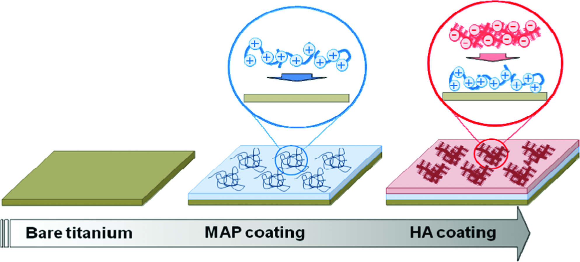
A scheme for hyaluronic acid (HA) surface immobilization using a mussel adhesive protein (MAP) coating. Color images available online at
Materials and Methods
Preparation of an HA-immobilized titanium surface using MAP
Pure titanium foils (≥99.5% purity; Alfa Aesar, USA) of 0.25 mm thickness were cut into 10×10 mm pieces, polished using 600 and 1200 grid sandpapers, and ultrasonicated in deionized water (DW) for 30 min. Then, the sample surfaces were washed with ethanol and acetone, immersed in a solution of methanol/HCl (1:1, v/v) for 30 min at room temperature, and rinsed five times with DW. The titanium surfaces were placed in a piranha solution containing a 4:1 (v/v) mixture of a 50% aqueous solution of H2SO4 and a 30% aqueous solution of H2O2 for 15 min, rinsed extensively with DW, boiled in DW for 15 min, and then dried under nitrogen gas.
For the immobilization of HA onto a titanium surface using MAP, purified fp-151 (>95% purity) 20 and HA (17 kDa MW; Lifecore Biomedical, USA) were each dissolved in a 10 mM NaCl (pH 5.0) solution to a final concentration of 1 mg/mL. The titanium surfaces were first incubated in the MAP solution for 30 min. After dispensing the MAP solution onto the titanium surface using a spin coater (Jaeseong Engineering, Korea) at 300 rpm for 10 s, excess and loosely tethered MAP was removed by washing with a 10 mM NaCl solution. Then, the HA solution was applied onto the MAP-coated titanium surface for 30 min. After dispensing the HA solution onto the MAP-coated surface using a spin coater at 300 rpm for 10 s, excess and loosely tethered HA was removed by washing with a 10 mM NaCl solution. A bare titanium surface was used as the negative control (NC) and solely HA-treated and/or solely MAP-coated titanium surfaces were used as comparative controls.
Analyses of HA immobilization on a titanium surface
The deposition of HA at each step was determined by contact angle measurement, immunostaining, and X-ray photoelectron spectroscopy (XPS). For contact angle determination, approximately 10 μL of DW was dropped onto the titanium surface. The contact angle between the droplet and the surface was measured by a CCD imaging system (Surface and Electro-Optics, Korea). The measurement was repeated at least three times and averaged for a given sample.
For immunostaining, a rabbit polyclonal anti-MAP antibody was produced (ABFrontier). Briefly, an 18-mer peptide, which was derived from a partial sequence of fp-151, was chemically synthesized and conjugated with the keyhole limpet hemocyanin carrier protein. To produce the anti-MAP antibody, two New Zealand white rabbits were immunized by subcutaneous injection using an emulsion of the antigen with adjuvant. After the third booster injection (2 weeks after the second and 6 weeks after the first immunizations), the final anti-MAP serum was obtained. Immunostaining of the titanium surface was performed using the anti-MAP antibody. Each sample-coated titanium surface was blocked using 1% (w/v) bovine serum albumin (BSA; Generay Biotech) in Dulbecco's phosphate buffered saline (DPBS; Hyclone) for 1 h at room temperature and washed three times with 0.02% (v/v) Tween 20 in DPBS. The sample surfaces were incubated with the anti-MAP antibody (1:5000), which was followed by incubation with a Texas red-conjugated anti-rabbit antibody (1:1000) (Abcam). The fluorescence was observed using an automated fluorescence stereo microscope (Leica, Germany).
To investigate the changes in the chemical composition of the titanium surface after sample coating, XPS analysis was performed using an ESCALAB 220-IXL system (VG Scientific). Nonmonochromatic MgKα radiation with a photon energy of 1253.6 eV was used to obtain the spectrum.
Preparation of other GAG-immobilized titanium surfaces using MAP
The immobilization of other GAG molecules [HS, CS, and DS, which were dissolved in a 10 mM NaCl (pH 5.0) solution, respectively] was also performed according to the HA immobilization procedure. The visualization of the GAG molecules was performed via Alcian blue staining. The GAG-coated titanium surfaces were rinsed with DW and stained with an Alcian blue 8GX (Sigma-Aldrich) solution dissolved in 3% acetic acid (pH 2.5) at room temperature for 15 min. After staining, images were obtained using an automated fluorescence stereo microscope.
Cell proliferation analysis
MC3T3-E1 (RIKEN Cell Bank) mouse preosteoblast cells were cultured in alpha-minimal essential medium (α-MEM; Hyclone) with 10% (v/v) fetal bovine serum (FBS; Hyclone) and penicillin/streptomycin (Hyclone) at 37°C in a humidified atmosphere with 5% CO2 and 95% air. All cells were collected by trypsinization, washed twice in DPBS, and diluted to a concentration of approximately 1×10⁵ cells per 1 mL of α-MEM without FBS. A total of 5×10⁴ cells (>95% viable) in serum-free medium were placed on each sample-coated titanium surface. The cells were allowed to adhere to the coated surfaces for 1 h, and unattached cells were removed from the coated surfaces by rinsing with PBS. Then, attached cells on each sample-coated titanium surface were incubated in α-MEM supplemented with 10% FBS at 37°C for 72 h. For a quantitative measurement of cell number after cell attachment and proliferation, we used the Cell Counting Kit-8 (CCK-8; Dojindo Laboratories), which contains 2-(2-methoxy-4-nitrophenyl)-3-(4-nitrophenyl)-5-(2,4-disulfophenyl)-2H-tetrazolium (WST-8) and produces a highly water-soluble formazan dye upon reduction in the presence of an electron carrier. The CCK-8 assay was performed in an appropriate manner at a given time: the assay was performed after a 1 hr incubation in serum-free α-MEM (attachment), and then it was given every 24 h during a 72 h incubation in serum-containing α-MEM (proliferation). A total of 25 μL of the CCK-8 solution was added to each well, and the plates were further incubated for 3 h at 37°C. After incubation, the absorbance was measured at 450 nm using a microplate absorbance spectrophotometer (Bio-Rad).
Cell morphology analysis
For the cell spreading analysis, a total of 5×10⁴ cells (>95% viable) were incubated in serum-free α-MEM on each sample-coated titanium surface for up to 24 h. The morphologies of the MC3T3-E1 cells on the sample-coated surfaces were analyzed by immunostaining. Briefly, cells on the sample-coated titanium surfaces were rinsed in DPBS, fixed in 4% (v/v) formaldehyde, and permeabilized with 0.2% (v/v) Triton-100 (USB) before blocking with a 1 mg/mL BSA solution for 30 min. Actin filaments were labeled with fluorescein isothiocyanate (FITC)-conjugated phalloidin (Sigma-Aldrich), and the nuclei were stained with 4′,6-diamidino-2-phenylindole (DAPI; Sigma-Aldrich). The specimens were analyzed using an automated fluorescence stereo microscope.
Cell differentiation analysis
MC3T3-E1 cells were seeded onto the sample-coated titanium surfaces and cultured as described above. The culture medium was replaced every 72 h. At 90% confluence, osteoblastic differentiation was induced with differentiation medium, which consisted of a mixture of 50 μg/mL ascorbic acid and 10 mM monobasic sodium phosphate in the culture medium.
The calcification of differentiated MC3T3-E1 cells was analyzed using Alizarin red S staining at 21 days after the induction of differentiation. After aspirating the media from the wells, cells were rinsed in DPBS and fixed with 4% (v/v) formalin. Then, cells were rinsed with DW and stained with a 1% Alizarin red S solution (adjusted to pH 4.2 with ammonium hydroxide; Sigma-Aldrich) at room temperature for 30 min. After the removal of the Alizarin red S solution, cells were rinsed three times with DW. Images of the Alizarin red-stained area were obtained using an automated fluorescence stereo microscope. Additionally, the retained dye was eluted into 20% acetic acid, and the absorbance was determined at 570 nm using a microplate absorbance spectrophotometer. The alkaline phosphatase (ALP) activity was also estimated in MC3T3-E1 cells at 15 days after the induction of differentiation using the SensoLyte® pNPP Alkaline Phosphatase Assay Kit (Anaspec) following the manufacturer's protocol.
Construction of an MAP/HA multilayer film
A quartz crystal microbalance (QCM; Stanford Research Systems) was used to confirm the formation of a polyelectrolyte multilayer of fp-151 and HA on a titanium-coated quartz crystal (Stanford Research Systems). Prior to the analysis, the quartz crystals were rinsed in ethanol, dried with nitrogen, and placed in a plasma oven for 10 min to ensure a clean surface was obtained. The crystal was excited at its fundamental frequency (∼5 MHz), and the observations were performed at the third (ν=3) and fifth (ν=5) overtones, which corresponded to 15 and 25 MHz, respectively. A 10 mM NaCl solution (pH 5.0) was circulated into the QCM flow cell (100 μL internal volume and 24°C temperature) for 40 min at a flow rate of 0.1 mL/min using a peristaltic pump. After stabilization of the signals, five cycles of polyelectrolyte multilayer formation were monitored by the shifts in frequency (Δf ). Each cycle consisted of the following steps: injection of 0.1 mg/mL fp-151 in the buffer for 10 min, a rinse with the buffer for 5 min, injection of 0.1 mg/mL HA in the buffer for 10 min, and a wash with the buffer for 5 min.
Statistical analysis
Independent experiments were performed at least three times with triplicate samples analyzed in each experiment. The statistical significance of the obtained data comparing the control and treated groups was analyzed using the paired Student's t-test. A p-value < 0.05 was considered to be statistically significant.
Results
Immobilization of GAGs onto a titanium surface using an MAP coating
We implemented an MAP linker for the simple and efficient surface immobilization of GAG molecules without modifications of the polysaccharides and/or the scaffold surfaces. Positively charged MAP, which has strong adhesion ability, was first coated onto the surface, and then negatively charged GAG molecules were directly immobilized onto the MAP-coated surface by charge interactions (Fig. 1). To examine the potential of our strategy, HA as a model GAG compound was firstly immobilized on a titanium scaffold surface using fp-151 as an MAP linker. Several analyses including contact angle measurement, immunostaining, and XPS were performed to check for the successful coating of MAP and the immobilization of HA on the titanium surface. While a bare titanium surface, which normally is negatively charged by oxidation, showed a 92° contact angle for the water droplet (NC in Fig. 2A), the coating of positively charged MAP onto the titanium surface resulted in a reduced contact angle (78°) due to a change in the hydrophobicity (MAP in Fig. 2A). Then, the contact angle returned to a larger value (95°) after the deposition of the negatively charged HA layer onto the MAP-coated surface (MAP/HA in Fig. 2A). These changes in surface contact angles indicate the successful immobilization of HA onto the titanium surface using an MAP coating.
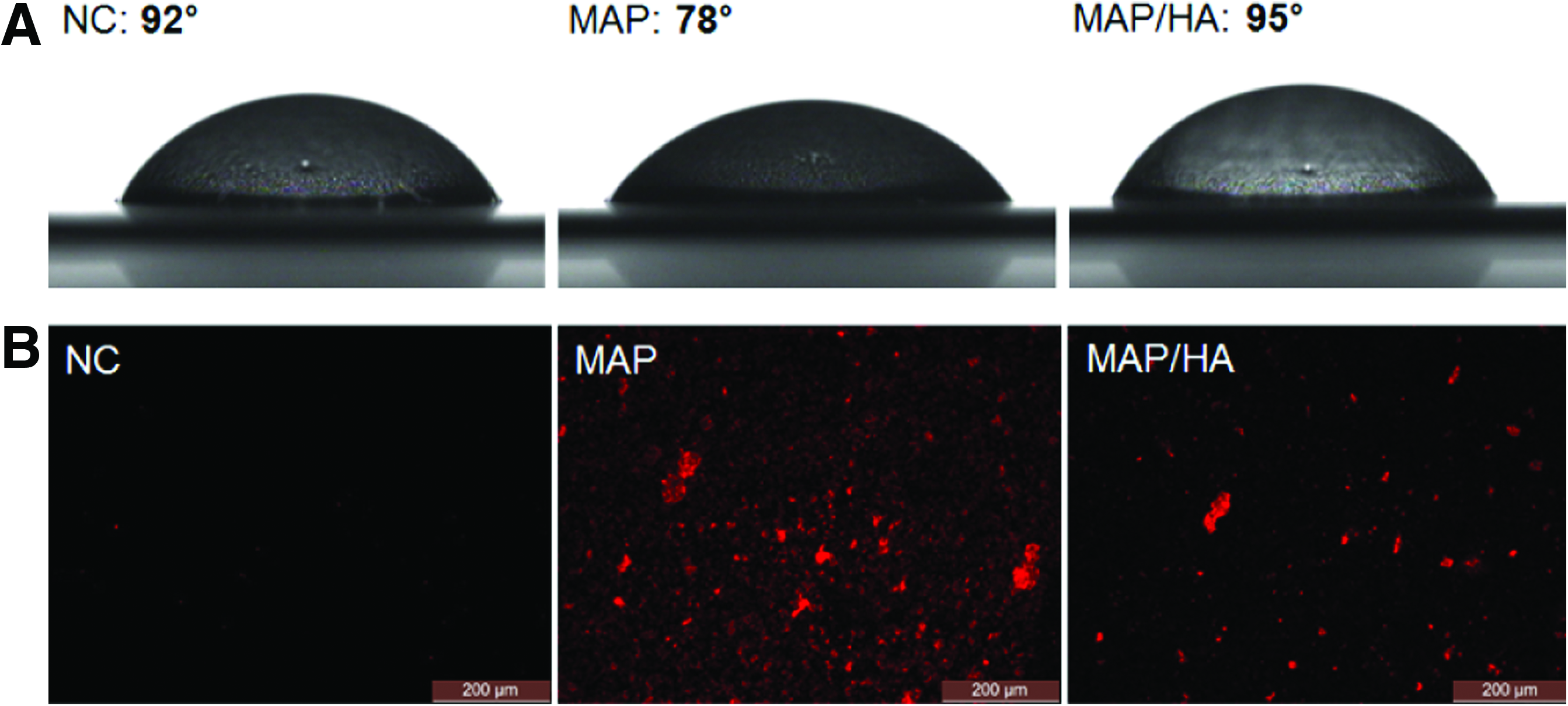
The characterization of bare (NC), solely MAP-coated (MAP), and HA-immobilized-MAP-coated (MAP/HA) titanium surfaces using
In immunostaining of fp-151, a considerable amount of red fluorescence (i.e., fp-151) was detected on the MAP-coated titanium surface (MAP in Fig. 2B) compared to bare surface (NC in Fig. 2B), and the fluorescence intensity was significantly decreased on the MAP/HA-coated surface (MAP/HA in Fig. 2B). In addition, while both elemental nitrogen ratios of the bare and HA-treated titanium surfaces measured at less than 3% in the elemental composition analysis using XPS, the nitrogen ratio of the MAP-coated surface was increased to ∼11% through the successful coating of MAP, which contains a nitrogen element (Table 1). Interestingly, HA immobilization onto the MAP-coated surface did not significantly change the nitrogen ratio (∼9%) compared to the MAP-coated surface (∼11%). This result was due to the nitrogen element in HA, which is composed of
XPS, X-ray photoemission spectroscopy; HA, hyaluronic acid; MAP, mussel adhesive proteins.
To demonstrate that our immobilization strategy is generally applicable to other negatively charged GAG molecules, we also performed the immobilization of HS, CS, and DS on titanium surfaces using a direct MAP coating. As expected, the three GAGs tested were also successfully immobilized onto the MAP-coated titanium surfaces based on the charge interactions between the positively charged MAP and the negatively charged GAGs (Fig. 3).
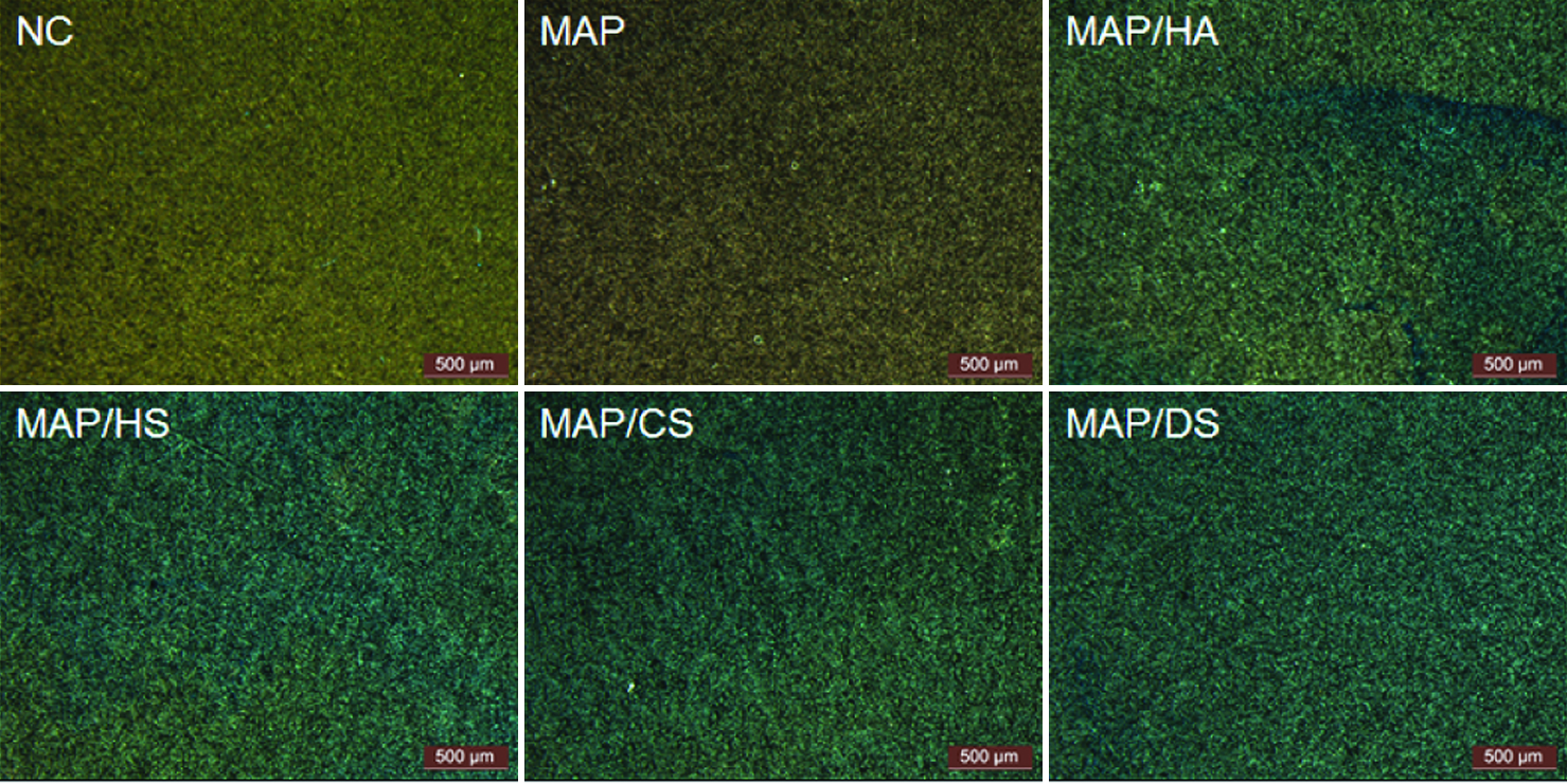
The visualization of the immobilized glycosaminoglycans (GAGs) [HA, heparin sulfate (HS), chondroitin sulfate (CS), and dermatan sulfate (DS)] on the MAP-coated titanium surfaces using Alcian blue staining. NC and MAP are bare and solely MAP-coated titanium surfaces, respectively. The scale bar represents 500 μm. Color images available online at
Cell behaviors on the MAP/HA-coated surface
The effects of HA immobilization on the proliferation of mouse preosteoblast MC3T3-E1 cells were first examined on an MAP-coated titanium surface. Bare, solely HA-treated and solely MAP-coated surfaces were used as controls. The proliferation of MC3T3-E1 cells on a MAP/HA-coated surface was significantly enhanced compared to the proliferation levels observed on the other surfaces (Fig. 4). Notably, the proliferation level on the HA-treated titanium surface was similar to the level observed on the bare titanium surface because HA could not be retained on the bare surface except by simple adsorption. The cell growth on the solely MAP-coated surface was also notably increased compared to the bare and HA-treated surfaces. This result was achieved due to the direct attachment of cells onto the MAP-coated surface, which we have reported previously.15,28 Importantly, due to our MAP-based immobilization strategy, the HA molecules that were efficiently immobilized onto the titanium surface were functional and thus promoted target cell proliferation.
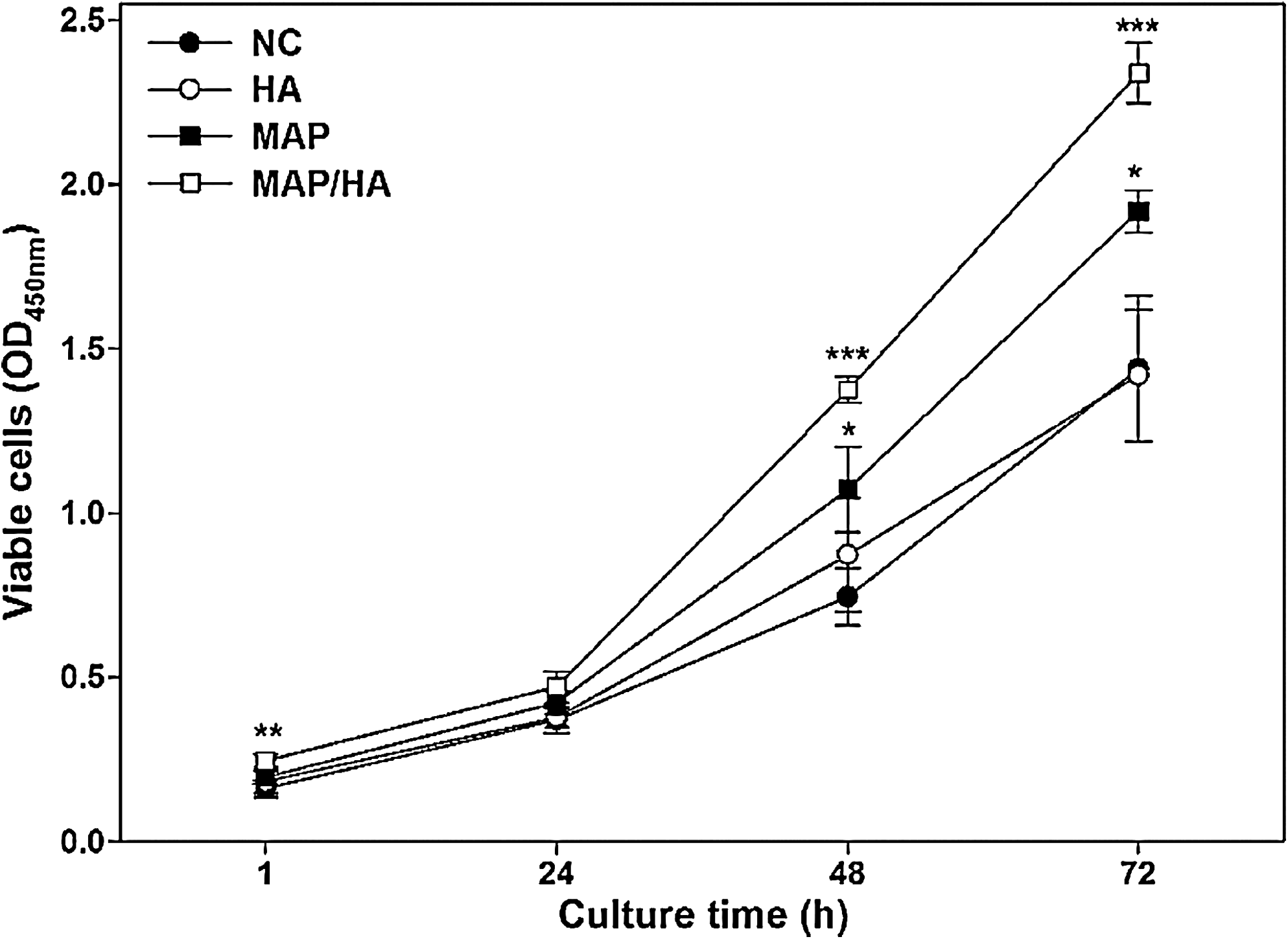
The proliferation of MC3T3-E1 preosteoblast cells on bare (NC), solely HA-treated (HA), solely MAP-coated (MAP), and HA-immobilized-MAP-coated (MAP/HA) titanium surfaces. Cells (5×10⁴) were incubated on sample-coated titanium surfaces without serum for 1 h at 37°C. After replacing the media with serum-containing medium, viable cells were measured with the CCK-8 assay every 24 h to determine proliferation. Values and error bars represent the mean of triplicate samples and standard deviation with statistical significance designated by the symbols *p<0.05, **p<0.01, and ***p<0.005.
When MC3T3-E1 cells were cultured without serum, immunostaining with FITC-conjugated phalloidin demonstrated the attached cells on the MAP/HA-coated surface showed a much better cell morphology and actin filament formation compared to the cells on bare, HA-treated, and MAP-coated titanium surfaces (Fig. 5). Because there are many factors for spreading such as fibronectin and vitronectin in a serum-containing environment, cell spreading experiments were performed under serum-free condition. The degree of cell spreading on the solely HA-treated titanium surface was similar to the cell spreading observed on the bare surface. In the case of the MAP-coated titanium surface, although the cells were partially spread, they did not form proper shapes, which may be due to a lack of cell recognition molecules. Therefore, these results indicate that the immobilized HA molecules on the MAP-coated titanium surface are biologically active and can serve as cell signaling materials for actin filament formation.
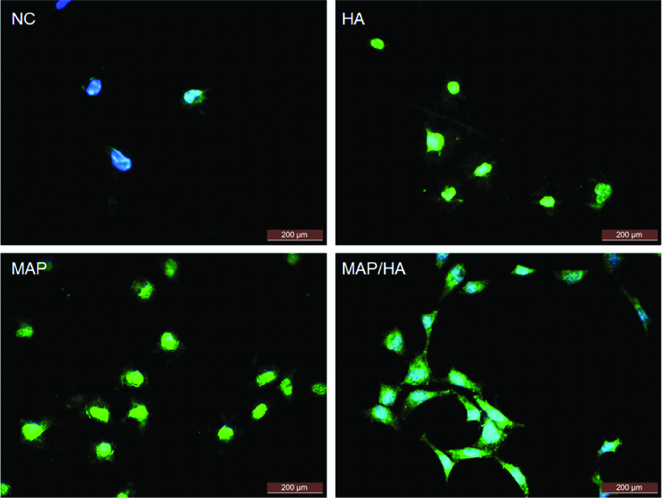
The spreading of MC3T3-E1 preosteoblast cells on bare (NC), solely HA-treated (HA), solely MAP-coated (MAP), and HA-immobilized-MAP-coated (MAP/HA) titanium surfaces. Cells (5×10⁴) were added to the sample-coated titanium surfaces without serum and cultured for 18 h at 37°C. Actin filaments were stained with a phalloidin-FITC-conjugated antibody (green), and nuclei were stained with DAPI (blue). The scale bar represents 200 μm. Color images available online at
The degree of differentiation on the titanium surfaces was investigated by measuring the matrix mineralization and ALP activity. Alizarin red S staining showed that the calcium incorporation of MC3T3-E1 cells on the MAP/HA-coated titanium surface was greatly improved (106%, 54%, and 12%) compared to the levels found on bare, HA-treated, and MAP-coated surfaces, respectively (Fig. 6A and B). Similar to the matrix mineralization result, the ALP activity of the MC3T3-E1 cells on the MAP/HA-coated surface was also significantly increased (64%, 40%, and 26%) compared to the activities measured on bare, HA-treated, and MAP-coated surfaces, respectively (Fig. 6C). Interestingly, the differentiation of the MC3T3-E1 cells on the solely MAP-coated titanium surface was also somewhat increased (Fig. 6A and B). This increase may be due to more attached and proliferating cells on the surface due to the adhesion ability of MAP (Fig. 4). However, ALP activity on the solely MAP-coated titanium surface was not notably influenced during the bone formation process (Fig. 6C), which was likely due to the lack of signaling factors for the differentiation of the preosteoblasts in MAP.
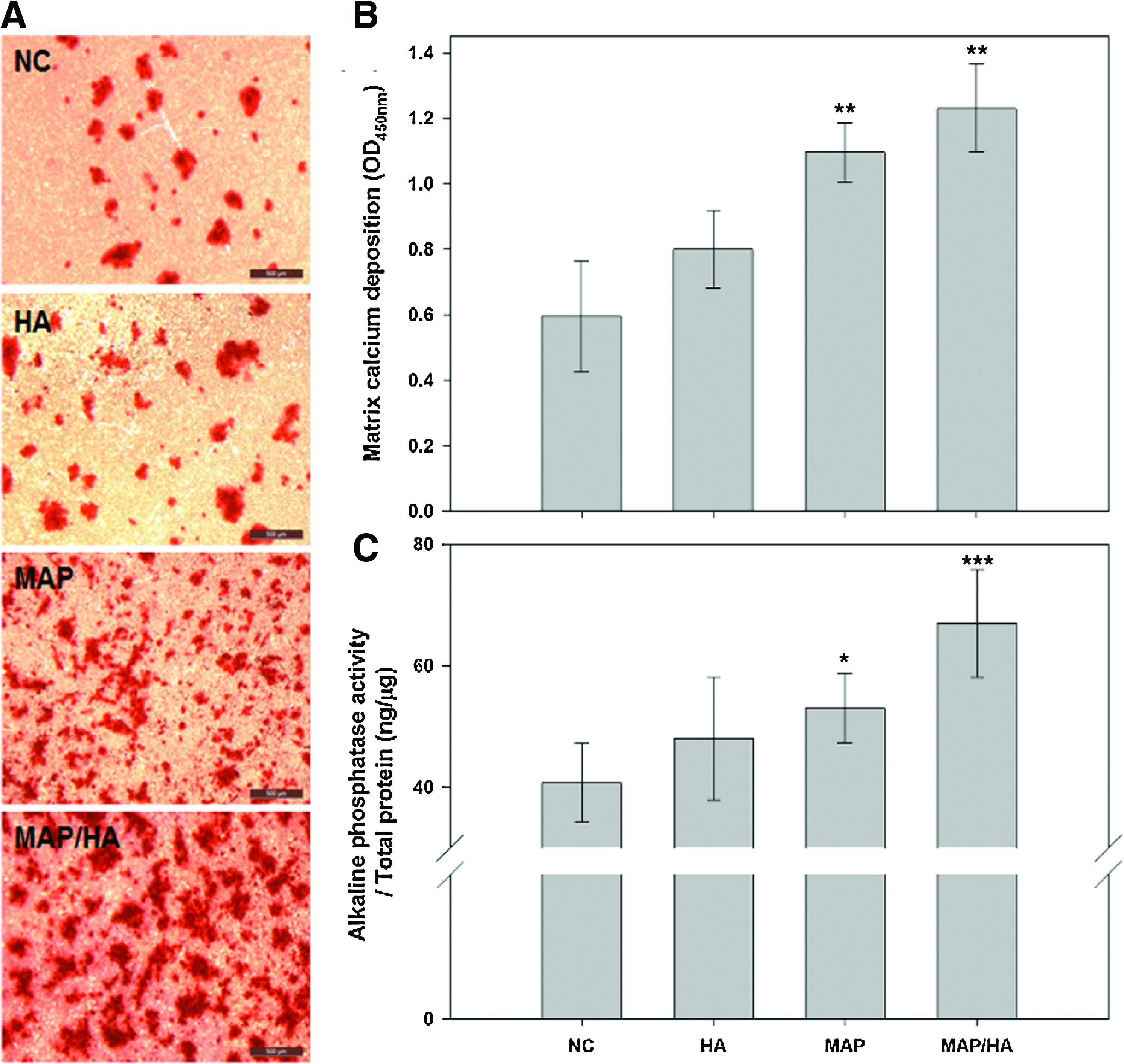
The differentiation of MC3T3-E1 preosteoblast cells on bare (NC), solely HA-treated (HA), solely MAP-coated (MAP), and HA-immobilized-MAP-coated (MAP/HA) titanium surfaces.
A multilayer fabrication of MAP and HA on a titanium surface
The layer-by-layer (LbL) technique is a versatile and simple coating method to fabricate multilayer constructs composed of oppositely charged molecules. It does not require chemical modifications of the surface and/or coating materials for the fabrication of multicomposite molecular assemblies.29–33 Because an oxidized titanium surface has a net negative charge, 34 positive charged MAPs are easy to access to the surface and eventually can induce a charge–charge interaction as well as their inherent adhesive property. Also, the strong positive charge from the high proportion of lysine residues and the adhesive property of MAPs can be directly applied for the fabrication of a multilayer with negatively charged molecules, such as the GAG compounds, using the LbL assembly method. Therefore, to examine the wider applications of our MAP-based immobilization method, we performed a potential fabrication of a polyelectrolyte multilayer (PEM) film on a titanium surface using the two oppositely charged molecules, MAP and HA. MAP was coated onto a bare titanium surface by simple spin coating. After washing away residual MAP, HA was then coated onto the MAP-coated layer by simple spin coating followed by washing. These steps were repeated to form the MAP n /HA n multilayer film (Fig. 7A). A frequency shift in the QCM analysis (Fig. 7B) and changes in the water contact angle measurement (Fig. 7C) clearly showed that the PEM film, which contained 10 layers, was well fabricated onto the titanium surface through the LbL method using MAP and HA.
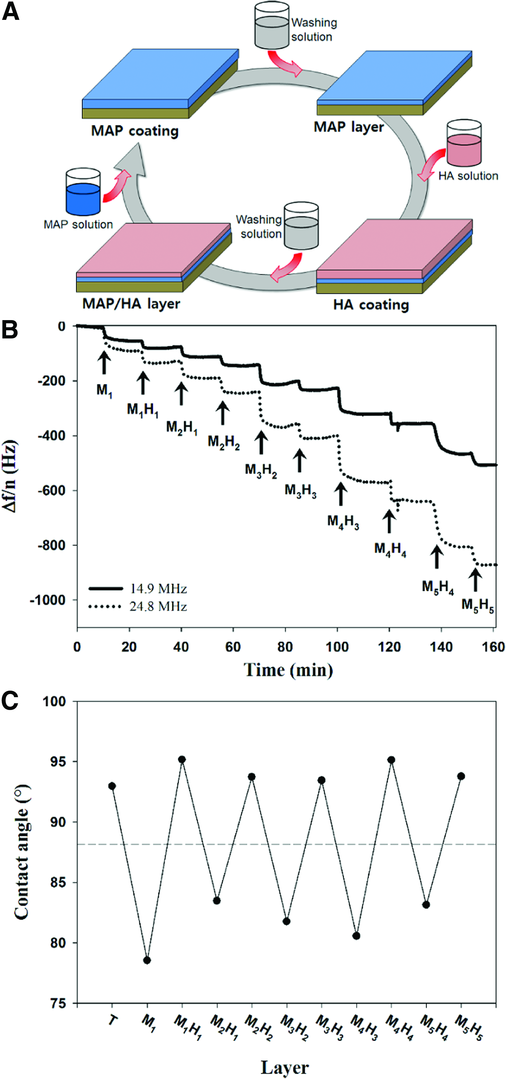
The fabrication of a polyelectrolyte multilayer using MAP and HA.
Discussion
GAGs found in a natural ECM are covalently and/or ionically linked to other ECM components. Thus, the immobilization of GAGs on surfaces for the preparation of an artificial ECM can mimic a natural environment. Moreover, the stable incorporation of GAGs onto surfaces without structural modifications may allow better biological activities of the GAGs for tissue engineering applications. HA, which is one of the major GAG components in the ECM, is an important substrate for cell surface receptors. It is known that HA stimulates the cellular signaling pathways that promote cell adhesion and proliferation in osteoblast cell culture.35,36 Thus, we reasoned that a functional HA coating on a titanium surface might enhance cellular behaviors. In detail, HA binds to CD44 and/or several other unknown osteoblast receptors, and these interactions stimulate the intracellular signal transduction for the organization of the cytoskeleton.37,38 This process leads to actin filament formation by a direct or indirect signaling pathway through the HA receptor as well as by integrin signaling through the focal adhesion kinase (FAK). Thus, cell morphology is regulated by the interactions between cell surface receptors and ECM molecules on the scaffold. In addition, osteoblast cell differentiation can also be enhanced by the interaction between HA and the HA receptor that activates osteogenic signaling transduction.36,39 Thus, the functionality of immobilized HA would also stimulate the differentiation of preosteoblasts.
Even though titanium has good biological and mechanical properties, the surface modification of titanium is needed to solve complications such as inadequate osteointegration. In the case of osteointegration, the fabrication of a multilayer is an alternative solution for the use of titanium in the dental and orthopedic fields as successful osteointegration usually occurs through ≤500 nm of the amorphous layer (some reports state that the thickness of the intermediate layer of fibrous connective tissue is 10 μm).40,41 The combination of cationic and anionic polyelectrolytes used to construct the PEM has also been used in a wide range of tissue engineering applications. 42 For example, one attempt focused on maintaining phenotypic cell behaviors through direct cellular interaction with the PEM. The incorporation of short bioactive peptides into the terminal layer of the PEM 42 or alternating with layers composed of various biomacromolecules 43 was shown to enhance cell adhesion, proliferation, and differentiation. Another trial used the incorporation of various growth factors and the controlled release of these growth factors from the PEM to modulate cellular behavior.44,45 In these studies, the growth factor was incorporated and controlled with the construction of the PEM using a positively charged synthetic polymer and negatively charged heparin.
The basic concept of both approaches was to engineer additional functionality into the multilayer films to improve their biological performance. Because MAP can be developed to contain bioactive peptides such as ECM- and growth factor-derived peptides through fusion technology, 15 additional processes are not required for peptide immobilization on the PEM. Moreover, in this study, we showed the successful surface immobilization of various macromolecules, especially the negatively charged GAGs, using an MAP coating method. Therefore, MAP and its derivatives can easily and successfully be used for the construction of a functional PEM that is able to modulate cellular behaviors for the tissue engineering fields.
Conclusions
Here, we performed the facile immobilization of GAG molecules, mainly HA, on a titanium surface using an MAP coating to improve the biocompatibility and biological activity of the titanium surface. Negatively charged HA was successfully immobilized onto the positively charged MAP-coated titanium surface by charge interactions without any chemical modifications. We found that preosteoblast behaviors, such as proliferation, spreading, and differentiation, were significantly increased on the MAP/HA-coated titanium compared to the bare, solely HA-treated, and solely MAP-layered surfaces. We also demonstrated that other negatively charged GAG molecules, including HS, CS, and DS, could be successfully immobilized on an MAP-coated titanium surface. Moreover, we expanded the MAP-based coating method to fabricate a multilayer film of the two counterionic molecules of MAP and HA onto a titanium surface using the LbL technique. Taken together, our MAP-based surface functionalization method could be generally applied for the simple and efficient immobilization of negatively charged bioactive molecules for tissue engineering and implanting applications.
Footnotes
Acknowledgments
This work was supported by the National Research Laboratory program (ROA-2007-000-20066-0) and the Brain Korea 21 program funded by the Ministry of Education, Science and Technology (Korea), the Marine Biomaterials Research Center grant from the Marine Biotechnology program funded by the Ministry of Land, Transport and Maritime Affairs (Korea), and the POSCO Steel Science program. We thank Dr. S.M. Jeon (POSTECH) for helping with QCM analysis.
Disclosure Statement
No competing financial interests exist.