
Abstract
Background:
In the immunohistochemical analysis of tissue-engineered structures, aggressive treatments for fixation and antigen retrieval can impair the quality of specimen staining and visualization.
Hypothesis:
We hypothesized that the adequate choice of fixative and antigen-retrieval method might improve the quality of immunohistochemical staining.
Methods:
Tissue-engineered vascular grafts were fixed using formalin, Carnoy's, or HOPE ® fixative. Antigen retrieval was performed where necessary and samples from each group were stained using hematoxylin and eosin to assess overall tissue preservation. For a set of proteins relevant to cardiovascular tissue development, immunohistochemical staining was applied to formalin-, Carnoy's-, and HOPE-fixed specimens to allow a comparative analysis.
Results:
In tissue-engineered constructs, antigen retrieval methods necessary after formalin fixation led to significant destruction of the overall tissue structure. Carnoy's fixation resulted in good overall tissue preservation and adequate results for immunohistochemical staining of alpha-smooth muscle actin (α-SMA), vimentin, type I collagen, elastin, and laminin. HOPE fixative led to a loosened tissue structure and a swollen appearance but showed adequate results for staining against type III collagen and elastin. Formalin fixation without antigen retrieval led to inadequate visualization of α-SMA, vimentin, type I- and type III collagen, and laminin.
Conclusion:
Based on the present study, we recommend that Carnoy's fixative is employed for the preservation of tissue-engineered constructs to allow immunohistochemical analysis of type I- and type III collagen, elastin, laminin, α-SMA, and vimentin. However, it is clear that the technique requires optimization based on the particular tissue engineering application.
Introduction
Formalin is the most widely used fixative for histological and immunohistochemical analysis of tissues.4,5 Formalin fixation is well established in routine clinical applications and its excellent results in most standard applications render it the gold standard. Formalin is a cross-linking fixative and chemically binds to protein molecules after denaturing them. 1 This aggressive procedure masks the antigen epitopes of proteins, thus rendering them inaccessible for further immunohistochemical marking. Thus, antigen retrieval by enzymatic pretreatment6,7 or heat conditioning8,9 is mandatory 10 for immunohistochemical analysis of formalin-fixed tissues, further adding to the aggressive nature of the method.11,12
Due to the delicate nature of tissue-engineered constructs prepared in vitro, they are prone to damage by this well-established standard fixation procedure. For this reason, the analysis of tissue-engineered constructs requires more gentle fixation methods that avoid further pretreatments such as antigen retrieval. To enhance tissue preservation and facilitate immunohistochemical analysis, alternative fixatives have been developed. Carnoy's fixative changes the shape or structure of protein molecules without binding to them and without masking the target epitopes, thus belonging to the group of coagulating fixatives. Carnoy's fixation results in a well preserved macroscopic tissue morphology. The novel HOPE ® (HEPES-glutamic acid buffer-mediated organic solvent protection effect) method of fixation results in good morphological preservation with reduced denaturation of proteins, nucleic acids, and antigen structures compared with other fixation methods. 2 Both Carnoy's- and HOPE fixative enhance the applicability of immunohistochemical methods and enable molecular analyses of fixed tissue without further pretreatment.
For the present study, we hypothesized that the appropriate choice of fixative in addition to a suitable antigen retrieval method might improve the quality of immunohistochemical staining of tissue-engineered constructs. We constructed vascular grafts using tissue engineering methods as described by Koch et al. 13 and subjected each graft to fixation using formalin, Carnoy's, or HOPE fixative, followed by antigen retrieval where necessary. The quality of tissue preservation and the immunohistochemical staining of selected cellular and tissue markers were compared between grafts processed using each of these fixatives.
Methods
All histological and immunohistochemical analyses were conducted using Vascular Composite Grafts developed within our group. 13 Explanted ovine carotid arteries served as native tissue controls. All animal procedures were conducted according to the European Convention on Animal Care.
Cell isolation and culture
For the fabrication of tissue-engineered vascular grafts, a mixed population of smooth muscle cells/fibroblasts was obtained from ovine carotid artery as described previously. 14 Cells were cultured in Dulbecco's modified Eagle's medium (DMEM; Invitrogen, Darmstadt, Germany) supplemented with 10% fetal calf serum (FCS; PAA, Cölbe, Germany), and serially passaged up to four times using 0.25% trypsin/0.02% EDTA (PAA). Cells were maintained in culture at 37°C in a humidified incubator (5% CO2 and 95% humidity). Prior to graft synthesis, cell phenotype was verified by a majority of alpha-smooth muscle actin (α-SMA)-positive cells and absence of von Willebrand factor—an endothelial cell marker (all from Dako, Glostrup, Denmark; data not shown).
Vascular composite graft construction
Fibrin synthesis
Fibrinogen (Sigma, Seelze, Germany) was dissolved in ultrapure water and dialyzed overnight against Tris-buffered saline (TBS; Sigma; pH 7.4) using a dialysis membrane (Novodirect, Kehl, Germany) with a molecular weight cut-off of 6–8 kDa. 13 Subsequent to sterile filtration, the fibrinogen concentration was optically determined by measuring the absorbance at 280 nm using a spectrophotometer (ThermoSpectronic Genesys™6; Thermo Fisher Scientific, Portsmouth, NH) and adjusted to 10 mg/mL with sterile TBS. The entire tissue-engineered graft (4.0 mL total volume) was composed of 2.0 mL fibrinogen solution (10 mg/mL), 1.4 mL TBS containing carotid artery-derived cells (10×106 cells/mL), 300 μL 50 mM CaCl2 (Sigma), and 300 μL thrombin solution (40 U/mL; Sigma).
Poly (L/D)lactide 96/4 mesh production
Poly (L/D)lactide 96/4 (PLA) fibers were manufactured at the Department of Biomedical Engineering, Tampere University of Technology in Finland, as previously described, 6 and were subsequently used to develop a macroporous textile mesh at the Institut für Textiltechnik, RWTH Aachen University in Germany. The mesh scaffold was based on a tubular warp knit structure due to the requirement for high elastic deformation under continuous dynamic stress. Following manufacture, the mesh scaffold was sterilized using low-temperature hydrogen peroxide gas plasma (STERRAD 100S Sterilization System; Ethicon GmbH, Hamburg, Germany).
Graft moulding/cell seeding
Vascular composite grafts (n=2) were moulded as described previously. 13 The PLA mesh was positioned over the inner cylinder of the mould before insertion into the outer cylinder, always ensuring that the mesh fibers were not under tension. Subsequently, the annular space between the inner and outer cylinders of the mould was filled with a cell/fibrin gel suspension (10×106 cells/mL), which provided a uniform wall thickness throughout the graft (final mass ratio of fibrin gel: PLA=14:1).
After 45 min, complete polymerization of the vascular composite graft was achieved. The inner casting cylinder was removed, and the graft was maintained in the outer cylinder. Subsequently, the graft was connected to a bioreactor system and transferred to an incubator at 37°C and 5% CO2 for mechanical conditioning. Medium flow through the graft was gradually increased from 20 mL/min to 200 mL/min over a 14-day conditioning `period, with physiological pressure conditions (120 mmHg, systolic pressure; 80 mmHg, diastolic pressure). The length and inner diameter of the grafts measured 80 and 5 mm, respectively (wall thickness, 1.5 mm).
The culture medium used in the bioreactor system consisted of DMEM with 10% FCS and 1% antibiotic-antimycotic solution (PAA), and it was changed once per week under sterile conditions. L-ascorbic acid-2-phosphate (1.0 mM; Sigma) was added to increase collagen synthesis. 15 Fibrin degradation was controlled by supplementation of the medium with 20 mg/mL aprotinin (Bayer Medical AG, Leverkusen, Germany). 16 At the end of the conditioning period, the vascular grafts were divided into three parts and fixed for microscopic analysis using the three fixatives described below.
Graft fixation
General considerations
Fixation time is determined by the efficiency of diffusion through the sample, which depends on probe size, temperature, tissue constitution, and agitation during fixation. Five millimeter segments were sampled from the tissue-engineered vascular grafts (wall thickness, 1.5 mm). At room temperature (RT), diffusion speed is determined to be 1 mm/h. 17 Correspondingly, an incubation time of 2 h at RT was determined to be sufficient.
Formalin fixation
Neutral buffered formalin was acquired as a ready-to-use 10% solution (PathoMed Logistik GmbH, Viersen, Germany). Vascular graft samples were incubated for 2 h at RT and stirred constantly. The incubation time of neutral buffered formalin was additionally based on its high diffusibility, which provides a sufficient number of cross-links while preserving the immunohistochemical properties of the tissue. 7
Carnoy's fixation
Carnoy's fixative contains ethanol, chloroform, and glacial acetic acid (all of pure quality) at a ratio of 6:2:1 (all from Sigma). Vascular graft samples were incubated for 2 h at RT and stirred constantly. All samples were subsequently washed twice in pure ethanol for 15 min.
HOPE fixation
HOPE fixative (DCS Diagnostics, Hamburg, Germany) was supplied to the end user as two separate solutions; HOPE I solution was supplied as a ready-to-use reagent, while HOPE II solution was diluted in acetone (Sigma) at a 1:200 ratio. The components of HOPE I and HOPE II solutions are subject to nondisclosure by DCS Diagnostics. Vascular graft samples were incubated in HOPE I solution for 4 h and subsequently immersed in HOPE II for 2 h at 4°C without stirring.
Dehydration
After both formalin and Carnoy's fixation, dehydration was conducted using an automated process (Leica TP 1020 Tissue Processor; Leica, Wetzlar, Germany) following standard conditions as outlined in Table 1.
The dehydration stage subsequent to HOPE fixation was performed according to the manufacturer's specifications. All tissue samples were subjected to three serial acetone incubations for 2 h at 4°C, followed by overnight paraffin (Vogel, Gießen, Germany) embedding at 55°C.
Analysis
Histology
All fixed tissue-engineered grafts were paraffin-embedded and sectioned perpendicular to their lumen at a thickness of 3 μm. Hematoxylin and eosin (H&E) staining allowed for analysis of overall tissue preservation. Sections were viewed using a routine bright field microscope (AxioImager D1; Carl Zeiss GmbH, Jena, Germany). Images were acquired using a digital color camera (AxioCam MRc; Carl Zeiss GmbH).
Antigen retrieval
Formalin-fixed samples had to undergo antigen retrieval methods to unmask the epitopes hidden by this fixation method.10,18 Heat-induced antigen (epitope) retrieval (HIER) was performed either by incubating samples immersed in 0.01 M citrate buffer (pH 6.0) in a 600 W microwave oven (3×5 min; HMT 72 G450; Bosch, Gerlingen, Germany) or a steam cooker at 95°C (40 min; Vitacuisine; Tefal, Offenbach, Germany). 10 For enzyme-induced antigen retrieval, enzyme-based pretreatment in Fast Enzyme (Zytomed, Berlin, Germany) for 5 min at RT was employed. 19
Immunohistochemistry
Nonspecific sites of the paraffin-embedded sections were blocked and the cells were permeabilized using 5% normal goat serum (Dako) in 0.1% Triton-phosphate-buffered saline (Sigma). Sections were incubated for 1 h at 37°C with the following primary antibodies: mouse monoclonal anti-α-SMA (1:1000; Sigma), mouse monoclonal anti-vimentin (1:50; Sigma), rabbit anti-type-I collagen (1:100; Acris Antibodies GmbH, Hiddenhausen, Germany), rabbit anti-type-III collagen (1:100; Acris Antibodies GmbH), rabbit anti-elastin (1:50; Research Diagnostics, Inc., Concord, MA), and rabbit anti-laminin (1:25; Sigma). The sections were then incubated for 1 h at RT with rhodamine-conjugated (Invitrogen), fluorescein-conjugated (Invitrogen), or biotinylated secondary antibodies (Dako). In the case of biotinylated staining, the procedure was pursued with a 1 h incubation of streptavidin/tetramethylrhodamine isothiocyanate (TRITC; Acris Antibodies GmbH) at 37°C. Cell nuclei were counterstained using DAPI (Sigma) nucleic acid stain. As negative controls, samples were incubated in diluent followed by the secondary antibody only. Samples were viewed using a fluorescence microscope (AxioObserver Z1; Carl Zeiss GmbH), and images were acquired with a digital color camera (AxioCam MRm; Carl Zeiss GmbH). As the grafts were divided into three parts for processing in each fixative, it was not possible to directly compare adjacent serial sections.
Results
Gross macroscopic appearance
Before fixation and dehydration, the tissue-engineered constructs exhibited a patent lumen and—subjectively—a mechanically stable tissue.
Formalin-, Carnoy's-, and HOPE fixation did not result in differences in macroscopic appearance, while the acetone-based dehydration process required after HOPE fixation led to tissue shrinkage, deformation, a distinct orange staining, and bubbling of tissue on the outer surface.
Histology
H&E staining
Formalin and Carnoy's fixative preserved overall tissue structure and thus allowed adequate assessment using H&E staining (Fig. 1). The sharpest images detailing structure and morphology were obtained from sections of Carnoy's-fixed tissue-engineered grafts. HOPE fixative led to a loosened tissue structure and a swollen appearance. The H&E process following HOPE fixation did not result in well-stained samples even after optimization of the staining process.
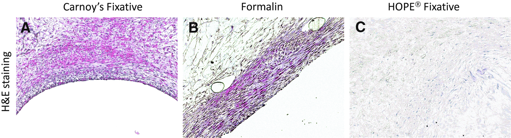
Hematoxylin and eosin (H&E) staining of Carnoy's-
Antigen retrieval (Fig. 2) resulted in significant destruction of the delicate tissue-engineered constructs. Where the vascular grafts showed more compact tissue, all retrieval methods resulted in loosening of the tissue structure. Fast-enzyme treatment completely destroyed the tissue structure and acquisition of distinct images was not feasible. HIER did not harm the tissue as severely as enzymatic treatment, but the more delicate parts of the tissue-engineered constructs were not preserved. All antigen retrieval methods hampered an adequate assessment of tissue structure and morphology. Consequently, further immunohistochemical analysis did not include samples thus treated.
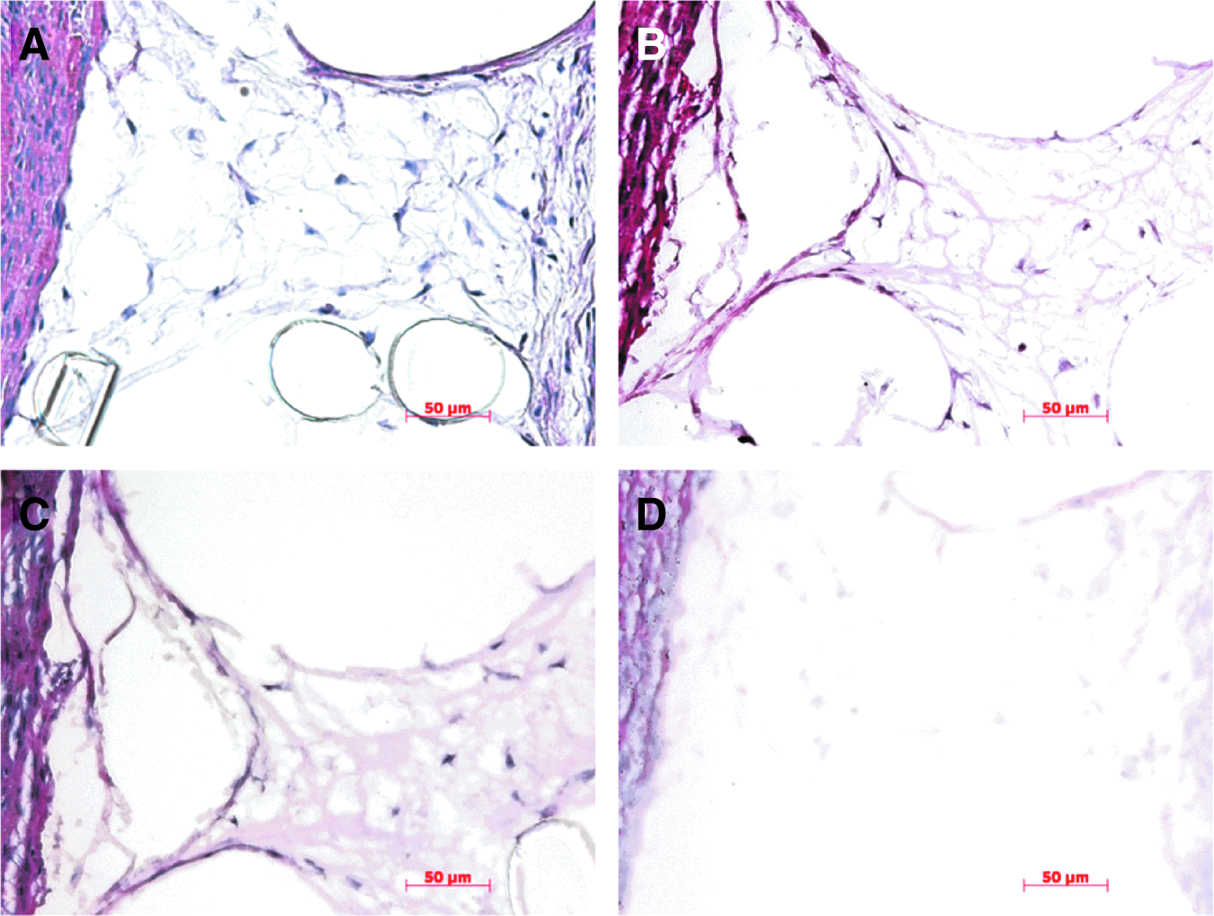
H&E staining of formalin-fixed tissue-engineered constructs before
Immunohistochemistry
Both α-SMA and vimentin (intracellular markers) were detectable throughout the tissue-engineered constructs following any fixation method (Figs. 3 and 4). The fluorescence intensity and proportion of cells stained strongly depended on the applied fixation procedure. Carnoy's-fixed samples showed the strongest signals for both α-SMA and vimentin. Formalin-fixed samples displayed reduced signals and a differently distributed expression of α-SMA than both Carnoy's- and HOPE-fixed samples, with highest expression levels in the subendothelial tissue layer, while a much lower proportion of cells stained positively for vimentin in formalin- and HOPE-fixed samples. To a lesser extent, this varied pattern of spatial distribution emerged in the analysis of type I collagen (Fig. 5). Again, formalin-fixed samples revealed highest expression levels in the subendothelial layer, while type I collagen signal levels were reduced throughout the remainder of the sample. The fluorescence intensity of type I collagen expression was similar among all samples.
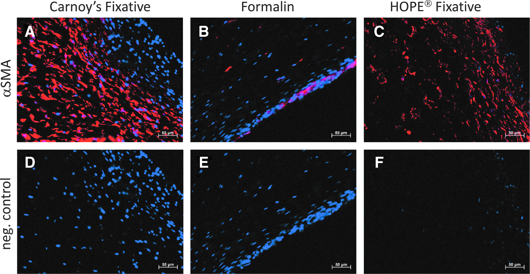
Staining against alpha-smooth muscle actin (α-SMA) after Carnoy's-
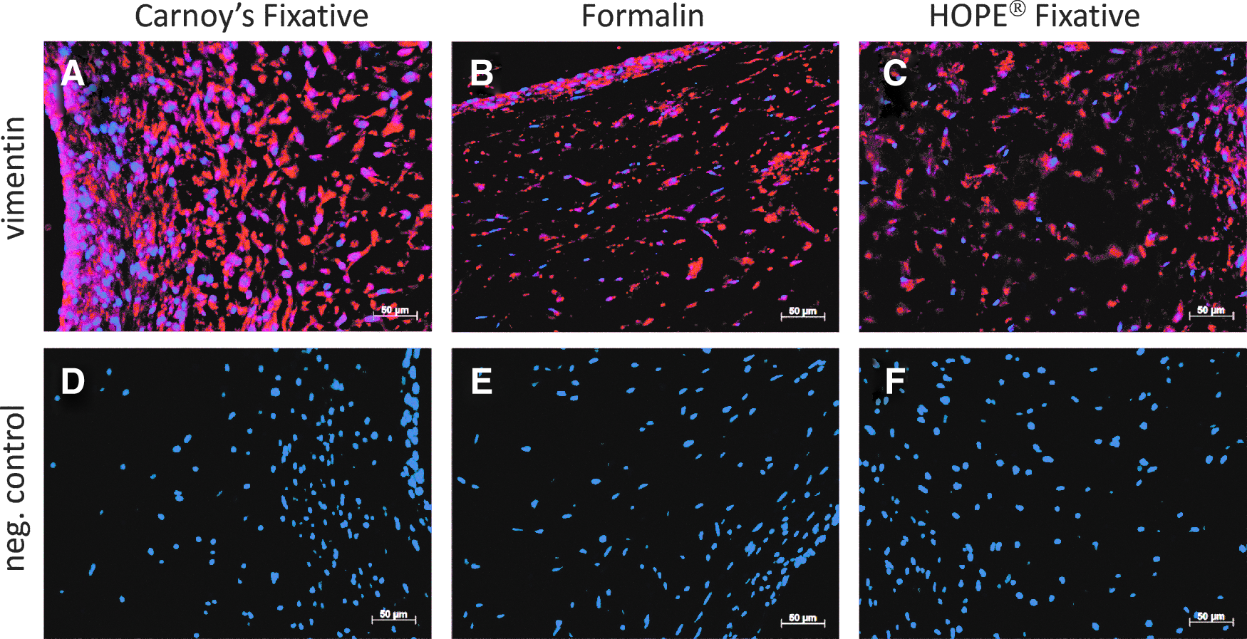
Staining against vimentin after Carnoy's-
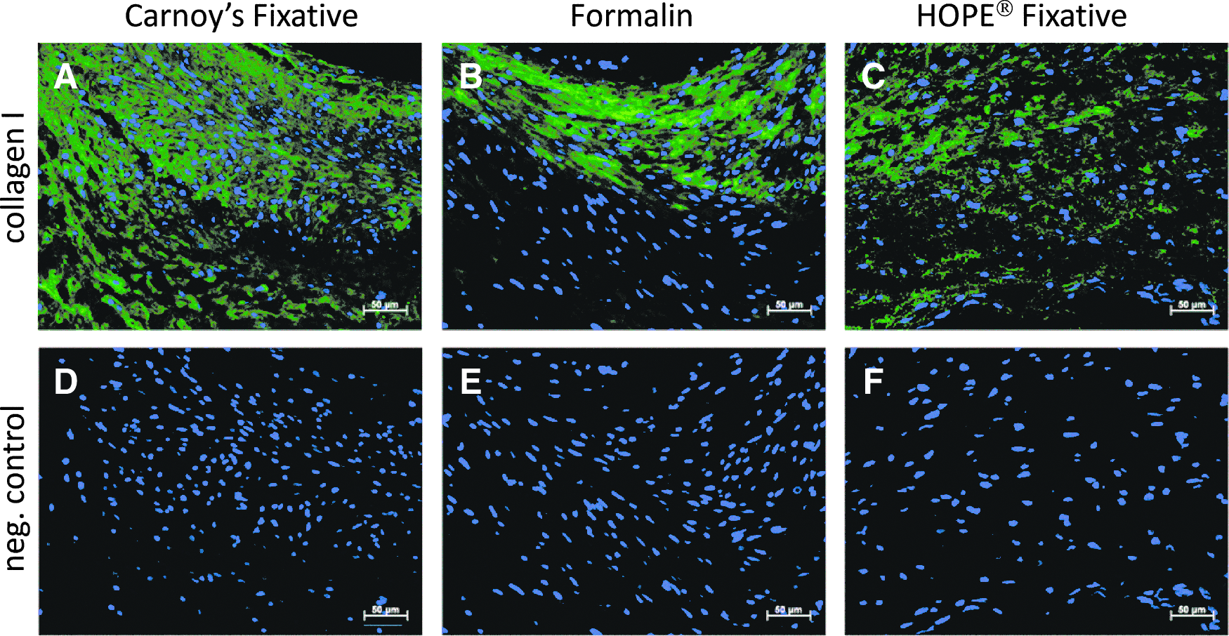
Staining against type I collagen after Carnoy's-
All samples stained positively for type III collagen (Fig. 6), although HOPE-fixed tissue displayed the most distinct staining. Carnoy's fixation resulted in unstructured, “smeared” fluorescence signals, independent of autofluorescence or false positive staining as indicated by the negative control. Formalin fixative preserved the structure of type III collagen better than Carnoy's fixative, but was still less distinct in comparison to HOPE-fixed tissue.
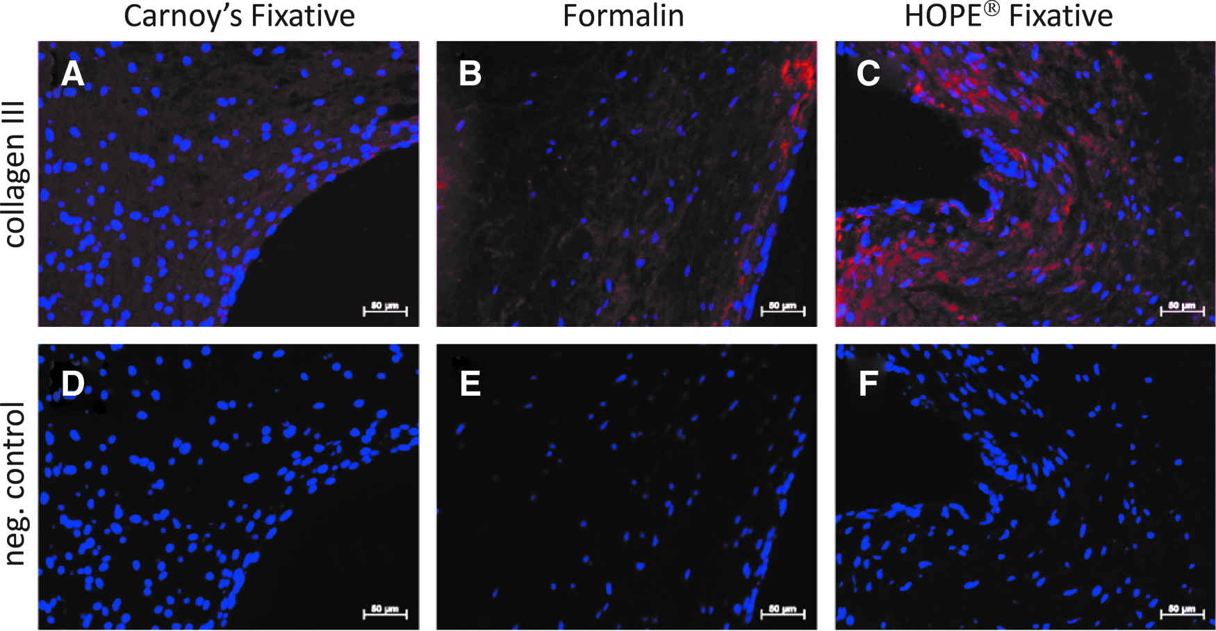
Staining against type III collagen after Carnoy's-
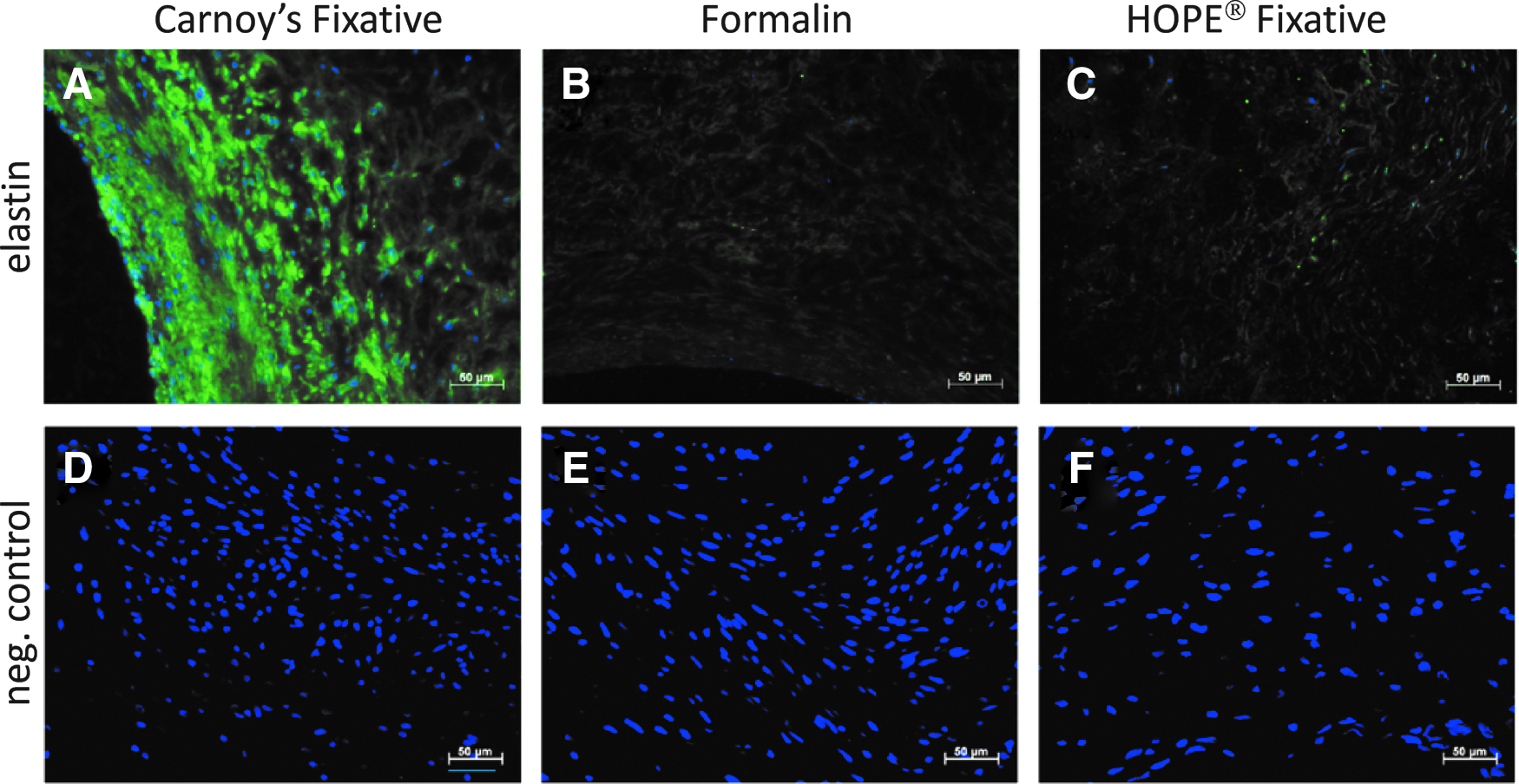
Independent of the fixation method, all samples stained positively for elastin (Fig. 7), with an equal level of fluorescence intensity. The spatial distribution of elastin expression was similar across all samples without any significant background staining observable.
Staining against elastin after Carnoy's-
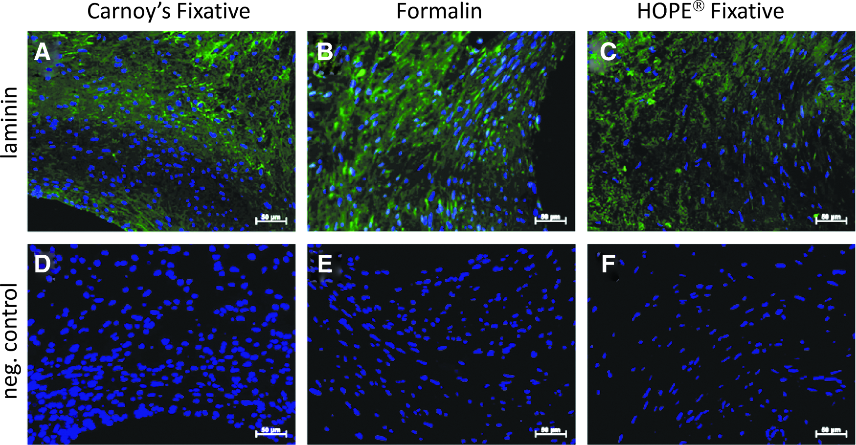
Carnoy's-fixed samples demonstrated a much stronger immunoflurescence signal for laminin, with only sparse staining of this antigen evident in both formalin- and HOPE-fixed samples (Fig. 8).
Staining against laminin after Carnoy's-
The DAPI-stained nuclei in formalin- and HOPE-fixed samples exhibited a stretched appearance, whereas nuclei in Carnoy's-fixed samples demonstrated a round to oval configuration.
Discussion
The gold standard for tissue preservation and subsequent processing is fixation with formalin and subsequent paraffin embedding. 5 However, formalin harbors several disadvantages: it alters nucleic acids and protein integrity and obscures antigens by structural modification, thus hampering adequate immunological analysis of these tissues. Still, formalin fixation is the easiest method for preserving tissue as formalin is available as a ready-to-use solution, while most other fixatives depend on the use of multiple solutions or have to be prepared just prior to use.
In cases of immunohistochemical analysis following formalin fixation, epitope retrieval methods are mandatory to make antigen sites accessible for analysis. The most popular unmasking techniques are enzyme digestion and HIER. 20 Different antigens may be selectively unmasked by one antigen retrieval method and not another, making this process complex and time consuming. Additionally, as shown in the present study, all tested and commonly used antigen retrieval methods significantly impair the delicate structure of in vitro-prepared tissue-engineered constructs, rendering them impractical for further histological analysis of tissue and cellular proteins.
Alternative fixation methods have been developed to enhance structural preservation and antigenicity, such that no additional destructive antigen retrieval methods are required. Carnoy's fixative does not mask the target protein epitopes in the manner of formalin, thereby preserving antigen immunoreactivity. The rapid action of Carnoy's fixative can save time in tissue fixation and the process can easily be automated on account of its similarity to formalin fixation. The composition of HOPE fixative is undisclosed intellectual property of DCS Diagnostics. In comparison with formalin- and Carnoy's fixation, the use of HOPE fixative is time consuming, and as processes are very different from those used for formalin fixation, it is less easily automatable.
Carnoy's fixative was shown to preserve overall macroscopic and microscopic structure in the same manner as formalin, as shown in Figure 1. HOPE fixative dramatically altered the macroscopic appearance of the tissue-engineered constructs. H&E staining of HOPE-fixed constructs also revealed distinct changes in overall tissue structure that rendered them impractical for further morphological analysis.
The results presented in Figures 3–8 clearly demonstrate that the quality of immunohistochemical staining significantly differs depending on the fixation and dehydration method employed. Unfortunately, none of the tested fixatives resulted in adequate quality of immunohistochemical staining of tissue-engineered structures for all antigens tested. Indeed, the use of formalin fixation without antigen retrieval was the least favorable technique employed for all antigens, except for type III collagen. Though immunohistochemical staining quality was high following HOPE fixation, showing bright fluorescence signals without disturbing background staining, the method seems inappropriate for the analysis of tissue-engineered constructs as its application leads to significant alteration of the tissue structure.
Conclusion
Histology and immunohistochemistry are the gold standard techniques in the evaluation of tissue-engineered constructs. The standard fixative, formalin, requires an antigen retrieval process that can significantly destroy delicate constructs produced in vitro. Based on the present study, we recommend that Carnoy's fixative is employed for the preservation of tissue-engineered constructs to allow for immunohistochemical analysis of types I- and III collagen, elastin, laminin,α-SMA, and vimentin. However, it is clear that the technique requires optimization based on the particular tissue engineering application. Due to its ease of use, formalin is an attractive alternative whenever routine histological staining is the prime goal.
Footnotes
Acknowledgment
Martina Tappe at the IZKF Aachen provided assistance on antigen retrieval methods.
Disclosure Statement
The authors declare that no competing financial or other interests have influenced this study. None of the authors is financially affiliated with any of the companies mentioned.