
Abstract
Though recent studies report decisive positive effects on cells, elicited by ultraviolet (UV)-induced bioactivation of biomaterial implant surfaces, they frequently employ cells other than of human origin or cells not representing oral implant targets. Therefore, the present study aims at exploring distinct cell functions of primary human alveolar bone osteoblasts (PHABO) in response to bioactivated microstructured titanium and zirconia implant surfaces with matched controls. UV-treatment significantly reduced surface carbon, while concomitantly increasing wettability. In case of titanium or zirconia biomaterial source of equal roughness, bioactivation did not significantly improve cell functions, including initial cell attachment, morphogenesis, proliferation, and gene expression of osteogenic biomarkers osteocalcin, alkaline phosphatase and collagen type I. However, cell functions discriminated surface roughness by either comparing titanium and zirconia or interindividual zirconia surfaces. While rough surfaces primarily favored primary adhesion, proliferation appeared improved on smooth surfaces, and gene expression seemed to be stronger modulated on the smoothest biomaterial. Our results show for the first time that bioactivation appears to be not the main causative for the observed modulation of the distinct cell functions analyzed in PHABO, but add to the body of evidence that they were more governed by surface architecture rather than by bioactivation.
Introduction
Another noncommercially applied method to increase surface hydrophilicity of titanium implants is the photochemical modification of titanium dioxide surfaces by ultraviolet (UV) light irradiation, which has been shown to improve initial cell response and early bone apposition on implant surfaces; therefore, often termed bioactivation.10,11 The UV-induced bioactivation is presumably caused by the removal of adsorbed hydrocarbons and other carbonaceous species by titanium dioxide-mediated photooxidation and/or by the photo-induced production of surface oxygen vacancies resulting in a higher reactivity of the titanium surface, which then is susceptible for dissociative water adsorption.12,13 Such a photochemical surface modification was recently reported for zirconia implant materials as well. The bioactivation of machined zirconia surfaces was thereby associated with increased surface hydrophilicity, progressive removal of hydrocarbons and enhanced initial osteoblast response. 14 Hence, UV-induced bioactivation represents a promising method to improve surface chemistry of zirconia-based implant materials. In this context, it should be noted that despite the gained knowledge concerning machined zirconia, zirconia-based UV-bioactivation studies employing other surface topographies do currently not exist.
However, the vast majority of studies aiming at examining cell response to implant materials utilize cells other than those of human origin or cells not representing oral implant targets. Frequently used cells in these experimental studies include human osteosarcoma1,2,15 and mouse myoblast cell lines, 11 human mesenchymal stem cells, 10 osteoblasts derived from rat bone marrow, 14 or primary rat osteoblasts. 16 Regarding this issue, numerous investigations have demonstrated significant differences in cell reactions in response to different culture conditions, such as the use of hormones and growth factors, as well as biomaterial surface characteristics. These differences were most likely related to the cell source and phenotype responding to the given experimental conditions.17–21 For instance, cell cultures using osteoblasts and mesenchymal stem cells derived from different species on microstructured titanium surfaces showed that cell reactions were influenced by surface chemistry and each cell type displayed different preferences in terms of chemical surface properties for optimal function, 22 while human bone marrow stromal cells derived from iliac crest and alveolar bone were shown to exhibit individual characteristics in terms of differentiation potential depending on their anatomical location. 23 In addition, preliminary experiments conducted by the own group revealed cell source-dependent differences in cell adhesion and morphogenesis between human mesenchymal stem cells and primary human osteoblasts in three-dimensional scaffolds. Therefore, the objective of this study was to examine the effect of UV-induced bioactivation of commercially available microstructured titanium- and two zirconia-based implant biomaterials on specific cell functions of implant-relevant target cells, that is, primary human alveolar bone osteoblasts (PHABO). Moreover, this is to date the first in vitro study characterizing the cell response of osteoblasts derived from human alveolar bone to zirconia-based oral implant surfaces. For this purpose, we first performed surface characterization of the microstructured titanium and zirconia implant biomaterials to describe their topography and chemical properties after UV-induced bioactivation. This was followed by the cultivation of PHABO under basal conditions devoid of stimulatory factors and/or osteogenic differentiation medium on UV-bioactivated and untreated control implant material surfaces, with subsequent examination of cell functions, including morphogenesis, initial cell attachment, proliferation and gene expression of osteogenic markers.
Materials and Methods
Implant materials and sample treatment
Titanium and zirconia disks (20 mm in diameter; 1.5 mm thickness) of the following commercially available implant materials were used for surface analysis and cell culture experiments: (1) electrochemical anodized titanium (TiUnite; NobelBiocare, Gothenburg, Sweden), (2) alumina sandblasted zirconia (Zit-Z; Ziterion, Uffenheim, Germany), and (3) alumina toughened zirconia with a porous zirconia surface layer (ZircaPore; Metoxit, Thayngen, Switzerland). Titanium and zirconia disks were sterilized by low-temperature hydrogen peroxide gas plasma sterilization STERRAD 100/100S (Advanced Sterilization Products [A.S.P], Johnson & Johnson Medical, Irvine, CA) and stored in dark for 7 days before usage. The surfaces of different materials were exposed to UV light with 345 J/cm2 UV-C and 17 J/cm2 UV-A light in a UV irradiation chamber (BS-02, Dr. Gröbel UV-Elektronik GmbH, Ettlingen, Germany) under controlled conditions.10,14 UV-treated disks were used immediately after complete bioactivation for surface analysis or cell culture experiments.
Scanning electron microscopy
The surface morphology of the implant materials and the morphogenesis of PHABO on UV-bioactivated disks and nonactivated controls were examined by scanning electron microscopy (SEM) using a LEO435VP scanning electron microscope (Zeiss, Oberkochen, Germany). The specimens from cell culture experiments were fixed with 4% formaldehyde in PBS for 20 min at room temperature. Specimen was dehydrated in ascending ethanol series (ranging from 30% to 100% ethanol, three times each for 20 min at room temperature), critical point dried (CPD030 Critical Point Dryer; Bal-Tec AG, Balzers, Liechtenstein) and immediately sputter coated with gold palladium for 60 s at 60 mA (SCD050; Balzers, Liechtenstein).
Interferometry
Surface topography of the titanium and zirconia implant materials was characterized by interferometry (IFM). Three disks of each implant surface were measured with an interferometer (MicroXam; Phaseshift, Tucson, AZ). The measuring area was set to 260×200 μm with a zoom factor of 0.62 resulting in a magnification of 31.6. Each disk was measured at three different areas; thus, 9 measurements were performed for each implant surface.
To characterize the surface in height, spatial and surface enlargement three parameters were selected: Sa describes the average height distribution in μm, Sds measures the density of summits over the evaluated area in 1/mm2, and Sdr describes the surface enlargement compared to a totally flat reference area in % or as a ratio. Before parameter calculation, a digital (Gaussian) filter of 50×50 μm was applied to remove errors of form and waviness.
X-ray photoelectron spectroscopy
The quantitative mean atomic composition of UV-bioactivated and nonactivated implant surfaces in terms of hydrocarbon and nitrogen contaminations was examined by X-ray photoelectron spectroscopy (XPS). A measuring area of 800 μm in diameter of each sample was analyzed in a Perkin Elmer PHI 5600 ESCA System (Perkin Elmer, Waltham, MA) with a magnesium X-ray source (1253.6 eV anode energy; 13 kV X-ray voltage; 300 W anode power). Survey spectra were measured (187.85 eV pass energy) with a step size of 0.8 eV and detail spectra (23.5 eV pass energy) of C1s and N1s peaks with 0.025 eV per step. All spectra were normalized to the binding energy scale of the C1s peak (285 eV).
Contact angle measurement
Surface wettability of bioactivated and nonactivated implant surfaces was examined by contact angle measurement. Contact angles of 1 μL water droplets were analyzed visually using the Dataphysics OCA 10 optical contact angle measuring system (Dataphysics GmbH, Filderstadt, Germany) by measuring the angles of each drop against the surface.
Isolation and cultivation of PHABO
Human osteoblasts were prepared from alveolar bone explants obtained from a 42-year old healthy male patient during implant site preparation procedure as previously described. 17 The collection of oral bone samples was approved by the Ethics Committee of the Albert-Ludwigs-University, Freiburg, Germany (vote Nr. 411/08). Osteoblasts derived from alveolar bone fragments were cultured in Dulbecco's Modified Eagle's Medium (PAA Laboratories, Coelbe, Germany) supplemented with 1% (w/v) glutamine (Gibco, Invitrogen, Karlsruhe, Germany), 10% (w/v) fetal calf serum (Biochrom AG, Berlin, Germany) and 0,2% (w/v) kanamycin (Sigma-Aldrich, Taufkirchen, Germany). The cells were maintained in a humidified 37°C incubator with 5% CO2. All experiments were carried out with osteoblasts of passage 5 and 6.
Indirect immunofluorescence
Cell adhesion and morphology of PHABO on UV-bioactivated and nonactivated zirconia and titanium disks were examined by indirect immunofluorescence (IIF) of vinculin and phalloidin-labeling of actin. Therefore, cells were fixed with 4% formaldehyde after 4 and 24 h incubation on UV-treated and untreated titanium- and zirconia-based implant surfaces.
For IIF, the following antibodies and staining reagents were used: mouse anti-vinculin (1:100; Abcam, Cambridge, UK), AlexaFluor 488-conjugated goat anti-mouse IgG (1:200; Invitrogen), Texas RedR-X phalloidin (1:40, Invitrogen), DAPI nucleic acid stain (300 nM, Invitrogen). Before incubation with the primary antibody, specimens were treated with 2% (w/v) bovine serum albumin (Sigma-Aldrich) and 0.2% TritonX-100 (Sigma-Aldrich) in PBS for 15 min at room temperature, and 2% (w/v) bovine serum albumin in PBS for further 15 min at room temperature. Primary and secondary antibodies, as well as phalloidin conjugate were diluted in PBS containing 0.5% (w/v) BSA and were then incubated for 1 h (antibodies) or 30 min (phalloidin conjugate) at 37°C. Nuclei were stained with DAPI for 5 min at room temperature. Optical evaluation was performed with the fluorescence microscope Biozero BZ-8000 (KEYENCE, Neu-Isenburg, Germany). For quantitative morphometric osteoblast analysis after 4 and 24 h of culture on examined biomaterials, we used the microscope software BZ Analyzer II from KEYENCE. To achieve statistically valid data, cell morphology of 30 cells per disk was characterized by measuring the software-innate Major axis, describing the long side of the smallest rectangle drawn around the cell body, and the Minor axis, presenting the rectangle width (Fig. 5B).
DNA quantification
Cell cultivation on UV-bioactivated and nonactivated titanium and zirconia disks was examined by determining the DNA content of cells which have successfully attached on the surfaces after 4 and 24 h incubation. For this, attached cells were washed once with PBS and lysed by a freeze-thaw cycle at −80°C in 400 μL TE-buffer (10 mM Tris-HCl, 1 mM EDTA, pH7.5). DNA quantification was performed with the Quant-iT™ PicoGreen assay (Invitrogen) according to the manufacturer's instructions.
Cell proliferation assay
Proliferation of PHABO on UV-bioactivated disks and nonactivated controls was measured by the alamarBlue assay. Therefore, culture medium was replaced after 3, 7 and 14 days culture with medium containing 10% (w/v) alamarBlue® reagent (AbD Serotec, Düsseldorf, Germany). After incubation for 2 h at 37°C, samples of the supernatant were analyzed by measuring fluorescence according to the manufacturer's instructions. Percentage of alamarBlue reduction in the samples was calculated using a 100% reduced alamarBlue control as reference, which was produced according to the manufacturer's protocol by autoclaving a sample containing culture medium with 10% (w/v) alamarBlue reagent for 15 min.
Gene expression analysis
The relative gene expression of bone-specific markers collagen type I α 1 (COL1A1; RefSeq# NM_000088), alkaline phosphatase (ALPL; RefSeq# NM_000478) and osteocalcin/bone gamma-carboxyglutamate-(gla)-protein (OC/BGLAP; RefSeq# NM_199173) was assessed on mRNA level by semi-quantitative real-time RT-PCR. Total mRNA of three samples per material and surface treatment was pooled and isolated after 7 and 14 days culture period (RNeasy Mini Kit; Quiagen, Hilden, Germany), and analyzed with the Experion Automated Electrophoresis System (Bio-Rad Laboratories, Munich, Germany). Synthesis of cDNA was performed with 500 ng mRNA using the RT2 PCR Array First Strand Synthesis Kit (SABiosciences, Qiagen, Hilden, Germany) according to the manufacturer's protocol. Real-time PCR reactions were carried out with the CFX96™ Real-Time PCR Detection System (Bio-Rad Laboratories) using 10 μM specific primers and RT2 SYBR Green qPCR Master Mix from SABioscience (Qiagen), and cDNA equivalent to 30 ng of total mRNA. Relative mRNA expression of COL1A1, ALPL and BGLAP was normalized to the housekeeping genes ribosomal protein L13a (RPL13A; RefSeq# NM_012423) and glyceraldehyde-3-phosphate dehydrogenase (GAPDH; RefSeq# NM_002046), and analyzed using the comparative ΔΔCT method. Concerning these two reference genes, the Gene Expression Macro software provided with the Bio-Rad CFX-System allows for calculation of a reference gene index, consisting of the averaged endogenous controls GAPDH and RPL13A. This gene index has been used as basis for the ΔΔCT method.24,25 Relative gene expression of examined biomarkers in osteoblasts on UV-bioactivated implant surfaces was compared with matched controls (gene expression of osteoblasts on untreated controls was defined as 1×).
Statistical analysis
Cell culture experiments were performed in triplicates in three independent experiments. Data of quantitative morphometry, DNA concentration, alamarBlue reduction, and gene expression were compared for statistically significant differences using the Student's t-test (p<0.05). For characterization of surface topography by IFM, we analyzed three disks of each implant surface and performed three measurements per disks; thus, 9 measurements were performed for each implant material. Surface parameters of each material were compared for statistically significant differences by the one way ANOVA test (p<0.05).
Results
Morphological and topographical characterization of titanium and zirconia surfaces
Since we used microstructured implant surfaces which were fabricated by different manufacturing processes, we were interested in the global surface architecture of the biomaterials and thus, characterized the morphology and topography of the three surfaces by SEM and IFM analysis.
In Figure 1, SEM micrographs of the TiUnite (Fig. 1A), Zit-Z (Fig. 1B) and ZircaPore (Fig. 1C) surfaces revealed significant differences in terms morphology and microtopography. TiUnite and ZircaPore displayed—different to Zit-Z—microporous topography with an overall heterogeneous distribution of the microstructures. The micropores of the anodized TiUnite surface varied thereby in diameter (0.56 to ∼2.45 μm), in shape and in the presence of distinct protrusions at the pore edges. Furthermore, the interspaces between the micropores showed a smooth surface texture with extended cracks (Fig. 1A, arrows). In contrast, the sandblasted Zit-Z surface had a homogenous surface topography with fractal architecture and a grainy texture (Fig. 1B). The ZircaPore surface consisted of a heterogeneous distributed porous ceramic layer exhibiting areas devoid of the porous surface coating (Fig. 1C, asterisk and Fig. 1F, arrows). In contrast to the TiUnite surface, the porous surface layer of ZircaPore exhibited high interconnectivity of the pores.
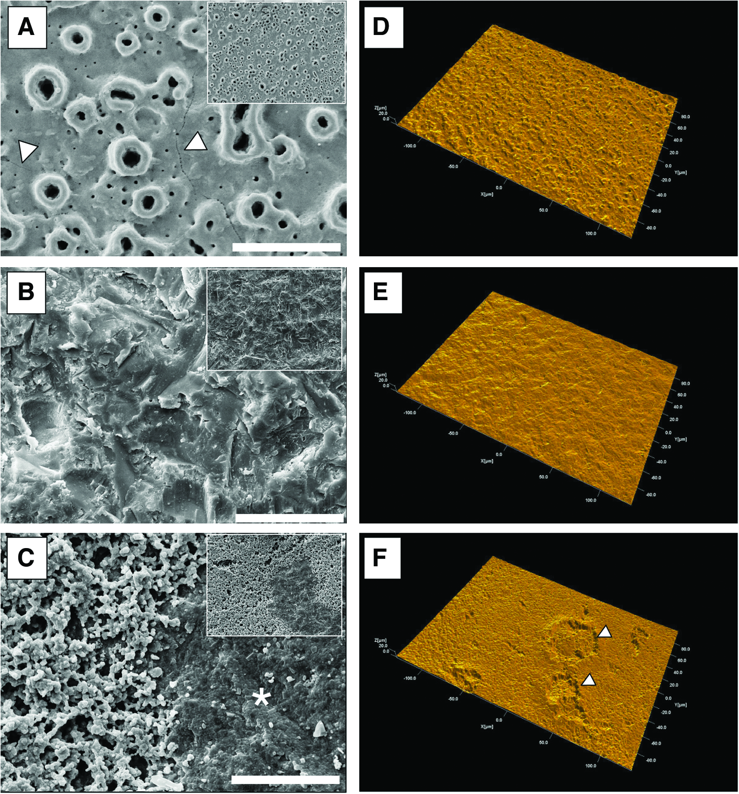
Scanning electron micrographs
Figures 1D–F show representative interferometer 3D reconstructed images of the TiUnite (Fig. 1D), Zit-Z (Fig. 1E) and ZircaPore (Fig. 1F) surface. In terms of height deviation (Sa values) and surface enlargement (Sdr values), TiUnite demonstrated the largest height deviation (Table 1, Sa=1.09±0.1) and surface enlargement (Table 1, Sdr=67.3±5.3), while Zit-Z demonstrated the smallest (Table 1, Sa=0.58±0.0 and Sdr=19.3±3.8). The ZircaPore surface had a medium range of height deviation (Table 1, Sa=0.77±0.2) but had significantly higher number of peaks per area unit and therefore, had the highest density of summits (Table 1, Sds=260632±6166) among the three investigated surfaces. A high density of peaks will contribute to surface enlargement. This explains that the Sa value was lower for the ZircaPore rather than for the TiUnite surface. However, the surface enlargement still was similar for these two surface modifications. In summary, Zit-Z demonstrated the smoothest surface, while TiUnite and ZircaPore had a more pronounced though similar overall surface roughness.
Surface parameters describing the topography of implant surfaces were, (1) Sa (average surface height deviation amplitude), (2) Sds (density of summits, i.e., number of peaks per area) and (3) Sdr (surface enlargement compared to a totally flat reference area). All mean values showed statistically significant differences between implant materials (One way ANOVA; p<0.05).
UV light treatment induces removal of surface carbon on titanium and zirconia
The UV light-induced bioactivation of titanium and zirconia implant materials is among others supposed to be associated with the removal of adsorbed surface hydrocarbons.13,14 For this reason, we evaluated the mean atomic composition of the bioactivated implant surfaces in terms of surface pollution by hydrocarbons and nitrogen contamination from the air by XPS analysis and compared them with untreated controls.
Figure 2 shows representative survey XPS spectra of the bioactivated TiUnite (Fig. 2A), Zit-Z (Fig. 2B) and ZircaPore (Fig. 2C) surfaces and the respective untreated controls. Irrespective from the implant biomaterial, XPS analysis revealed significant reduction of the carbon-specific peak (Fig. 2A–C, inset: surface carbon peak C1s with (red) and without (blue) bioactivation) after UV-treatment of the surfaces. The XPS spectra of the examined implant materials revealed a significant reduction of the surface carbon ratio after UV light treatment of up to 32%. Thereby, the atomic percentage of carbon on TiUnite and ZircaPore surfaces declined to a similar extent from 39% and 38% to 7% and 8%, respectively. The Zit-Z surface showed an overall higher carbon ratio compared to TiUnite and Zit-Z, which was 48% for untreated disks and 18% for UV-treated samples. UV light treatment showed no effect on surface nitrogen ratio, which was 2% for untreated TiUnite and ZircaPore surfaces, and 3% and 2% for UV-treated surfaces, respectively. Irrespective of UV light treatment no surface nitrogen was detectable on Zit-Z surfaces.
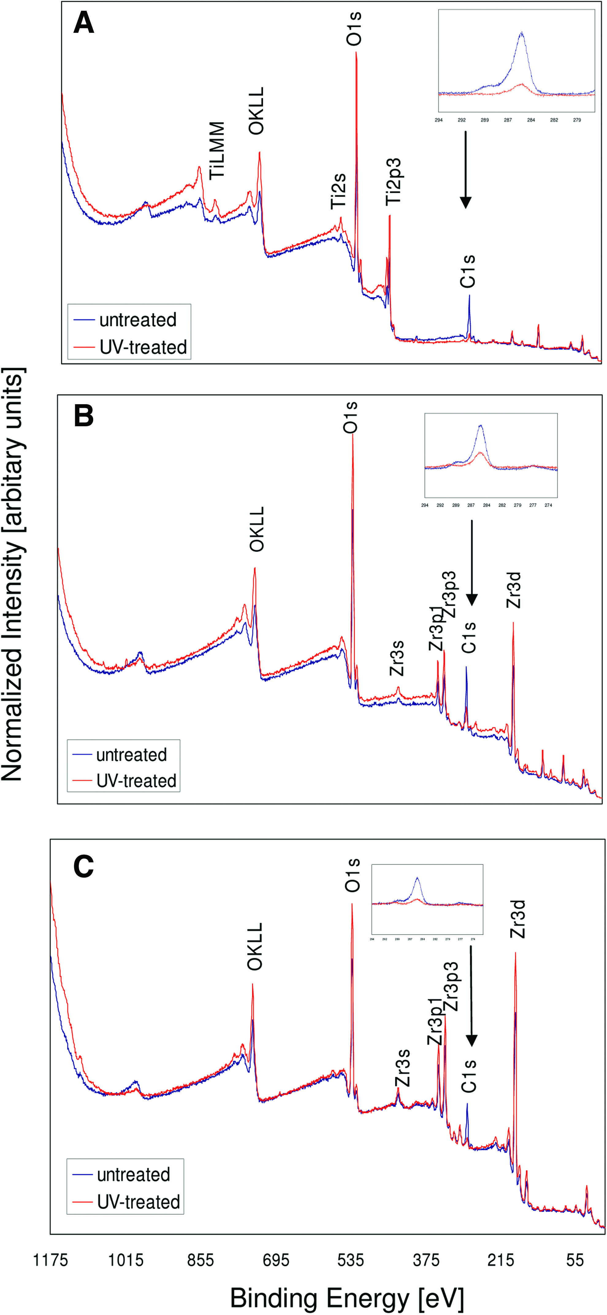
X-ray photoelectron spectroscopy (XPS) survey spectra of ultraviolet (UV)-treated (red) TiUnite
UV light treatment increases surface wettability of titanium and zirconia
Since XPS analysis showed effective removal of surface carbon on implant materials by UV light treatment, we were next interested in the hydrophilic status of the bioactivated implant materials. Therefore, contact angle measurements were performed with UV-treated and untreated titanium and zirconia disks.
The contact angle of water droplet on untreated implant surfaces was 32.5° for TiUnite, 87° for Zit-Z and 91.9° for ZircaPore, and decreased after UV light treatment to 8.5°, 41.7°, and 18.1°, respectively. All untreated implant surfaces showed remarkable differences in surface hydrophilicity, with TiUnite demonstrating the highest surface wettability and ZircaPore the lowest. UV light increased surface wettability on the different implant material surfaces to a different degree. The contact angle on the TiUnite surface was reduced by 74%, on Zit-Z by 52% and on ZircaPore by 80%. This resulted in Zit-Z showing the lowest surface wettability after UV light treatment, while TiUnite retained the highest and ZircaPore had an intermediate hydrophilic status.
Bioactivation of implant surfaces yields no obvious differences in osteoblast morphogenesis
Surface characterization of TiUnite, Zit-Z and ZircaPore by SEM, XPS and contact angle measurement revealed significant differences in surface topography between the examined implant materials, while UV light-mediated removal of surface carbon contaminations accompanied by an increase in surface wettability. Since surface topography and chemistry may have serious impact on initial cell response to implant surfaces, we next focused on morphogenesis of PHABO on bioactivated and nonactivated TiUnite, Zit-Z, and ZircaPore surfaces during the first 24 h of culture.
As revealed by SEM, cell morphology on UV-treated implant surfaces did not differ considerably from untreated controls after 24 h, indicating that osteoblast morphogenesis on microstructured implant surfaces was not affected by UV light-induced bioactivation (data not shown). In contrast, as illustrated in Figure 3, intersurface comparison between titanium and zirconia surfaces following bioactivation yielded different osteoblast morphologies as documented by SEM micrographs. Cells on TiUnite (Fig. 3A) demonstrated a dendritic spreading, branched spindle shaped extensions on Zit-Z (Fig. 3B) and a homogeneous elongated morphology on ZircaPore (Fig. 3C). Furthermore, osteoblasts cultured on Zit-Z, representing the smoothest of the examined surfaces, seemed to display the most flattened morphology compared to TiUnite and ZircaPore (compare Fig. 3B with Fig. 3A, C). These results give a first hint that osteoblast morphogenesis was dependent from surface topography, rather than from UV light-mediated bioactivation.
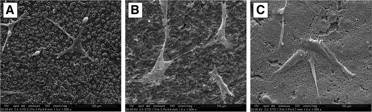
Scanning electron micrographs of primary human alveolar bone osteoblasts (PHABO) incubated for 24 h on bioactivated TiUnite
A similar situation was observable by fluorescence microscopy of green-labeled focal adhesion (FA) protein vinculin and red-labeled actin cytoskeleton in osteoblasts on bioactivated and nonactivated titanium and zirconia biomaterial implant surfaces. While after 4 h red-labeled actin cytoskeleton and green fluorescent adhesion site constituent vinculin exhibited fairly similar distribution patterns irrespective from bioactivation (representative data from UV-treated surfaces are shown in Fig. 4) and surface roughness (Fig. 4A: TiUnite, B: Zit-Z, and C: ZircaPore), culture periods of 24 h showed discriminative patterns regarding surface topography. For rough biomaterial surfaces TiUnite and ZircaPore, actin fluorescence appeared preferentially at the lateral and apical cell border sites (Fig. 4D, F, arrows), concomitant with vinculin oriented along the longitudinal axis of cell orientation (Fig. 4D, F, merge of red-labeled actin and green-labeled vinculin depicted as insets). This pattern suggests existence of actin stress fibers due to tension sites at the lateral sites and apical cell margins, coinciding with cell adhesion by potential focal contacts. By contrast, the smoother Zit-Z biomaterial implant surface generally displayed actin and vinculin abundance with a more cytoplasmic distribution throughout the entire cell soma (Fig. 4E, arrows in the large icon and merge of red-labeled actin and green-labeled vinculin in the inset), thereby suggesting a less tensing cell adhesion substratum, as also reflected by a more spread triangular morphology (Fig. 4E). Furthermore, vinculin distribution on TiUnite reflected the porous surface topography of the implant surface (Fig. 4A, arrows in the inset), indicating that vinculin was missing in areas coinciding with large pores. Vinculin is an adaptor protein and key regulator of FA sites, which is recruited from the cytoplasm to FA sites upon activation and interacts directly with talin and actin, leading to clustering of activated integrin. 26 Since the vinculin localization in PHABO is lacking in spots reflecting large pores it can be speculated that the pores are devoid of FA sites and thus, primary cell adhesion, thereby suggesting bridging of the pore gap by the corresponding parts of the cell soma.
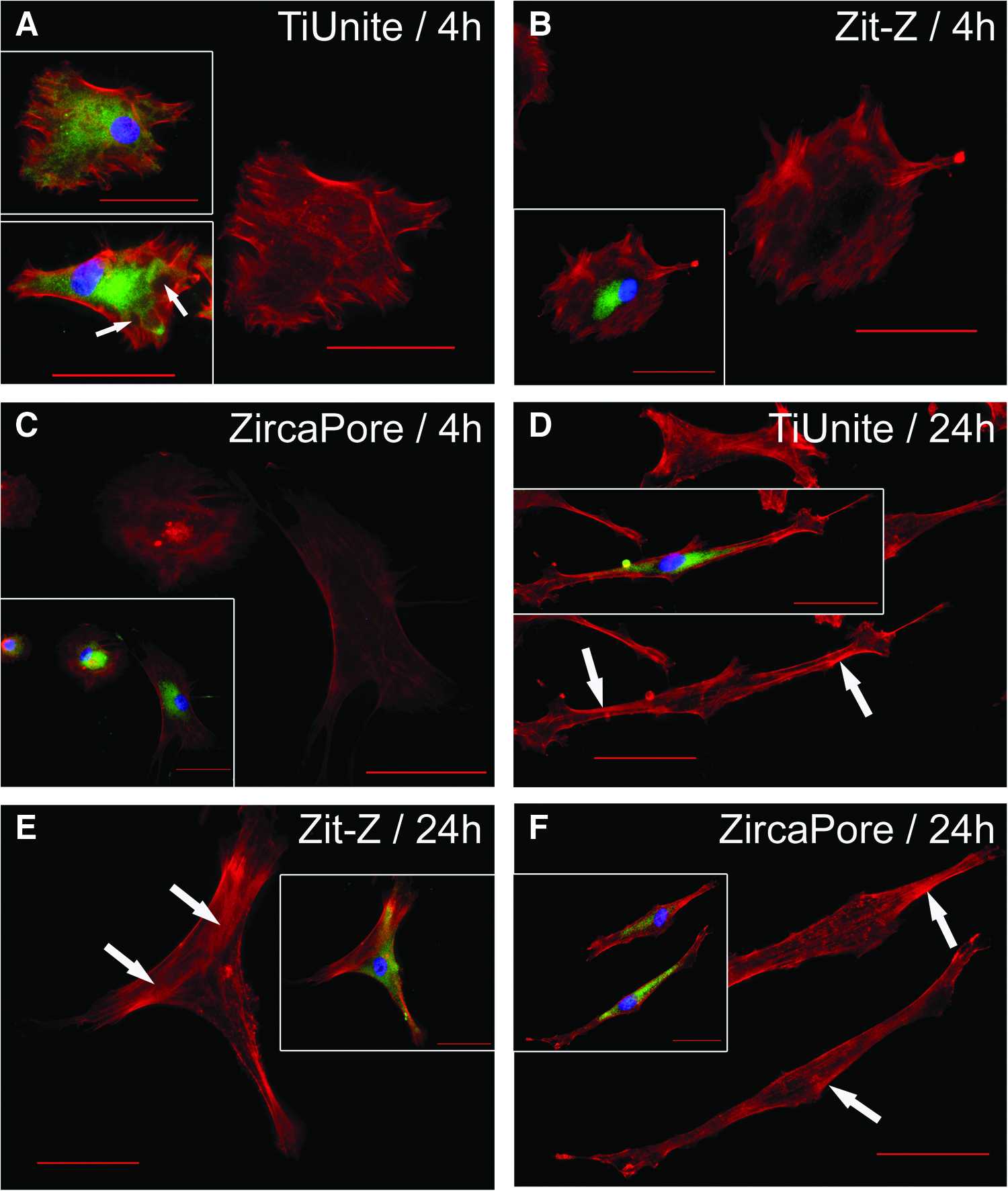
Images show indirect immunofluorescence of focal adhesion protein vinculin (green fluorescence, insets) and phalloidin-labeled actin (red fluorescence, large images) in PHABO on UV-treated TiUnite
The SEM and IIF data provide evidence for morphological differences of PHABO depending on surface topography rather than on UV-treatment. To back up this suggestion, we performed fluorescence imaging software-based quantitative morphometrical cell measuring, revealing comparable values of cell morphology-indicating parameters, namely Major and Minor axis (as summarized in Table 2) of the cell borders (Fig. 5B), on all examined biomaterials after 4 h of culture (Table 2). Irrespective of UV light treatment, osteoblasts at 24 h discriminated between the different surface topographies, clearly showing an elongated morphology on the rougher surfaces TiUnite and ZircaPore, represented by high Major axis and low Minor axis values, and a more spreading morphology on the smoother Zit-Z surface, as indicated by lower Major axis and higher Minor axis values with matched rougher implant surfaces (Table 2). These morphological differences are highlighted in Figure 5A, showing representative data from UV-treated surfaces.
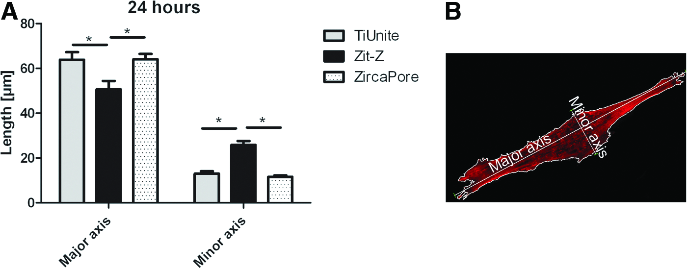
The morphology-indicating parameters Major and Minor axis describe the long side and width of the smallest rectangle drawn around the cell border, and represent the dimension of the cell spreading. Data from 30 cells per disk (mean values±S.E.M.; n=30) are listed. Bold data show statistically significant differences between implant materials (Student's t-test).
S.E.M., standard error of the mean; UV, ultraviolet.
Initial osteoblast attachment on microstructured implant materials is rather influenced by surface topography than by UV-induced bioactivation
Qualitative evaluation of cell adhesion and morphogenesis of PHABO on bioactivated titanium and zirconia implant surfaces by SEM and IIF indicated no obvious effect of UV light treatment on the aforementioned cell functions. Therefore, we further performed quantitative analysis of initial cell attachment on examined implant surfaces by determining the DNA concentration in PHABO cultures after 4 and 24 h incubation on UV-treated and untreated disks. Since the DNA amount correlates directly with cell number, DNA concentration in our samples provided information about the number of adhered cells on the different implant biomaterials.
As shown in Figure 6, DNA concentration generally revealed increased cell numbers at 24 h (Fig. 6A–C). This increase applied to both the intersurface comparison (Fig. 6A–C) as well as bioactivation (UV-treated, black columns) versus nonbioactivation (untreated, grey columns). However, regardless from UV-treatment, significant differences in DNA concentration were detectable between individual implant surfaces at 4 and 24 h. As exemplified for bioactivated surfaces, DNA concentration was significantly lower on Zit-Z (Fig. 6D, E: black columns; 4 h: 6.4±1 ng/mL and 24 h: 11.9±2 ng/mL) when compared to TiUnite (Fig. 6D, E: grey columns; 4 h: 10.4±0.4 ng/mL and 24 h: 17.6±0.8 ng/mL) and ZircaPore (Fig. 6D, E: white columns; 4 h: 11.6±1.4 ng/mL and 24 h: 18.2±2.7 ng/mL) after 4 and 24 h incubation, while the latter two showed comparable DNA amounts at these time points. The DNA amounts for both implant biomaterial treatment modes are summarized in Table 3. Thus, the amount of adherent cells was lowest on the smoothest of the three surfaces (Zit-Z), suggesting that cell adhesion was rather influenced by surface topography than by UV-induced bioactivation or implant core material. In this context, rougher surfaces may appear the more suitable substrate for cell functions, such as primary adhesion.
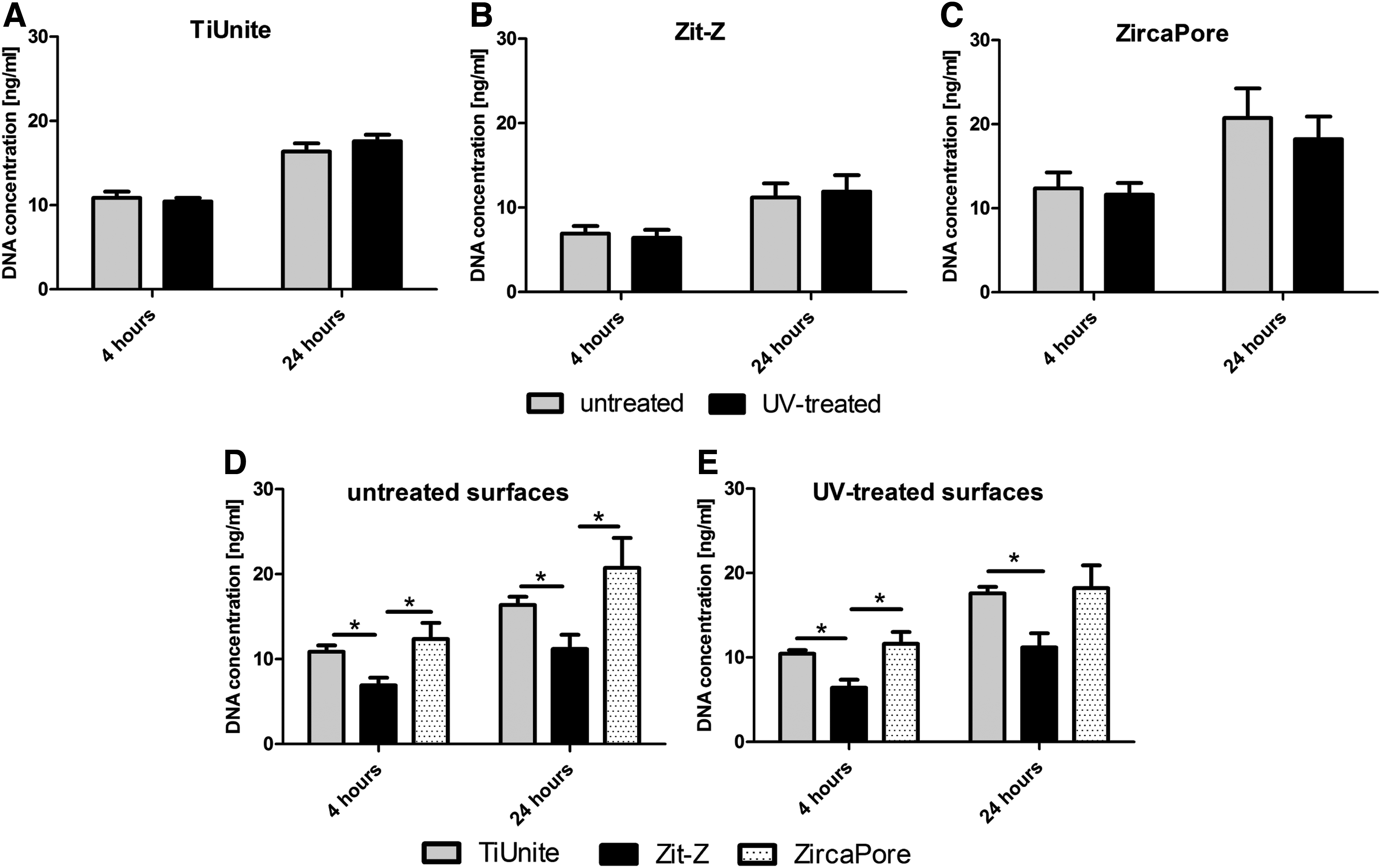
Quantitative analysis of initial cell attachment of PHABO on UV-treated (black columns) and untreated (grey columns) TiUnite
Data from three independent experiments (mean values±S.E.M.; n=9) are listed.
PHABO, primary human alveolar bone osteoblasts.
Osteoblast proliferation appeared favored by smooth surfaces but not by bioactivation
Since quantitative analysis of initial cell attachment yielded comparable results regarding surfaces with and without bioactivation, we were next interested in examining putative UV-dependent effects on cell proliferation. PHABO proliferation was therefore, analyzed by the resazurin-based alamarBlue assay by employing the same culture for the analyzed time points, namely 3, 7 and 14 days.
Assay-based metabolic resazurin reduction indicated PHABO proliferation increase in chronological order on both bioactivated and nonactivated implant surfaces (Fig. 7 and Table 4, giving resazurin reduction in%). For TiUnite no differences were detectable on bioactivated surfaces with matched controls (Fig. 7A, compare black/UV-treatment with grey/untreated columns and Table 4). Interestingly, though proliferation was favored by UV-treatment at day 7 on ZircaPore (Fig. 7C, compare black/UV-treatment with grey/untreated columns and Table 4: 12%±1.5%/+UV, 7.8%±0.7%/−UV), bioactivation appeared adverse in case of Zit-Z at day 14 (Fig. 6B, compare black/UV-treatment with grey/untreated columns and Table 4: 35%±2.5%/−UV, 27%±0.7%/+UV), thereby suggesting that this cell function is not obligatory supported by UV-application.
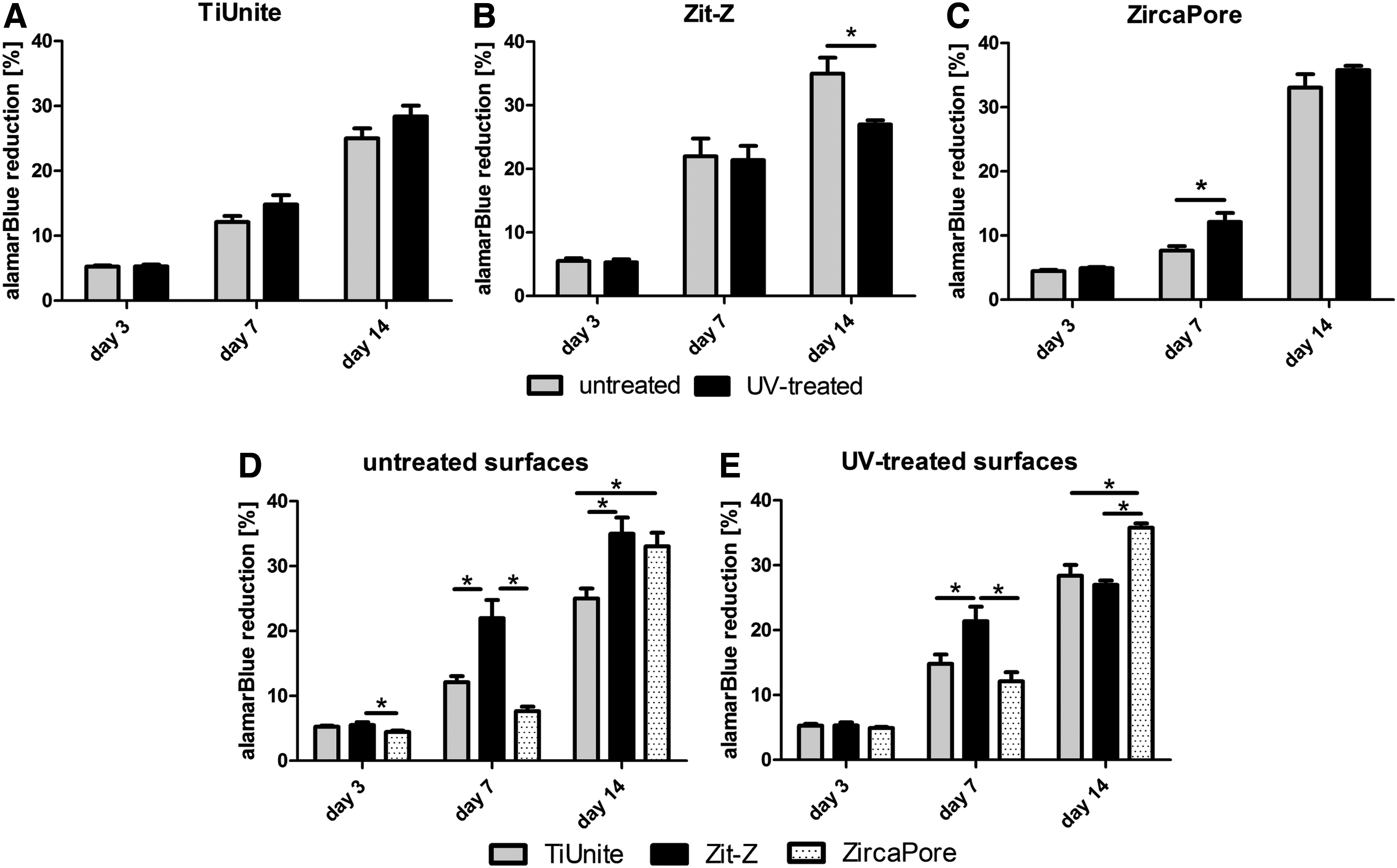
Cell proliferation of PHABO cultured on UV-treated (black columns) and untreated (grey columns) TiUnite
Percentage of alamarBlue reduction in the samples was calculated using a 100% reduced alamarBlue control as reference. Data from three independent experiments (mean values±S.E.M.; n=9) are listed.
Comparing osteoblast proliferation on individual biomaterials (Fig. 7D, E), our data revealed that at early culture periods, as indicated at day 3, cell proliferation was comparable on all bioactivated surfaces (Fig. 7E, compare grey/TiUnite, black/Zit-Z and white/ZircaPore columns and Table 4) and nonactivated implant biomaterials, except ZircaPore (Table 4 and Fig. 7D, compare grey/TiUnite and black/Zit-Z columns with white column/ZircaPore). Intriguingly, when compared to primary adhesion, proliferation may be favored by the smoother Zit-Z biomaterial, as suggested by significance of proliferation increase, preferentially detected at the nonactivated surface stage at day 7 (Table 4, alamarBlue reduction: 22% Zit-Z and 12.1% TiUnite and 7.7% ZircaPore) and in parts at day 14, respectively (Fig. 7D, compare black Zit-Z with grey and white columns, and see also Table 4). This proliferation situation also held true for UV-activation at day 7 (Fig. 7E), while for day 14 UV-treatment appeared unfavorable for smoother Zit-Z, thereby strongly suggesting that also in case of proliferation the surface topography might be of more importance rather than bioactivation (Fig. 7E, compare black/Zit-Z with white/ZircaPore columns, and see also Table 4).
Gene expression analysis for osteogenic biomarkers revealed independence from UV-treatment of microstructured biomaterial implant surfaces
Our previous experiments support the growing evidence that the microstructured implant topography may be superior over bioactivation in governing the yet analyzed cell functions, including morphogenesis, initial cell adhesion, and proliferation. To test for putative effects on osteogenic differentiation by UV-induced bioactivation of microstructured implant surfaces, we quantified mRNA synthesis of respective biomarkers, including COL1A1, ALPL and OC/BGLAP in PHABO at day 7 and 14. While BGLAP encodes for the protein OC, which plays an important role in regulating bone-matrix mineralization and is considered as late osteoblast differentiation marker, 27 COL1A1 is another important bone-matrix protein, representing the predominant collagen in mineralized bone, and is characteristic for the osteogenic lineage. 27 The ALPL enzyme, known as early bone differentiation marker, hydrolyses monophosphate esters, and is involved in bone-matrix mineralization by providing osteoblasts with inorganic phosphate for continuous formation of hydroxyapatite crystals.27,28
Figure 8 and Table 5 summarize the relative gene expression levels of osteoblasts on bioactivated TiUnite (Fig. 8A), Zit-Z (Fig. 8B) and ZircaPore (Fig. 8C) surfaces at day 7 (grey columns) and 14 (black columns) with references to corresponding nonactivated controls (Fig. 8: gene expression of controls was defined as 1×and indicated as dotted line). Our results from the gene expression analysis revealed that UV-bioactivation obviously yielded only moderate modulation, as reflected by up and downregulation of osteogenic biomarkers on almost all biomaterials, except ZircaPore, with effects most pronounced on the smoothest Zit-Z surface. This observation indicated that the smooth surface may be most sensitive in response to UV-treatment. In detail, while fairly equal mRNA levels were detected for all three osteogenic genes under study in case of PHABO cultured on the ZircaPore surface (Fig. 8C; expression value range from 0.88–0.99, Table 5), the other rough surface, TiUnite, displayed significant down-regulation for ALPL (0.68±0.12, Table 5), and marginal down-modulation for COL1A1 (Fig. 8A; 0.78±0.22, Table 5) at day 7. For the smooth Zit-Z implant biomaterial, UV-bioactivation yielded significant up-regulation in gene expression for COL1A1 at day 14 (1.45±0.24, Table 5), while for ALPL at day 14 (1.48±0.88, Table 5) and BGLAP at day 7 (1.3±0.57, Table 5) only slightly elevated levels could be seen, respectively (Fig. 8B).
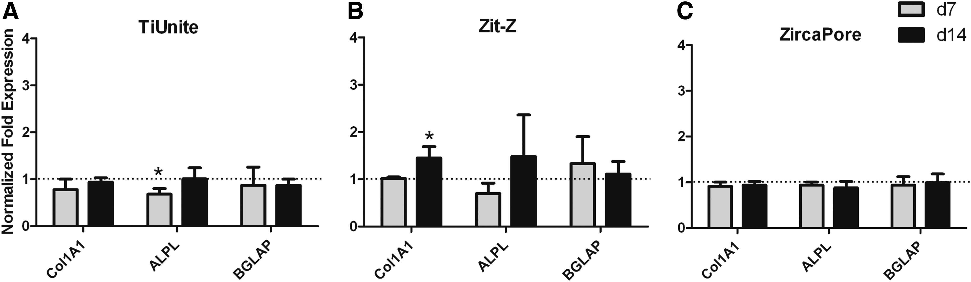
Relative gene expression of collagen type I α1 (COL1A1), alkaline phosphatase (ALPL) and osteocalcin/bone gamma-carboxyglutamate (gla) protein (OC/BGLAP) in PHABO at 7 (grey columns) and 14 (black columns) days culture on UV-bioactivated TiUnite
Expression levels were normalized to the calculated reference gene index, consisting of the averaged endogenous controls ribosomal protein L13a (RPL13A) and β-actin (ACTB), and to the matched untreated controls (gene expression of osteoblasts on untreated controls was defined as 1×). Data from three independent experiments (mean values±SD; n=3) are listed.
COL1A1, collagen type I α 1; ALPL, alkaline phosphatase; OC/BGLAP, osteocalcin/bone gamma-carboxyglutamate-(gla)-protein.
Discussion
Since it is a well established concept now that initial bone cell response to implant materials is modulated by the surface structure and chemical properties of the materials, many efforts have been made to improve the bioactivity of implant biomaterial surfaces. To improve osseointegration of oral implant materials, surface modification techniques for commercially available systems involve primarily surface topography,29–33 particularly surface roughness, and to a lesser extend chemical surface properties like hydrophilicity.6,9 Recent studies have shown that UV-C light treatment, that is, bioactivation of titanium- and zirconia-based implant materials resulted in increased surface hydrophilicity and improved cell functions of human mesenchymal stem cells, 10 and rat bone marrow-derived osteoblasts. 14 UV light-induced hydrophilicity of titanium surfaces is thereby, supposed to be generated by the removal of adsorbed hydrophobic hydrocarbon contaminations by titanium dioxide-mediated photooxidation and/or by the UV-mediated creation of titanium surfaces with increased oxidation potential susceptible to hydrolytic chemisorption of water.12,13 Similarly, UV light treatment of zirconia, exhibiting semiconductive properties as well, 34 was reported to mediate the elimination of hydrocarbon contaminations from machined zirconia surfaces, thereby yielding increased hydrophilicity of zirconia implant materials. 14 In this context, it is noteworthy that up till now, only sparse information exists on responses of PHABO as hard tissue target cells of oral implant materials. Furthermore, to our best knowledge no data are so far available for PHABO on zirconia-based materials. Against this background, the aim of our study was to investigate the influence of UV-induced bioactivation in conjunction with surface topography of commercially available microstructured titanium and zirconia implant biomaterial surfaces on PHABO cell functions.
For this purpose, the employed microstructured implant biomaterials were first characterized in terms of surface morphology and topography, since each surface modification arose from different manufacturing technologies. Biomaterial surface analysis by SEM and IFM thereby revealed significant differences in surface structure resulting from the different fabrication processes, as characterized by microporous surface properties of the TiUnite and ZircaPore surfaces with a heterogeneous distribution of the microstructures, and homogeneous fractal surface architecture of the Zit-Z biomaterial. As indicated by the analyzed surface parameters, namely: average height deviation (Sa), density of summits (Sds) and surface enlargement (Sdr), TiUnite and ZircaPore biomaterials displayed similar overall surface roughness, while Zit-Z demonstrated the smoothest surface characteristic compared to TiUnite and ZircaPore. UV light irradiation of examined biomaterials significantly reduced the percentage of surface carbon by up to 32%, while UV light concomitantly increased the surface wettability, as indicated by contact angle reduction by up to 80%, suggesting effective bioactivation of all examined implant surfaces. Since, in our study, removal of surface carbon contaminations correlated well with improvement of the surface wettability on titanium and zirconia biomaterials, it can be concluded that UV-induced bioactivation was irrespective of the different implant biomaterials primarily mediated by the observed elimination of adsorbed hydrocarbons. UV-induced bioactivation of the Zit-Z surface appeared thereby less effective in terms of surface carbon removal and improvement of surface wettability when compared to TiUnite and ZircaPore. Since Zit-Z demonstrated the smoothest surface characteristics compared to TiUnite and ZircaPore, exhibiting comparable surface roughness and degree of UV-induced bioactivation, it can be speculated that the efficiency and/or persistence of UV-induced bioactivation was related to surface topography, or more specifically to surface roughness. The results from the biomaterial surface characterization are summarized in Table 6.
Surface topography was examined by interferometry, mean atomic composition of biomaterial surfaces in terms of hydrocarbon (C1s peak) and nitrogen (N1s peak) contaminations was analyzed by XPS, and surface wettability was determined by water contact angle measurement.
XPS, X-ray photoelectron spectroscopy; +, less pronounced surface roughness; ++, high surface roughness.
The biological evaluation of oral implant biomaterial UV-treatment further revealed that initial osteoblast response in terms of morphogenesis and distribution of potential primary FAs was not affected by UV-mediated bioactivation, but rather seemed to be influenced by surface topography of the microstructured titanium and zirconia biomaterials, as indicated by discriminative PHABO adhesion patterns. Independent from UV-treatment, rough TiUnite, and ZircaPore surfaces seemed likely to be cell tightening cell substrates by yielding longitudinal osteoblast morphology, as corroborated by quantitative morphometrical analysis, concomitant with tension sites and primary adhesion contacts, preferably located at the longitudinal cell borders and apical sites. This assumption is backed up by visualization of fluorescent actin stress fibers at the respective cell margins, and a distinct more longitudinal fluorescence distribution for the FA protein vinculin. By comparison, the smoother Zit-Z surface may represent a less tensing cell adhesion substratum, since cytoskeletal actin and FA protein vinculin here displayed a more homogeneous distribution in the entire cytoplasm in conjunction with a morphometrically-proven clearly spread osteoblast morphology after 24 h culture. Our observations are supported by another in vitro study reporting serious effects of implant surface topography on cell shape and adhesion pattern of osteoblasts, resulting in elongated and strained cell morphology with increasing level of surface roughness. 35
In addition to morphogenesis and the per se adhesion ability of a respective target cell on a biomaterial implant surface, adhesion efficiency is pivotal for implant success, regarding osseointegration. In this context, primary PHABO adhesion measured by DNA cell content revealed the rougher surfaces TiUnite and ZircaPore being the more efficient adhesion substratum when matched with the smoother Zit-Z biomaterial. Interestingly, though UV-bioactivation resulted in the aforementioned remarkable reduction of surface carbon on titanium and zirconia-based biomaterials too, it had no striking enhancing effect on primary cell adhesion. Hence, previous surface roughness-depending adhesion adds to the body of evidence that bioactivation responding increase in implant wettability is not the only parameter and may be outrivaled by surface architecture, regarding this critical biological cell function. Our findings appear to be in line with a current report, describing, enhanced osteoblast adhesion on rough biomaterials when compared with smooth implant surfaces. 16
Despite their favoritism of primary adhesion, the rough TiUnite and ZircaPore surfaces did not exhibit improved PHABO proliferation when matched with the smooth Zit-Z implant biomaterial. This applied to the early 3 days culture period, where proliferation behave almost equal, and also for the later stages of 7 and 14 days, displaying cell cycling rates being increased on the smoother rather than on the rougher surface. Since this effect was restricted to surface architecture and not to bioactivation, the cell functions analyzed so far give a strong hint that UV-treatment here appears of being of minor priority, while PHABO primary adhesion and proliferation are a priori more governed by the surface topography and here in an obviously surface-dependent manner. More specifically, this means that with focus on our study PHABO primary adhesion is supported by the rough surfaces, while proliferation seems to be assisted by smoother implant biomaterials. Such a supportive effect of smooth surface characteristics on cell proliferation was recently also demonstrated for human osteoblast-like MG63 cells on titanium dioxide films with different nano-roughness. 36
For the achievement of successful osseointegration, target biomaterial jaw osteoblasts should not only adhere properly, and show satisfactory proliferation performance on the desired implant surface, but also display expression of tissue-innate genes, which contribute to bony hard tissue homeostasis. Therefore, we proceeded analysis of osteogenic biomarkers on the gene expression level, which strongly gives evidence that bioactivation had no enhancing effect on the expression of BGLAP (OC/BGLAP), COL1A1, and ALPL. Again, modulation of gene expression became most evident by comparison of surface topography and not UV-treatment, reflected by modulation of genes being most pronounced on the smooth Zit-Z surface with matched rough implant biomaterials. Regarding BGLAP, our findings have been corroborated by previous studies, employing bone marrow-derived osteoblasts.14,37 Although not employing target cells of oral implant biomaterials, as carried out in the present study, the work published by other groups also provides evidence for the leading role of surface topography in terms of cell function modulation, when compared with surface hydrophilicity,15,38,39 shown to be improved by UV irradiation in our study. Such a negligible role of UV irradiation in modulating cell responses has been also demonstrated in another in vitro study on osteoblasts, established on nano-structured photocatalytic titanium dioxide coated surfaces. 40
However, recent studies describing improved cell functions on UV-bioactivated implant surfaces employed exclusively cell cultures with immature progenitor cells treated with the glucocorticoid dexamethasone to induce and maintain the osteoblastic phenotype in vitro. In contrast, the present work involved primary human osteoblasts already displaying an osteoblastic phenotype cultured under basal culture conditions devoid of stimulatory factors and/or osteogenic differentiation medium to avoid a possible interference of the effects arising from biomaterial surface properties and dexamethasone treatment. Since dexamethasone modulates osteoblast proliferation and differentiation depending on the dose rate and exposure time, as well as cellular phenotype,41–43 the absence of this factor in our cell culture system might be a possible reason for these contradicting results. Therefore, further studies are necessary to elucidate the influence of dexamethasone on PHABO functions in conjunction with UV-mediated bioactivation of implant surfaces.
Footnotes
Acknowledgments
We thank Dr. Mifsud for performing contact angle measurements. TiUnite and ZircaPore disks were provided by Nobel Biocare and Metoxit, respectively. For her work, Brigitte Altmann was supported by a grant from SIC implant system”SIC invent AG” Basel, CH-Switzerland”received by Ralf Kohal (ID# ZVK20090511a).
Disclosure Statement
No competing financial interests exist.