
Abstract
Intervertebral disc (IVD) degeneration is a common cause of low back pain. Testing potential therapeutics in the regeneration of the disc requires the use of model systems. Although several animal models have been developed to investigate IVD degeneration, they are technically challenging to prepare, expensive, present with limitations when performing biomechanical studies on the disc, and are impractical in large-scale screening of novel anabolic and scaffolding agents. An IVD organ culture system offers an inexpensive alternative. In the current paradigm, the bony endplates are removed to allow for nutrient diffusion and maintenance of disc cell viability. Although this is an excellent system for testing biologics, it results in concave cartilage endplates and, as such, requires special platens for loading purposes in a bioreactor as flat ones can overload the annular disc region leading to improper loading. Furthermore, the absence of bone makes it unsuitable for applying complex cyclic loading, a topic of interest in the study of chronic progressive degeneration, as multiaxial loading is more representative of daily forces encountered by the IVD. We have developed and validated a novel long-term IVD organ culture model that retains vertebral bone and is easy to prepare. Our model is ideal for testing potential drugs and alternate-based therapies, in addition to investigating the long-term effects of loading paradigms on disc degeneration and repair.
Introduction
C
IVD degeneration is influenced by a multitude of factors: poor nutrition, aging, biomechanical,8–11 biochemical,12–16 genetic factors,17–20 and injury of the spine. Thus, mechanisms that are responsible for disc degeneration lead to biochemical changes in the composition and structure of the extracellular matrix due to both reduced synthesis and increased degradation, with a particularly pronounced net loss of aggrecan. 21
Several models are employed to study the processes and treatments of IVD degeneration. They include in vitro 2- and 3-dimensional culturing of IVD cells, in vitro culturing of IVD tissues (NP and AF), whole-disc organ culturing of IVD, and in vivo animal models. Each system has its limitations. For instance, culturing IVD cells in isolation may be inexpensive, however, this system does not replicate the IVD environment, and mechanical studies are not possible. Small animals such as rabbits, rats, and mice have been used for metabolic studies22–26 and disc repair.27–29 Advantages of small animal models include affordability and a rapid degeneration process. However, there are issues with managing the small size of the IVDs and the NP cells that remain notochordal throughout life, unlike the human, and many large animal models.30,31 Alternatively, large animals such as dog and sheep have a disc structure, cell type, and size similar to the human,32–34 they undergo degeneration slowly, are expensive, and not appropriate to establish new experimental conditions. Motion segment organ models, using larger discs that preserve the native IVD structure and adjacent vertebral bodies, have an invaluable role in developing new therapies, but loss of cell viability when cultured for long periods limits their usefulness.35–37
Although advancements in organ culture model techniques with larger discs have been achieved, they are hampered by the inability to achieve long-term cell viability due to hindrance in nutrient diffusion through the bony endplate (BEP).34,37,38 IVD cell viability is maintained in a large animal disc by removal of vertebral bone and the adjacent calcified portion of the EP.33,36,39,40 Although this partially improves problems with nutrient diffusion, it results in concave EPs that makes applying complex loading, such as torsion in a bioreactor, a major challenge.
Complex loading, which consists of axial compression, torsion, flexion, and extension within the spine, is becoming an increasingly important topic of investigation, as it has been shown to affect disc biology.41–47 Ideally, a long-term disc organ culture model with a flat surface would be preferable to a curved one, although at present there is no established procedure to achieve this result beyond 3 weeks. Recently, a method was established that maintains a flat surface by retaining the BEP. 35 However, cell viability and proteoglycan synthesis declined after 21 days.
The objective of the present study was to develop a large animal disc organ culture model that maintains long-term cell viability while retaining the BEP and vertebral bone (vIVDs). To achieve this, we developed an easy-to-use media system named PrimeGrowth™ that when applied to isolated vIVD motion segments can extend and maintain viability for up to 5 months. Our media system for vIVD preparation and culture is highly reproducible, making it amenable for investigation of regenerative disc therapies and mechanobiology in a well-controlled environment.
Materials and Methods
Disc isolation and culture
Tails of 22- to 28-month-old steers were obtained from the local abattoir within 4 h of slaughter. Tails were immersed in Dovidine Solution (10% Povidone–Iodine) (Laboratoire Atlas, Inc.) before removal of the skin and excess soft tissue. The largest four IVDs were prepared for organ culture by parallel cuts through the adjacent vertebral bodies at ∼1 cm from the EPs (vIVDs) using an IsoMet®1000 precision sectioning saw (Buehler) (Fig. 1). Sixteen vIVDs were washed in phosphate-buffered saline 1 × (PBS) (Cat# 311-010-CL; Wisent, Inc.) and divided into two groups: eight Control (Dulbecco's modified Eagle's medium [DMEM]) and eight PrimeGrowth™. vIVDs were placed into sterile 60-mL LeakBuster™ Specimen Containers (Starplex Scientific, Inc.) followed by incubation in either 30 mL DMEM (Cat# 319-005-CL; Wisent, Inc.) or 30 mL PrimeGrowth Isolation Medium (Cat# 319-511-EL; Wisent, Inc.) for 1 h on an orbital rocker at 37°C. To neutralize the reaction with PrimeGrowth Isolation Medium, vIVDs were washed thrice for 2 min with PrimeGrowth Neutralization Medium (Cat# 319-512-CL; Wisent, Inc.). The control vIVDs were washed thrice for 2 min in DMEM. Both groups of vIVDs (DMEM and PrimeGrowth) were placed in newly prepared 0.2 μm filter-vented 60-mL LeakBuster Specimen Containers and cultured in 30 mL standard growth medium (DMEM, 10% heat-inactivated fetal bovine serum, 1 × penicillin–streptomycin, and 50 μg/mL ascorbic acid) or 30 mL PrimeGrowth Culture Medium (Cat# 319-510-CL; Wisent, Inc.), respectively, under standard culture conditions (37°C, 5% CO2). Medium was replaced every 3 days for each group of vIVDs. A summary of the procedure and a representative vIVD dissected demonstrating vertebral bone thickness and CEP, NP, and AF content is presented in Figure 2.
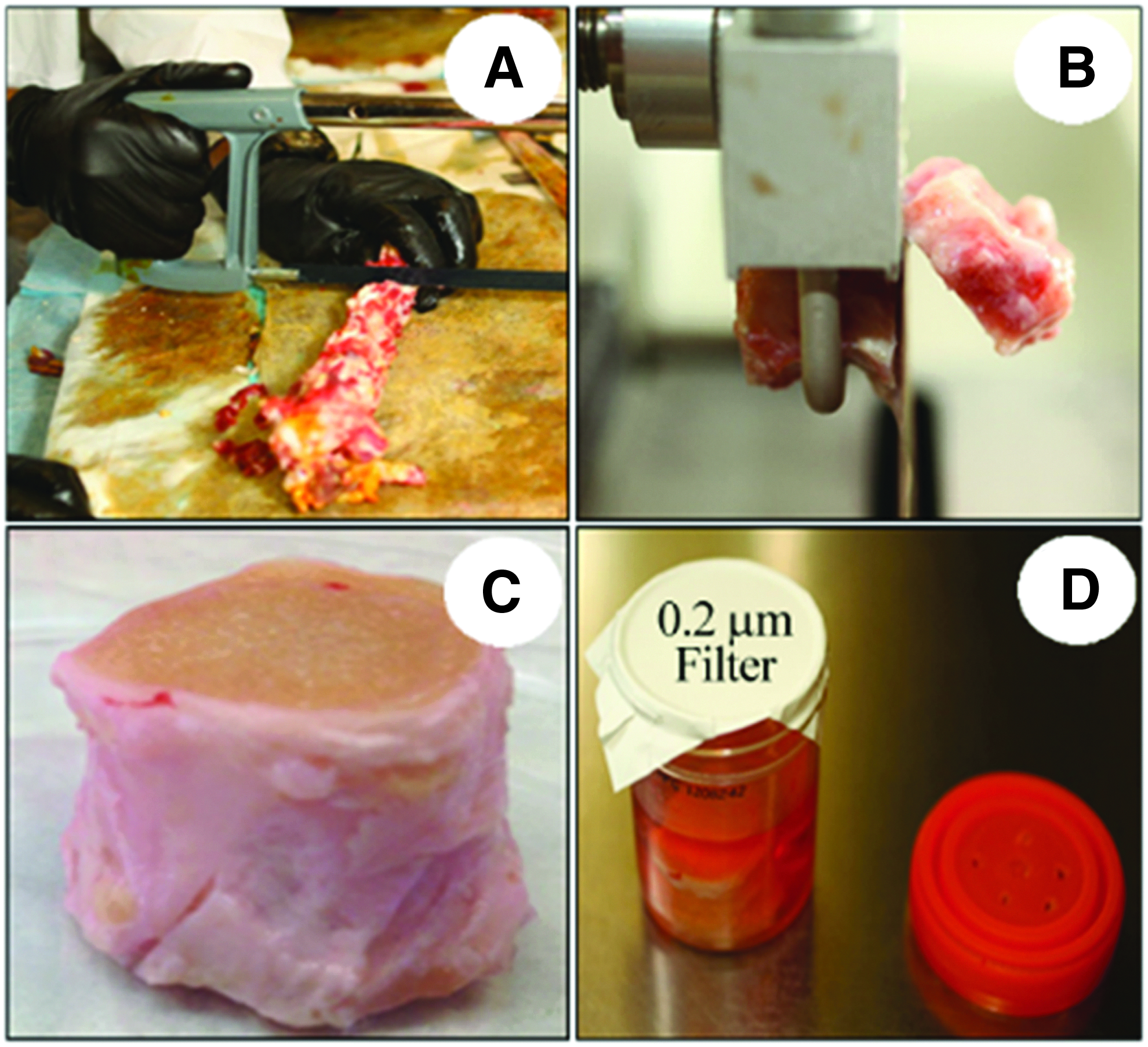
Images demonstrating IVD preparation.
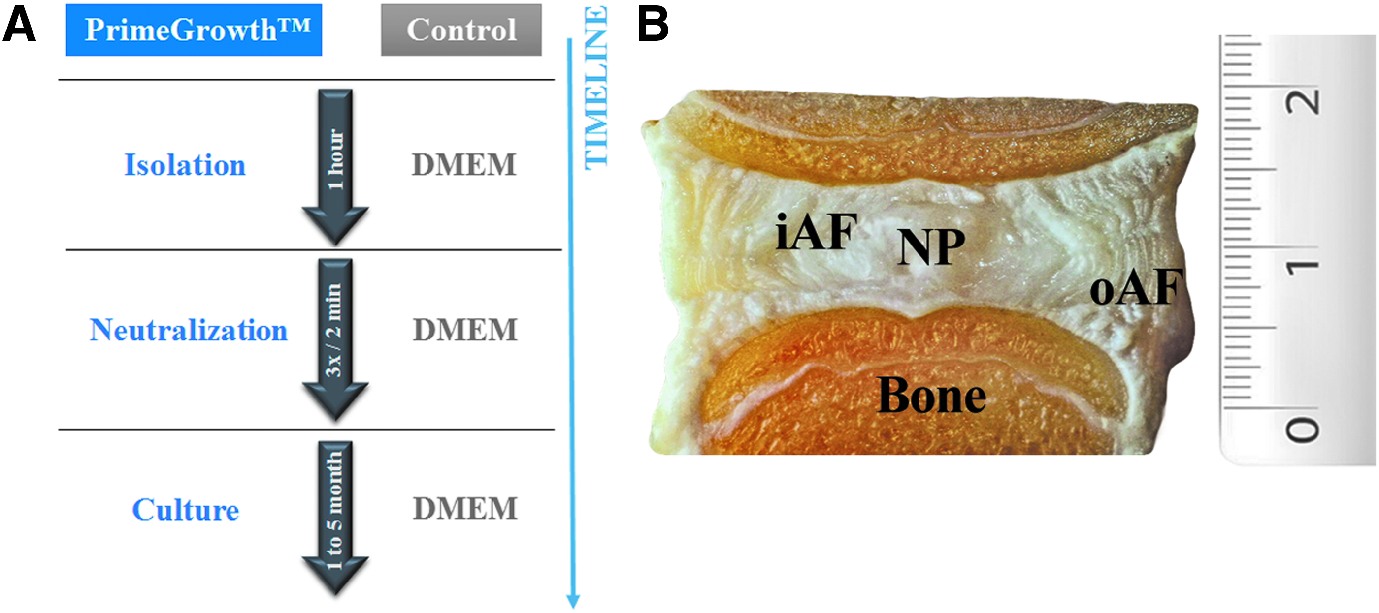
Schematic of vIVD processing with PrimeGrowth.
Live/dead assay and cell density
vIVDs cultured for 1 or 5 months were dissected to separate the NP, inner AF (iAF), and outer AF (oAF) regions. A 4 mm biopsy punch was used to prepare specimens for cell viability using a live/dead fluorescence assay (Live/Dead® Viability/Cytotoxicity Kit, Cat# L3224; Thermo Fisher Scientific, Waltham, MA). Bone sections were prepared by cutting a portion of the vertebral bone adjacent to the disc. Tissue punches and bone sections were washed once in PBS before incubation with the Live/Dead Viability/Cytotoxicity reagent for 30 min following the manufacturer's guidelines. Tissue punches were placed on a slide and visualized by confocal microscopy using a Zeiss LSM confocal laser-scanning microscope equipped with 488 and 543 nm laser lines. Ten Z-stacked images were merged representing 100 μm tissue thickness. Percent viability was determined by counting the total number of green (live) and red (dead) cells from the merged images using the cell counter function in ImageJ (National Institute of Health, Bethesda, MD) and calculating the ratio of the two (live/dead).
To determine cell density, NP, iAF, and oAF tissue punches of freshly prepared (Day 0) and 5-month PrimeGrowth®-cultured vIVDs were incubated with the Live/Dead Fluorescent Assay Kit as indicated above. Tissues were imaged by confocal microscopy using a 488 nm laser line and Z-stacked images representing a volume of 0.1 mm3 were developed. Cells were counted using the cell counter function in ImageJ software (National Institute of Health, Bethesda, MD).
Glucose diffusion
vIVDs from either the DMEM or PrimeGrowth groups were cultured for 1 month. Discs were washed in PBS and incubated for 72 h in diffusion medium containing PBS (1 × ), CaCl2 (1 mM), MgCl2 (0.5 mM), KCl (5 mM), 0.1% BSA, and 0.5 mM 2-[N-(7-nitrobenz-2-oxa-1,3-diazol-4-yl)amino]-2-deoxy-
Histology
IVDs were dissected from the vertebra, fixed in Accustain (Sigma-Aldrich), paraffin embedded, and 5 μm sections were prepared on slides. Sections were rehydrated before immunohistochemistry or staining by deparaffinization in xylene followed by sequential incubation in decreasing concentrations of alcohol and water. Proteoglycan content was visualized by staining tissue sections with Safranin O solution (0.1%) for 5 min. Slides were washed in water before mounting. To determine collagen content, specimens were stained with 0.1% Picrosirius red for 1 h and washed twice in acidified water (0.5% acetic acid). All sections were dehydrated by sequential alcohol concentrations and xylene, and mounted in Permount (Thermo Fisher Scientific). Slides were imaged using a Leica DM LB2 microscope (Leica Microsystems Inc.).
Glycosaminoglycan analysis
Sulfated glycosaminoglycans (GAGs) were quantified in tissue extracts by a modified dimethyl methylene blue (DMMB) dye-binding assay.48,49 Samples were diluted to fall within the middle of the linear range of the standard curve.
Statistical analysis
Statistical analysis was performed using two-way analysis of variance followed by Bonferroni multiple comparison test with GraphPad Prism 5.0 software.
Results
Current methods of isolating and culturing IVDs for extended periods that retain vIVD result in extensive disc cell death. To determine if PrimeGrowth media system improves the viability of disc cells in vIVDs, we cultured vIVDs in PrimeGrowth or standard growth (Control) medium for 1 month. As shown in Figure 3A, when vIVDs were processed with PrimeGrowth, cell viability (green cells) was maintained in the IVD. Greater than 90% cell viability was achieved in all regions of PrimeGrowth-treated discs, compared with <25% in the NP, and <5% in the iAF and oAF, respectively, when vIVDs were cultured in control medium (Fig. 3B). Interestingly, PrimeGrowth also maintained the viability of cells in the vertebral bone of vIVDs (Fig. 3A, B). To determine if PrimeGrowth improved the nutrient availability of disc cells, we prepared vIVDs using either PrimeGrowth or control media, cultured them for 1 month in the respective media, followed by 72 h incubation with the stable fluorescent glucose analog, 2-NBDG. Accumulation of 2-NBDG in vIVDs suggests nutrient diffusion. Using 3D confocal Z-stack fluorescent intensity mapping of the NP, iAF, and oAF of dissected vIVDs, the presence of 2-NBDG was markedly reduced or even absent in various regions of the control discs. Contrarily, in the PrimeGrowth group, 2-NBDG was found to be widely diffused throughout the disc (Fig. 4A). Similarly, when vIVDs were dissected and 2-NBDG was extracted from NP, iAF, and oAF tissues, and analyzed by fluorescent spectroscopy, significantly higher concentrations of 2-NBDG were found in all regions of the PrimeGrowth-treated vIVDs when compared with controls (Fig. 4B).
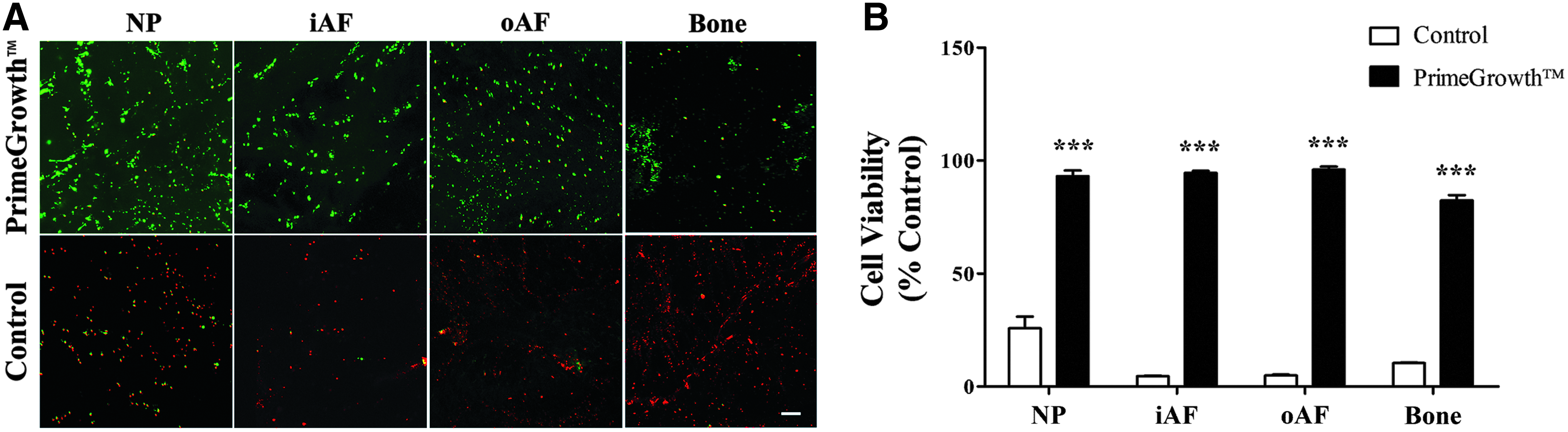
Comparison of cell survival in discs isolated with PrimeGrowth or control medium. Cell viability was measured in a 1-mm-thick tissue section after vIVDs were incubated in serum-free medium containing fluorescent dyes (Live/Dead, Invitrogen).
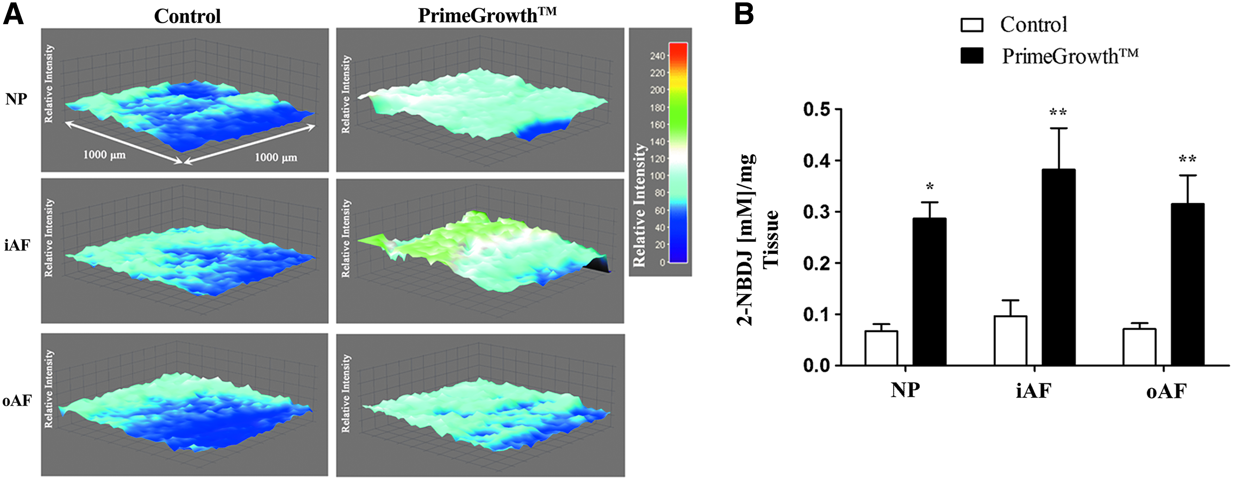
Glucose diffusion in vIVDs treated with PrimeGrowth or control. Bovine IVDs with vertebrae were prepared with control or PrimeGrowth medium and cultured for 1 month. Discs were incubated for 72 h in diffusion medium containing 2-[N-(7-nitrobenz-2-oxa-1,3-diazol-4-yl)amino]-2-deoxy-
Monitoring regeneration of vIVDs requires extended periods of culturing, as the disc environment presents with reduced oxygen tension and nutrient permeability rates effectively altering the metabolism of IVD cells compared with other cell types.50,51 To determine if PrimeGrowth-treated vIVDs can remain viable for extended periods of time, we cultured them for up to 5 months. As shown in Figure 5, cell viability persisted in all regions of the vIVD (NP, iAF, and oAF) similar to freshly processed vIVDs (Day 0). Notwithstanding cell viability, long-term culturing of vIVDs did not affect disc cellularity. There were no differences in NP, iAF, or oAF cell numbers between Day 0 and 5-month cultured vIVDs (Fig. 5C).
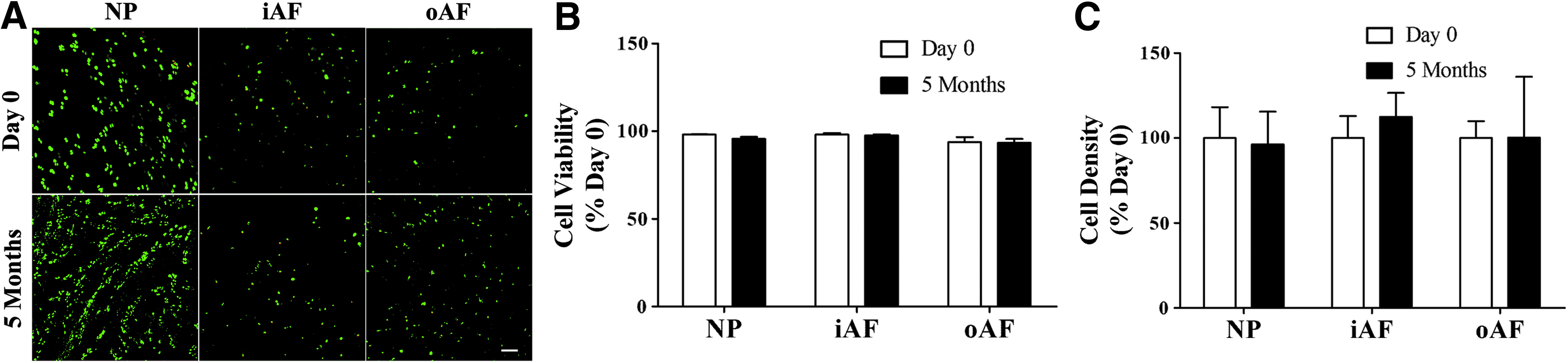
Comparison of cell survival in discs isolated with PrimeGrowth after 5 months in culture and discs before culture (Day 0). Cell viability was measured in a 1-mm-thick tissue section after incubation in serum-free medium containing fluorescent dyes (Live/Dead, Invitrogen).
To determine if the matrix composition of PrimeGrowth-treated vIVDs was affected following extended culturing periods, we measured proteoglycan and collagen content. Histological examination of freshly prepared vIVDs (Day 0), and those cultured for 1 or 5 months in PrimeGrowth demonstrated no apparent difference in Safranin O staining for proteoglycans in either the NP or AF regions of the discs (Fig. 6A). Similarly, using the DMMB dye-binding assay to measure GAGs, an indicator of proteoglycan content in NP and AF tissues, no significant differences were determined in freshly prepared (Day 0) compared with 1 or 5 month(s) cultured vIVDs, respectively (Fig. 6B). In addition to proteoglycan, collagen content, as determined by Picrosirius red staining, also remained similar in the NP and AF regions of Day 0, 1-, and 5-month cultured vIVDs (Fig. 6C).
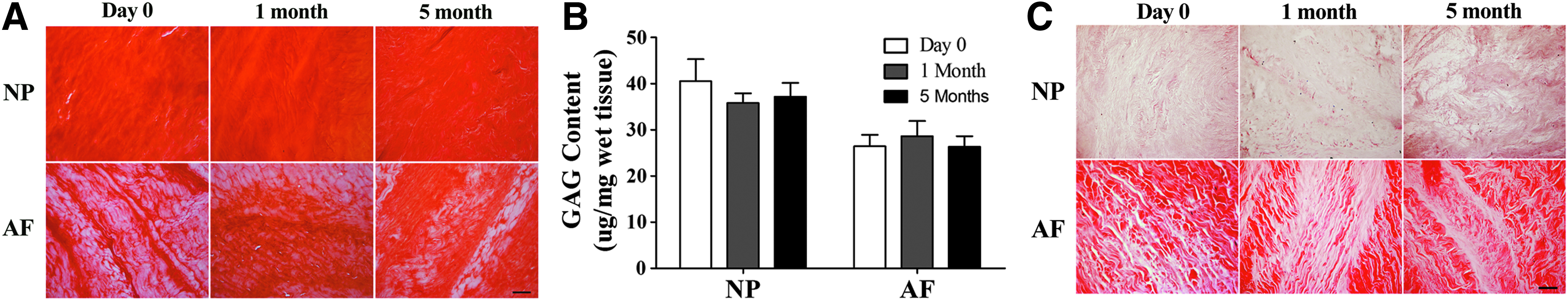
Proteoglycan content in vIVDs following prolonged culturing. Discs isolated with the PrimeGrowth media were cultured for 1 or 5 months.
Discussion
Large animal IVD organ culture models are an effective means of investigating regenerative therapies in disc degeneration and the biological responses of loading on disc cell physiology and matrix composition in a controlled environment. Although several organ culture models have been developed, limitations on the long-term culturing of IVDs that retain vertebrae persist.34–37,52,53 The vertebrae, and particularly the BEP, are infiltrated with blood vessels that terminate at the CEP—the membrane barrier that filters nutrients entering the disc. It is believed that these vessels undergo clotting during IVD isolation, obstructing, and thereby limiting nutrient supply to disc cells eventually resulting in cell death.
In the present study, we characterize the use of a three-step media system (isolation, neutralization, and culture) named PrimeGrowth, which was specifically optimized for the isolation and culturing of vIVDs. Our unique media formulation was demonstrated to sustain greater than 90% NP and AF cell viability in free-swelling vIVDs cultured for up to 5 months. To our knowledge, this is the longest reported survival period of any IVD organ culture model retaining vertebral bone in the literature.
In earlier attempts at culturing bovine IVDs retaining BEPs for studying mechanobiology, cell viability was markedly decreased after 1 week. 37 To overcome drastic declines in cell viability in large animal IVDs retaining BEPs, Gantenbein et al. 34 injected sheep with heparin before sacrifice and isolation of their discs. IVD cell viability was greatly improved in this model; however, decreases were apparent after 7 days in culture. Although this method of preparing IVDs was possible in ovine, heparinization is not a feasible option for culturing bovine IVDs since tissues are often obtained from abattoirs.33,35,37,39,40,46,47,54–57
An alternative method derived to overcome barriers to nutrient diffusion by removing the vertebrae and the BEP all together, but retaining the CEP to prevent disc swelling, has been effective in maintaining long-term cell viability in cultured discs.36,40 Although an excellent system for investigating biologics in disc repair, 57 this organ culture model is limited when considering various loading paradigms in repair strategies. Vertebral bone and the BEP provide flat surfaces amenable to static and dynamic loading paradigms. The use of this method in a bioreactor drew attention to the fact that the CEPs were concave and not ideal for flat platens, resulting in overloading of annular disc regions leading to cell death. 39 Thus, special platens and analysis of stress profilometry were required to evaluate load distribution in the discs with different geometries to find a shape that mimicked native load transfer. 39 Notwithstanding, the absence of bone makes it unsuitable for applying complex loading, a topic of interest in chronic progressive disc degeneration.42,46,47,56
Recently, Chan and Gantenbein-Ritter have demonstrated sustained NP and AF cell viability in bovine IVDs that retain both BEP and vertebral bone for a period of 2 weeks by adopting a surgical jet lavage system in their disc isolation method. 35 The success of their model was attributed to the penetrating abilities of the jet lavage capable of removing blood clots within the vertebral bone. Although this approach greatly improved the viability of both NP and AF cells in vIVD cultures, cell viability had begun to decline at the end of a 21-day culture period. Therefore, any experiments on these vIVDs must be performed within a 14-day period to be certain that the physiology and viability of IVD cells are not comprised due to culturing limitations.
In our organ culture model, treatment of vIVDs with PrimeGrowth isolation solution permitted diffusion of nutrients into the disc, as determined by incorporation of the glucose analog 2-NBDG in NP and AF tissues. When vIVDs were conditioned in PrimeGrowth isolation solution followed by culturing in PrimeGrowth culture medium, specifically optimized to represent the native physiological environment of the disc, long-term cell viability was achieved. Although long-term culturing of vIVDs may affect the integrity of disc tissue independent of cell viability, we found no significant differences in proteoglycan content upon either histological examination or GAG assay of NP and AF tissues. A similar result was obtained following histological examination of collagen fibers in vIVDs. PrimeGrowth also maintained viability of cells in the bony vertebrae. Taken together, these results suggest that our vIVD organ culture system not only retains the characteristic features of the disc for up to 5 months in culture, but maintains a viable vertebral bone. This organ model system provides a unique opportunity to investigate the interplay between the vertebrae and disc following long-term biomechanical loading.
In conclusion, we provide a novel ready-to-use disc preparation and culturing approach for vIVDs that can be used to investigate long-term biological repair of large animal discs in bioreactors, where complex loading paradigms can be applied.
Footnotes
Acknowledgments
The authors thank Intervertech, Inc. and Wisent Bioproducts, Inc. for production and supply of PrimeGrowth™ media. They are grateful to Jill Urban, PhD and Peter Roughley, PhD for their helpful discussions. This work was funded by the Canadian Institute of Health Research (CIHR).
Disclosure Statement
A percentage of the sales of PrimeGrowth™ media will be used for research funding in Dr. Mwale's laboratory.