
Abstract
While transplantation is a viable treatment option for end-stage lung diseases, this option is highly constrained by the availability of organs and postoperative complications. A potential solution is the use of bioengineered lungs generated from repopulated acellular scaffolds. Effective recellularization, however, remains a challenge. In this proof-of-concept study, mice lung scaffolds were decellurized and recellurized using human bronchial epithelial cells (BEAS2B). We present a novel liquid ventilation protocol enabling control over tidal volume and high rates of ventilation. The use of a physiological tidal volume (300 μL) for mice and a higher ventilation rate (40 breaths per minute vs. 1 breath per minute) resulted in higher cell numbers and enhanced cell surface coverage in mouse lung scaffolds as determined via histological evaluation, genomic polymerase chain reaction (PCR) analysis, and immunohistochemistry. A biomimetic lung bioreactor system was designed to include the new ventilation protocol and allow for simultaneous vascular perfusion. We compared the lungs cultured in our dual system to lungs cultured with a bioreactor allowing vascular perfusion only and showed that our system significantly enhances cell numbers and surface coverage. In summary, our results demonstrate the importance of the physical environment and forces for lung recellularization.
Impact statement
New bioreactor systems are required to further enhance the regeneration process of bioengineered lungs. This proof-of-concept study describes a novel ventilation protocol that allows for control over ventilation parameters such as rate and tidal volume. Our data show that a higher rate of ventilation is correlated with higher cell numbers and increased surface coverage. We designed a new biomimetic bioreactor system that allows for ventilation and simultaneous perfusion. Compared to a traditional perfusion only system, recellularization was enhanced in lungs recellularized with our new biomimetic bioreactor.
Introduction
End-stage lung disease continues to be a leading cause of death worldwide, with transplantation as the only viable treatment solution. This approach, however, remains limited due to a shortage of transplantable organs1–3 and potential complications with allograft rejection. 4 The use of transplantable ‘off the shelf’ bioengineered lungs is an exciting alternative that is currently under investigation.5–10
A bioengineered lung is a previously harvested organ that has been decellularized and subsequently repopulated with cells of interest. 11 The process of decellularization aims to remove all the cellular and immunologically active components while maintaining extracellular matrix, as well as the macroscopic and microscopic lung architecture. 5 Decellularization is achievable via physical, chemical, and enzymatic means, among which, the enzymatic approach is the most commonly used.10,12–14 The decellularization protocols, depending on the species of interest, can be carried out either in static13,15 or dynamic bioreactor systems.12,14,16,17
Similarly, there has been effort put forth toward identifying suitable cell types for repopulation of acellular lung scaffolds,9,18 however, effective recellularization remains a challenge. Examples include challenges in isolation and expansion of appropriate cell types, optimization of ex vivo conditions for maintenance, differentiation and/or maturation of cells, achievement of complete scaffold cell coverage, as well as recapitulation of epithelial and endothelial cell function in the scaffold.19–22
Recellularization of lung scaffolds can be divided into two procedural stages, which include cell seeding, in which the cells are delivered into the scaffold; and culture and propagation, in which cells are maintained in a bioreactor system, typically conducted over days and weeks.9,23
Previously, we have shown that significant enhancement of reepithelialization can be achieved using a novel negative pressure cell seeding protocol. 24 Herein, we investigated the effect of ventilation in the enhancement of recellularization during the bioreactor-based culture and propagation stage. Specifically, we have developed a novel negative pressure liquid ventilation system that allows for control over tidal volume and facilitates higher ventilation rates. Our results demonstrated that higher rates of ventilation combined with a physiological tidal volume resulted in improved reepithelialization.
Finally, we developed a new lung bioreactor that incorporated our new wet ventilation system into the design, which allows for physiological ventilation and vascular perfusion and accommodates rodent lungs' in vivo spatial orientation. In these proof-of-concept experiments, we used this system for reepithelialization, with results demonstrating enhanced cell surface coverage and higher cell number in comparison to traditional airway recellularization methods using vascular perfusion.
Methods
A commercially available programmable two-channel syringe pump, model NE-4000 (New Era), was used to design the negative pressure liquid ventilation device. Decellularization protocol was performed for 3 days and was completed via enzymatic and chemical treatment with reagents, including triton X100, sodium deoxycholate (SDC), sodium chloride, DNase, and phosphate-buffered saline (PBS) (Supplementary Fig. S1). Human bronchial epithelial cells were delivered to the lungs intratracheally using our previously established negative pressure cell seeding method. 24 After a static period, tissues were cultured in a bioreactor using either vascular perfustion alone or with simultaneous liquid ventilation. After 3 days of culture, lungs were fixed in formalin and evaluated for cell number and scaffold coverage. Details of all procedures are discussed in the proceeding sections.
Lung-heart block extraction and decellularization
Animal work was performed as per the guidelines and established methods provided by the Institutional Animal Care and Use Committee of the University Health Network. C57BL/6 male mice (Jackson Laboratory) between 14 and 16 weeks were euthanized by CO2, the thoracic cavity was exposed, and blood flushed via perfusion of PBS (Thermo Fisher Scientific, CA) through the right ventricle. Trachea and pulmonary artery (PA) were cannulated, and extracted heart-lung blocks were stored in a solution of PBS and 1% Antibiotic-Antimycotic (Thermo Fisher Scientific) at 4°C. The decellularization protocol was conducted per established protocols.13,25
In brief, tissues were stored in distilled water (dH20) at 4°C for 1 h, followed by perfusion of airways and vasculature with dH20. Tissues were then stored in a 0.1% Triton X100 solution at 4°C overnight. This was followed by another wash with dH20 and storage in 2% SDC solution at 4°C for 24 h. On the third day, samples were washed with dH20 and stored in Sodium Chloride solution at room temperature (RT) for 1 h. Subsequently, lungs went through a final round of a dH20 wash and were stored in DNase (Thermo Fisher Scientific) solution at RT for an hour. Decellularized lungs were washed and stored in PBS and 1% Antibiotic-Antimycotic solution (Thermo Fisher Scientific) at 4°C.
Cell culture
For reepithelialization of the airways, human bronchial epithelial (BEAS2B) cells (ATCC) were used. BEAS2B cells were cultured in high glucose content Dulbecco's modified Eagle's medium (Thermo Fisher Scientific) containing 10% fetal bovine serum (Thermo Fisher Scientific) and 1% Antibiotic-Antimycotic solution. Cells were stored in a standard incubator (95% air and 5% CO2 at 37°C). Cells were passaged once they reached 85% confluency (1 × per week). Before seeding, BEAS2B cells were trypsinized, counted using Vi-CELL™ Cell Viability Analyzer (Beckman Coulter Life Sciences), and 2.5 million cells were resuspended in 1.5 mL of cell culture media for scaffold seeding.
Negative pressure cell seeding
The negative pressure cell seeding system was set up and used as previously described by Ahmadipour et al.. 11 In brief, the cell suspension was connected to the tracheal inlet via a tube (‘ID = 3.1 mm), and the other inlet of the main reservoir was connected to the syringe pump. The reservoir was then sealed, and the syringe pump withdrew air from the main reservoir (up to 60 mL of air was removed at a 50 mL/min rate). The generated pressure gradient between the cell suspension (exposed to atmospheric pressure) and sealed lung pulled the cells into the lung airways.
Setup of negative pressure wet ventilation system
The wet negative-pressure liquid ventilation system was set up by filling up the main reservoir (125 mL flask) with cell culture media while keeping the ventilation reservoir half full, exposed to atmospheric pressure at the same height as seeded lung. The main reservoir was then sealed. The syringe pump was connected to the main reservoir, and the rate was adjusted to achieve ventilation rates of 1 breath/min (control group), and 40 breaths/min while setting the inhalation and exhalation volumes to 300 μL (approximately the tidal volume of an adult mouse). 26 By eliminating the air-liquid interface and ensuring that the entirety of the main reservoir was filled with cell culture media, the removal of liquid from the main reservoir resulted in an equal amount of liquid moving toward the tracheal inlet. Thus, less volume was required to initiate the ventilation, and faster ventilation rates were possible than current ventilation systems. 27
In addition, the “Y” shaped connector, placed inside the chamber, and the arrangement of the tubing and one-way valves facilitated a constant mixture of cell culture media between the two reservoirs while simultaneously releasing excess pressure in the main reservoir (due to leakage of the decellularized scaffold during the inhalation). During the 3 days culture period, bioreactors were placed in a standard incubator (95% air and 5% CO2 at 37°C), and cell culture media was changed every other day.
Setup of vascular perfusion system
The setup of the vascular perfusion system was previously described by Ahmadipour et al.. 11 In brief, two set of tubings (one connected to the PA and one circulating the cell culture media), were connected to a persitalic pump which continuously moved the cell culture media through the vasculature. After delivering the BEAS2B cells through the trachea, lung scaffolds were maintained in static conditions for 18 h and then transferred to the bioreactor. The lungs served as controls when compared to those repopulated using the newly developed biomimetic bioreactor system (further described in the following section).
Lungs scaffolds were maintained in an upright position as per previously described vascular perfusion systems.13,24,25,28–34 A Masterflex L/S Precision Modular Driver with a Bench Top Controller (Cole-Palmer, CA) was used to achieve perfusion at the rate of 1.5 mL/min for 3 days. During this culture phase, bioreactors were placed in a standard incubator (95% air and 5% CO2 at 37°C), and cell culture media was changed every other day.
Setup of biomimetic lung bioreactor system
Seeded lungs were transferred to the bioreactor and cultured under simultaneous vascular perfusion and wet negative-pressure ventilation conditions. The bioreactor design combined the traditional vascular perfusion and our newly developed ventilation system. This system had multiple tubings, through which simultaneous vascular perfusion and ventilation were achieved. Similar to our ventilation system discussed above, one set of tubings accommodated the connection of the main chamber to the syringe pump, one facilitated the movement of cell culture media from the secondary chamber to the main chamber during the ventilation, and finally, one line was there to equilibriate the main and secondary reservoirs by allowing transfer of leaked cell culture media from the scaffold in the main chamber to that of the secondary reservoir.
To obtain vascular perfusion, one tubing line was connected to the PA, while the other line circulated the cell culture media from the main chamber. These two lines were passed through the peristaltic pump, and the cell culture media was continuously perfused through the PA line and scaffold vasculature. To facilitate the flow of cell culture media, the described directions, multiple one-way valves were used (Fig. 3A). The system's spatial placement alongside tubing network arrangement allowed for vascular perfusion, high-rate negative-pressure liquid ventilation, and in vivo supine orientation (Fig. 3).
This bioreactor orientation was in accordance with rodent physiological supine orientation, which normalized the effect of gravitational forces over the scaffold during the recellularization process. The vascular perfusion rate was set at 1.5 mL/min, and the ventilation rate was set to 40 breaths/min with a tidal volume of 300 μL. During the 3 days of the culture, the bioreactor was placed in a standard incubator (95% air and 5% CO2 at 37°C), and cell culture media was changed every other day.
Histology and fluorescence staining
For fixation, a 10% formalin solution was injected intratracheally into scaffolds and stored at RT overnight. Fixed scaffolds were then transferred into 70% ethanol solution and processed using the Excelsior ES Tissue Processor (Thermo Fisher Scientific). Processed samples (N = 3 for each group) were embedded in paraffin blocks, and for each lung, three to four 5 μm thick whole-lung sections at different depths were prepared. Sections were then stained by hematoxylin and eosin (H&E) and 4′,6-diamidino-2-phenylindole (DAPI) nuclear stain, using established protocols.10,15
In brief, H&E staining was achieved by deparaffinization via two washes in xylene followed by two washes in 100% ethanol, one wash in 95%, and one wash in 70% ethanol. Deparaffinization was followed by incubation in hematoxylin (3 min) and eosin (1 min). Staining was followed by one wash in 70% ethanol, one in 90% ethanol, two washes in 100% ethanol, and two washes in xylene. Slides were covered with coverslips.
Cell nuclear staining was performed by a similar deparaffinization procedure followed by incubation in 2 mg/mL DAPI (Sigma) for 5 min. Slides were then washed in PBS followed by additional washes in ethanol and xylene as described above. Slides were covered using a standard coverslip and scanned using the Aperio slide scanner (Leica Biosystems).
Immunohistochemistry
Immunohistochemistry slides were processed and prepared at the Drug Development Program (DDP) biomarker laboratory located at Princess Margaret Hospital using methods previously described. 35 In brief, after deparaffinization, antigen retrieval was performed using 10 mM sodium citrate buffer at 100°C for 20 min. Sections were permeabilized with 0.1% Triton X solution for 15 min and washed with two 10 min period incubations in PBS. Blocking was performed with 10% goat serum for 1 h at RT. After blocking, rabbit anti-human Ki67 polyclonal antibody (NB110-90592SS; Novus Biologicals), (1:50) was added to evaluate proliferation. Sections were stored for 4°C overnight and washed with two 10 min periods of incubation in PBS. Slides were coated with Signal Stain 3,3′-Diaminobenzidine (DAB) chromogen and washed two times in PBS for 10 min each. Slides were then mounted and scanned using the slide scanner.
Image analysis of histology sections
Histology slides were analyzed using HALO software™ (Indica Labs) using our previously established methods.
11
In brief, for each whole-lung section, five examples of cells and scaffolds were introduced and highlighted to the software manually. A classifier was set at a resolution of 1.04 μm/pixel and minimum object size of 20 μm2. Cell surface coverage was calculated using:
Resulting value was subsequently normalized to the surface coverage percentage calculated for native lungs. 11
Semiquantitative histology image scoring
Images analyzed by HALO™ were randomized and given to five blinded observers. Each observed was asked to score the H&E images based on cell surface coverage based on a scale of 1 (0–10% coverage) suggesting little to no coverage, to 10 (90–100%) indicating high coverage. Results from observes were collected, tabulated, and averaged.
Real-time polymerase chain reaction
Real-time polymerase chain reaction (PCR) was completed on gDNA to determing the level of human Glyceraldehyde-3-Phosphate Dehydrogenase (GAPDH F-CTGGGCTACACTGAGCACC, R-AAGTGGTCGTTGAGGGCAATG, Primerbank ID 378404907c3) in recellularized mouse lung scaffolds using PowerUp SYBR Green Master Mix (Thermo Fisher Scientific). Estimated cell numbers in recellularized lungs were calculated by processing sixteen 10-μm thick sections, taken from paraffin blocks and pooled for genomic DNA extraction using QIAamp DNA FFPE Tissue Kit (Qiagen, CA) as per the manufacturer's protocol. The values were then normalized to tissue sections' surface area.
Statistical analysis
Paired Student's t-tests were conducted to determine the significance of cell surface coverage and gene expression analysis. Statistical significance was defined as p-value <0.05.
Experiment
A novel negative-pressure-based wet ventilation culture system allows for precise control over tidal volume and ventilation rate
We designed a novel negative pressure liquid ventilation system that allowed for high ventilation rates and control over tidal volume (Fig. 1). Liquid ventilation was achieved by completely filling the main reservoir with cell culture media. Removal of any volume from the main reservoir using the syringe pump was compensated by flowing an equal amount of liquid through the ventilation line.
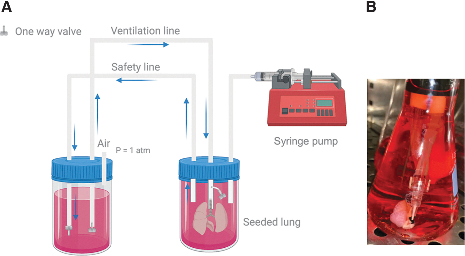
Physiological negative-pressure wet ventilation system allowing control over tidal volume and ventilating rate.
Our system included the addition of a safety line transferring liquid from the main chamber to the ventilation reservoir, thus enabling the maintenance of pressure equilibrium in the system. This is particularly important, as the decellularized lung is “leaky,” meaning that cell culture media seeps through the pleura and exhalation line during inhalation and exhalation, respectively. The addition of the safety line also allows for constant mixing of cell culture media between the main and ventilation reservoirs.
Higher ventilation rate results in greater cellularity and enhanced cell surface coverage in acellular mouse lung scaffolds
We studied the effects of ventilation rate on recellularization, explicitly comparing a low ventilation rate (1 breath per minute-control group) based on prior published systems7,8,36,37 and a higher ventilation rate. In these studies, the tidal volume was set to 300 μL, the physiological tidal volume of mouse lungs. 26 As per our previous studies, mouse lungs were decellularized and seeded with BEAS2b cells via negative-pressure cell delivery. 24 Scaffolds were ventilated for 3 days.
Recellularized lungs were then fixed, tissue sections were processed and evaluated for cell surface coverage and morphology, viability, and proliferation. The histological assessment suggested that cell morphology was similar in both groups and increased cell surface coverage in lungs ventilated at 40 breaths per minute (Fig. 2A, B, E, F). Nuclear DAPI staining confirmed cell localization in the scaffold with markedly higher cell numbers in scaffolds belonging to the high-rate ventilation group (Fig. 2C, G).
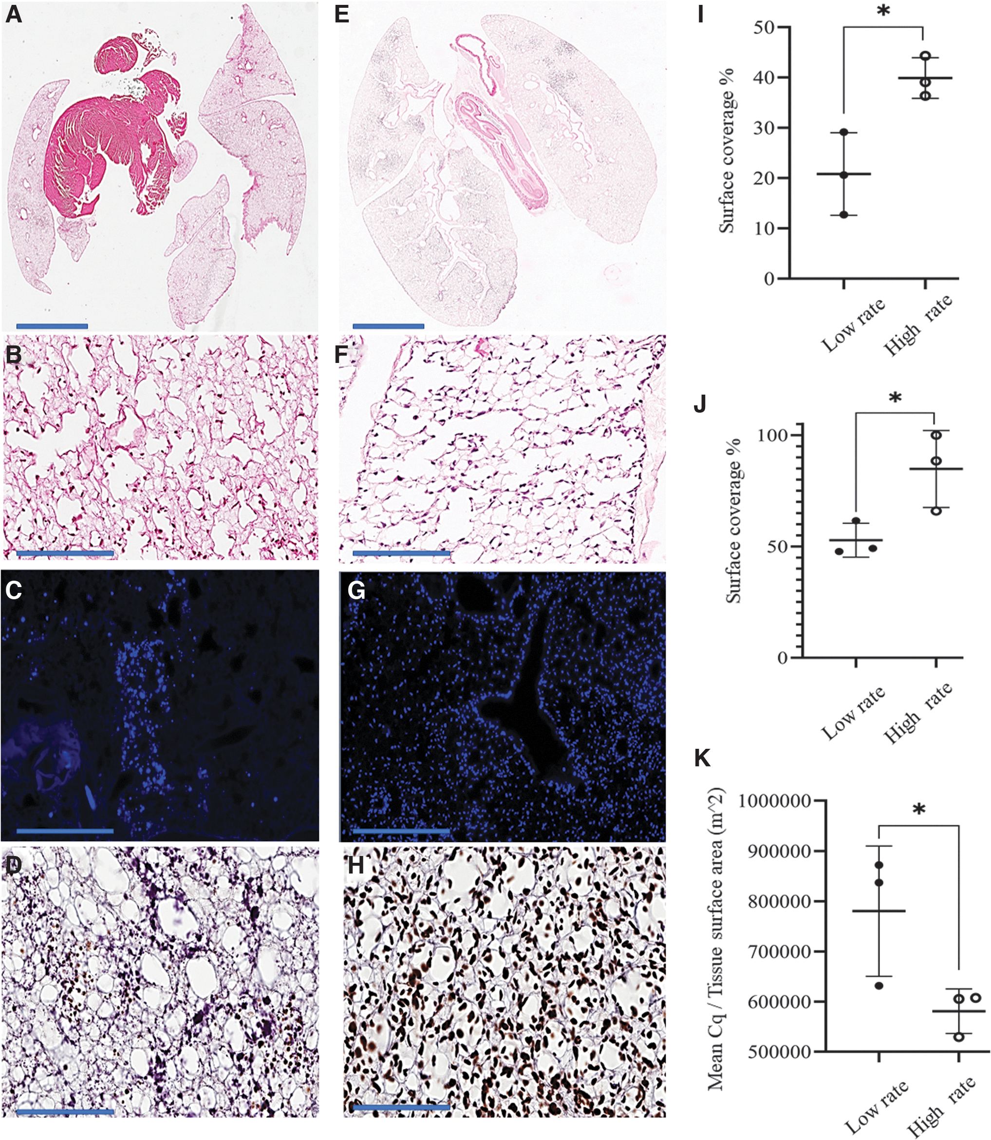
A high rate of ventilation results increased cellularity in decellularized lung scaffolds after 3 days of culture.
In addition, scaffolds recellularized under high ventilation rate conditions showed a significantly higher number of proliferative cells as visualized by Ki67 positivity (Fig. 2D, H). Quantitative assessment of cell surface coverage using HALO and histology image scoring indicated that cell surface coverage was significantly higher at higher rates of ventilation than the lower ones (Fig. 2I–J). Moreover, PCR showed that the presence of human GAPDH genomic DNA was substantially higher in lungs ventilated at higher rates in comparison to lower ones (Fig. 2K). These results suggest that high-rate ventilation during the recellularization process results in greater cellularity in lung scaffolds.
A biomimetic lung bioreactor system
We designed a new bioreactor system encompassing our novel negative-pressure liquid ventilation system. Our system also includes a vascular perfusion circuit allowing for simultaneous perfusion and ventilation. In addition, unlike existing systems,7,8,28–30,36–38 our bioreactor can accommodate rodent lung scaffolds to be placed in a physiological supine orientation and the upright position. Combining these features results in a more biomimetic rodent ex vivo lung culture system (Fig. 3). A graphical representation of the bioreactor system, in the upright position, can be seen in Figure 3A, highlighting the placement of lung scaffold fully submerged in media, the direction of the flow of media through the circuit and the position of one-way valves. AutoCAD drawings (Fig. 3B) show the actual dimensions and specifics of the design.
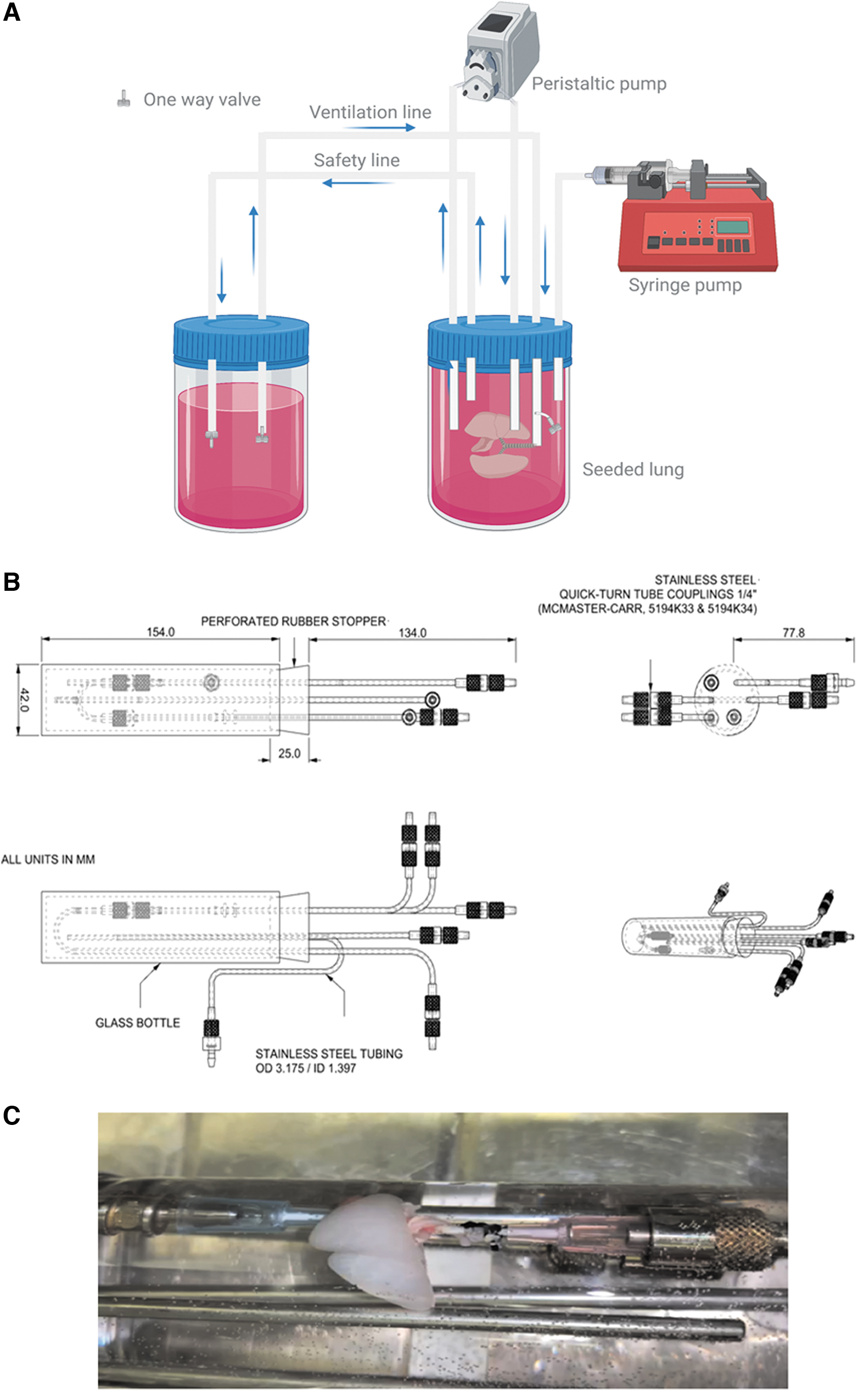
Physiological biomimetic lung bioreactor facilitating vascular perfusion, controlled ventilation, and in vivo spatial orientation.
Of note, the glass cylinder allows for efficient use of media, and the transparency enables visualization of the scaffold throughout the setup and recellularization process. Also, unlike rubber tubing, which can move and cause ruptures in the pulmonary artery and trachea, stainless steel tubing allows for fixed tubing positions connected to cannulas, thus preventing movement. The bioreactor system can be placed horizontally, enabling the physiological supine position of the lung scaffold, as shown in Figure 3C.
Use of biomimetic lung bioreactor for recellularization of lung airways
We evaluated our biomimetic bioreactor system for recellularization of lung airways. We compared our system (experimental group) to standard bioreactor methods using vascular perfusion only (control group). In the experimental group, lungs were seeded as described 24 with the bioreactor in the upright position and incubated under static conditions for 18 h. Subsequently, the bioreactor was placed horizontally, enabling a physiological supine position. Repopulated lung scaffolds were then perfused at the rate of 1.5 mL/min and ventilated at a tidal volume of 300 μL and 40 breaths/min rate. In the control group, lungs were seeded as per the experimental group and perfused via the vasculature as previously described24,28–30 at a rate of 1.5 mL/min. All lungs were maintained in culture for 3 days postseeding with continuous ventilation and perfusion. Repopulated scaffolds were then fixed and stained for further analysis.
The histological evaluation shows a greater number of cells in the airways in the lungs that were both perfused and ventilated (Fig. 4A, B, D, E). These findings were confirmed with DAPI nuclear staining (data not shown). Assessment of proliferation via Ki67 staining showed that most of the cells in both groups were proliferating (Fig. 4C, F).
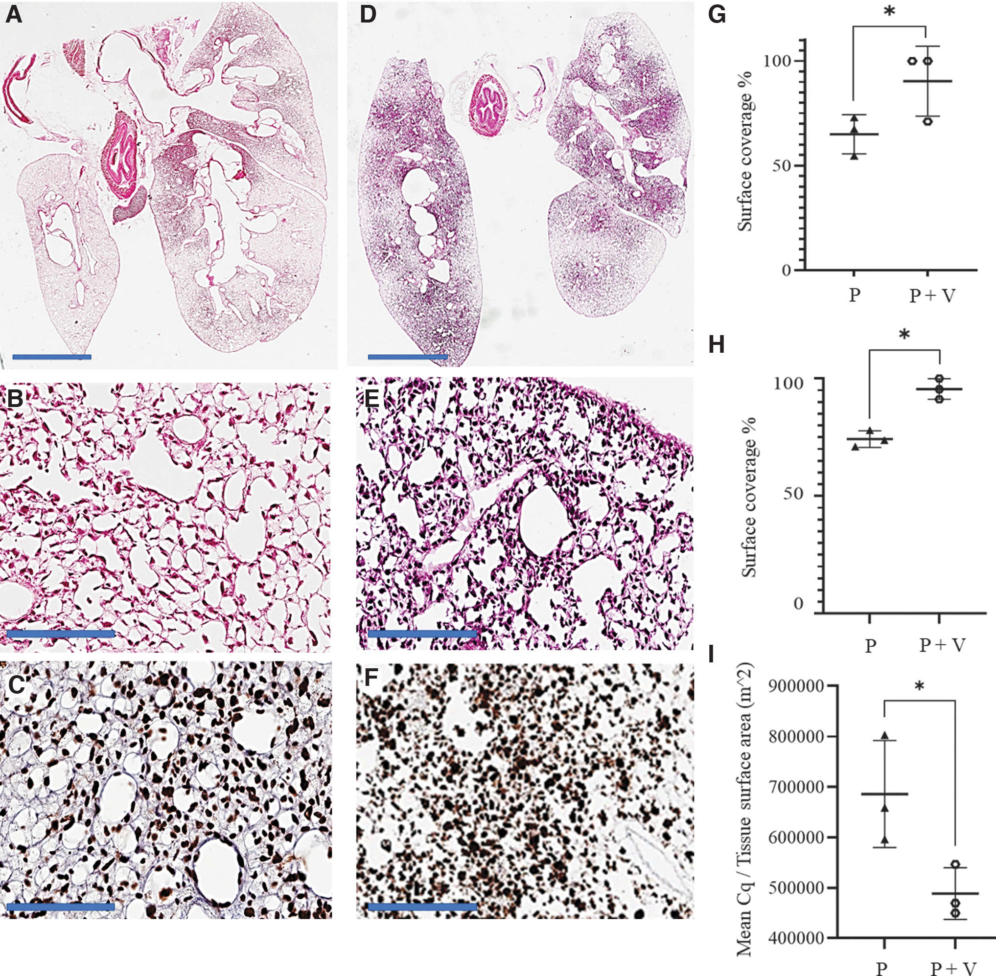
Addition of high-rate ventilation enhances cellularity in acellularized lung scaffolds.
Quantitative assessment of cell surface coverage using HALO and semiquantitative histology image scoring showed significantly higher cell surface coverage in lungs going through simultaneous perfusion and ventilation in comparison to perfusion only (Fig. 4G, H). Moreover, the amount of human GAPDH genomic DNA was also higher in lungs that were simultaneously perfused and ventilated (Fig. 4I). Taken together, results show that a bioreactor system that allows for both perfusion and ventilation results in enhanced recellularization of lung airways.
Discussion
In this study, we designed a novel ventilation system that allows for control over tidal volume and facilitates high ventilation rates. We found out that using a physiological tidal volume (300 μL) for mice and a higher ventilation rate (40 breaths per minute) resulted in enhanced cell surface coverage and higher cell numbers obtained during airway recellularization. We incorporated this ventilation system in designing a new biomimetic lung bioreactor system, which facilitates simultaneous vascular perfusion and can maintain the rodent lung in a physiological supine orientation during the recellularization process. We compared the lungs cultured in this system with those recellularized using vascular perfusion only and showed that the cell surface distribution and number were significantly higher for the lungs cultured using our biomimetic lung bioreactor systems.
Several studies have reported bioreactor-based reepithelialization using vascular perfusion.13,24,25,28–34 However, vascular perfusion alone does not facilitate the ventilation-induced mechanical and physical environment forces to which epithelial cells are naturally exposed in vivo. Previous studies have incorporated ventilation during the culture phase, after which epithelial and endothelial cells have been delivered to the acellular lung scaffolds.7,8,27,29,36,37,39–41 Current ventilation systems, however, do not allow for precise control over respiratory parameters such as tidal volume and cannot accommodate physiological ventilation rates with the majority using a low rate of ventilation, typically 1 breath per minute for liquid ventilation protocols 36 and as high as 5 breaths per minute in dry ventilation protocols. 38
In our system, by filling the entirety of the main reservoir and system with liquid, we were able to precisely control the tidal volume. Specifically, as liquid was removed by the syringe pump, it was compensated by an equal amount of liquid from the secondary reservoir thus allowing for control over tidal volume. This is in contrast to previously reported air-liquid ventilation protocols where the removed air cannot be fully compensated by liquid from secondary reservoir due to differences in the compliance of the air and liquid. This discrepancy in compliance requires a greater amount of air to be removed from the main reservoir to generate enough force to deliver the liquid to the airways.
Our results show that a higher rate of ventilation resulted in enhanced recellularization. We believe that is partly due to better recruitment of the collapsed airways, thus allowing for improved nutrient delivery to the cells and increased waste removal from the scaffold. Therefore, we designed, manufactured, and validated a novel biomimetic lung bioreactor system that incorporated the wet negative ventilation system. An added feature of our bioreactor is the spatial orientation of the lung scaffold during the recellularization process, which can normalize gravitational forces and result in a more homogeneous distribution of cells in the scaffold. 42
In addition, in humans, the lung is an asymmetric organ with the long axis, superior to inferior, and the short axis anterior to posterior. The supine position orients the effects of gravity to the shorter of the two axes. Therefore, we feel that the supine position of scaffold during the recellularization will result in more homogeneous and less gravity-dependent distribution of cells and will be an advantageous feature when applied to human lung scaffolds.
Our study has a number of limitations. First, BEAS2B cells are a cell line, and while they are derived from normal bronchial epithelium and commonly used for studies involving airway epithelial cell biology, 43 further experiments are needed to validate our negative-pressure liquid ventilation system and a biomimetic lung bioreactor using primary lung epithelial cells. We have ongoing studies in the laboratory, which assess the use of more appropriate cell populations, specifically airway progenitor-like cells for recellularization of the acellularized scaffolds.44,45
Moreover, this study focused on recellularization of the airways only using one cell type. Given the complexity in the structure and cell composition of the lung, 46 complete recellularization will likely require region-specific cell seeding. This is supported by recent studies investigating different approaches for targeted recellularization.
Nichols et al. 9 have reported a sequential cell seeding protocol using a number of cell types delivered to the scaffold in a specific sequence with the aim to target specific cells to different regions of the scaffold. Similarly, Novakovic and colleagues show that by manipulating the cell suspension flow through the scaffold, different lung regions can be targeted. 47 In our own previous studies, we have shown that we can enhance the cell surface coverage in airways and vasculature using a novel negative pressure cell seeding method, in which we can control cell suspension flow rates. 24
One of the advantages of our herein described bioreactor system, is that it allows for simultaneous perfusion and ventilation and control over those parameters. In the future, we envision the use of these parameters in establishing cell seeding protocols that will specifically target cells to the different regions of the lung.
While we expect to see similar findings using primary cells, ventilation parameters may require some optimization. To enable ventilation at the mouse scale, we made a conscious choice of using a syringe pump, which allows precise control over tidal volume, which is imperative to prevent overstretching the scaffold during the regeneration process. However, the existing commercialized pumps cannot tolerate very high-frequency ventilation, and when we tried to achieve ventilation rates closer to physiological norms in the mouse (>200 bpm), several mechanical failures were observed. This syringe pump ventilation system allowed us to achieve 300 μL of tidal volume and reach a consistent ventilation rate of 40 breaths per minute over the 3 days of the culture phase.
We believe a new system of ventilators that can tolerate a high frequency of oscillation could overcome this barrier, enabling even higher ventilation rates than those achieved in this study, although the current approach is more than adequate to achieve physiologic ranges for human or porcine lungs. Moreover, the culture period in the bioreactor (with prolonged ventilation) will need to be extended past the 3 days used in this study. This will be particularly important in work with primary epithelial cells and/or stem cell-derived epithelium, in which we plan to investigate how ventilation and perfusion can influence the phenotype and function of primary epithelial cells, and differentiation and maturation of pluripotent stem cell-derived epithelial cells.
In these future studies, we also plan on evaluation of other relevant parameters in addition to proliferation, including apoptosis, epithelial phenotype, and barrier function. Another limitation of this study is that we have not performed revascularization, a necessary step for generating a clinically applicable graft. Future studies will include scaffold reendothelialization as it will be important to evaluate the impact of higher ventilation rates on endothelial cells within the scaffold.
Conclusion
We believe that incorporating physiologically relevant mechanical cues during recellularization of the airways is necessary to produce and maintain a functional epithelium..12,14,21,22,30,48,49 We present a novel negative pressure liquid ventilation system with which we can achieve high ventilation rates while still allowing control over tidal volume. Results from this study demonstrated that the use of a physiological tidal volume and inclusion of a high ventilation rate enhance lung scaffold airway recellularization. Inclusion of a higher ventilation rate may have a significant effect on the fate of cells during the recellularization and warrant further exploration.
Footnotes
Authors' Contributions
M.A. designed the project, performed experiments, analyzed data, and wrote the article. P.D. and D.T. assisted in conducting experiments and analysis. F.G.A., G.P., and C.S. contributed in designing the apparatus used in the project. T.K.W. and G.K. conceptualized and designed the project, supervised the work, and contributed to writing the article. All authors reviewed the article.
Acknowledgment
Authors thank Dr. Roya Navab for her assistance in preparation and processing of slides for immunohistochemical evaluation of Ki67.
Disclosure Statement
M.A., Dr. T.K.W., and Dr. G.K. have applied for a patent for the biomimetic lung bioreactor system through University Health Network. The remaining authors declare no competing interests.
Funding Information
This research was funded from the Canada First Research Excellence Fund (CFREF; C1TPA-2016-18).
References
Supplementary Material
Please find the following supplemental material available below.
For Open Access articles published under a Creative Commons License, all supplemental material carries the same license as the article it is associated with.
For non-Open Access articles published, all supplemental material carries a non-exclusive license, and permission requests for re-use of supplemental material or any part of supplemental material shall be sent directly to the copyright owner as specified in the copyright notice associated with the article.