
Abstract
This work employs nitrogen plasma immersion ion implantation (PIII) to modify electrospinning polylactic acid membranes and immobilizes basic fibroblast growth factors (bFGF) by forming crosslinking bonds. The study investigates the modified membranes’ surface characteristics and the stimulatory effects of crosslinked bFGF polylactic acid membranes on osteoblast and fibroblast proliferation. The PIII process occurs under low vacuum conditions and is controlled by processing time and power pulse width. The experimental results indicate that, within a 400-second N2-PIII treatment, the spun fibers remain undamaged, demonstrating an increase in hydrophilicity (from 117° to 38°/36°) and nitrogen content (from 0% to 7.54%/8.05%). X-ray photoelectron spectroscopy analysis suggests the formation of a C-N-C=O crosslinked bond. Cell culture and activity assessments indicate that the PIII-treated and crosslinked bFGF film exhibits significantly higher cell growth activity (p < 0.05) than the untreated group. These intergroup differences are attributed to the surface crosslinking bond content. In osteogenic induction, the results for each day show that the treated group performs better. However, the intergroup disparities within the crosslinked bFGF group disappear with prolonged culture time due to the rapid osteogenesis prompted by bFGF. The findings suggest that PIII treatment of electrospinning polylactic acid membranes holds promise in promoting osteogenesis in bone tissue scaffolds.
Impact Statement
The study describes the modification of polylactic acid (PLA) electrospinning by plasma immersion ion implantation (PIII). After treatment, the spinning surface forms chemical crosslinking bonds, thereby improving hydrophilicity and obtaining the ability to fix cytokines. For this purpose, we tested osteoblasts’ growth activity and osteogenic induction ability to grow on the three electrospinning membranes (original, treated, and immobilized cytokine samples). The results proved that this method can fix growth factors on PLA electrospinning and promote cell proliferation and osteogenic induction of osteoblasts. This could provide some inspiration for the development of clinical implants.
Introduction
Due to an aging population and orthopedic diseases, bone injury repair has become a global medical concern, and the demand for materials to repair bone defects continues to grow. 1 In the field of bone tissue, biodegradable polymer materials are considered ideal materials that will not cause long-term foreign body reactions in the body and avoid secondary surgical removal of implants. It also provides mechanical support in the early postoperative stage and transfers this effect to newly formed bone tissue as bone healing and degradation occur. 2 Biologically implanted therapy is mainly free from drug supply and administration challenges, as well as limitations on infusion plans, compared with continuous infusion therapy. 3 For example, polylactic acid (PLA) is nontoxic, has high-strength characteristics, and is biocompatible. PLA has been proven to have good plasticity and mechanical properties,4–8 making it a good tissue scaffold material.9–11 Synthetic polymers are often used for cell adhesion and activity analysis to evaluate their medical potential applications, and this process almost always occurs on the surface of polymers. Researchers generally want to synthesize polymer materials containing biological factors to help cell proliferation, wound healing, and reduce immune rejection and inflammatory reactions. 12 After being implanted into the human body, polylactic acid hydrolyzes into its component lactic acid, a natural byproduct of muscle contraction. Then, it is metabolized through the tricarboxylic acid cycle and ultimately excreted from the body as carbon dioxide and water. 13
Scaffolds in tissue engineering generally simulate the extracellular matrix (ECM) microenvironment, providing growth sites for cells or inducing tissue repair. The natural ECM is a three-dimensional porous elastin, collagen, and proteoglycans network, which helps cells maintain their morphology and perform specific functions. Electrospinning is a technology that can turn polymers into nanofibers, with a fiber diameter similar to natural ECM collagen fibers (5–500 nm), which form a three-dimensional porous structure identical to ECM. 14 Therefore, electrospinning polylactic acid into a simulated ECM tissue scaffold is feasible. Still, its hydrophobic surface properties and biological inertness are not conducive to early postoperative healing. 15 To avoid this situation, researchers have used various methods to improve the surface of PLA, among which plasma modification has received significant attention.
Plasma modification on the polymeric surfaces would make it possible to create functional groups with no need to use chemical solvents while it is simpler.16,17 Also, the treatment can be controllable through adjusting parameters while hindering any adverse effect on the scaffolds. 18 Therefore, plasma treatment tends to be an appropriate strategy for polymer modification. Plasma immersion ion implantation (PIII) is a practical, biocompatible, and flexible approach for plasma modification on the material surface,19,20 which could perform comprehensive surface treatment on the sample. Meanwhile, some research studies have demonstrated that PIII-treated surfaces can covalently immobilize cytokines without additional linkers. Similar methods and materials have been used to research coagulation and antibacterial and anti-inflammatory effects.21–23
The basic fibroblast growth factors (bFGFs) are endogenous polypeptide growth factors, significantly promoting bone repair. 24 It has been reported that bFGF may be involved in pulpal repair by regulating gene and protein expression related to cell proliferation, differentiation, and matrix production. 25 Immobilizing bFGFs on the PLA electrospun membranes seems viable to help bone repair.
In this study, we made PLA electrospinning membranes. We modified them with PIII treatment under different parameters to improve their hydrophilicity and create crosslink bonds that could immobilize bFGF on them. The effects of PIII treatment with varying parameters on PLA membrane were investigated using a scanning electron microscope (SEM), contact angle measuring instrument, and X-ray photoelectron spectroscopy (XPS). In vitro culture of osteoblasts on the samples, we could compare the effects of polymer samples on osteoblast growth and understand how PIII treatment and cytokine immobilization modification polymer electrospinning membranes select the optimal parameters.
Material and Methods
Materials
Polylactic acid (PLA) was purchased from NatureWorks LLC (United States); organic solvents, chloroform and dimethylformamide (DMF), were supplied by Sinopharm Chemical Reagent Co., Ltd.; and FGF basic/bFGF Protein (146a.a) was bought from MedChemExpress (Shanghai, China). The bFGF ELISA Assay Kits were from Elabscience (Wuhan, China); Minimum Essential Medium (MEM) and osteoblastic cell line (MC3T3-E1) were from iCell (Shanghai, China); and fetal bovine serum was brought from Gibco (CA, USA), originated from Australia. Enhanced Cell Counting Kit-8 (CCK-8) was from Biosharp (Beijing, China) as well as Alkaline Phosphatase (ALP) Assay Kit. The osteogenic induction and differentiation medium was from Pricella (Wuhan, China).
Synthesis of PLA membranes
The polymer solution was prepared by dissolving 1 g of PLA in 3 mL of dimethylformamide and 7 mL of chloroform to maintain specific viscosity. The solution was stirred with a magnetic stirrer bar until the solution became completely clear. The resulting solution was placed into a 10-mL syringe with a metal needle. A voltage of 15 kV was applied to the syringe needle, and the collector was set at 20 cm from the needle tip. The solution flow rate was set at 5 μL/h. The electrospinning was conducted at room temperature (22 ± 2°C) and a relative humidity of 50 ± 5%. The collected nanofiber membranes were kept overnight in a desiccator to ensure complete evaporation of the solvent.
PLA membranes treated by PIII and immobilization of protein
PLA membranes treated by PIII
Place the PLA membranes on the vacuum chamber substrate, which connects to the cathode voltage. Plasma is excited by a radiofrequency (RF) power supply connected to the vacuum chamber. When the vacuum degree is 3 × 10⁻³Pa, set up a nitrogen flow rate of 20sccm, RF at a power 100W, the cathode power supply frequency to 50 Hz, and voltage to 20 kW. Control the duration to 100 s, 200 s, and 300 s, respectively, when the pulse width is 20 μs. Parallelly, control the pulse width to 10 μs, 20 μs, and 30 μs, respectively, when the duration is 200 s, and group them accordingly.
Immobilization of protein
To immobilize proteins, each sample received 250 ng/mL of recombinant in FGF basic/bFGF Protein (146a.a) sterile PBS for 2 h at room temperature before being rinsed in sterile PBS.
Experiment
Surface characterization
Surface morphology
A scanning electron microscope (SEM, SU8220, Hitachi, Japan) was used to observe the morphology of the PLA electrospun nanofiber membrane treated with plasma. The fiber membranes were carefully fixed on stubs, and then gold coated by a sputtering device before SEM observation.
A total internal reflection fluorescence microscope (TIRFM, OLYMPUS, IX73) was also used to observe the morphology of the PLA electrospun nanofiber membrane treated with plasma. ImageJ analyzed the pictures.
Wettability measurement
The hydrophilicity of the samples, which is known to correlate closely with cell behavior, was evaluated using a sessile drop method. A distilled water droplet was placed onto each sample surface, and photos were taken using a contact angle measuring instrument record. The contact angle between the distilled water and the surface was calculated using image analysis software matching the device.
Element determination
XPS (Nexsa, Thermo Fisher, America) examined the surface chemical states of the samples. All XPS data were imaged by Origin.
A Fourier Transform Infrared Microscope (FTIR, Thermo Scientific, Nicolet 8700) was used to examine the chemical changes of the samples.
Tensile test
A cupping machine (Shanghai, LD24) was used to test the functional change of the samples treated by plasma. The samples were cropped to a dumbbell shape as long as 9 cm. The data were imaged by Origin.
In vitro cellular response
Enzyme-linked immunosorbent assay (ELISA) of bFGF
All samples were incubated in the ELISA plate with PBS for 2 h; then the supernatant was determined using the ELISA Assay Kits as per the manufacturer’s instructions. The standard curve was calculated based on the standard sample’s results and each piece’s content was obtained. Three duplicates of each piece were tested.
Cell culturing
An osteoblastic cell line (MC3T3-E1) and a fibroblast line (HDF-a) were cultured in MEM supplemented with 10% fetal bovine serum and 1% penicillin–streptomycin in 5% CO2 at 37°C. The medium was replaced every 2–3 days.
Cell proliferation and differentiation
The viability of osteoblastic cells in different samples was evaluated by the CCK-8 method. After osteoblastic cells were seeded in 96-well plates with PLA membranes at 3500/well for 1 day, 3 days, and 5 days, respectively, 10 µL of CCK-8 was added and cultured for 30 min. The absorbance of samples was measured at 450 nm with a microplate reader (Qingdao, China). The medium was changed every two days.
The fibroblasts were seeded in 96-well plates with PLA membrane-immobilized bFGF at 3500/well for 1, 2, and 3 days; 10 µL of CCK-8 was added and cultured for 30mins. The absorbance of samples was measured at 450 nm with a microplate reader. The medium was changed every two days.
The Alkaline Phosphatase Assay Kit evaluated the osteogenic ability of osteoblasts in different samples. After cells were seeded in 96-well plates with PLA membranes at 3500/well for 7 days, the medium was changed into an osteogenic induction and differentiation medium and kept culturing for 1 day, 3 days, and 5 days, respectively. Following the ALP Assay Kit manual for operation, the absorbance of samples was measured at 405 nm with a microplate reader. The medium was changed every two days.
Cell adhesion assay
After osteoblastic cells were seeded at 3500/well in 96-well plates for planned days, the samples were washed three times with PBS and then fixed in 4% paraformaldehyde for 12 h, and dehydrated in gradient ethanol (50%, 75%, 90%, and 95%). Then, they were dried at −60°C for 24 h using a vacuum freeze dryer (Shanghai, China). SEM observed the cell morphology.
After fibroblasts were seeded at 3500/well in cell climbing sheets with PLA membrane-immobilized bFGF for 2 days, cells were permeabilized with 0.25% Triton X-100 for 10 min after being fixed with 4% paraformaldehyde for 5 min. The nuclei were stained with 4′,6-diamidino-2-phenylindole (DAPI), and the actin cytoskeleton was labeled with TRITC–phalloidin for fluorescence imaging.
Statistical Analysis
All experiments were conducted in at least three independent cultures. Results were expressed as mean ± standard error (SE) from three replicates. Data were analyzed using Dunn–Sidak analysis to determine significant differences between groups. The mean difference was significant at p < 0.05.
Results
Surface characterization
Every PLA membrane was made by electrospinning under the same conditions, such as applied voltage and solution flow rate. Ideally, we could ignore individual differences in PLA membranes and focus on the differences after different PIII treatments. These samples are encoded according to the treatment parameters: untreated PLA is “a”. While duration is 200 s, the pulse width is 10 μs, 20 μs, and 30 μs, respectively, encode “b,” “c,” and “d”. While pulse width is 20 μs, duration is 100 s, 200 s, and 300 s, respectively, encode “e,” “c,” and “f”.
Representative scanning electron microscopy (SEM) images of PLA surfaces before and after PIII treatment with different parameters are shown in Figure 1A. After PIII treatment, the PLA electrospinning morphology becomes more irregular within a specific parameter range, with larger pores, twisted fibers, and more apparent boundaries among the layers. At the same time, the duration is 400 s with a pulse width of 20 μs; electrospinning fibers are broken. Under that parameter, 300 s duration with 20 μs pulse width would be the limitation to keep the sample structure.
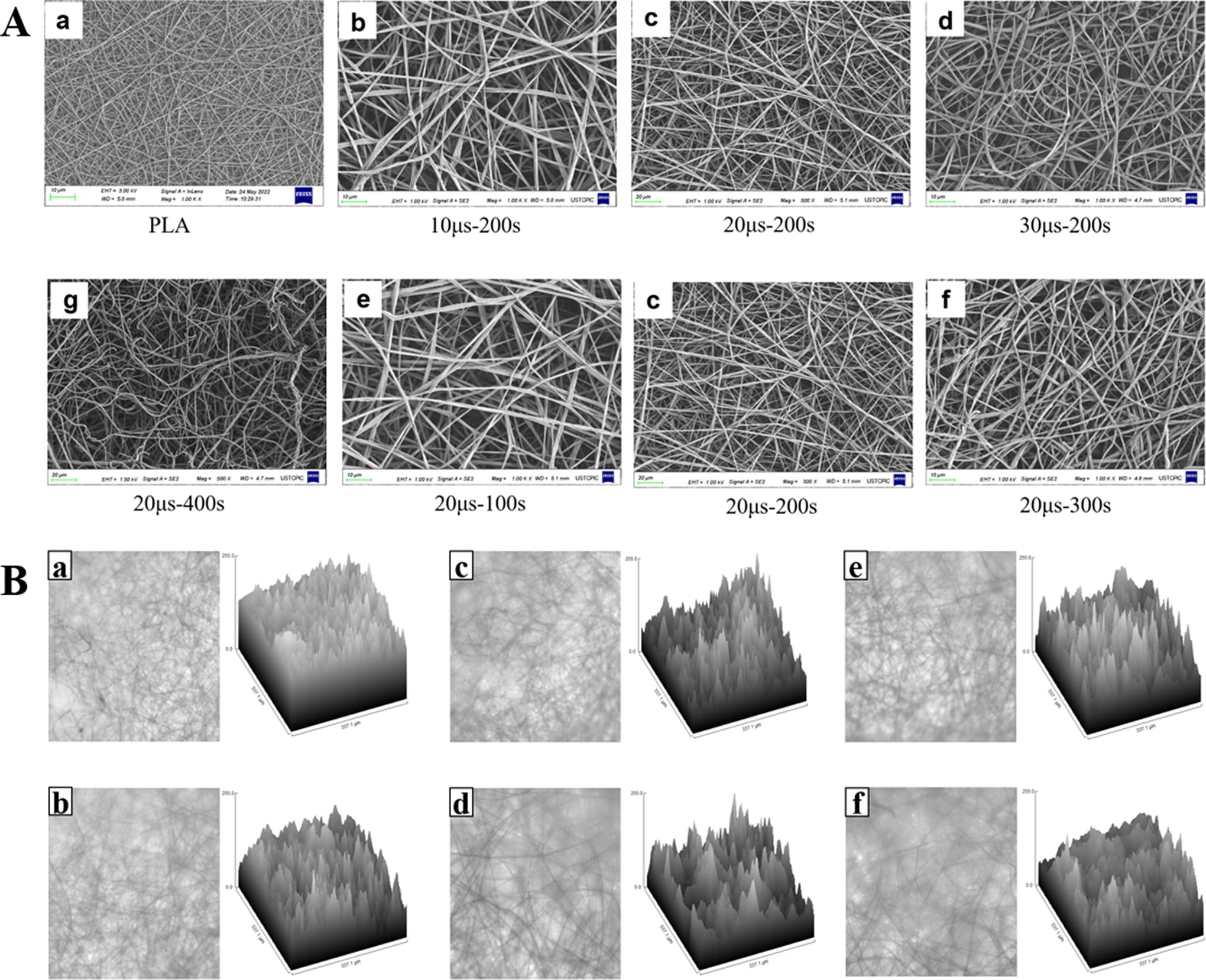
Total internal reflection fluorescence microscope (TIRFM) images and analyzed images of PLA surfaces before and after PIII treatment with different parameters are shown in Figure 1B. The films treated by PIII seem to be thinner and rougher.
PLA surface wettability is altered from hydrophobic original PLA (117°) to hydrophilic (Fig. 2) after N2 PIII by increasing duration and pulse width. While keeping the time change in the pulse width, the water contact angles were 87° (b), 53° (c), and 38°(d). While keeping the pulse width change the duration, the water contact angles were 88° (e), 53° (c), and 36°(f). The changes in surface hydrophilicity of the N2 PIII-treated samples are evident, which could be attributed to the change in surface chemical bonds.
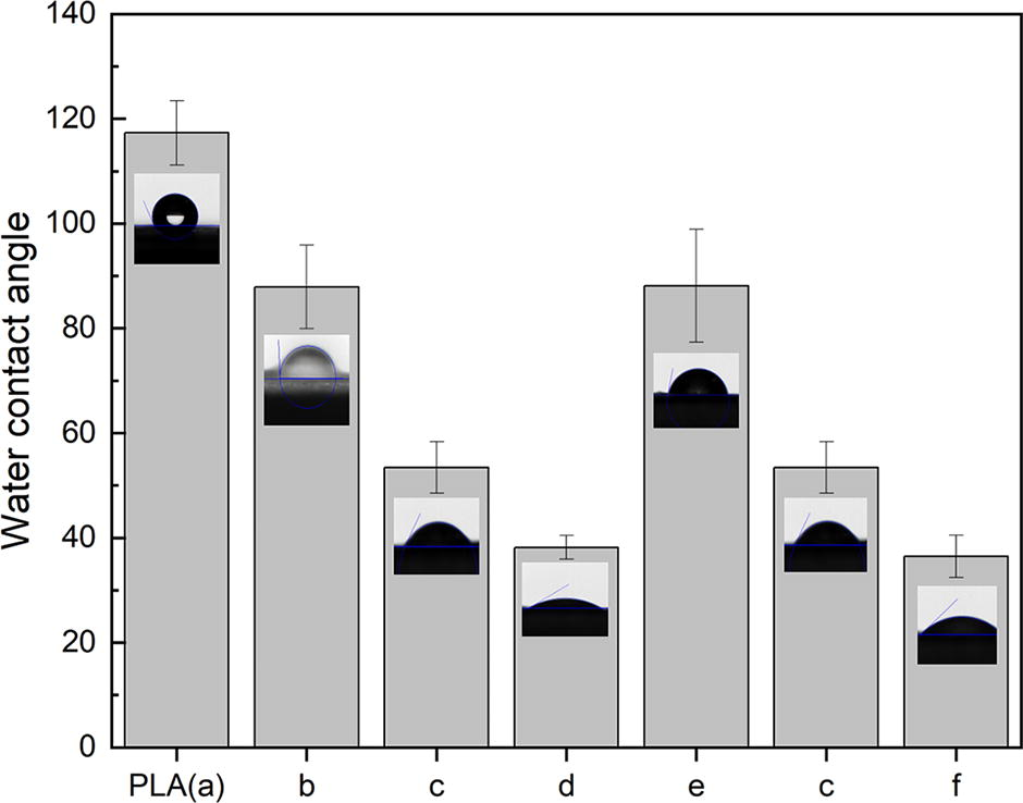
Water contact angles of the electrospinning membranes: a PLA (117°), b (87°), c (53°), d (38°), e (88°), and f (36°). PLA, polylactic acid.
Figure 3 presents the high-resolution C 1 s and N 1 s spectra of the pristine and one N2 PIII-treated sample, as the others are similar. The prominent C-OH/C-O-C and O-C=O peaks of the pristine PLA decrease in intensity after N2 PIII, and there is an emergence of C-N-C=O. 26 This result shows that the C-O/C=O bond is disrupted during the N2 PIII process and forms a new C-N-C=O bond with the N element, accompanied by the breakdown of polymerization bonds. It seems new C-N-C=O bonds increase hydrophilicity. The difference in hydrophilicity between groups is due to the different content of N elements. Groups with longer duration and broader pulse width are more hydrophilic (d, f). The surface chemical alteration on the N2 PIII-treated samples is evident.
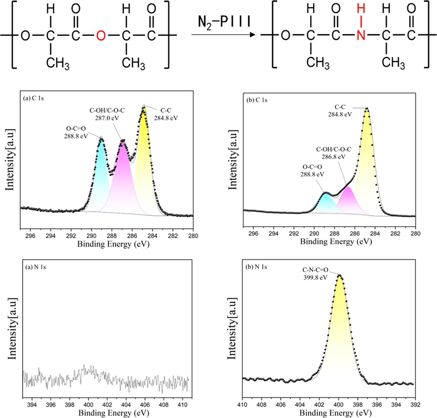
XPS obtained high-resolution C 1 s and N 1 s spectra. The result of PLA (
Table 1 shows the element content of PLA electrospun film analyzed by XPS after the above treatment. As analyzed above, C-OH/C-O-C and O-C=O fracture and form C ≡ N and C-C during the treatment process. The oxygen content decreases from about 30% to about 20% after treatment (the oxygen-containing chemical bonds on the surface are generally hydrophilic, indicating that the increase in hydrophilicity of the sample in this study is not caused by a decrease in oxygen content 27 ). The increase in N element varies between 6–8% depending on the processing method, while the total amount of C element remains unchanged, but its proportion increases due to the decrease in oxygen element. The groups with the highest increase in N element are d and f, which are 7.54% and 8.05%, respectively. The increase in time and pulse width increases the sample’s nitrogen content.
Elemental Compositions in Each Sample Semiquantitative Analyzed by XPS Data
XPS, X-ray photoelectron spectroscopy; PLA, polylactic acid.
FTIR is shown in Figure 4A. There is no new peak in the results of PIII-treated samples. One explanation is that the concentration of implanted ions in the treated polymer surface layer is significantly lower compared with polymer target atoms. 28
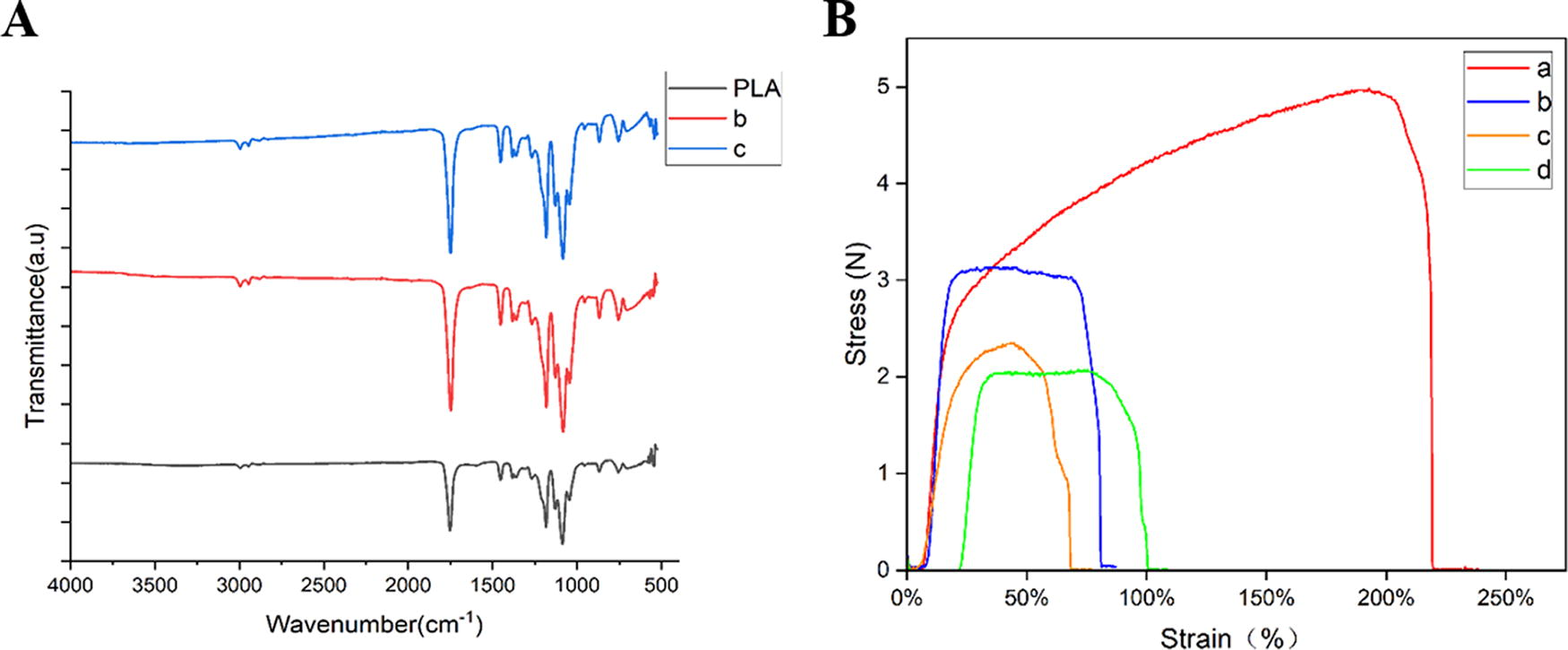
However, the tensile ability of samples treated by PIII changes obviously, as shown in Figure 4B. This may be due to the breaking of polymerization bonds during the modification process.
In vitro cellular response
The groups that mobilized bFGF protein encode with an extra “P.”
The cell proliferation was evaluated by the CCK-8 method, as shown in Figure 5A. On day 1, there is only a statistical difference (*p < 0.05) between the PLA (a) group and the f group. Meanwhile, statistical difference (*p < 0.05) exists in a–p, b–p, and f–p. Contrary to expectations, the group with the highest absorbance on the third day was group c. Although there are statistical differences in d and f (*p < 0.05) and significant differences in c (**p < 0.01) compared with PLA. On day 5, significant differences were also observed between the PIII-treated groups like b and f (*p < 0.05), e and d (*p < 0.05), f (**p < 0.01), etc. This means that different treatment parameters can also impact the growth of cells on treated PLA electrospun membranes. The increase in time and pulse width is beneficial and is consistent with previous research results. 29 For extra “P”, each group has almost statistical differences. This may be because bFGF immobilized on the PLA membranes increases osteoblastic cell growth rate 30 and expands the differentiation.
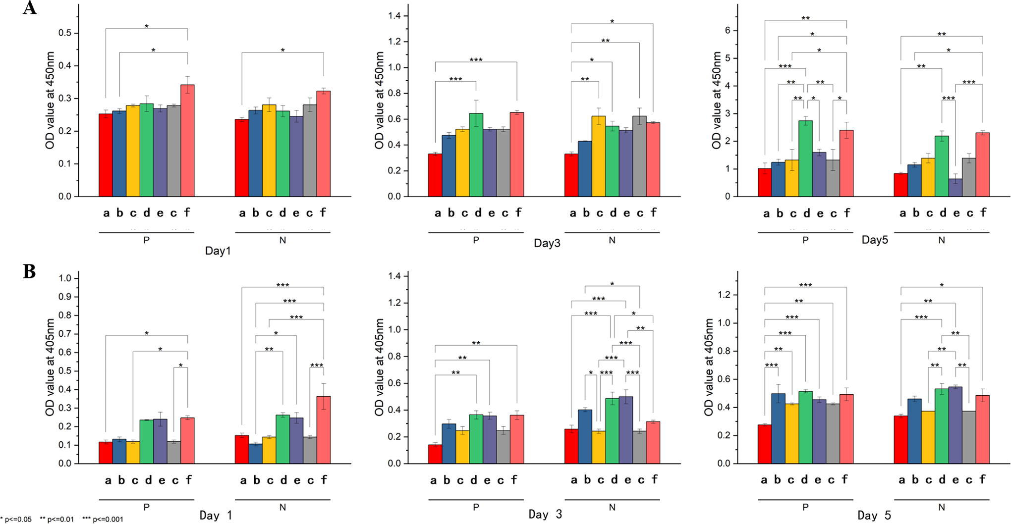
Alkaline phosphatase is one of the in vitro osteogenesis markers. Its osteogenic ability can be evaluated and compared by calculating the average expression intensity of alkaline phosphatase in each sample. 31 As shown in Figure 5B, as the time for replacing with osteogenic induction culture medium increased (1 to 3 days, 3 to 5 days), the ALP content in the samples treated with PIII and immobilized bFGF (P group) grew rapidly. The group had no statistically significant difference by the third and fifth days. However, there have always been intragroup differences within the N group. This may be because the role of the bFGF protein is more pronounced, covering the differences between samples, so that every sample of immobilized bFGF could undergo rapid osteogenesis.
Based on the results of CCK8, select group a and group f for cell fixation. In Figure 6, we can observe that compared with group a, group f has more cell adhesion per unit area. This once again confirms that the plasma treatment of N2 is beneficial for the growth of cells in polymers.
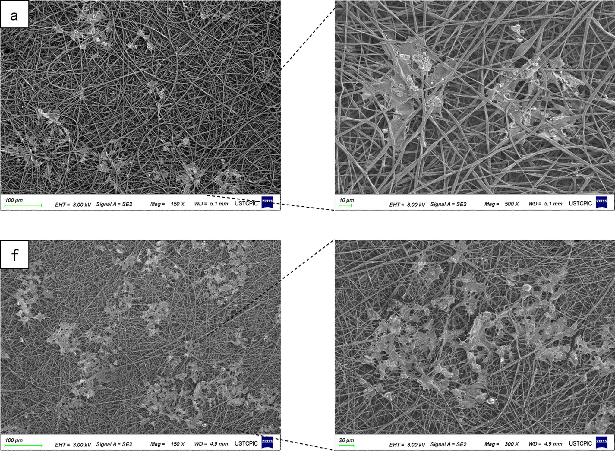
SEM of MC3T3-E1 cells cultured on a, and f groups for 5 days. SEM, scanning electron microscope; MC3T3-E1, MEM and osteoblastic cell line.
The following results are all from the “P” group and will not be annotated separately.
The ELISA results can roughly display the bFGF content of each sample. The PIII treatment time or pulse width of groups d and f were the longest, so the bFGF content was also the highest. The correlation between N2 PIII treatment and protein content was determined based on significance calculation as shown in Figure 7. Combined with other results, we are confident that the effect of bFGF in the extra P group covers the impact of N2 PIII treatment. However, they are both beneficial for cell culture.
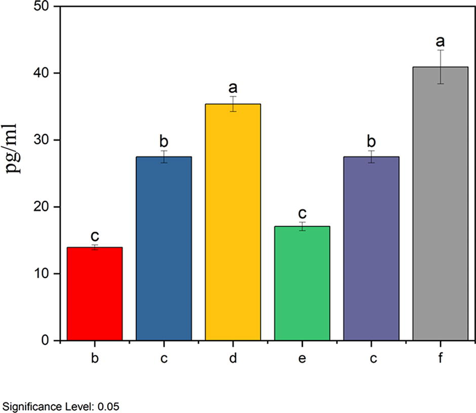
The bFGF content of each sample is based on the absorbance standard curve obtained by ELISA (Trace residual bFGF was detected in a group). Groups with the same letter indicate no statistical difference (p < 0.05), while groups with a different letter indicate a statistical difference. bFGF, basic fibroblast growth factor.
To ensure that bFGF fixed on the PLA membrane can still exert its biological ability, fibroblasts were seeded on PLA membranes and immobilized bFGF. The growth vitality of fibroblasts on different membranes is shown in Figure 8. Similar to osteoblastic cells, the result of groups d and f was statistically different from the others (day 1). As the growth time increased, other groups also showed statistical differences compared with PLA.
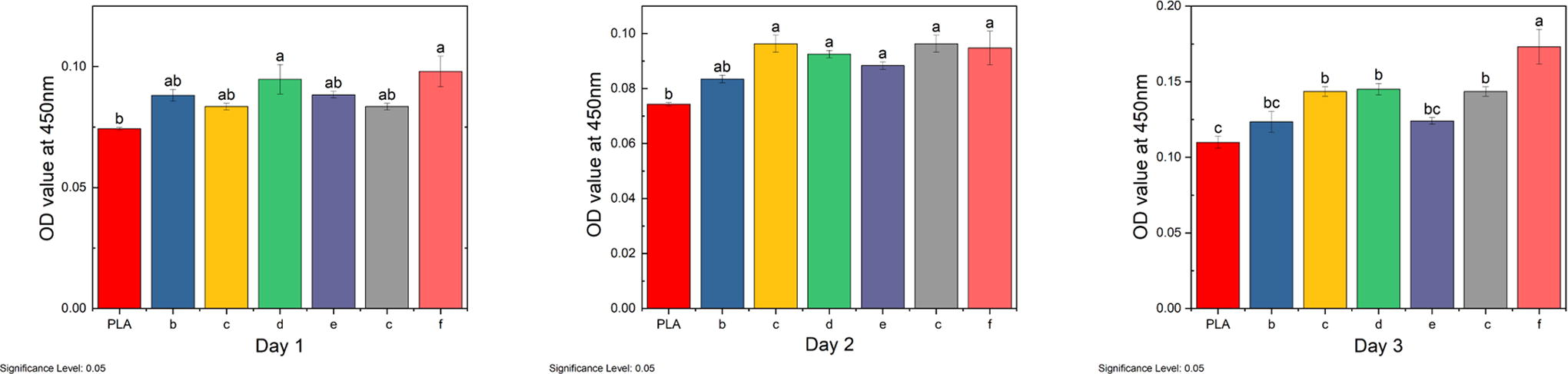
The growth vitality of fibroblasts in the first 3 days. Groups with the same letter indicate no statistical difference (p < 0.05), while groups with a different letter indicate a statistical difference.
The adhesion of fibroblasts on PLA membrane-immobilized bFGF is shown in Figure 9. Phalloidin staining showed that the fibroblasts had a flattened morphology, and were widely dispersed on PIII-treated surfaces, which indicated good cell adhesion. At the same time, fibroblasts had a shrunken morphology on untreated PLA membrane and were rarely distributed on the surface.
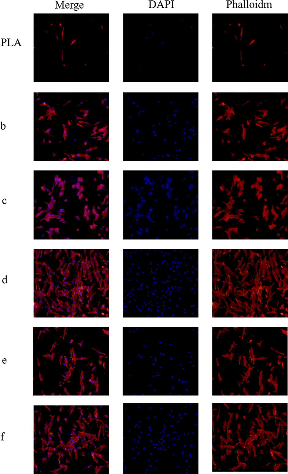
Representative confocal microscopy images of fibroblasts were cultured on various surfaces and stained with TRITC phalloidin (red) and DAPI (blue).
Discussion
N2 plasma in PIII is composed of many species of nitrogen radicals. When the bias voltage is applied, 32 cation will aggregate on the surface of the polymer and collide and react with the polymer chains. The chemical and physical properties of the treated polymer surface can be adjusted based on electrical parameters (processing time, power supply, pulse width, frequency), gas type, and air pressure.33,34 In this study, we performed N2-PIII treatment on PLA electrospinning nanofibers. XPS analysis showed that nitrogen-containing functional groups were formed on the surface of the polymer treated with N2-PIII, and the chemical bond content of the original C element changed. It is speculated that N plasma may have disrupted these chemical bonds and generated C-N-C=O bonds. These changes formed on PLA are undoubtedly due to the complex reactions during PIII. When treating polymers with ion irradiation, there are two competing reactions. One is chain breakage when the atoms in the polymer main chain are replaced by high-energy ions (nuclear stopping), while the second is branching/crosslinking, which is caused by free radicals formed by electron excitation (electron stopping).35,36 Generally speaking, two reactions occur simultaneously, but one dominates depending on the type of polymer and irradiation conditions. According to the XPS spectrum analysis, it should be the first reaction that dominates the process of this experiment. The contact angle and bFGF content seem to be mismatched to the N element content of each group, as the first two terms are more determined by polar groups on the surface of samples.37,38 Combine the results of different surface water contact angle changes and electron microscopy images, except for group g, the fiber structure on the surface of the samples treated with N2-PIII was not damaged and underwent specific chemical changes, including changes in element content and the formation of new chemical bonds C-N-C=O. Meanwhile, the formation of new chemical bonds is accompanied by the breaking of polymerization bonds, which causes reduced stretching ability and rougher film surface (Fig. 2).
Plasma treatment of polymers has been proven to increase the biocompatibility of samples,39,40 and polymers, after partially ionized particle treatment, have the characteristic of connecting biomolecules without chemical reagents. The fixed material can exert the biological ability of the original biological factors.12,21 Basic fibroblast growth factor (bFGF) preserves the viability of various cells,41–43 including cell growth and binding to multiple protein receptors, which play a role in signaling pathways such as regulating intracellular calcium.
This experiment used PLA electrospinning crosslinked bFGF for osteoblast growth and osteogenic induction (MC3T3-E1) and the development of fibroblasts (HDF-a). Compared with the original samples, these experimental samples showed significant improvements, and the differences between the experimental groups disappeared over time. On the contrary, the intergroup differences between the PIII-treated groups of the uncrosslinked bFGF group increased or remained unchanged over time, indicating that the influence of bFGF covered the effect of plasma treatment. According to the electron microscopy images and fluorescence staining images after cell fixation, cells on the PLA membrane-immobilized bFGF can grow better: better extension, flat morphology, and more individual cells per unit area.
The results obtained from this study demonstrate that N2-PIII treatment not only enhances the biocompatibility of PLA electrospinning membranes but also significantly promotes the proliferation of osteoblasts on the modified membrane. Furthermore, applying N2-PIII treatment to the electrospinning membranes results in a crosslinked bond, C-N-C=O, immobilizing the growth factor bFGF. The immobilization of bFGF on the PLA electrospinning membrane ensures that the growth factor retains its biological functionality. The biological effects and cellular responses elicited by the immobilized bFGF exceed those achieved solely through N2-PIII treatment of the PLA electrospinning membranes. This finding is particularly noteworthy as it highlights the potential of combining plasma treatment with growth factor immobilization to achieve superior osteogenic outcomes.
It is important to emphasize that the plasma treatment used in this study did not compromise the PLA electrospinning membranes’ structural integrity or functional properties. This suggests that the modification process is gentle and nondestructive, preserving the inherent advantages of the electrospinning membranes. The electrospinning membranes could be used for implant coating, scaffolding, or postoperative fillers.10,44,45 Researchers try to improve the properties of electrospinning, such as mechanical properties or affinity to cells and tissues. From the experimental results, the PIII is an excellent modification method; the electrospinning membrane’s biocompatibility is improved, while the bFGF could be immobilized. Crosslinking PLA nanofibers with bFGF appears more effective than plasma treatment alone when using these modified membranes as bone tissue scaffolds. However, it is worth noting that the cost of bFGF may be a limiting factor in its widespread application. Future research should explore alternative, cost-effective growth factors or immobilization techniques that can achieve similar osteogenic outcomes. Additionally, further investigation is needed to determine if other biological processes of bFGF can be harnessed on the fixed membranes. Understanding the full range of bFGF’s functional capabilities on these modified surfaces could lead to developing even more effective bone tissue scaffolds.
In conclusion, the results of this study provide valuable insights into the potential of combining N2-PIII treatment with growth factor immobilization to enhance the osteogenic properties of PLA electrospinning membranes. While the cost of bFGF remains a consideration, the promising osteogenic outcomes achieved through this combined approach suggest that further exploration in this area is warranted.
Conclusion
Using N2-PIII to modify PLA electrospinning and immobilize the biological factor, bFGF, for bone repair is a feasible solution. With the increase of processing time and pulse width, the structure of polylactic acid electrospun is not destroyed within a specific range, and the N content increases, resulting in a corresponding increase in its biological affinity for cells. However, prolonged or high-energy treatment can damage the structure of nanofibers, rendering them unable to serve as tissue scaffolds. The effect of crosslinked cell growth factor bFGF can function normally and outweighs the impact of N2-PIII. The surface of the polymer treated with N2 PIII is quickly completed, with little effect on the overall polymer’s intrinsic physical and chemical properties, which could be applied for various biodegradable polymer modifications. Therefore, this method for PLA-immobilized bFGF modified by PIII has the potential as a bone tissue implant.
Footnotes
Acknowledgments
The authors thank the University of Science and Technology of China and The Second Affiliated Hospital of Anhui Medical University for the experimental instruments.
Authors’ Contributions
Q.L. performed experiments and data analyses and drafted the article. Z.H. and W.C. experimented. Q.X. and Z.W. gave technical and material support. Z.W. managed projects and obtained funding. All authors discussed the analyses and have contributed to, seen, and approved the article.
Disclosure Statement
None of the authors has any conflicts of interest to disclose, and all authors support submission to this journal.
Funding Information
This work was supported by the Fundamental Research Funds for the Central Universities (No. USTC 20210079), the Funding for Joint Laboratory of Applied Plasma Technology (No. JL06120001H), and the ITER Project of the Ministry of Science and Technology (No. 2022YFE03080001).