
Abstract
Only few published data are available on ticks and tick-borne zoonotic pathogens in Bolivia. To evaluate rickettsial seroprevalence and infection in dogs and ticks, during February–April 2007, we collected whole blood, sera, and ticks from dogs living in the rural, peri-urban, and urban areas of Cochabamba, Bolivia. Dog sera were subjected to enzyme-linked immunosorbent assay test to detect IgG antibodies against Rickettsia rickettsii and 68.2% of samples were found to be positive (n = 30; 95% confidence interval [CI]: 52.4–81.4). Blood samples and ticks were tested using polymerase chain reaction to detect spotted fever group (SFG) rickettsiae. One blood sample was positive for Rickettsia parkeri (2.3%; 95% CI: 0.06–12.3). Ticks were collected from 10 dogs and were identified as Amblyomma tigrinum (n = 44) and Rhipicephalus sanguineus (n = 1). All A. tigrinum ticks were collected from resident dogs from the rural areas of Cochabamba, whereas R. sanguineus was from a dog originating from Santa Cruz. Of 42 DNA samples extracted from ticks, 23 (54.8%; 95% CI: 38.7–70.1) were polymerase chain reaction positive for Rickettsia spp. Sequencing analysis identified 22 samples as R. parkeri and one as Rickettsia aeschlimannii. Positive ticks (all A. tigrinum) were collected from six dogs, all of which were seropositive. This is the first report of SFG rickettsiae in A. tigrinum, suggesting that this tick—like others species in the Amblyomma maculatum group—may play a role in the biological cycle of Ri. parkeri. The high infection prevalence of SFG rickettsiae in ticks and the even higher seroprevalence in dogs suggest an active circulation of agents of rickettsiosis in the study area, although there are no confirmed cases of infection in humans. Our study supports the use of canine serology as risk indicator for SF rickettsioses.
Introduction
Because of extensive cross-reactivity among various SFG rickettsiae (Raoult and Paddock 2005), the species infecting seropositive patients is assumed based on geographic location. Rickettsia rickettsii is endemic to North and South America, where it causes Rocky Mountain spotted fever. Therefore, spotted fever-type illness in the western hemisphere is commonly attributed to this bacterium in seropositive patients. Because of new biomolecular techniques and an increased interest in tick-borne zoonoses, the number of rickettsial species recognized as potential pathogens is growing. Rickettsia parkeri, for example, previously considered nonpathogenic, was recently identified as a human pathogen (Parola et al. 2005). In fact, it is suspected that several cases of spotted fever-like illness in the United States that were presumed to be caused by Ri. rickettsii may have been caused by infection with Ri. parkeri (Paddock et al. 2004, Paddock 2005). Unlike Rocky Mountain spotted fever, infection with Ri. parkeri is characterized by an eschar at the tick-bite site. Therefore, it has been hypothesised that atypical eschar-associated SF rickettsioses in South America may be caused by Ri. parkeri rather than Ri. rickettsii (Parola et al. 2005). Domestic animals can be good indicators for the distribution of Rickettsia spp. and animal serology can be used for human rickettsiosis surveillance. In Latin America, serological surveys of horses and dogs were used for the monitoring and control of Brazilian spotted fever in areas where humans are exposed to the tick vector Amblyomma cajennense (Sangioni et al. 2005). Moreover, serological evidence of canine infection with Ri. parkeri was reported in Brazil (Horta et al. 2007, Labruna et al. 2007). Dogs are excellent sentinels, as they are susceptible to a wide range of emerging human infections and frequently exposed to potential vectors. Moreover, they are ubiquitous, widespread, and easily accessible (Cleaveland et al. 2006).
In Bolivia, only few data are available on the hard ticks fauna and on distribution and incidence of tick-borne zoonoses because of the lack of valid diagnostic methods and epidemiological surveillance systems. Our work was aimed at identifying rickettsiae in dogs and in infesting ticks and estimating their prevalence in rural, peri-urban, and urban areas of Cochabamba.
Materials and Methods
Study area
The Central Valley (Valle Central) of Cochabamba is situated in the Department of Cochabamba, Bolivia. It is an upland area 1150 km2 wide, located at 2500–2800 m of altitude in the Oriental Cord of the Andes (Cordillera Oriental). The weather is temperate, with a mean annual temperature of 17.5°C. The rainy season is from November to April, with a mean annual rainfall of 400–500 mm (Anonymous, unpublished data). The department is surrounded northward by the Cordillera of Tunari (5035 m above sea level), where the Tunari National Park (5,684 hectares) is located. The southern border of the park overlaps with the 2750 m above sea level altitude contour (Quinteros Condoretty, unpublished data).
The study area (Fig. 1) includes the municipalities of Cochabamba and Las Lomas (Cercado province), Sacaba (Chapare province), and Quillacollo and Tiquipaya (Quillacollo province). It accounts for a population of more than 900,000 inhabitants, 700,000 of them living in the urban area, mainly in Cochabamba city (17°23′S, 66°09′W).
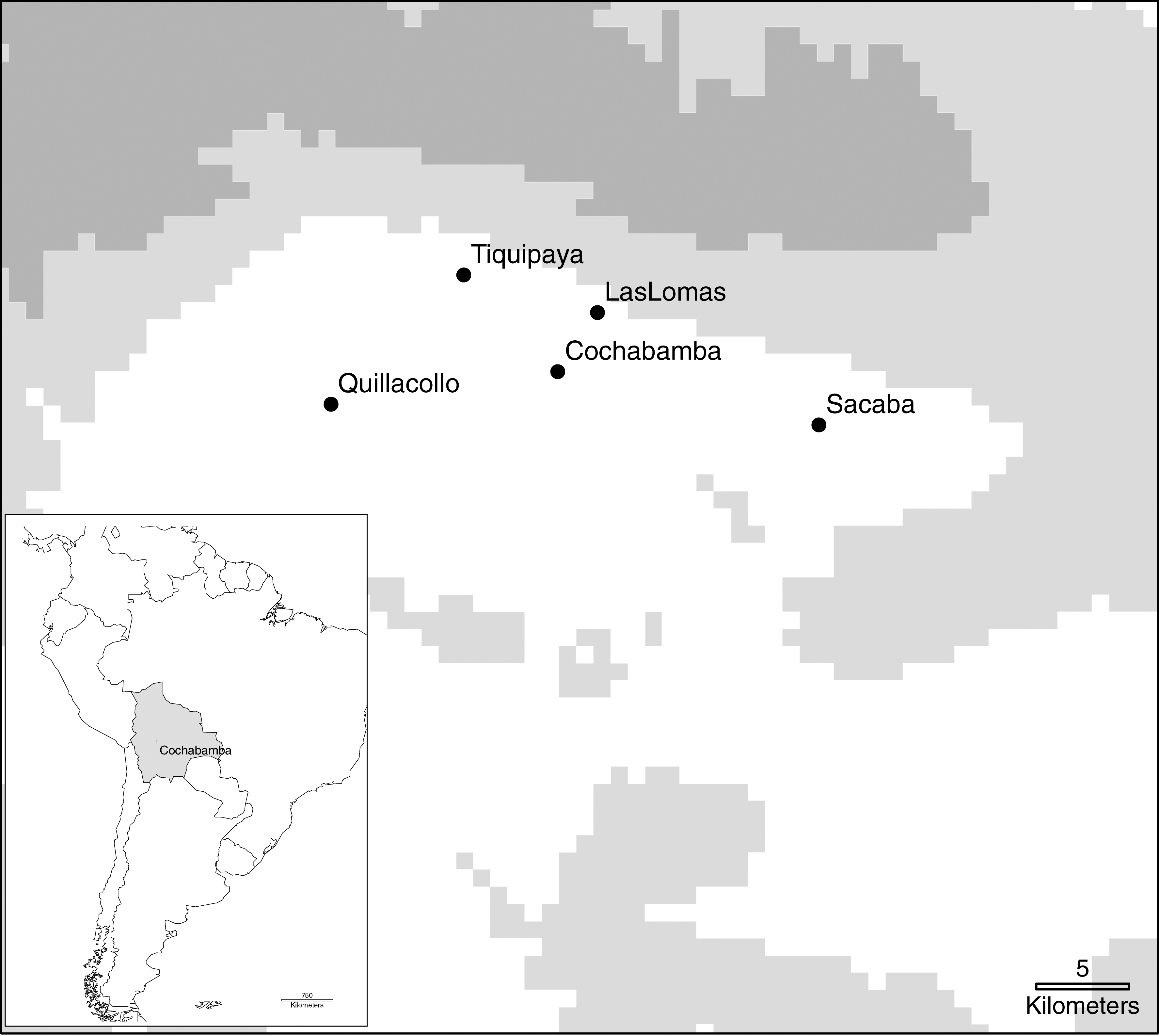
Map of the Central Valley of Cochabamba with the municipalities included in the study (white area: 2000–3000 m above sea level [asl]); light gray areas: 3000–4000 m asl; dark gray areas: 4000–5000 m asl). The bottom left box shows the map of Latin America with Bolivia (gray) and Cochabamba city.
Agriculture is the principal economic activity in the rural areas, where population is mainly constituted by Quechua (indigenous population of the Andes high plateau), generally living in poor conditions.
No estimates on livestock population are available, but small-scale farming (cattle, small ruminants, pigs, and poultry) is common in rural areas. The dog population in Cochabamba Department is estimated at one dog every three inhabitants, totaling around 400,000 animals (Anonymous, unpublished data). In rural areas, especially in Las Lomas and Tiquipaya, resident dogs often wander inside the Tunari National Park, where the sylvatic fauna is abundant and comprises several mammal (rodents, opossums, bats, foxes) and avian species (Anonymous, unpublished data).
Moreover, cases of tick bites in humans in the rural area—especially in children—were reported to the authors by local physicians and residents, but neither published data nor public health records on tick infestation and tick-borne diseases are available.
Biological sample collection
Between February and April 2008, whole blood and sera samples were collected from 44 resident dogs. The animals were included in the study for being infested by ticks or having a history of tick infestation, according to their owners. Most of the dogs lived in the rural area (n = 28), while 11 of them were from the suburban area and 5 from the urban center of Cochabamba.
Blood samples were either collected from dogs at the veterinary clinic “Las Huellas,” Cochabamba city (n = 19), or directly in the field in the rural areas (n = 25). An aliquot of each sample was stocked in LongMire Buffer (Randi et al. 2002) and preserved at 2–4°C in labeled microtubes, while the remaining blood was centrifuged and sera aliquots were stored at −20°C until serological testing.
Seven of the 44 dogs were infested and a sample of ticks was collected from them. Moreover, ticks were taken from other two resident dogs in the rural area of Cochabamba and from a dog that had just returned from Santa Cruz, a department located more than 400 km away and characterized by tropical climate (Table 1); blood/sera were not taken from these three dogs. Ticks were all stored in 70% alcohol until identification and polymerase chain reaction (PCR) testing.
Blood/sera were not collected from these three dogs.
n.t., not tested; PCR, polymerase chain reaction.
Informed consent to collect blood and ticks, and to perform the tests, was obtained from the dogs' owners.
Serologic and molecular biology assays
A commercial enzyme-linked immunosorbent assay (Helica®; Biosystems, Fullerton, CA) targeting R. rickettsii IgG was used to test dog sera. Positive and negative controls were provided with the kit and consisted of dog serum reactive and nonreactive with R. rickettsii, respectively. The tested sera were diluted at 1:100 and absorbency (optical density) was read under a spectrophotometer. The assay procedure was performed and results were assigned (positive and negative samples, and respective optical density and cutoff values) according to the manufacturer's instructions.
The ticks were identified at species level by stereoscopic microscope using identification keys according to Estrada-Peña et al. (2005) and Onofrio et al. (2006).
DNA was extracted from single blood samples and individual ticks using Qiagen DNeasy tissue kit (Qiagen GmbH, Hilden, Germany). Negative controls (distilled water) were added to verify the potential contaminations of samples during this phase.
To confirm tick taxonomic identification, a 456-bp fragment of 16S mitochondrial gene of hard and soft ticks was amplified by PCR (d'Oliveira et al. 1997).
The presence of Rickettsia spp. DNA in our samples was investigated by PCR assays targeting the citrate synthase gene (gltA, 401-bp product; Labruna et al. 2004). Positive samples were further tested to detect the ompA gene, specific for the SFG rickettsiae, using Rr190.70F and Rr190.602R primers (530-bp product; Regnery et al. 1991). In case of a negative result to this second PCR, samples were subjected to a seminested PCR to identify the same gene as described by Sumner et al. (2007). Positive gltA and ompA samples were also tested for the presence of ompB gene (856 bp; Roux and Raoult 2000). In all PCR reactions, 5 μL of DNA sample was tested. Distilled water was used as negative control and DNA from a Rickettsia conorii isolate was used as positive control.
Purified PCR products were sequenced. Sequences were analyzed by Chromas 2.0 software (Technelysium, Helensvale, Australia) and submitted to BLAST (
Prevalence of serology and PCR-positive results were calculated, with 95% exact binomial confidence intervals (95% CI). Chi-square test was used to study the association among categorical variables. Analyses were performed by R software (R Development Core Team 2008).
Results
Antibodies against Rickettsia spp. were detected by enzyme-linked immunosorbent assay test in 30 of 44 dog sera, thus showing a seroprevalence of 68.0% (95% CI: 52.4, 81.4). The remaining sera were considered negative. No statistical difference in serology results was detected among dogs living in different municipalities (chi-square test: p > 0.05).
One of the 44 whole blood samples tested positive using the PCR targeting the gltA gene (2.3%; 95% CI: 0.06, 12.0). The sequence showed 99% similarity with the gltA gene of R. parkeri strain At24 isolated in Brazil (EF102236). The PCR assays targeting ompA and ompB genes yielded negative results. The positive sample belonged to dog H, a seropositive animal living in the rural area of Las Lomas that was also infested by PCR-positive ticks (Table 1).
Overall, 45 adult ticks were collected from 10 dogs: 44 were identified as Amblyomma tigrinum (19 male ticks, 25 female ticks) and 1 as Rhipicephalus sanguineus male tick. This latter tick was collected on the dog moved from Santa Cruz, an area with different ecological characteristics. Other dogs resided in rural areas (Las Lomas and Tiquipaya) and one in Cochabamba city (Table 1). The taxonomic identification of A. tigrinum was confirmed by PCR, as the sequence (GenBank accession no.: FJ965339) showed 99% similarity with the corresponding 16S ribosomal RNA gene sequence of A. tigrinum (AY836004).
PCR for Rickettsia spp. detection was performed on 42 ticks (41 A. tigrinum and 1 Rh. sanguineus), while 3 A. tigrinum were kept in our laboratories as reference specimens. SFG rickettsiae DNA was detected in 23 tick samples, all A. tigrinum (54.8% infection prevalence; 95% CI: 38.7–70.1), by amplifying gltA, ompA, and ompB genes. The positive ticks, 13 males and 10 females, were collected from six dogs living in Las Lomas. Five of these dogs were seropositive to SFG rickettsiae; only dog F, a 5-month-old puppy, was seronegative (Table 1).
Sequence analysis of ompA and ompB genes showed the identity of 22 of 23 specimens (GenBank accession nos.: ompA FJ986616, ompB FJ986617), which were 100% similar to the reference ompA and ompB sequences of R. parkeri (EU715288 and AF123717, respectively). One A. tigrinum was infected by a rickettsia which was identified as R. aeschlimanii. Indeed, the sequences of ompA and ompB genes amplified from this tick (GenBank accession nos: ompA GQ180862, ompB GQ180863) were, respectively, 98% and 99% similar to the reference R. aeschlimannii sequences (DQ379980 and AF123705).
Discussion
Our study demonstrates the presence and circulation of SFG rickettsiae in the Central Valley of Cochabamba, namely R. parkeri, and indicates a potential zoonotic risk for humans.
The high rickettsial seroprevalence detected in dogs suggests a situation of endemicity for rickettsiosis. In our study area, adult A. tigrinum are highly infected with Ri. parkeri. Dogs are their primary hosts and may thus be easily exposed to SFG rickettsiae and become seropositive. Most of the seropositive dogs were older than 2–3 years, thus suggesting a previous exposure to Rickettsia spp.-infected ticks. This is in agreement with Pinter et al. (2008), who stated that the odds of a dog being parasitized at least once by a R. rickettsii-infected tick are higher in older animals. All dogs that were seropositive and/or infested by ticks resided in the rural area or—according to their owners—got infested by ticks in the rural areas. Most of the infested dogs were living in Las Lomas, a municipality bordering the Tunari National Park. According to veterinarians and pet owners, ticks are abundant especially in August in the rural areas, so during our study period (February–April) only a small percentage of examined dogs was found infested. Dogs carrying Rickettsia spp.-positive ticks were all seropositive except dog F. Possibly, being a puppy, it was not yet exposed to the peak tick period and did not yet develop a IgG response by the time of the survey.
Positive serologic titers can indicate an active infection or a previous exposure of the dog to rickettsiae, as antibodies can last for extended periods of time after infection (Tesouro et al. 1998, Parola et al. 2005). PCR may discriminate between active or previous infections; in fact, detection of rickettsial DNA in blood can only be achieved during rickettsemia. Moreover, it detects infection in dogs before seroconversion (Kidd et al. 2008). Unfortunately, rickettsemia lasts for a few days or weeks (Burgdorfer et al. 1988), so rickettsial DNA detection in vertebrate blood is considered a rare event (Horta et al. 2007). Moreover, Rickettsia spp. infect endothelial cells and concentration of the organism in blood is low (La Scola and Raoult 1997). Lack of positive PCR results in dog blood may be due to the absence of circulating rickettsiae at the time of testing. Moreover, a low sensitivity in our PCR assay may explain why we could only detect the gltA gene of R. parkeri in the blood sample of dog H. However, the PCR-positive result indicates that the dog was rickettsiaemic at the time of the study and allowed the identification of the specific rickettsial agent responsible for the infection. Our serologic assay employed a single antigen (R. rickettsii) and was thus not sufficient for rickettsial speciation (Raoult and Paddock 2005). Our study is the first report of Ri. parkeri amplification from dog blood.
The finding of R. parkeri in Bolivia follows its detection in Amblyomma triste in Uruguay (Pacheco et al. 2006), Brazil (Silveira et al. 2007), and Argentina (Nava et al. 2008). We report for the first time the infection by SFG rickettsiae in A. tigrinum. This supports the idea that, in Latin America, R. parkeri may be associated with ticks belonging to the Amblyomma maculatum group (Parola et al. 2009). The possibility of A. tigrinum as vector for rickettsiae must be further investigated, but our study indicates the implication of this tick in the transmission cycle of R. parkeri. It was the sole tick species collected in the resident dogs of Cochabamba. These results are particularly important as A. tigrinum adults also feed on humans (Guglielmone et al. 2006).
The identification of the ticks circulating in the study area is an important aspect of our work, as no information on tick fauna was available. Our finding of A. tigrinum at such altitudes, in an area characterized by dry temperate climate, supports previous studies indicating the high ecological plasticity of this tick species, which is distributed in different phytogeographical regions (Guglielmone et al. 2000). Suitable habitat characteristics supporting the tick life cycle are available in Cochabamba, especially in the rural area, where vertebrate hosts for adult ticks (carnivores; Guglielmone et al. 2000) and immature stages (wild birds and rodents; Nava et al. 2006) do live.
Finally, a sequence showing high similarity to Rickettsia aeschlimannii was detected in one DNA specimen obtained from A. tigrinum. This rickettsial species has been detected in several countries in Europe and Africa, where it is usually associated with Hyalomma marginatum marginatum and Rhipicephalus appendiculatus ticks. Its pathogenic role is suspected; in fact, it has only been detected in patients with eschar-associated infections in Morocco and South Africa (Parola et al. 2005). In South America, R. aeschlimannii was only detected in Ixodes boliviensis in Peru (Blair et al. 2004), so our finding is quite unexpected and deserves further investigations.
In conclusion, our work suggests that SFG rickettsiae are widespread in the Central Valley of Cochabamba and may infect dogs and potentially other animal species, including humans. In addition, our results support the use of animal serology as risk indicator for possible SFG rickettsiae infection in humans. As R. parkeri spotted fever is a newly identified disease and shows a low virulence, it can be less easily recognized compared with lethal pathogens such as R. rickettsii (Horta et al. 2007). It is thus possible that R. parkeri spotted fever cases in humans have been occurring in Cochabamba without a proper diagnosis. This is especially probable in the rural areas, where people lack medical services and can easily be exposed to ticks, because of the closeness to sylvatic habitats that can support the ticks' life cycle.
Footnotes
Acknowledgments
The authors thank Marcelo Labruna (Faculty of Veterinary Medicine, University of São Paulo, Brazil) for fruitful suggestions during the study, Alberto Guglielmone (INTA, Rafaela, Argentina) for assistance in tick identification, Gervásio Bechara (Sao Paulo State University, Brazil) and the ICTTD-3 project (EU-INCO 6th FP, no. 510561) for providing the tick identification manual for the Neotropical region. The research was partially funded by the University of Turin, research grant “fondi ex-60%,” and by the International Mobility Student Program of Turin University. Field activities were facilitated through the SAPUVETNET II project (no. II-0438-A), an international network on Veterinary Public Health formed by Latin-American and European Universities/Faculties of Veterinary Medicine, cofinanced under the ALFA Program, European Union.
Disclosure Statement
No competing commercial associations or financial interests exist in connection with the submitted manuscript for all authors concerned.