
Abstract
Essen regimen, the Thai Red Cross two-site ID regimen, Zagreb schedule, and the eight-site ID regimen are the standard rabies vaccines recommended by the World Health Organization (WHO). In this study, a liposomal rabies vaccine (LipoRV) was developed, which was found to facilitate the production of rabies virus neutralizing antibody (RVNA) in BALB/c mice. Liposome solution was prepared with hydrogenated soya phosphatide and cholesterol. LipoRV composed of liposome solution and inactivated rabies vaccine (IRV). The immune responses were compared between the mice treated with either LipoRV or IRV. Higher levels of interleukin-2 (p < 0.05), interferon-γ (p < 0.01), and natural killer cell activity (p < 0.05) were observed in the mice immunized with LipoRV than those with IRV. The potency of LipoRV was significantly higher than that IRV (p < 0.05). In addition, three injections of LipoRV on days 0, 3, and 14 could elicit similar RVNA levels as the five shots of IRV. Our data also showed a higher survival rate in mice treated with three shots of LipoRV (56.2%) than five shots of IRV (40.6%). In conclusion, liposome enhances the immune response of mice to rabies vaccine and could be applied as a potential immunopotentiator.
Introduction
R
The intradermal and intramuscular PEP recommended by the WHO includes the Essen regimen, Zagreb schedule, Thai Red Cross schedule, and Oxford regimen. The Essen regimen has been used over the last three decades as the “gold standard” among all PEP, which is administered by repeated injections on days 0, 3, 7, 14, and 28 (WHO). Fourteen days after repeated injections, a previously unvaccinated subject is able to produce protective antibodies. However, the expense of Essen regimen is also the highest among all of the PEP regimens. The Zagreb schedule (IM 2-0-1-1) is similar to the Essen regimen, but patients receive no dose of vaccine on day 3 when most tropical wound infections become apparent. A comparable number of antibodies are produced by the Thai Red Cross schedule (2-2-2-0-1-1) in comparison with the Essen regimen (4) on a precondition of seven shots in 30 days. As for the eight-site intradermal “Oxford” regimen, patients need a booster dose on day 90, leading to limited use so far (11).
Immunopotentiator, also called immunologic adjuvant, is one class of substance capable of nonspecifically promoting the specific immune response after being injected with antigen precedingly or simultaneously. They are divided into storage type and center type according to the mechanism of their action. The storage type such as aluminum hydroxide, oil emulsion, and aluminum phosphate assists in eliciting durative immune stimulation and effective immune response by prolonging persistence of antigen. Bacterium components, endotoxin, liposome, and several Chinese herbal medicines belong to the center type, directly stimulating the immune system and the activity of antigenic substance. A number of previous studies have demonstrated the immunoadjuvant activity of liposomes acting as an inducer of cell-mediate immune response with high efficiency (3,10,12). Here, we investigated the effects of rabies vaccine with liposome adjuvant (LipoRV) on BALB/c mice. The productions of RVNA were compared between different schedules of LipoRV and IRV.
Materials and Methods
Cell culture
Vero cells obtained from the WHO were cultured in Bioreactors (40 L each, CelliGen 510 Bioreactor; New Brunswick Scientific Co. Ltd) using culture medium containing 10% calf serum (Gibco, Grand Island, NY) in a culture flask. BHK-21 cells obtained from NICPBP were cultured in Dulbecco's modified Eagle's medium (DMEM; Gibco) containing 10% (v/v) heat inactivated fetal bovine serum (Gibco) and 100 mg/mL penicillin and streptomycin at 37°C in a 5% CO2 humidified incubator.
Viruses
Nonpassage rabies viruses (CNX8601) and fixed rabies viruses (challenge virus standard, CVS-11 strain) were donated from the Chinese Center for Disease Control and Prevention (China CDC, Beijing, China). CNX8601 was isolated from the cerebrospinal fluid of a patient and used to challenge BALB/c mice. The CVS11 strain was used to infect BHK-21 cells for a rapid fluorescent focus inhibition test (RFFIT). The aGV strain purchased from Changchun Institute of Biological Products Co., Ltd (Changchun, China) was used for vaccine production. The National Institute for the Control of Pharmaceutical and Biological Products, China, provided national reference standard serum, CVS, and national reference standard rabies vaccine.
Mice
Female BALB/c mice aged 6–8 weeks were used in this study. To avoid the interference of gender on the observed results, only female mice were used in the current study. Mice purchased from Changchun Institute of Biological Products Co., Ltd were used for the potency tests. Serum was collected from orbital veins of anesthetized mice. All mice were sacrificed by cervical dislocation. The experimental manipulation of mice was performed according to the National Institute of Health Guide for the Care and Use of Laboratory Animals, and was approved by the Scientific Investigation Board of Science Technology of Jilin province, China, with a permit number, SCXK(ji)2016-0008.
Inactivated rabies vaccine
Vero cells cultured for 5 days were infected with the aGV virus, and viruses were harvested continuously from 3 to 11 days after infection. Clarification filtration and ultrafiltration concentration pipelines were used to concentrate the harvested viruses for 30 times in a whole-course aseptic condition. Inactivation of the viruses was achieved using β-propiolactone (1/4,000 dilution) at 4°C. Chromatographic purification was performed to eliminate impurities. Then, the purified viruses were freeze-dried and stored in 1.0 mL/ampul for further experiments.
Liposomal rabies vaccine
All glasswares were sterilized at 180°C for 4 h to inactivate endotoxin. All solutions were prepared in sterile pyrogen-free 20 mM sodium phosphate buffer saline (PBS, pH 7.7). A class 100 horizontal laminar flow cabinet was used to complete all manipulations. Hydro soy phospholipids and cholesterol were purchased from Avanti Polar Lipids, Inc. (Alabaster, AL). The blank liposomes were prepared by reverse evaporation and contained 1% hydro soy phospholipids. They were then mixed with the IRV prepared as described above. LipoRV contained 0.2 mL blank liposome per dose. They were freeze-dried and packaged in 1.0 mL/ampul. For all comparison experiments, the LipoRV and IRV contained the same amount of rabies antigen (50–60 μg/dose).
Morphologic observation and particle size determination of liposome rabies vaccine
Liposome rabies vaccine was diluted with PBS and added on copper screen. For negative staining, 2% phosphate was used. A transmission electron microscope was used to observe the ultramicro-morphous of the liposome rabies vaccine. The size distribution and the average grain size of liposome rabies vaccine were determined by the Malvern Zetasizer 3000 HS laser diffraction particle size analyzer at room temperature.
Entrapment rate assay of EPI liposome
Liposome rabies vaccines were diluted in water at a concentration of 15 mg/mL and centrifuged at 4,000 g for 10 min. The Lowry method was used to determine the content of free protein (Cf) and whole protein (Cw) in 1 mL of supernatant. The entrapment rate (ER) was calculated by the following formula: ER = [Cw − Cf]/Cw × 100%.
Immunization
The BALB/c mice were immunized with the vaccines (0.5 mL) by intraperitoneal injection. Blood samples were collected by retro-orbital bleeding with capillary tubes and centrifugation at 5,000 rpm for 10 min to separate sera. Sera were incubated at 56°C for 30 min for inactivation and stored at −20°C for further tests. In all experiments, preimmune sera were collected 2 days before immunization of the mice, as negative control.
Potency of the vaccines
The potency of IRV alone, LipoRV, and a reference vaccine (provided by the National Institute for Control of Pharmaceutical and Biological products, China) was measured according to the NIH (National Institutes of Health) test protocol used. Sixteen NIH mice in each group were treated with 0.5 mL of the diluted (from 1:25 to 1:625) rabies vaccines by intraperitoneal injection on days 0 and 7. Mice were challenged intracerebrally with CVS (40LD50 per 0.03 mL) after 7 days since the last vaccination and were observed for 14 days. The Reed and Muench method was used to calculate the ED50 (50% efficient dose) presented in international units (IU) per dose.
The RFFIT
The RFFIT was recruited to test RVNA production in mouse sera according to the WHO. The collected preimmune sera and national reference standard serum were used as negative controls. All serum samples were serially diluted threefold in duplicate with DMEM and loaded in a 96-well microtiter plate. CVS11 (5 × 10⁶ FFU/1.0 mL) was added followed by incubation at 37°C for 1 h. Then, 6 × 10⁵ BHK-21 cells were added into each well and incubated for 24 h at 37°C. Acetone solution (80%, V/V) was used to fix the cells, which were further incubated with the fluorescent isothiocyanate (FITC)-conjugated anti-nucleoprotein antibody (Chemicon, Temecula, CA) for 30 min in the dark. The number of fluorescent foci presenting residual rabies virus was counted by a fluorescence microscope. The titer of RVNA in the serum was calculated in IU/mL, normalized to the titer of national reference standard serum (14).
Cytokine assays
Mice were separated into three groups immunized with 0.5 mL LipoRV, 0.5 mL IRV, or 0.5 mL PBS, respectively. Six mice from each group were sacrificed on day 5 after immunization. The serum of each mouse was collected for cytokine analysis. Interleukin (IL)-2 and interferon (IFN)-γ were evaluated by enzyme-linked immunosorbent assay according to the protocol recommended by the manufacturer (Sigma Chemical Company, St. Louis, MO).
Natural killer cell activity
To test the activity of natural killer (NK) cell, the three groups of mice were immunized with IRV, LipoRV, or PBS, respectively. Five mice from each group were sacrificed on day 3 after immunization. Splenocytes from mice were stained with PE-labeled anti-NK and FITC-labeled anti-CD3 mAb and then analyzed by an FACS Calibur (Becton Dickinson, San Jose, CA).
Immunogenicity test
Mice were divided into five groups. Each group comprised 10 BALB/c mice. For control group, mice were vaccinated with IRV on days 0, 3, 7, 14, and 28 by intraperitoneal injection. Four doses of LipoRV were intraperitoneally injected to mice on days 0, 3, 10, and 20, or days 0, 3, 14, and 28. Moreover, three doses of LipoRV were given intraperitoneally on days 0, 3, and 10, or days 0, 3, and 14. Blood samples were collected on days 7, 10, 14, 20, 28, 35, 42, 49, 56, and 61.
Postexposure prophylaxis
To test the efficacy of PEP against rabies virus, three groups of mice (32 mice per group) were exposed to rabies virus (CNX8601) through masseter muscle. A dose of IRV was given in one group after 3 h of exposure and sequentially administered on days 3, 7, 14, and 28. The other groups of mice received a regimen of three injections of LipoRV or PBS as control on days 0, 3, and 14. Serum was collected on days 5, 7, 10, 14, 18, 22, 26, 33, 37, and 44. RVNA production was assessed using the RFFIT test. The mice were monitored daily for abnormal clinical signs associated with rabies. Abnormal clinical signs of the central nervous system were recorded. Survival rate was calculated according to the records of 44 days after challenge.
Statistical analysis
The antibody levels were presented as geometric mean titer (GMT) ± standard deviation. Comparisons between the groups were analyzed by unpaired two-tailed Student's t-test or analysis of variance. A p < 0.05 was considered statistically significant.
Results
The characteristics of liposome rabies vaccine
The morphology of the liposome rabies vaccine was determined by a transmission electron microscope. It was found that the vaccine formed spherical or nearly spherical particles with similar diameters (12.25 μm). The ER was 60–70%.
Potency of the vaccines
As shown in Table 1, the potency of IRV was >4.0 IU per dose, which was lower than the potency of LipoRV (≥7.0 IU per dose, p < 0.05).
IRV, inactivated rabies vaccine; LipoRV, liposomal rabies vaccine.
The production of RVNA
IRV and LipoRV vaccines induced similar immune responses and both stimulated detectable RVNA after 5 days of vaccination. GMT increased steadily and reached peaks on day 10 and 13 after vaccination for LipoRV and IRV, respectively (Fig. 1). Until 32 after vaccination, GMT was remarkably decreased. Despite the GMTs of RVNA induced by the LipoRV vaccine being slightly higher than that of IRV, it was only significant on day 10 (p < 0.05).
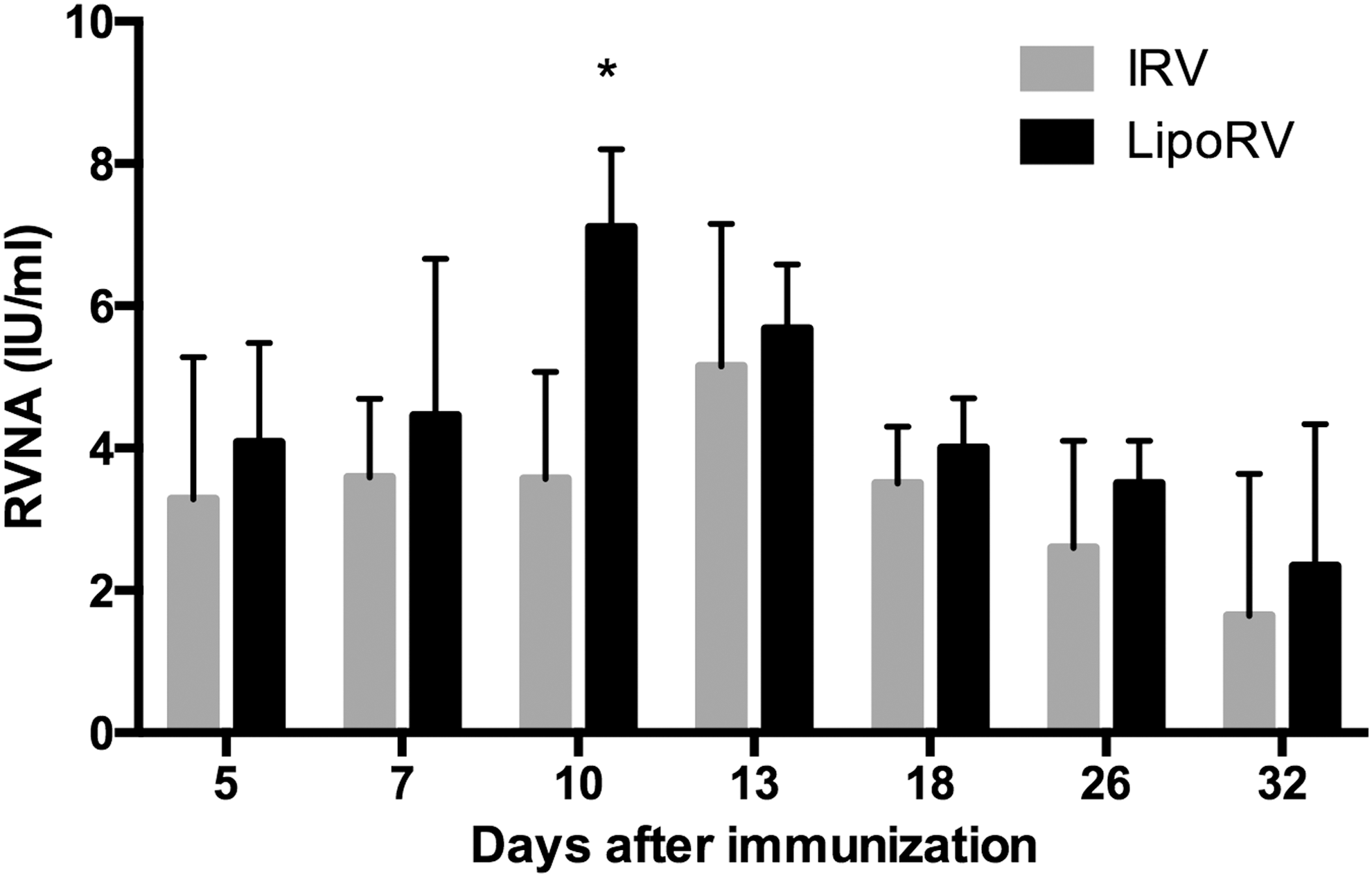
RVNA production induced by LipoRV or IRV. The RVNA values induced by IRV or LipoRV vaccine from 5 to 32 days after vaccination. *p < 0.05 represents significant difference. IRV, inactivated rabies vaccine; LipoRV, liposomal rabies vaccine; RVNA, rabies virus neutralizing antibody.
IL-2 and IFN-γ productions
For both of the vaccines, IL-2 and IFN-γ were detectable on day 15 after vaccination with a single injection (Fig. 2). Significantly higher levels of IL-2 and IFN-γ were observed in the mice treated with LipoRV compared to IRV and PBS (control).
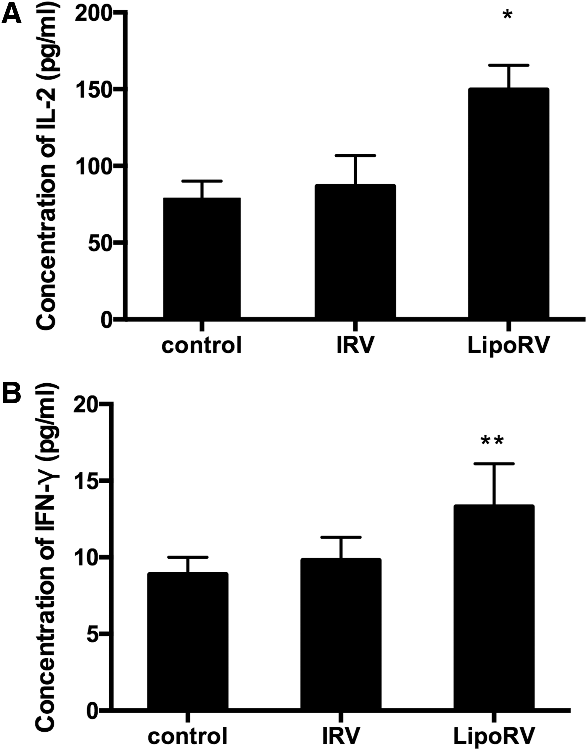
The effect of IRV and LipoRV on the production of IL-2 and IFN-γ in BALB/c mice.
Immunogenicity of LipoRV and IRV
To assess the immunogenicity of LipoRV and IRV, mice were immunized with IRV or different doses of LipoRV by intraperitoneal injection. Serum was collected for testing the RVNA level. As shown in Figure 3A, RVNA (27.1 ± 10.57 IU/mL) induced by LipoRV was significantly elevated on day 7 compared with IRV (7.01 ± 4.21 IU/mL, p < 0.05). Although RVNA levels reached peaks for both LipoRV and IRV groups on day 20, its value for the LipoRV 0/3/14/28 (89 ± 21.9 IU/mL) group was markedly higher than that for the IRV group (p < 0.05) and remained high until day 42 (p < 0.05). Despite the decrease of RVNA shown after day 42 of LipoRV injection, it was still significantly higher than that of IRV group (p < 0.05). We further reduced the dose of LipoRV by removing the last injection and found that RVNA levels were significantly reduced after the peaks on day 20 in all groups (Fig. 3B). However, LipoRV 0/3/14 still stimulated the highest level of RVNA compared to LipoRV 0/3/10 and RV 0/3/7/14/28.
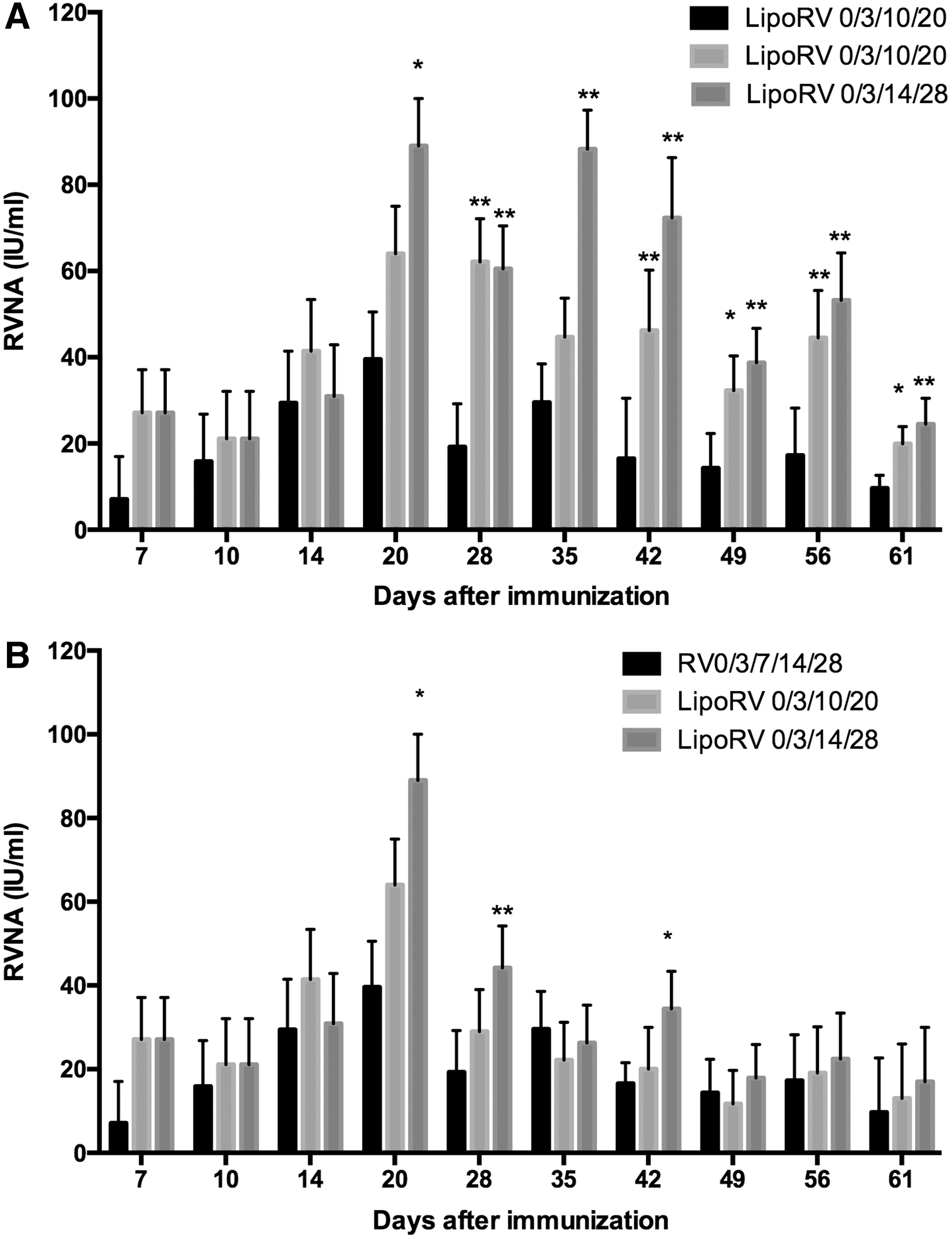
Comparison of the immunogenicity between LipoRV and IRV.
NK cell activity assay
We further compared the activity of NK cells among IRV, LipoRV, and PBS (control) vaccination. It was observed that LipoRV vaccination induced the highest level of NV cell activity in comparison with IRV or PBS, indicating that LipoRV is more effective than IRV in the induction of NK cells in the BALB/c mice (Fig. 4).
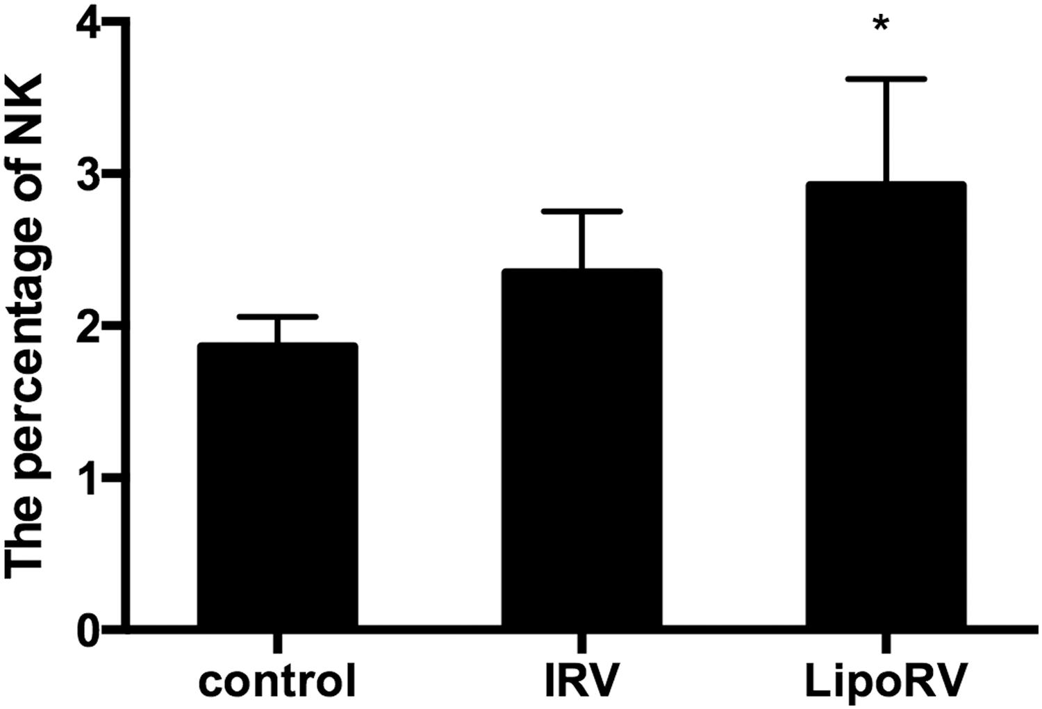
NK cell activity induced by IRV or LipoRV in BALB/c mice. NK cell activity was presented as the percentage of FITC-negative and PE-positive cells in a lymphocyte population. *p < 0.05, LipoRV group vs. the group immunized with IRV alone. NK, natural killer.
Postexposure prophylaxis
For PEP, the efficiency of LipoRV 0/3/14 and IRV 0/3/7/14/28 in the induction of RVNA was measured. Similar changes of RVNA were shown from 5 to 44 days for both vaccines (Fig. 5A). The GMT reached peaks at day 18 and 14 after the vaccination of LipoRV0/3/14 and IRV 0/3/7/14/28, respectively. The survival rate of LipoRV 0/3/14 (56.2%) was higher than IRV 0/3/7/14/28 (40.6%) after the challenge of street rabies virus in BALB/c mice (Fig. 5B).
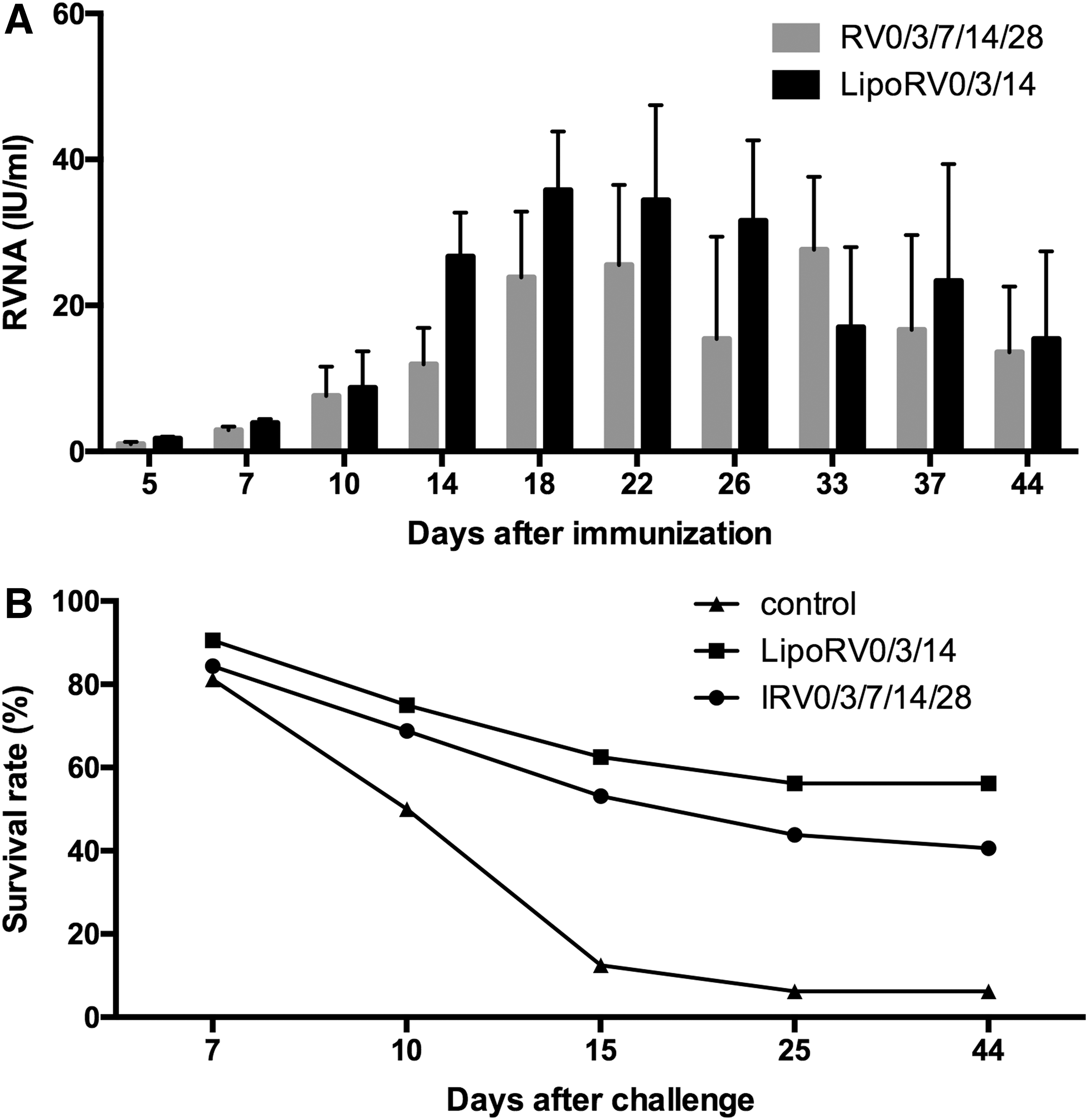
The efficiency of LipoRV in postexposure prophylaxis (PEP).
Discussion
Several methods have been developed for the preparation of liposome, including film dispersion, high-pressure homogeneous method, reverse-phase evaporation, and surface-active agent. In this study, we recruited the reverse-phase evaporation for the preparation of liposome rabies vaccine, which greatly reduces the costs compared to film dispersion and the high-pressure homogeneous method. However, according to our results, the liposome rabies vaccine prepared by this method showed high ER and particle size.
A number of studies have demonstrated the immune adjuvant activity of liposome, which may direct target to antigen-presenting cells in the spleen and lymph nodes (22). In this study, we found that rabies vaccine with liposome induced an intense immune response with promoted production of RVNA response in the mice challenged with rabies virus. Therefore, this form of vaccine may be potentially efficient in clinical, but further studies are necessary to confirm its effect on humans. Nevertheless, liposome exerted its function as an adjuvant in the present study rather than delivery vehicles. Our unpublished data showed that the liposome did not exhibit the ability of sustained release or prolong RVNA production.
In China, IRV produced by the Changchun Institute biological products has been extensively applied for rabies prophylaxis and PEP. Generally, IRV is administered in an individual after the exposure of rabies virus. To prevent the symptom of rabies, inoculation of rabies vaccine promptly is the only method and the potency of the vaccine needs to be ≥2.5 IU/dose. Given the long length of the schedule and multitimes of injections required for the current vaccine, it is necessary to develop novel vaccines with less injections and shortened schedule, which may be dependent on a higher potency. We compared the potency of IRV and LipoRV and found that LipoRV showed significantly increased potency (7.30, 12.27, 10.29 IU/dose) compared with IRV (4.35, 4.72, 5.25 IU/dose, p < 0.01). This result indicates that liposome could enhance the rabies vaccine to induce effective protection.
For postexposure prophylaxis in humans, rabies vaccines are given intramuscularly (IM), usually on the upper arm deltoid. In our study, mice were immunized with the rabies vaccines by intraperitoneal injection. A previous study reported both IM and intraperitoneal injection of IRV showed higher ability in the induction of serum RVNA and protective effect compared to other routes of injection (23). Poor immune responses and less protective effects were observed in the mice even with increased doses of IRV administered subcutaneously.
Cell-immediate immunity plays an important role in protecting the body against pathogens. It involves the releases of cytokines such as type 1 IFN and chemokines, the activation of complement, and the attraction of macrophages, neutrophils, and NK cells into the infected tissues. IFN-γ is the signature cytokine for Th 1 cells. Th 1 cells produce IFN-γ to limit the proliferation of pathogens and assist the production of antibody by B lymphocytes (18,19). It also secrets IL-2 that is known to induce the proliferations of activated T and B cells, and enhance NK cytotoxicity. We measured the serum levels of these cytokines and found that liposomes enhanced the immune response by promoting productions of IL-2 and IFN-γ, and the activation of NK cells.
RVNA is the golden standard for rabies vaccine efficacy (8,16). The present study found that LipoRV facilitated the induction of more vigorous production of RVNA by a single injection in BALB/c mice. More importantly, based on the measurements of RVNA in various doses of LipoRV, we found that less injections could still reach the efficacy of five-shot regimen of IRV. As the LipoRV (0/3/14) schedule showed the highest efficiency in inducing RVNA, it was used for our further experiments.
In this study, we also evaluated LipoRV in mice as a postexposure treatment in the absence of specific immunoglobulin commonly utilized as important postexposure therapies. We tested whether the three-shot regimen of LipoRV was as efficient as the classic five-shot IRV without the use of anti-rabies immune serum. The results indicate that the three-shot LipoRV regimen induced similar RVNA titer levels as the five-shot IRV regimen. In addition, the three-shot LipoRV regimen showed a higher survival rate in comparison with the five-shot IRV regimen in the challenged mice.
Based on the comparison between the two vaccines, LipoRV may be more efficient with low cost. It is possible that the similar constituents of liposome to the cell membrane assist absorption, fusion, lipid exchange, and endocytosis of cells. It was reported that rabies vaccine encapsulated by liposome was easily absorbed by cell (1,2). Consistently, numerous studies have already confirmed a significantly higher efficacy of the vaccine encapsulated by liposome compared to nonencapsulated vaccine (3,9,15).
In conclusion, liposome enhances the immune effect of rabies vaccine and could be a potential immunopotentiator. By improving the potency of vaccines, the doses of the vaccines could be reduced from five to three doses, whereas other PEP regimens normally require 21–90 days. This improved schedule may substantially increase health economics and the benefits of prophylaxis.
Footnotes
Acknowledgment
The authors are grateful to Qing Tang from the Chinese Center for Disease Control and Prevention for the provision of nonpassage rabies virus (CNX8601).
Authors' Contributions
L.M., Y.Y., M.Y., Y.L., J.Z.: Did the experiments and analyzed the data; J.G., D.Z.: Designed the study and wrote the article.
Author Disclosure Statement
No competing financial interests exist.