






Abstract
Significance:
The skin serves as the primary defense against external stimuli, making it vulnerable to damage. Injuries can cause a dysregulated environment, resulting in chronic inflammation and inhibition of cell proliferation and migration, which delays recovery. Innovative approaches, such as three-dimensional (3D) bioprinting, can foster a controlled healing environment by promoting synergy between the skin microbiome and cells.
Recent Advances:
Traditional approaches to wound healing have focused on fostering an environment conducive to the interplay between cells, extracellular proteins, and growth factors. 3D bioprinting, a manufacturing technology with applications in tissue engineering, deposits biomaterial-based bioink containing living cells to fabricate custom-designed tissue scaffolds in a layer-by-layer fashion. This process controls the architecture and composition of a construct, producing multilayered and complex structures such as skin.
Critical Issues:
The selection of biomaterials for scaffolds has been a challenge when 3D skin tissue engineering. While prioritizing mechanical properties, current biomaterials often lack the ability to interact with environmental stimuli such as pH, temperature, or oxygen levels. Employing smart biomaterials that integrate bioactive molecules and adapt to external conditions could overcome these limitations. This innovation would enable scaffolds to create a sustainable wound-healing environment, fostering microbiome balance, reducing inflammation, and facilitating cellular recovery and tissue restoration, addressing critical gaps in existing wound care solutions.
Future Directions:
Novel bioink formulations for skin injury recovery are focused on improving long-term cell viability, proliferation, vascularization, and immune integration. Efficient recovery of the skin microbiome using bioactive molecules has the potential to create microenriched environments that support the recovery of the skin microbiome and restore immune regulation. This promising direction for future research aims to improve patient outcomes in wound care.
Graphical Abstract
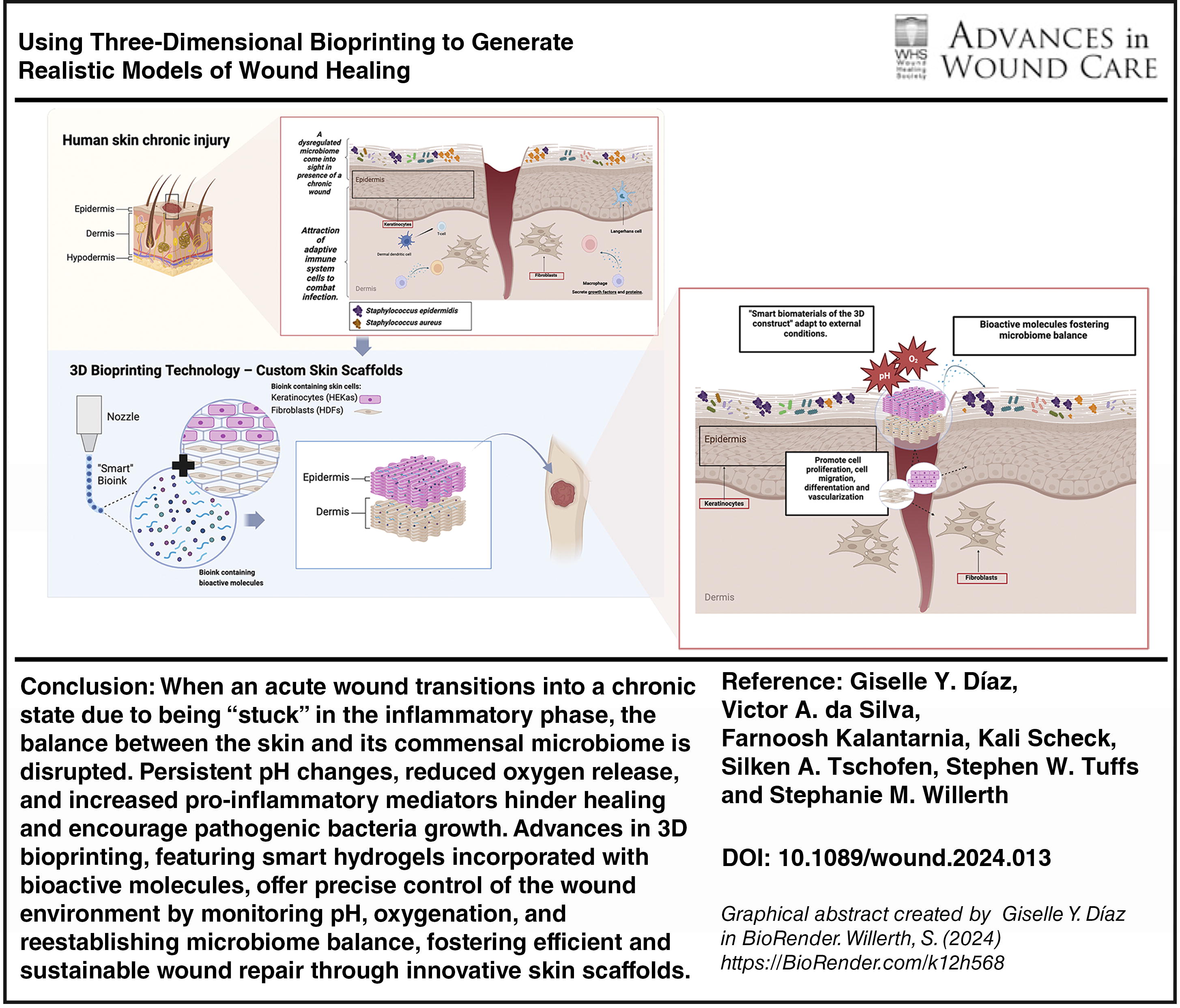
SCOPE AND SIGNIFICANCE
Skin wounds are categorized as acute or chronic based on the time required for healing. Chronic wounds take significantly longer to heal, causing persistent pain and restricted mobility. They can escalate into severe complications, including diabetic foot ulcers (DFUs), which lead to amputations in 20% of cases. These wounds not only impose a substantial financial burden on health care systems but also present higher risks, prolonged care requirements, and elevated overall costs compared with other skin wounds. Traditional wound care often falls short in addressing the complexities of wound healing, such as controlling inflammation, promoting cell proliferation, and migration by maintaining homeostasis between the microbiome and skin cells. This review examines the potential of three-dimensional (3D) bioprinting in wound healing, emphasizing its ability to create biologically relevant, sustainable skin substitutes that restore tissue functionality, promote hemostasis, and improve healing outcomes.
Stephanie M. Willerth, PhD

TRANSLATIONAL RELEVANCE
Wound healing initiates an intricate set of complex mechanisms within the skin microbiome while promoting the restoration of the native tissue. If these systems are not symbiotic, acute wounds can become chronic and cause long-term complications for the patient. The design and implementation of smart bioinks using 3D bioprinting can help maintain the microbiome by keeping a balanced pH and oxygen-rich environment in the wound site. Furthermore, these bioinks can aid in the restoration of the skin microenvironment, which is imperative for patient recovery. Understanding the mechanisms of wound healing and how to effectively recover the microbiome can drive therapies that modulate these factors for improving wound-healing outcomes.
CLINICAL RELEVANCE
Effective management of chronic wounds can improve patient outcomes and reduce health care costs. The restoration of the skin microbiome plays a vital role in this process, as microbiome imbalances can lead to infections and delayed healing. Smart hydrogels and 3D bioprinting technologies introduce new possibilities for creating biocompatible and functional tissue constructs. These advancements support better wound care by enhancing cell proliferation, differentiation, and survival. These technologies could revolutionize clinical treatment protocols, offering more precise, personalized, and effective wound-healing strategies, thereby improving the quality of life for patients with chronic wounds.
BACKGROUND
A wound is defined as any disruption to the integrity of skin, mucous membrane, or organ tissue. 1 Wounds are classified as acute or chronic depending on the timeframe required for healing. Acute wounds are newly formed and exhibit normal physiological healing. 2 Regardless, an acute wound can become “stuck” in the chronic inflammatory phase, failing to progress to subsequent healing stages. This delay extends the healing timeframe to weeks or longer, resulting in the wound becoming chronic. 3
The inappropriate management of skin tears can lead to complex chronic wounds, including DFUs, infected lacerations, burns, and pilonidal or perianal abscesses, which are frequently seen in Canadian emergency departments. 3 Approximately 15% of individuals with diabetes develop a foot ulcer, and untreated DFUs result in lower-extremity amputations in 20% of cases. 4 DFU care places a significant financial burden on health care systems, costing Medicare $30,186 annually per case. 5 With diabetes cases projected to rise to 643 million globally by 2030, 6 early interventions that address initial healing stages are crucial to preventing further complications. 7
Healing a skin wound requires an extraordinary mechanism of cellular function that is distinctive in nature. The repair process consists of four stages: homeostasis, inflammation, proliferation, and remodeling (Fig. 1). At the beginning of the healing process, a blood-filled wound is first closed by a fibrin coagulum that acts as a scaffold for cell infiltration. 9 Second, inflammation occurs simultaneously as neutrophils infiltrate the wound site, where they are replaced by monocytes that differentiate into macrophages. Proliferation begins 2–10 days after the lesion was made and consists of “rebuilding” involving re‐epithelialization, angiogenesis, collagen synthesis, and extracellular matrix (ECM) deposition. 10 In this phase, endothelial cells that constitute the inner cellular lining of the blood vessels (arteries, veins, and capillaries) form new capillaries, bringing oxygen and nutrients to the growing tissue and removing waste products. 11 The formation of granulation tissue allows re-epithelialization, where the keratinocytes roll over from the basal layer to restore the physiological features of the multilayered epithelial tissue. 12 Remodeling starts about 3 weeks after injury and can last for over a year, where all the activated processes are silenced, leaving the region rich in collagen, recovering the ECM of the damage, and increasing the tensile strength of the tissue. 8,13
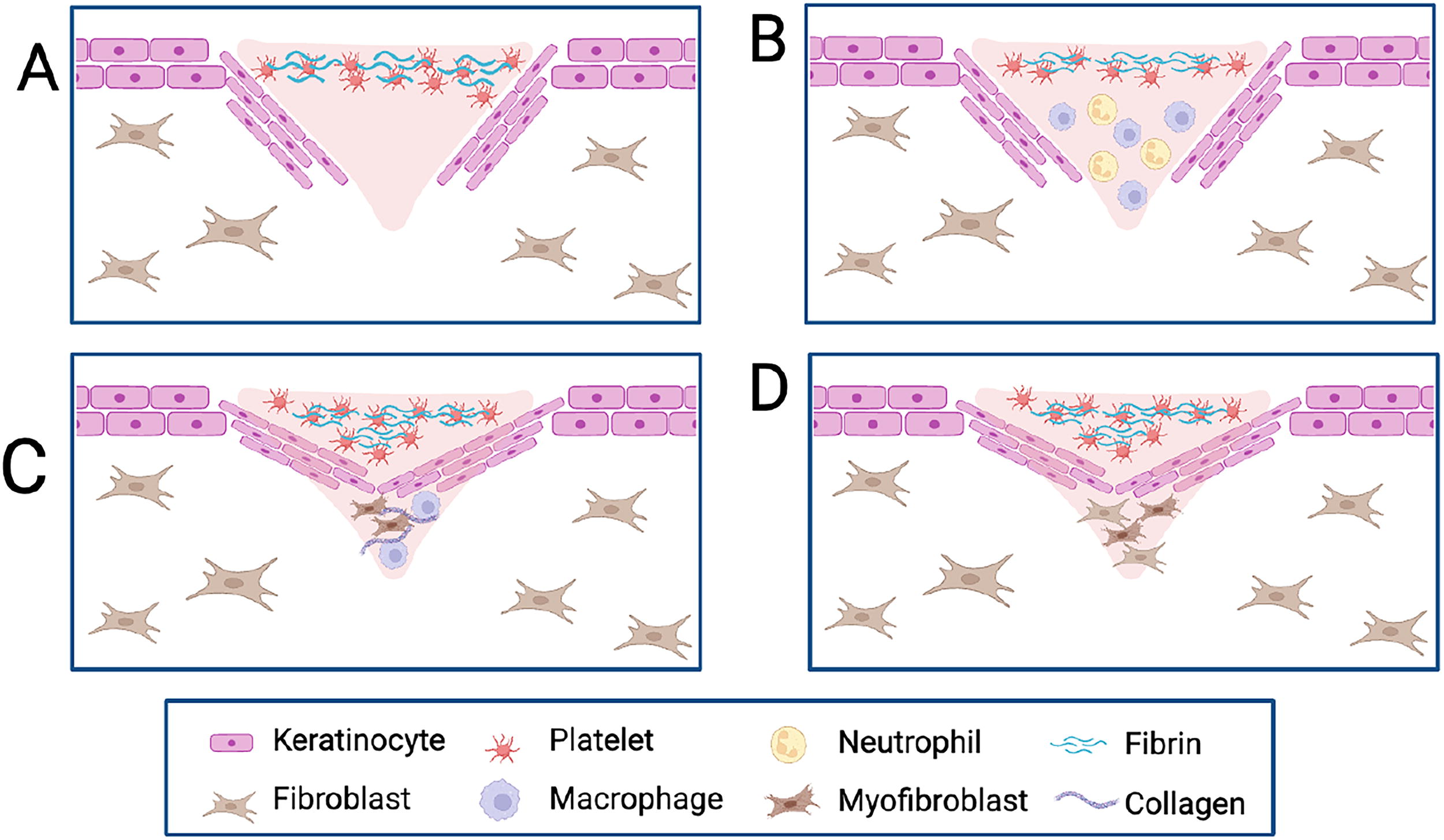
Schematic overview of the stages of wound healing. (
As previously mentioned, acute wound healing follows the normal physiological repair process, whereas chronic wounds are marked by a persistent inflammation phase due to immune dysregulation, which delays healing. The microenvironment of a chronic wound is characterized by high quantities of proinflammatory macrophages, overexpression of inflammatory mediators such as tumor necrosis factor-α and interleukin-1β (IL-1β), and increased activity of matrix metalloproteinases (MMPs), namely MMP-2 and MMP-9, which degrade the ECM and prevent the commencement of the proliferative stage of healing. 10,14
The skin is naturally colonized by transient, temporarily resident, and permanently resident bacterial species, including Staphylococci, Micrococcus spp., and Coryneform bacteria, which coexist in a mutualistic symbiotic relationship with the host, contributing to skin health and maintaining homeostasis. 15 One way this balance is maintained is through the acidic pH of the stratum corneum (pH 4.5–5.3). However, when a chronic injury occurs, the exposure of internal tissue and interstitial fluid (pH ∼7.40) causes the wound area to become alkaline (pH 7.42–8.90), favoring the growth of pathogenic bacteria like Staphylococcus aureus 16,17 (Fig. 2). In addition, oxygen tension within the wound is a systemic factor strongly influenced by wound pH. Under acidic conditions, hemoglobin releases more oxygen due to the Bohr effect. Fibroblast growth and collagen synthesis require oxygen during wound healing. Any disruption in oxygen tension directly affects the proliferation and migration behavior of these cells, impeding the healing process. 16,18
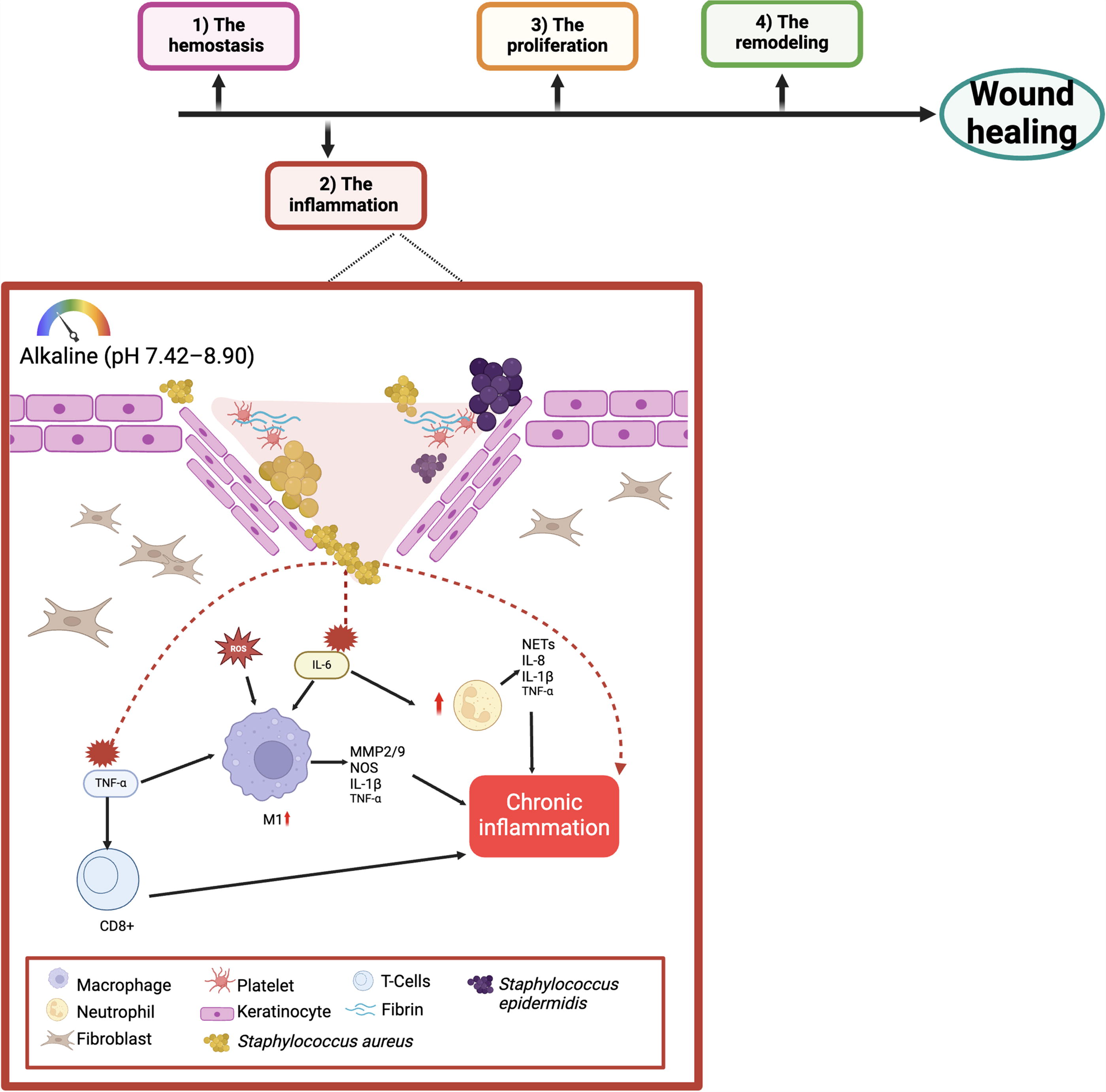
Immune dysregulated system in chronic wound healing. Common features of chronic wounds include high levels of proinflammatory macrophages, overexpression of inflammatory mediators such as tumor necrosis factor-α and interleukin-1β, increased activity of matrix metalloproteinases (MMPs), and an abundance of reactive oxygen species. These factors collectively contribute to defective re-epithelialization. Additionally, the alkaline pH around the wound area promotes the growth of pathogenic bacteria, such as Staphylococcus aureus, which activates the immune system by recruiting neutrophils and proinflammatory macrophages. This activation results in the accumulation of inflammatory cytokines and MMPs. The presence of a dysregulated immune system creates a vicious cycle, where pathogenic bacteria continue to proliferate, perpetuating chronic inflammation. Adapted from Raziyeva et al., 2021. 14 Created in BioRender. Willerth, S. (2024) https://BioRender.com/k12h568.
Becoming stuck in the inflammatory phase during chronic wounds initiates a chain of interrelated events. And one of the ways that could directly and efficiently decrease the inflammation is achieving skin homeostasis with the commensal microbiome. 19 For instance, Staphylococcus epidermidis suppresses excessive inflammatory responses through a Toll-like receptor (TLR)-crosstalk phenomenon between TLR2 and TLR3. 20 In addition, S. epidermidis plays a beneficial role by inducing keratinocytes to release antimicrobial peptides (AMPs), such as β-defensins and cathelicidin, which enhance the skin’s defense against pathogens such as S. aureus. 19
Traditional approaches to wound healing, such as autografts, allografts, and xenografts, are not the best methodologies for addressing the complex process of chronic wound healing. They focus more on managing the skin injury rather than fully restoring tissue integrity. 21 Therefore, there is an urgent need to develop new methods capable of restoring skin integrity by maintaining the acidic pH of normal skin and efficiently restoring homeostasis between skin cells and the microbiome. 3D bioprinting is an advanced tissue technology that can fabricate skin scaffolds with biological materials, cells, and other bioactive molecules that could be inserted in the wound area and work as treatment delivery systems. 22 This review explores the importance of employing smart biomaterials with bioactive molecules to develop smart bioinks for 3D bioprinting, providing efficient and sustainable treatment solutions for chronic wound inflammation.
DISCUSSION
The main purpose of tissue engineering is the development of biological substitutes to restore, maintain, or improve tissue function or as an in vitro drug screening platform. 23 This can be accomplished using advanced tools such as bioprinting bioinks via various bioprinting techniques. Replicating native tissue structures remains challenging due to the need for biomaterials that provide structural support while being biodegradable, nontoxic, and immune compatible. 24 Moreover, these materials must exhibit good rheological properties to ensure proper 3D printing without inducing shear stress on the cells or causing the construct’s deformation. 25 Therefore, obtaining useful and effective tissue-engineered skin that mimics the natural microarchitecture of the skin and enhances cell viability, migration, interaction, and maturation depends on the correct selection of biomaterials and the appropriate 3D bioprinting strategy. 26,27
Restoring skin tissue after an injury is a complex, interconnected process involving dynamic interactions between the wound and its external and internal microenvironments. 15 This underscores the importance of smart biomaterials, which compose the bioinks used in 3D bioprinting. These biomaterials can sense, respond, and adapt to changes in the biological surroundings, making them valuable tools for creating dermal substitutes. 28 By promoting optimal pH, temperature, and oxygenation and incorporating bioactive molecules, they can efficiently restore the microbiome and accelerate wound healing. 27
Various bioprinting techniques have been explored for creating functional skin constructs, broadly classified into three main categories: extrusion-based, 29 jetting (or inkjet)-based, and light-based (or vat photopolymerization) methods. 30 The following section describes these methods, highlighting their specific contributions to advancing 3D bioprinting for skin regeneration, as well as their limitations (Fig. 3).
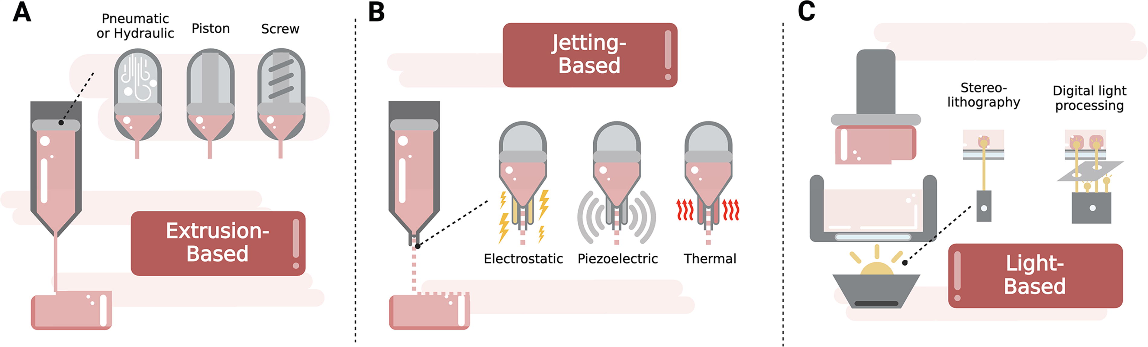
Bioprinting techniques for creating functional skin constructs. The skin is a complex organ with three distinct layers—epidermis, dermis, and hypodermis—each with specific functions that support tissue restoration. 3D-bioprinting technologies, utilizing layer-by-layer deposition of biocompatible bioinks loaded with biologics such as skin cells, growth factors, and live biotherapeutic products, have the potential to replicate the complex structure of native skin. Created in BioRender. Willerth, S. (2024) https://BioRender.com/k12h568.
3D Bioprinting strategies
The skin is a complex organ with three distinct layers—epidermis, dermis, and hypodermis—each with specific functions. 31 The epidermis, the outermost layer, acts as a protective barrier and continuously renews itself through keratinocytes. 32 Below, the dermis comprises fibroblasts and fibrous proteins such as elastin and collagen, providing the skin with strength. The hypodermis, the deepest layer, contains adipocytes that support the ECM, secrete cytokines, and facilitate wound repair. 27 Therefore, 3D bioprinting technologies, utilizing layer-by-layer deposition of biocompatible bioinks loaded with biologics such as skin cells, growth factors, and live biotherapeutic products (LBPs), have the potential to replicate the complex structure of native skin.
Extrusion-based bioprinting is a widely utilized technique, where bioinks are extruded through a nozzle to form continuous filaments that create the desired structure. 33 The extrusion process is driven by pneumatic, mechanical (piston or screw), or hydrodynamic systems, making it suitable for high-viscosity bioinks. These bioinks maintain structural integrity, prevent cell sedimentation, and ensure uniform cell distribution. 34 However, higher-viscosity bioinks require greater pressure during printing, increasing shear stress at the nozzle tip, which can reduce cell viability and affect protein signaling and cell phenotype maturation. 35 Optimizing factors such as bioink viscosity, nozzle diameter, and extrusion pressure is essential to balance these effects. Approaches to predict cell viability based on shear stress have been explored. Zhang et al. summarized the main mathematical models that can be used to quantify the cell resistance to shear stress for optimizing bioprinting systems. 36 In addition, shear stress was also demonstrated to affect printability; excessive stress can cause nozzle clogging and uneven extrusion, while insufficient stress can lead to poor flow properties and a compromised structure. 37 To mitigate these effects, researchers can adjust printing parameters, such as extrusion speed and nozzle size, or modify bioink rheology with thickeners, crosslinking agents, or the use of sacrificial and support bioinks to enhance structural integrity without compromising cell health. 38 –40 According to Tian et al., some of the most important factors for achieving good cell viability in extrusion-based bioprinting are porosity, permeability, stiffness, and diffusivity. 41 However, because these parameters can inversely and simultaneously affect both the biological and mechanical performance of bioprinted tissues, it is crucial to strike a balance to achieve optimal biological functionality and structural integrity. 41 Bebiano et al. demonstrated that by adjusting crosslinker and polymer concentrations in the bioink, the stiffness of bioprinted skin constructs could be tailored, leading to site-specific differences in fibroblast morphology and spreading. 29 The versatility of extrusion-based bioprinting also allows for the creation of complex, multilayered skin grafts. For instance, implantable skin grafts have been generated using multiple bioinks: one containing fibroblasts and endothelial cells to form the dermis, followed by another containing keratinocytes to form the epidermis. When implanted in immunodeficient mice, these grafts became perfused through both graft and host microvessels, indicating successful integration with the host tissue. 42
Jetting-based bioprinting technology is classified into inkjet printing, microvalve printing, acoustic printing, laser-assisted printing, electrospinning, and electrohydrodynamic jet printing. These methods provide advantages such as high precision, rapid throughput, single-cell printing, and droplet-continuous capabilities for fabricating tissue constructs. 43 The technique’s ability to eject singular bioink droplets ensures accuracy and facilitates the simultaneous deposition of multiple cell types and bioactive molecules. However, the heat- or piezoelectric-based energy required for droplet formation may cause cell damage or lysis, and the reliance on low-viscosity bioinks limits the diversity of biomaterials that can be utilized. 44 Therefore, careful optimization is required to ensure cell viability and functionality. Ng et al. identified two primary factors influencing the quality of jetting-based bioprinting: droplet impact velocity and droplet volume. 45 Their study, utilizing high-speed imaging, demonstrated that slower droplet impact velocities reduce splashing, thereby improving printing accuracy and enhancing cell viability, while higher velocities can induce mechanical stress on cells. For thermal inkjet bioprinters, the group emphasized that controlling droplet volume is critical to maintaining cell viability, particularly when printing duration must be limited to prevent excessive evaporation. 45 The same group also investigated the influence of bioink properties on printing performance and cell health by playing with the concentration of polyvinylpyrrolidone. It was observed that higher bioink viscoelasticity was found to stabilize droplet filaments and help maintain the integrity of printed droplets. For accuracy and printability, the group observed that the increase in bioink viscosity was shown to facilitate droplet deposition on the substrate surface without splashing. 46 In the context of skin models, Albanna et al. developed a mobile inkjet bioprinter capable of depositing autologous fibroblasts and keratinocytes in situ. This device featured integrated imaging technology that allowed for real-time management of wound sizes and topologies, enabling researchers to print directly over the skin wounds of mice and adjust the thickness to achieve uniform wound closure. This approach not only replicated the layers of skin but also demonstrated quicker healing and faster growth of normal skin due to its precise control. 47 To further illustrate the effectiveness of inkjet-based bioprinting, Yanes et al. successfully deposited endothelial cells in specific locations to build capillary-like networks within a collagen scaffold containing fibroblasts and keratinocytes. This bilayer skin graft with printed microvasculature was implanted into mice, resulting in improved wound healing and the formation of neoskin closely resembling normal skin. 48
Light-based bioprinting, also known as stereolithography or vat photopolymerization, utilizes light to cure or solidify photopolymerizable bioinks layer by layer, enabling the creation of highly detailed tissue structures. 49 This technique excels in achieving extremely high resolution, making it ideal for fabricating intricate tissue architectures. However, the requirement for photopolymerizable bioinks restricts the range of usable materials. Moreover, exposure to light and photo initiators can negatively affect cell viability, necessitating careful selection and optimization of bioinks and printing parameters. 50 As highlighted by Chartrain et al. and Ng et al., the success of vat photopolymerization depends on critical factors such as the type and wavelength of the light source, which must be compatible with the photo initiator to ensure efficient polymerization. The wavelength is particularly important as it influences the depth of light penetration and the resolution of the printed layers. Additionally, key printing parameters such as layer thickness, exposure time, and printing speed must be finely tuned to optimize the resolution, surface finish, and mechanical properties of the printed scaffold. 30,51 To address the challenges associated with light-based bioprinting, Hafa et al. developed a system that simultaneously prints and images the constructs in real time. This innovation allows for continuous monitoring and adjustment during the printing process, significantly improving the speed, resolution, and viability of printed cells such as fibroblasts and keratinocytes. 52
To harness the advantages of different bioprinting techniques, combining multiple approaches is becoming increasingly popular. In a recent study, Lee et al. demonstrated this by integrating extrusion and inkjet-based bioprinting to create a skin model composed of fibroblasts and keratinocytes. The process began with a pneumatic microextrusion technique to produce a stable dermal layer of collagen and primary human dermal fibroblasts. This was followed by piezoelectric inkjet printing of primary human epidermal keratinocytes to form an epidermal layer with high viability and uniformity. 53 This approach underscores the importance of integrating multiple cell types, biomaterials, and technologies to accurately replicate the complex, layered architecture of human skin.
Current skin substitutes, such as skin grafts, fail to fully replicate the functionality of native skin. They are mechanically fragile, prone to contamination, expensive, and exhibit low engraftment rates. 54 Additionally, many substitutes are acellular or lack diverse cell types, providing only temporary coverage. The absence of key fibrous proteins such as collagen and elastin, responsible for tensile strength, contributes to mechanical fragility. 8 Furthermore, the alkaline pH of the wound microenvironment promotes pathogen growth, impairs fibroblast activity, and delays wound remodeling and healing. 16,18 These limitations can be addressed with 3D bioprinting, as this technology allows precise positioning of cells in specific patterns and orientations within layered constructs. Kim et al. demonstrated that densely populated fibroblasts align along ridges and elongate more compared with less dense regions. Their study highlighted that cell migration and morphology are influenced by the density and orientation of patterns, underscoring the importance of topography in cell–matrix interactions. 55 This is particularly crucial in wound healing, where fibroblasts naturally migrate to open wounds. 56 Therefore, 3D bioprinting holds the potential to create customized skin constructs with enhanced functionality and compatibility. By precisely placing bioactive molecules and cells at wound sites, this approach could promote efficient hemostasis recovery, limit the development of pathogenic bacteria, and enhance the wound-healing process. This would be achieved by facilitating cell growth, differentiation, migration, and proliferation around the area where the construct is located.
Biomaterials in wound healing
The appropriate selection of biomaterials directly influences the functionality and success of bioinks encapsulating living cells or bioactive factors. 57 When selecting a biomaterial, several key factors must be considered, including mechanical strength, rheological properties for printability, and biological compatibility with skin tissue. Bioinks must also support cellular growth and proliferation without adversely affecting cell phenotypes. 26 For skin bioprinting, a suitable bioink should mimic the natural ECM found in the skin, providing an optimal tissue microenvironment that enhances cellular processes such as migration, nutrient exchange, and tissue viability maintenance. 58 Biomaterials can be designed not only as structural frameworks but also as delivery systems for bioactive molecules. 59 They can be derived from natural sources such as collagen, fibrin, and hyaluronic acid (HA), which form hydrogels with inherent biocompatibility and biodegradability, making them ideal for in vivo applications. 60 Alternatively, synthetic materials, engineered to mimic the mechanical properties of natural tissues, can offer greater customization. 61 Combining natural and synthetic materials allows bioinks to fulfill the three essential properties required for effective bioprinting: (i) biocompatibility with the host to maintain cell viability and reduce the risk of immune rejection, (ii) rheological properties for smooth deposition, and (iii) rapid crosslinking to retain 3D structure postbioprinting. 62,63 This approach enables the creation of customizable platforms that provide an ideal microenvironment for skin cells, promoting their proliferation, migration, and differentiation while maintaining the mechanical strength, rigidity, and shape needed for wound healing. 64
The importance of in vivo wound healing highlights the need for developing vascularized skin substitutes. Vascularization is critical for supplying oxygen and nutrients, removing metabolic waste, and facilitating angiogenesis—the formation of new blood vessels from existing ones—which is essential for the survival and integration of bioprinted skin. 65 Biomaterials that support the incorporation of cells and growth factors to promote proper vascularization are, therefore, crucial. Dai et al. utilized biodegradable polyurethane–gelatin (4:1) to extrude human fibroblasts, endothelial progenitor cells (EPCs), and keratinocytes, creating bilayered dermoepidermal constructs. Incorporating EPCs into the dermal layer enhanced vascularization, as demonstrated by improved wound healing and microvessel formation when grafted onto skin wounds in immunodeficient mice. 66
As we discussed for appropriate bioprinting process, the biomaterials that composed the bioink should be low viscous to protect the cells from shear stress and prevent clogging and solidify rapidly to maintain shape. Hydrogel-based bioinks are commonly used for skin regeneration and wound healing, with key features including their remarkable capacity to retain significant amounts of water, without losing structural integrity, due to hydrophilic components in their polymer structure. 67,68 This unique attribute of hydrogels highlights their suitability for replicating ECM properties and promoting tissue regeneration.
The commonly used biomaterials for skin bioprinting are HA, chitosan, alginate, collagen, gelatin, and fibrin. 26,69 HA derives from the ECM, offering inherent biocompatibility and supporting cell adhesion and tissue regeneration. 70 HA oligomers (∼6 − 20 monomers) alleviate the inflammatory response, enhance re-epithelialization, and facilitate angiogenesis by stimulating growth factors such as transforming growth factor beta, epidermal growth factor (EGF), basic fibroblast growth factor (bFGF), and vascular endothelial growth factor (VEGF), which recruit and activate keratinocytes, endothelial cells, and fibroblasts. 71 However, HA has poor mechanical strength and limited viscoelasticity, 72 requiring combination with other biomaterials to enhance its performance. For instance, Maiz-Fernandez et al. improved HA’s mechanical stability by integrating it with chitosan, forming a robust, self-stabilizing hydrogel. 73
Chitosan, a linear polysaccharide derived from the deacetylation of chitin, is biocompatible, biodegradable, and antibacterial, making it effective against pathogens such as S. aureus while promoting hemostasis recovery of the rest of the microbiome. 44,74 However, chitosan’s weak mechanical integrity, rapid dissociation under physiological conditions, and lack of cell-binding domains limit its utility in tissue engineering. 75 Ng et al. addressed these issues by formulating a chitosan-gelatin polyelectrolyte hydrogel, which improved cell viability. 76
Alginate, a polysaccharide-based hydrogel, is widely utilized due to its biocompatibility, low toxicity, and excellent printability. Moreover, alginate is utilized as a shear-thinning solution, wherein its viscosity decreases as the shear rate increases. This property protects cells from excessive shear stress during bioprinting. Once deposited, the bioink rapidly regains rigidity, allowing the scaffold to maintain its structural integrity. 36
Collagen is a fundamental component of the skin ECM, offering structural integrity and mechanical support. 77 Its porous structure and permeability enable efficient nutrient exchange and waste removal when used as a scaffold. 78 However, as a standalone biomaterial ink, collagen lacks stability and is challenging to pattern. 33 To address this, chemical crosslinking is employed, but it compromises biocompatibility and increases antigenicity. Using high-purity collagen, as demonstrated by Osidak et al., enhances mechanical properties in bioprinted structures. 79
Gelatin, a biodegradable polymer derived from the partial hydrolysis of collagen, demonstrates versatility in forming hydrogels via a sol-gel transition at low temperatures, which supports the maintenance of scaffolds at specific printing temperatures. 33,80 However, optimizing printing conditions, particularly temperature and viscosity, presents challenges when gelatin is used as a standalone bioink due to its inherent limitations. 81
Fibrin plays a pivotal role in promoting cell proliferation, differentiation, and vascularization. 82 Fibrinogen polymerizes into fibrin gel upon activation by thrombin. However, the weak mechanical properties of fibrin gel limit its ability to sustain 3D structures, requiring its combination with other biomaterials to address its mechanical deficiencies and enhance structural stability. 83
Understanding the advantages and limitations of each biomaterial is crucial for developing hydrogel-based bioinks. Recognizing that some materials enhance mechanical properties, while others are vital for supporting cell growth, differentiation, and vascularization, simplifies the selection of complementary biomaterials. This approach enables the creation of an optimized skin scaffold that can be effectively bioprinted, offering structural strength, biocompatibility, and a supportive microenvironment. These scaffolds foster cell viability, proliferation, maturation, and differentiation, enabling vascularization, ECM production, and cell migration to wounds, ultimately improving the healing process and tissue repair. However, chronic wounds are often characterized by persistent inflammation, pH alkalinization, and oxygen limitation, which disrupt the homeostasis between the host and microbiome, prolonging the injury and making it more susceptible to pathogen colonization. Therefore, refining current biomaterials by incorporating bioactive molecules such as LBPs or growth factors can help protect the wound environment, preventing or delaying enzymatic, pH-mediated degradation, and supporting oxygenation. 84 This approach would make them “smart hydrogels” that respond to stimuli from the wound microenvironment, triggering the release of bioactive molecules to develop an advanced therapeutic system. 85
Smart hydrogels
The terminology of “smart hydrogels” refers to materials that can recognize natural or pathological cues in the tissue microenvironment and using those as passive triggers such as pH or enzymes. 86 These hydrogels can monitor wounds in real time, deliver drugs tailored to different healing stages, and adjust their degradation rate for optimal bioactive molecule release 87 (Fig. 4). In the following sections, we review several approaches for using intelligent hydrogels to enhance both wound healing and the effectiveness of skin microbiome management.
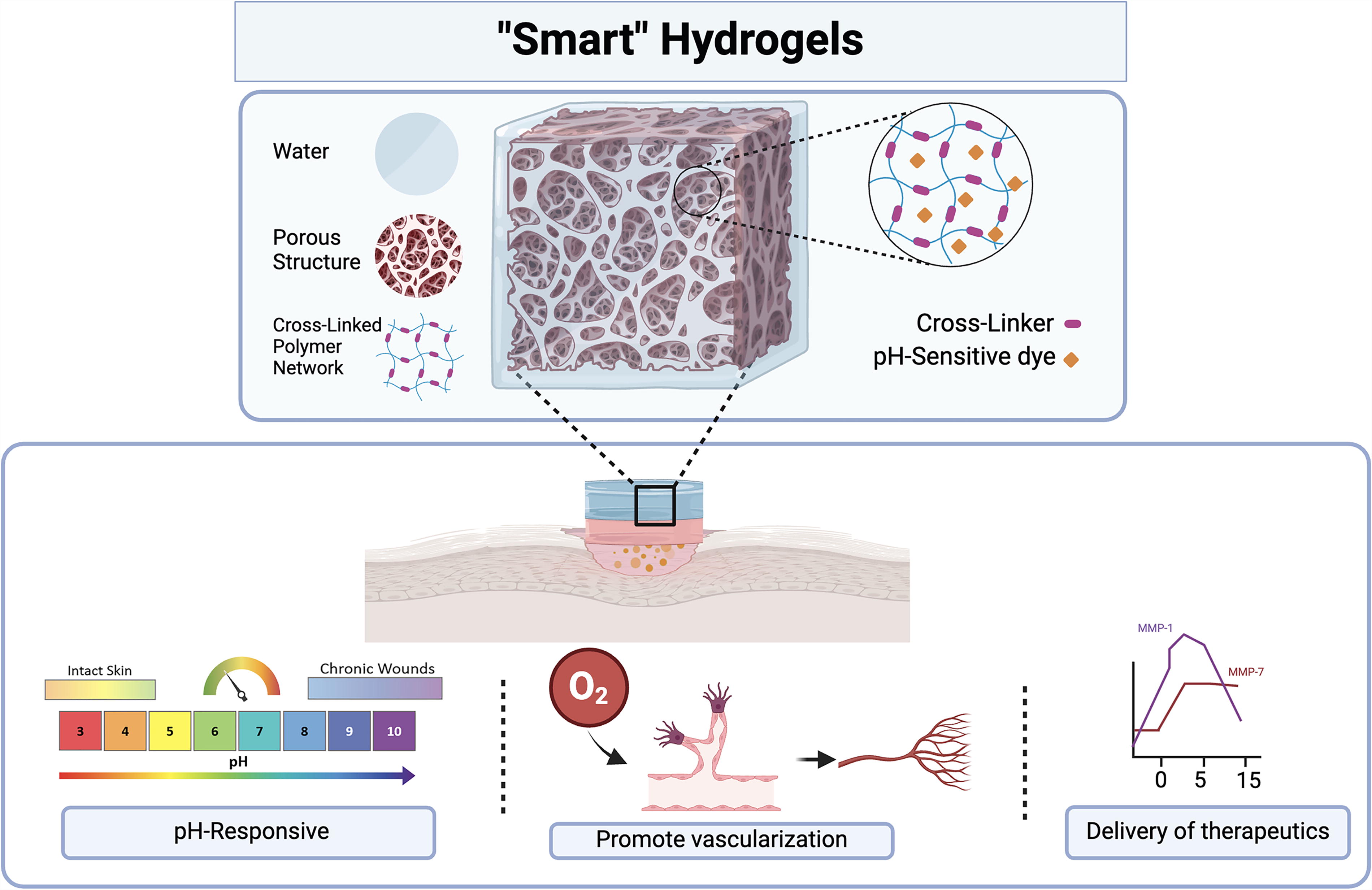
Smart hydrogels for wound-healing applications. Smart hydrogels are materials designed to adapt to external conditions by responding to natural or pathological signals, such as changes in pH or the presence of enzymes. These cues serve as passive triggers, allowing the hydrogels to adjust their properties, promote vascularization, or interact with surrounding enzymes. For instance, during the inflammatory phase of the healing process, hydrogels can react with matrix metalloproteinase-1 (MMP-1) to release bioactive molecules such as antibiotics, while, in the granulation phase, they respond to MMP-7 to release growth factors trapped within the biomaterials. Adapted from Olivia et al., 2020. Created in BioRender. Willerth, S. (2024) https://BioRender.com/k12h568.
pH sensitive
One of the main characteristics of a dysregulated immune environment of chronic wounds is the maintenance of the alkaline pH on the wound site, impeding the healing process and favoring the invasion of bacteria becoming pathogenic. 88 While an acidic pH is crucial during the initial stages of acute healing, the optimal pH for keratinocyte and fibroblast proliferation ranges from 7.2 to 8.3, making slightly alkaline conditions ideal for the epithelization and granulation phases of wound repair. 89 Interestingly, several growth factors induce a 0.1–0.3-unit intracellular pH increase upon interaction with cells. 90 For example, potent mitogens such as EGF and platelet-derived growth factor cause a sustained increase in intracellular pH in fibroblasts. 91 However, fluctuations in chronic wounds pose a challenge to tunability, emphasizing the need for real-time pH monitoring to assess healing progress.
Colorimetric methods employing pH-indicator dyes provide a fast, cost-effective solution for pH monitoring, although hydrogel crosslinking must retain the dye within the structure. 92,93 To address the dye-leaching problem, Zepon et al. developed a smart wound dressing using carrageenan and locust bean gum containing cranberry extract as a natural dye. 94 This hydrogel film served as a pH sensor with discernible color changes at pH 5.0, 7.3, and 9.0 and demonstrated antibacterial properties against Pseudomonas aeruginosa and S. aureus. Similarly, Mirani et al. employed 3D bioprinting with a coaxial extruder to fabricate fiber-based pH sensors, incorporating dye-loaded beads and crosslinked alginate fibers. 95 By varying fiber diameters, they achieved tunable responses to specific pH levels, enabling real-time wound monitoring. Building on this work, the integration of growth factors such as bFGF and VEGF with pH-sensing hydrogels further enhanced wound healing, particularly in diabetic wounds. 96
By understanding and monitoring pH fluctuations, researchers can identify wound-healing phases, detect bacterial contamination, and develop intelligent systems for controlled release of growth factors. Delivering these factors precisely during appropriate healing phases prevents overstimulation and ensures a balanced response, creating a synergistic interaction with the wound environment to promote recovery effectively.
Oxygenation
Oxygen is vital in the wound-healing process. Adequate oxygen supports immune response, cellular metabolism, angiogenesis, collagen synthesis, and tissue strength. Insufficient oxygen, in contrast, delays healing, emphasizing the importance of addressing factors affecting oxygen delivery for optimal wound recovery. 97,98 In healthy tissue, at 3–4 mm below the skin, the oxygen level is around 50 mmHg. However, chronic wounds experience severe hypoxia when the oxygen level drops below 8 mmHg. 99 Furthermore, reactive oxygen species (ROS) play a dual role in wound healing. They are essential for immune defense and act as signaling molecules for tissue repair. However, excessive ROS can lead to inflammation and oxidative stress, hindering the healing process. Therefore, some studies focused on developing smart hydrogels for ROS scavenging and wound oxygenation. 100 –102 Hydrogels containing natural polyphenols such as epigallocatechin gallate, resveratrol, tannic acid, and gallic acid demonstrate ROS scavenging properties. 103,104
A supply of oxygen should be provided to guarantee cell survival and efficient vascularization of tissue implants before angiogenesis occurs. 105 Erdem et al. demonstrated prolonged oxygen generation by adding the biomaterial calcium peroxide to their cardiomyocyte-laden gelatin methacrylate (GelMA) hydrogel. This approach showed an improvement in the metabolic activity and viability of cells in the bioprinted constructs under hypoxic conditions. 106 The development of oxygen-generating hydrogels that provide an oxygen-rich environment improves cell survival and function during and after the bioprinting process. This property contributes to the formation of new blood vessels in translational applications of 3D-bioprinted tissues by supplying cells with oxygen and nutrients, thereby enhancing cell proliferation in wound healing.
Bioactive molecules
Approximately 1 million bacteria reside per square centimeter, amounting to over 1010 bacterial cells on human skin across the 1.8 m2 surface of the skin. 107 The skin’s surface comprises diverse microenvironments with distinct pH, moisture, temperature, and topography that regulate the delicate balance between the host and its resident microbes. Through immune tolerance, the skin can distinguish between harmless commensal microorganisms and harmful pathogens. TLRs may become desensitized by prolonged exposure to commensal microorganisms, either through reduced TLR expression on the cell surface or activation of inhibitors such as IL-1 receptor-associated kinase 3 and suppressor of cytokine signaling 1. 108 Additionally, commensals can create an environment hostile to pathogens that invade, as seen with antagonism against S. aureus, by modulating the local pH. 109
Among commensals, S. epidermidis has been shown to modulate the host’s innate immune response. Phenol-soluble modules produced by S. epidermidis selectively inhibit pathogens such as S. aureus and Group A Streptococcus while cooperating with host AMPs to enhance pathogen-killing. 19 Recent studies highlight that commensal-induced TLR signaling is essential for cell survival and repair during infections. 20
LBPs are emerging as an innovative class of therapeutics, incorporating engineered living microorganisms such as yeast. The integration of LBPs into smart hydrogels represents a major advancement in wound-healing technologies. These microorganisms act as natural probiotics with anti-inflammatory and immunomodulatory properties, degrade toxins, and enhance the overall healing process. 110
TAKE HOME MESSAGES
Inappropriate wound management often causes acute injuries to progress into chronic wounds, which may require weeks or months for recovery. Wound healing involves four phases: homeostasis, inflammation, proliferation, and remodeling. Chronic wounds remain “stuck” in the inflammatory phase, delaying recovery. The microenvironment of a chronic wound is characterized by high levels of pro-inflammatory macrophages, overexpression of inflammatory mediators, and increased matrix metalloproteinase (MMP) activity, which degrades the extracellular matrix essential for cell attachment, communication, migration, and proliferation. The skin’s microbiome, such as S. epidermidis, coexists in a mutualistic symbiotic relationship with the host. It suppresses excessive inflammation and promotes the release of antimicrobial peptides by keratinocytes, enhancing the skin’s defense against opportunistic pathogens like S. aureus. Scaffolds created using 3D bioprinting technology must exhibit key biomaterial properties: mechanical strength mimicking the extracellular matrix, biodegradability, non-toxicity, immune compatibility, and good rheological properties to ensure smooth printing without causing shear stress on cells or deformation of the construct. “Smart hydrogels” incorporate bioactive molecules within biomaterials that respond to stimuli from the wound microenvironment, such as pH or enzymes. These stimuli act as passive triggers to release drugs and adjust the hydrogel’s degradation rate for optimal bioactive molecule delivery across different healing stages. Angiogenesis, the formation of new blood vessels from existing ones, is crucial for promoting tissue vascularization. The skin hosts approximately 1 million bacteria per square centimeter. Through immune tolerance, it distinguishes between harmless commensal microorganisms and harmful pathogens. LBPs (Live Biotherapeutic Products) are bioactive molecules with probiotic, anti-inflammatory, and immunomodulatory properties that degrade toxins, reducing inflammation when incorporated into smart hydrogels for chronic wound healing. Restoring the balance between the microbiome and host homeostasis is crucial for regulating microbial invasion, controlling bacterial proliferation, and preventing excessive inflammation.
Etter et al. demonstrated the potential of bioprinting by encapsulating yeast within a GelMA hydrogel patch to assess drug delivery parameters and nutrient availability, allowing microbial growth and sustained protein secretion. 111 Similarly, Schaffner et al. bioprinted hydrogels immobilizing P. putida and A. xylinum, which degraded pollutants and produced medically relevant bacterial cellulose. 112 These studies suggest the possibility of incorporating microbiota into hydrogels to restore homeostasis, thereby reducing the likelihood of dysbiosis, inefficient skin cell proliferation, and opportunistic infections during the wound-healing process.
This highlights the critical importance of the synergistic interaction between the microbiome and the host in restoring homeostasis. Such balance is essential to regulate microbial invasion and bacterial proliferation, preventing excessive inflammatory responses. By creating an environment that naturally regulates bacterial behavior—similar to healthy skin—this approach paves the way for biotherapeutic treatments with smart hydrogels that constitute the 3D-bioprinted dermal substitutes (Fig. 5).
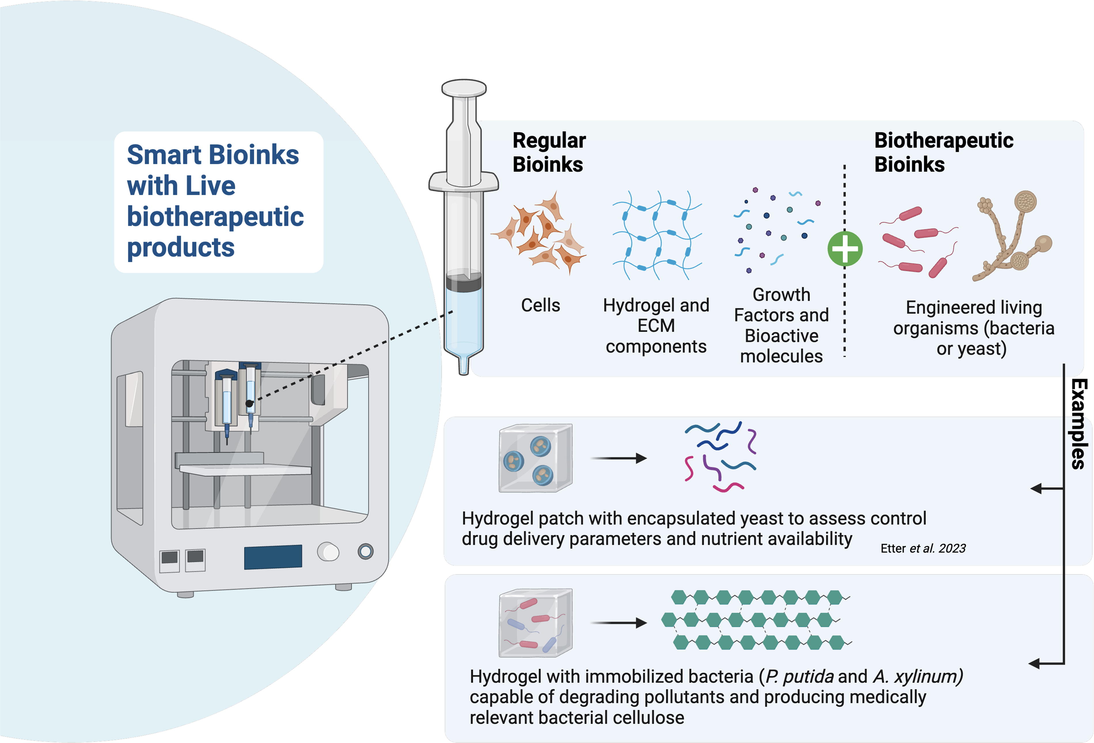
Smart bioinks with live biotherapeutic products. The skin’s diverse microenvironments host over 1010 bacterial cells, maintaining a delicate balance between commensal microorganisms and pathogens through immune tolerance mechanisms. Commensals like Staphylococcus epidermidis modulate the host’s immune response by producing phenol-soluble modulins that inhibit pathogens such as Staphylococcus aureus while cooperating with antimicrobial peptides to enhance pathogen elimination. The integration of live biotherapeutic products into smart hydrogels and bioinks offers innovative wound-healing strategies, enabling immune modulation, toxin degradation, and enhanced healing. Bioprinted hydrogels incorporating engineered microorganisms further advance this approach by restoring homeostasis, promoting efficient skin cell regeneration, and preventing infections. Created in BioRender. Willerth, S. (2024) https://BioRender.com/k12h568.
SUMMARY
This review explored various bioprinting techniques and hydrogels used for creating bioprinted skin constructs, emphasizing their complementary roles in achieving optimal mechanical properties, printability, cell viability, and biocompatibility. Additionally, we highlighted the potential of smart hydrogels incorporating bioactive molecules capable of adapting to external stimuli, offering promising solutions for monitoring wound pH, oxygenation, and reestablishing microbiome balance. Such materials not only regulate microbial invasion and bacterial proliferation but also promote cell growth, migration, and maturation, facilitating a more efficient and sustainable wound-healing process. The integration of biomaterials with LBPs presents an emerging frontier, although further research is required to ensure their effective interaction with host cells. These microorganisms, functioning as natural probiotics and anti-inflammatory agents, hold the potential to support healing without disrupting cell development.
Inappropriate wound management often leads acute injuries to progress into chronic wounds, characterized by persistent inflammatory cycles, increased susceptibility to infections, and more complex treatment challenges. By integrating knowledge of skin architecture, wound-healing phases, and the interaction between biomaterials and key physiological factors—such as pH regulation, oxygenation, growth factor release, and the native microbiome—we can design advanced 3D-bioprinted skin substitutes that have the potential to deliver biomolecules at controlled rates, enhance vascularization, and personalize treatment, ultimately transforming wound care and advancing more effective, sustainable treatment solutions.
Footnotes
ACKNOWLEDGMENTS AND FUNDING SOURCES
This work was supported by NSERC
AUTHOR’S DISCLOSURE AND GHOSTWRITING
S.M.W. is the founder and CEO of Axolotl Biosciences, a biotechnology company focused on 3D printing. The authors have no other relevant affiliations or financial involvement with any organization or entity with a financial interest in or financial conflict with the subject matter or materials discussed in the article apart from those disclosed. No ghostwriters were used to write this article.
ABOUT THE AUTHORS
FUNDING INFORMATION
No funding was received for this article.