
Abstract
Abstract
N-Heterocyclic carbene (NHC) ligands have attracted great interest over the last decade for their use in the design of homogenous catalysts. NHC-based metal complexes have interesting potential biomedical applications, such as in antimicrobial and cancer therapy, which are beginning to be explored more fully. We have studied here the oxidant activities of a series of Ru(II) complexes in vitro and zebrafish (Danio rerio) have been used as a model in vivo to investigate and characterize the toxicity of some of these compounds. Dual behavior was observed for the NHC-based complexes as they behaved as antioxidants at low concentrations but showed pro-oxidant capacity at higher concentrations. Zebrafish embryos were exposed to Ru(II) complexes under several different conditions (0 or 24 h postfertilization, with or without the chorion) and various parameters, such as viability, edema, heart rate, blood coagulation, pigmentation, scoliosis, malformation, and hatching, were tested. In general, zebrafish embryos were not harmed by exposure to Ru(II) complexes whatever the experimental conditions. Several toxicity profiles were observed depending upon the chemical structure of the compound in question. Their characteristics as pro-oxidant and/or antioxidant agents together with their biosafety may point to their having biomedical applications as antitumoral or neuroprotective drugs.
Introduction
N-Heterocyclic carbene (NHC)-based metal complexes are being proposed as potent antimicrobial [Ag(I)-NHC],5–7 radiotherapic [105Rh(III)-NHC],8,9 and antitumoral [Au(I)-NHC] agents.10–13 Within the same context, Ray and coworkers have described the antimicrobial activity of Au(I) and Ag(I)-NHC complexes (Fig. 1A), and the potential antitumoral activity of two trans-dichloro Pd(II)-NHC complexes (Fig. 1B, C). 14 The cytotoxicity 15 and antimicrobial activity 16 of other Au(I)-NHC compounds have been studied by Horvath et al. and Ozdemir et al., respectively.
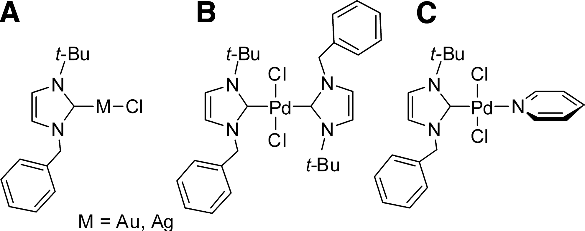
Structures of antimicrobial (
Although the literature shows an increasing biomedical interest in NHC-metal complexes, only a few reports regarding their toxicity are currently available and these limit themselves to analyzing their potential to kill tumoral cells or pathogens6,14,17; according to our knowledge, no animal models have been used for this purpose. As far as ruthenium compounds are concerned, it has been shown 18 that exposure to a chiral organoruthenium half-sandwich compound gives rise to differential zebrafish (Danio rerio) embryo phenotypes according to which enantiomer is used. The effects ranged from no symptoms to pathological effects (decrease in head size, heads without eyes, stunted and crooked tail, or enlarged, misshapen yolk). The popularity of zebrafish as an animal model in toxicology as well as in developmental biology is due to the fact that they are a vertebrate species, their genome is sequenced, and their embryos are mainly fully transparent, so that any change can be observed directly through an optical microscope. 19 Indeed, Eimon and Rubinstein 20 have recently reviewed several assays conducted with zebrafish, including cardiotoxicity, ototoxicity, seizure liability, developmental toxicity, and gastrointestinal motility, the results of which suggest that such assays are acceptably predictive as far as human drugs are concerned, ranging from “sufficient” (65–75% predictability) to “good” (75–85% predictability).
In this work we have studied in vitro the antioxidant/pro-oxidant activity of a series of ruthenium(II) arene complexes with different NHC ligands (NHC = imidazolin-2-ylidene, imidazolin-4-ylidene, and pyrazolin-3-ylidene). For comparative purposes we also studied the oxidant activity of two ruthenium precursors, [RuCl2(p-cymene)]2 and RuCl2(dmso)4. We assessed the biosafety of three complexes selected from the series in zebrafish embryos, an established animal model for studying toxicity.
Materials and Methods
Preparation of the ruthenium compounds
[RuCl2(p-cymene)]2,
21
RuCl2(dmso)4,
22
and complex
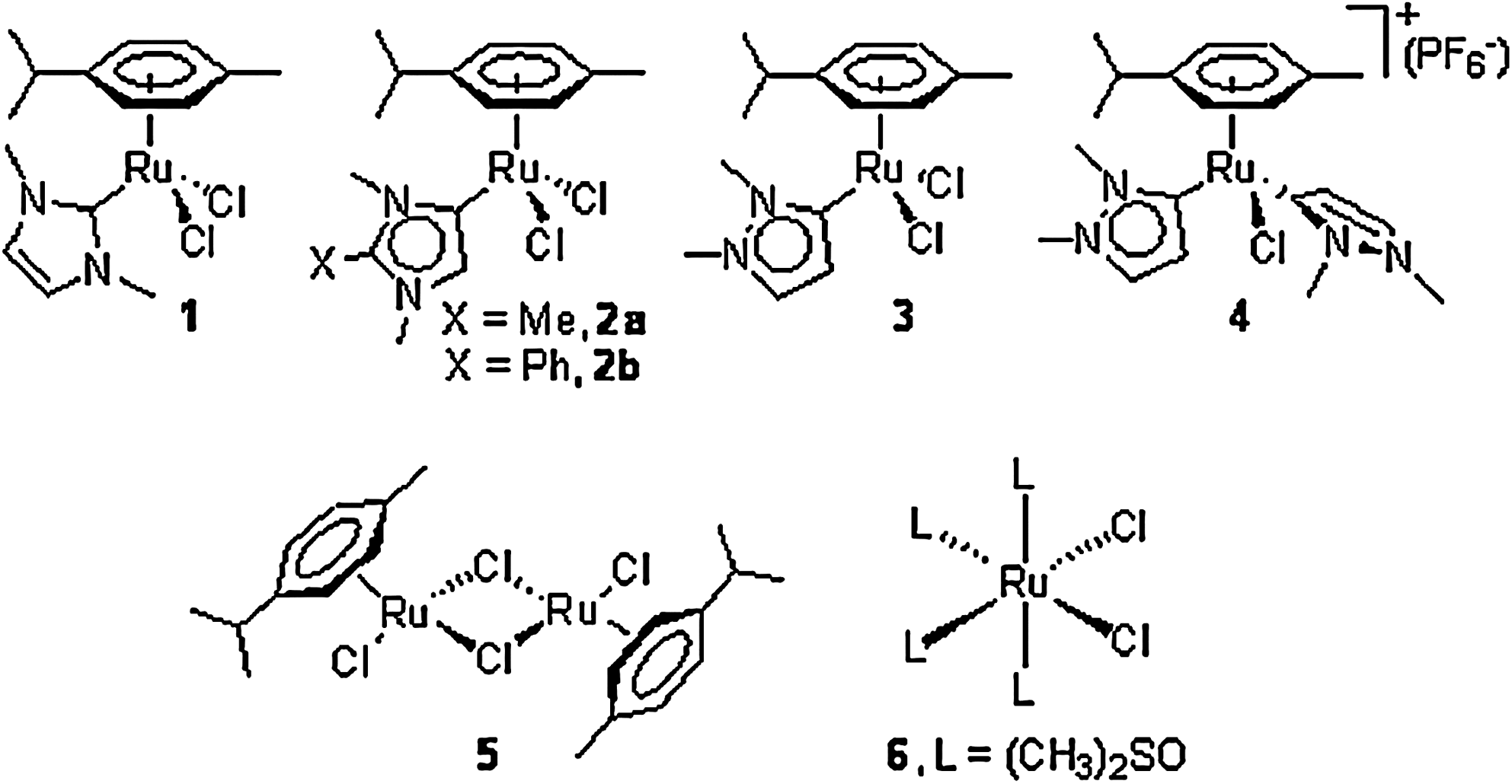
Structures of the studied Ru(II)-N-heterocyclic carbene compounds
Pro-oxidant assay
The radical cation of
Trolox equivalent antioxidant capacity assay
In the Trolox equivalent antioxidant capacity (TEAC) assay, first reported by Miller and Rice-Evans 25 and later modified by Re et al., 26 Trolox (6-hydroxy-2,5,7,8-tetramethylchromane-2-carboxylic acid) serves as a standard or control for the antioxidant capacity. The TEAC assay measures the ability of a compound to reduce the ABTS radical cation, giving useful information regarding its antioxidant activity. As described earlier, the ABTS radical cation is formed by interaction of ABTS with the ferrylmyoglobin radical species, generated by the activation of metmyoglobin with H2O2. It has a characteristic long-wavelength absorption spectrum maximum at 405 nm. A standard curve for Trolox was constructed by plotting the absorbance of different concentrations (in μM) of Trolox at 405 nm. Antioxidants suppress the production of the ABTS radical cation in a concentration-dependent way and the color of the solution decreases proportionally. The antioxidant capacity of the compound in question is obtained by comparison with the concentration of Trolox, using the standard curve, in μM Trolox equivalent (TE).
We performed the TEAC assay in 96-well microtiter plates with Trolox standard concentrations or ruthenium compounds at 1, 10, and 100 ng/mL. We assayed 3.5 μg/mL of myoglobin (Sigma), used as the source of metmyoglobin, which was oxidized by H2O2 (2 mM) in the presence of ABTS (1.8 mM) dissolved in 0.05 M phosphate-citrate buffer solution. ABTS was then oxidized by the ferrylmyoglobin radical thus generated, producing the radical cation of ABTS. After 3 min incubation at room temperature, the end point of absorbance at 405 nm was measured using a Benchmark Plus spectrophotometer (Biorad).
Zebrafish egg production and treatments
Fertilized eggs were obtained by natural mating of adult zebrafish (AB line) in our laboratory, carried out according to the Zebrafish book. 27 A total of 4–5 pairs were set up for each mating and, on average, 200–250 embryos per pair were generated. Eggs were collected within 1 h of spawning, rinsed, and placed into a clean Petri dish, where they were kept at 28.5°C. Twenty-four hours after spawning they were rinsed in dilution water (CaCl2 · 2H2O, 0.294 g/L; MgSO4 · 7H2O, 0.12325 g/L; NaHCO3, 0.06475 g/L; and KCl, 0.00575 g/L) at pH 7.8 ± 0.2 according to Organization for Economic and Co-operation Development (OECD) TG 212, placed into a clean Petri dish, and kept at 28.5°C. Twenty-four hours after spawning, only the fertilized eggs (a minimum of 10 embryos) were exposed to 24-well plates containing either the compound vehicle (dimethyl sulfoxide [DMSO], 0.1%) or different concentrations of the compounds being assayed (ruthenium compounds or t-butyl hydroperoxide [TBH], ranging from 0.001 to 1.000 μg/mL). Apart from this, to complete the toxicology study we used two reference toxic compounds: sodium dodecyl sulfate (SDS) and copper (II) sulfate (CuSO4). The assays were performed using the same procedure as described earlier (with DMSO, 0.1%) and using a range of concentrations between 0.01 and 100 μg/mL. Strict precautions were taken to prevent contamination during manipulation. Each experiment was performed in triplicate.
Early-stage testing to assess teratological effects of the compounds
To observe any possible teratological effect, the fish-embryo test was applied and endpoints for the assessment of acute chemical toxicity such as coagulation of embryos, alteration of heart rate, edema, blood coagulation, weak pigmentation, scoliosis, malformations, and hatching success were recorded. For this assay, 30 min postfertilization embryos were incubated for 48 h with various compounds (ruthenium compounds, TBH, and reference products) or solvent (control), at which time the endpoints were observed. Trolox was used as a positive antioxidant control.
Zebrafish embryo test: embryotoxicity
The development of embryos from blastula to early larval stages was monitored at 24, 48, 72, 96, and 120 h postfertilization (hpf ) throughout the experiment. For the assays, 24 hpf embryos were incubated with various compounds (ruthenium compounds, TBH, and reference products) and solvent (control) for 48 h. At the time of observation, dead embryos were removed from the exposure chambers to prevent contamination of the surviving embryos. Endpoints used for assessing the effects of the compounds included egg and embryo mortality, alteration of heart rate, edema, blood coagulation, weak pigmentation, scoliosis, malformations, and hatching success. The morphological effects were observed and recorded among the larvae from both control and treated groups using a stereomicroscope (7–115×) connected to a camera.
Toxicity of the ruthenium compounds toward dechorionated embryos
The chorions of 24-h-old embryos were disrupted mechanically using two very fine pairs of forceps and experiments were conducted as described earlier.
Results
Antioxidant/pro-oxidant capacities
The oxidant/pro-oxidant capacities of a series of NHC-based Ru(II)-arene complexes (NHC = imidazolin-2-ylidene, imidazolin-4-ylidene, and pyrazolin-3-ylidene [
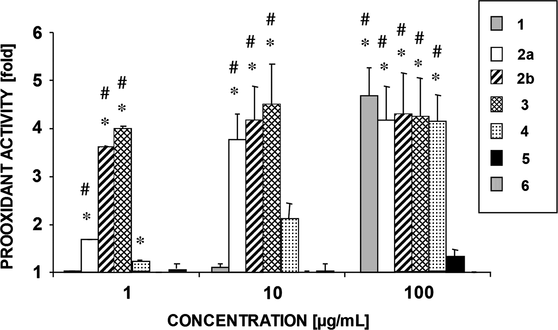
Pro-oxidant activity in vitro of the ruthenium compounds in the range 1–100 μg/mL. The results are shown as the oxidation of the ABTS radical cation caused by the compounds compared with oxidation by 2 mM H2O2. The data are the mean ± SD of two independent experiments. The statistical significance of the differences was examined with Student's t-test (*p < 0.05 compared with
With respect to antioxidant activity, complex
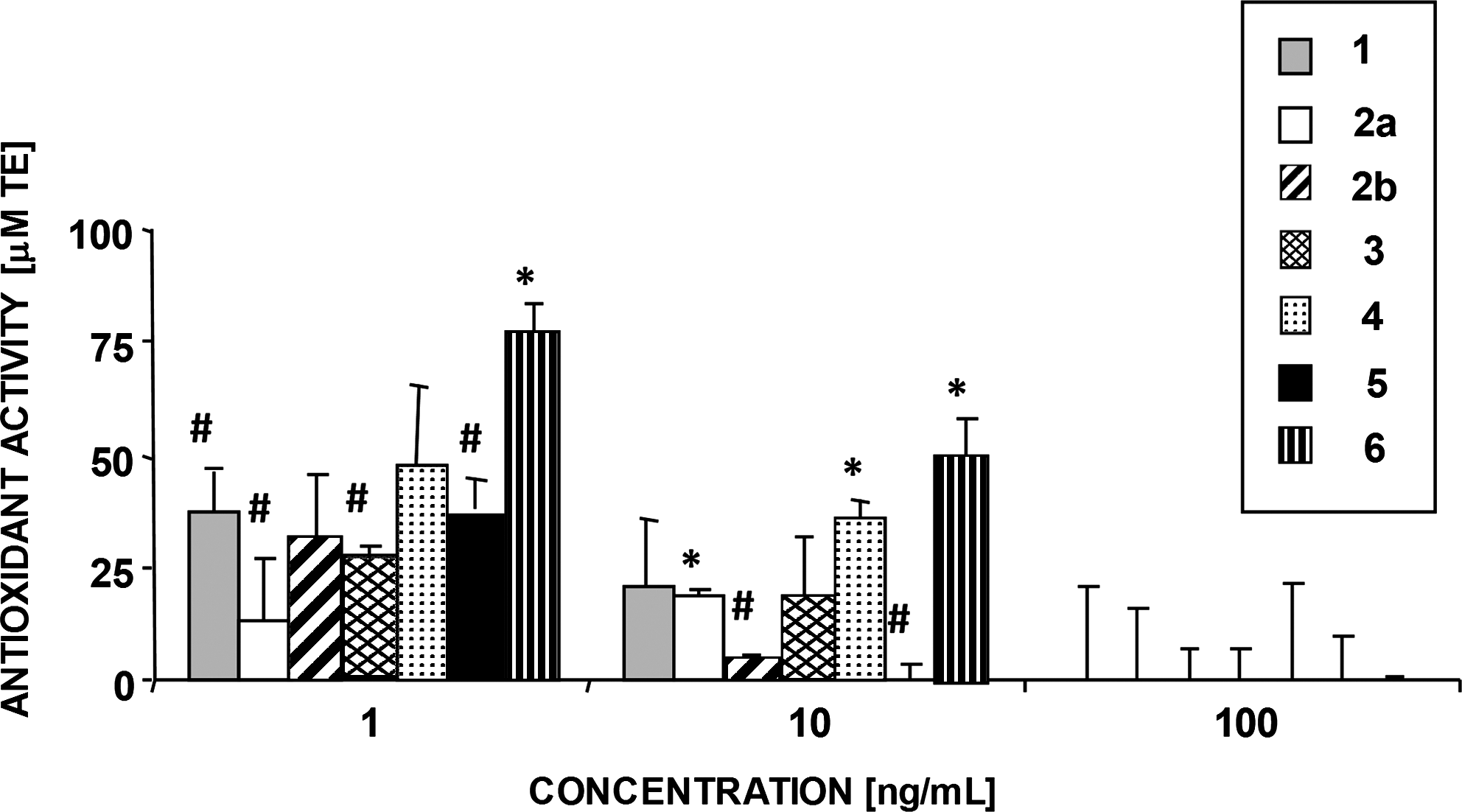
Antioxidant activity in vitro of the ruthenium compounds in the range of 1–100 ng/mL. The antioxidant activity of the compounds is expressed as TE antioxidant capacity values (μM TE) measuring the capacity of the compounds to prevent the formation of the ABTS radical cation by 2 mM H2O2 and quantified using the Trolox standard curve (see Materials and Methods). The data are the mean ± SD of two independent experiments. The statistical significance of the differences was examined with Student's t-test (*p < 0.05 compared with
Embryotoxicity of the ruthenium compounds
To demonstrate the utility of these organometallic compounds for experiments in whole organisms we conducted phenotypic experiments in zebrafish embryos following the OECD Test Guideline 212 (with minor changes). Complexes
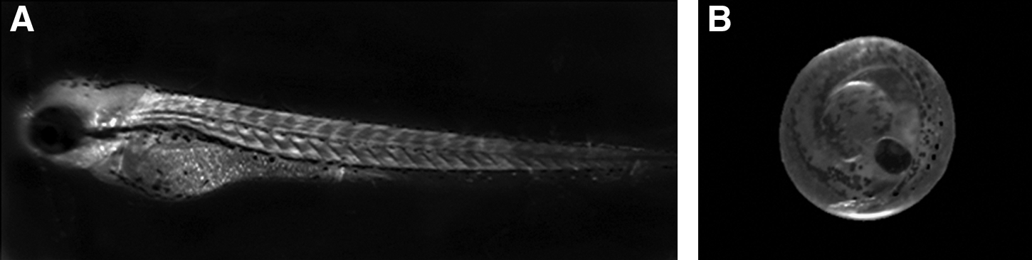
Representative example of an early control larva (72 hpf ) (
On the basis of the results of these initial assays, the dependence of embryotoxicity on the concentration of ruthenium compounds
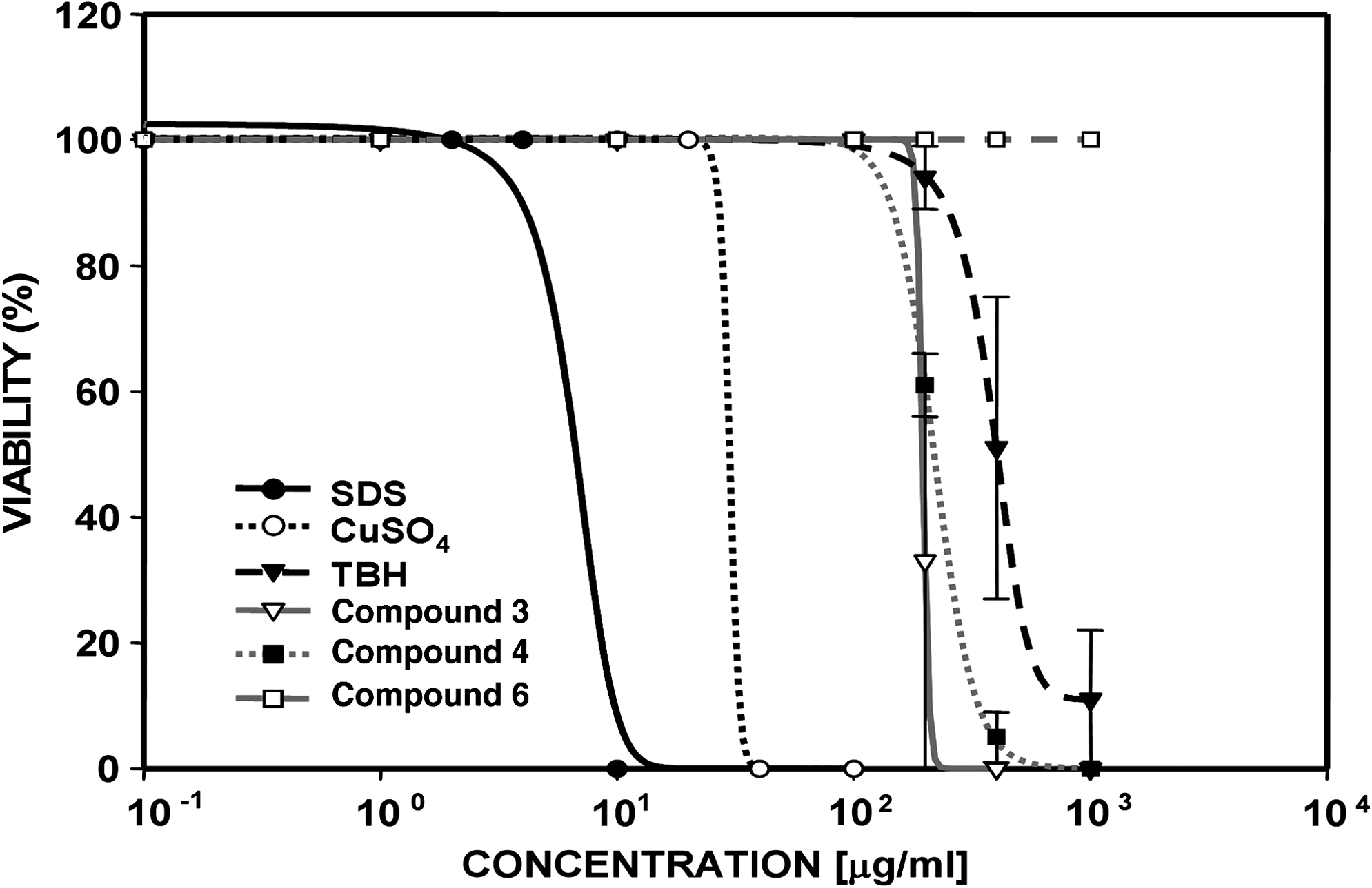
Percentage of zebrafish-embryo viability after exposure to different concentrations of compounds. The embryos were exposed at 24 hpf + 48 hpt to ruthenium compounds or reference substances. Values are the mean ±SD of two independent experiments performed in triplicate.
Dose of a chemical that kills 50% of a sample population.
Dose of a chemical that kills 5% of a sample population.
Dose of a chemical that kills 95% of a sample population.
The lowest tested concentration of a substance with a significant effect compared with the control.
The concentration immediately below LOEC. These data were obtained in embryos exposed to the compounds at 24 hpf and 48 hpt.
SDS, sodium dodecyl sulfate; CuSO4, copper (II) sulfate; TBH, t-butyl hydroperoxide; hpf, hours postfertilization; hpt, hours posttreatment.
Morphological changes in embryos incubated with ruthenium compounds
Once the viability curves had been obtained and the toxicological parameters defined, we studied the induction of morphological changes in embryos. Embryos were incubated with three concentrations of all six compounds (10, 100, and 1000 μg/mL), corresponding to low, mid, and high toxicity (Fig. 7). At 10 μg/mL, the embryos incubated with ruthenium compounds or with TBH were in no way different from the controls, having emerged from the chorion at the early larva stage (72 hpf = 24 hpf + 48 hpt) and clearly showing a well-developed body. At the same concentration, however, the embryos treated with CuSO4 remained inside the chorion, whereas the embryos treated with SDS had died and turned opaque because of coagulation of their organs. At a concentration of 100 μg/mL the embryos treated with compound
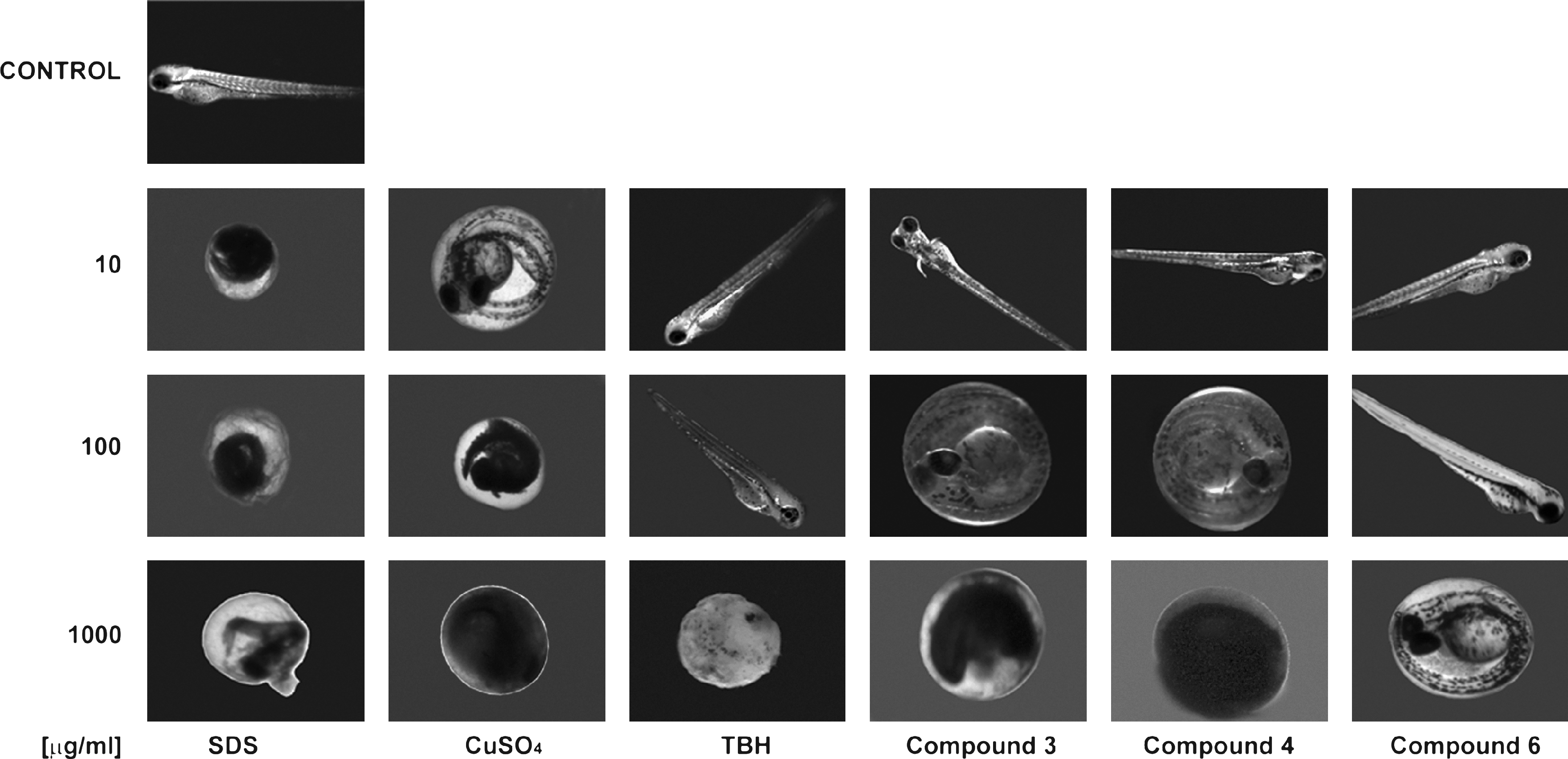
Representative images of zebrafish embryos after various treatments. The embryos were exposed at 24 hpf + 48 hpt to different concentrations of compounds and any morphological change was duly recorded.
To discover whether the embryos remaining within the chorion was the result of a delay in egg development or a stage previous to the death, we undertook additional experiments. Embryos incubated with 100 μg/mL of TBH or ruthenium compounds
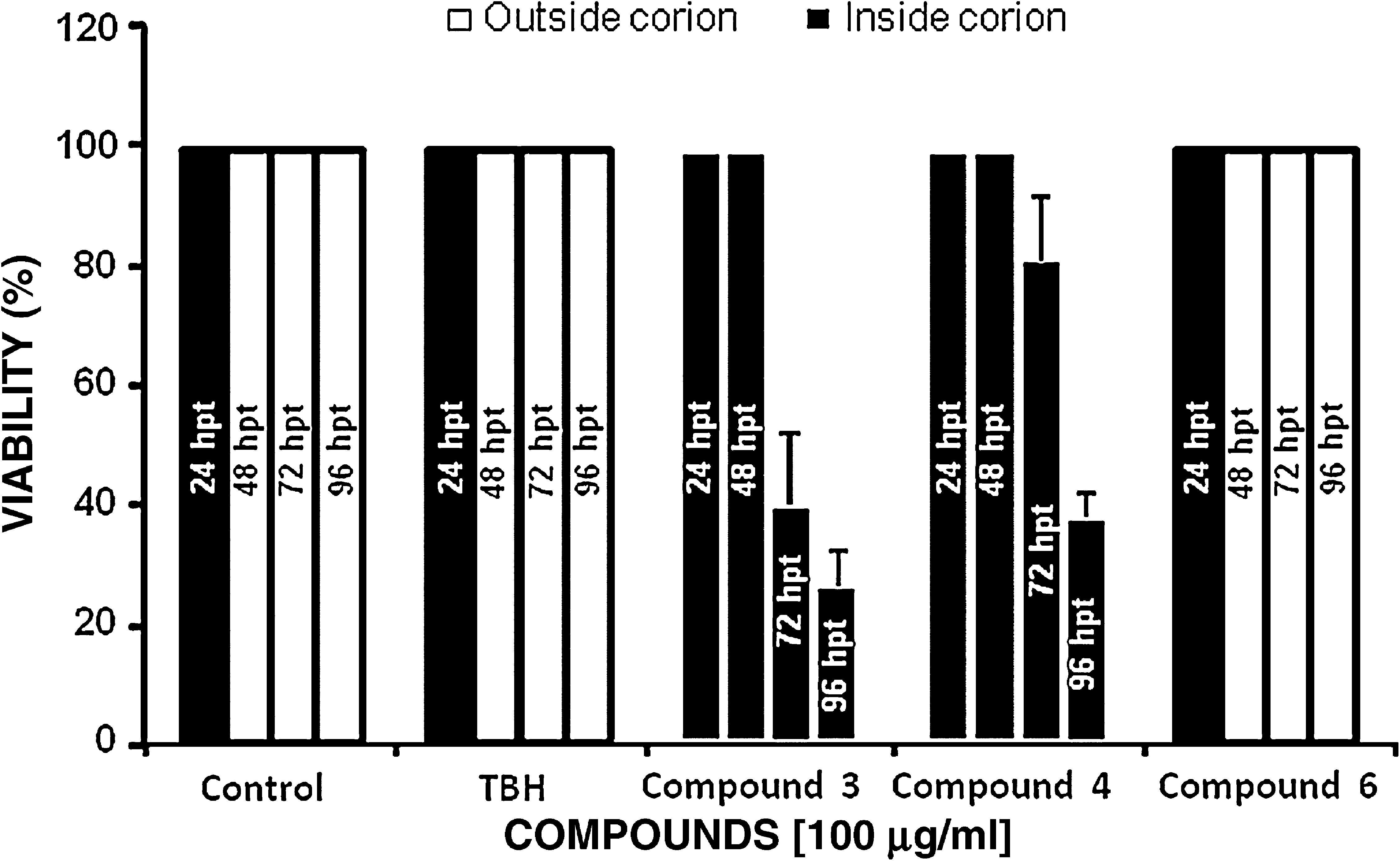
Percentage of viability and cumulative hatching of zebrafish embryos after exposure to different compounds (at 100 μg/mL) and recorded at different times: 24 hpf +24 hpt, 24 hpf + 48 hpt, 24 hpf + 72 hpt, and 24 hpf + 96 hpt.
Cardiotoxicity of ruthenium compounds
The influence of ruthenium compounds upon the heart rate of zebrafish embryos was compared with the reference compounds (Fig. 9). The results indicate that the most toxic compound was SDS, followed by CuSO4, thus confirming the results of the viability assays (Fig. 6 and Table 1). The safest substance assayed was once again compound
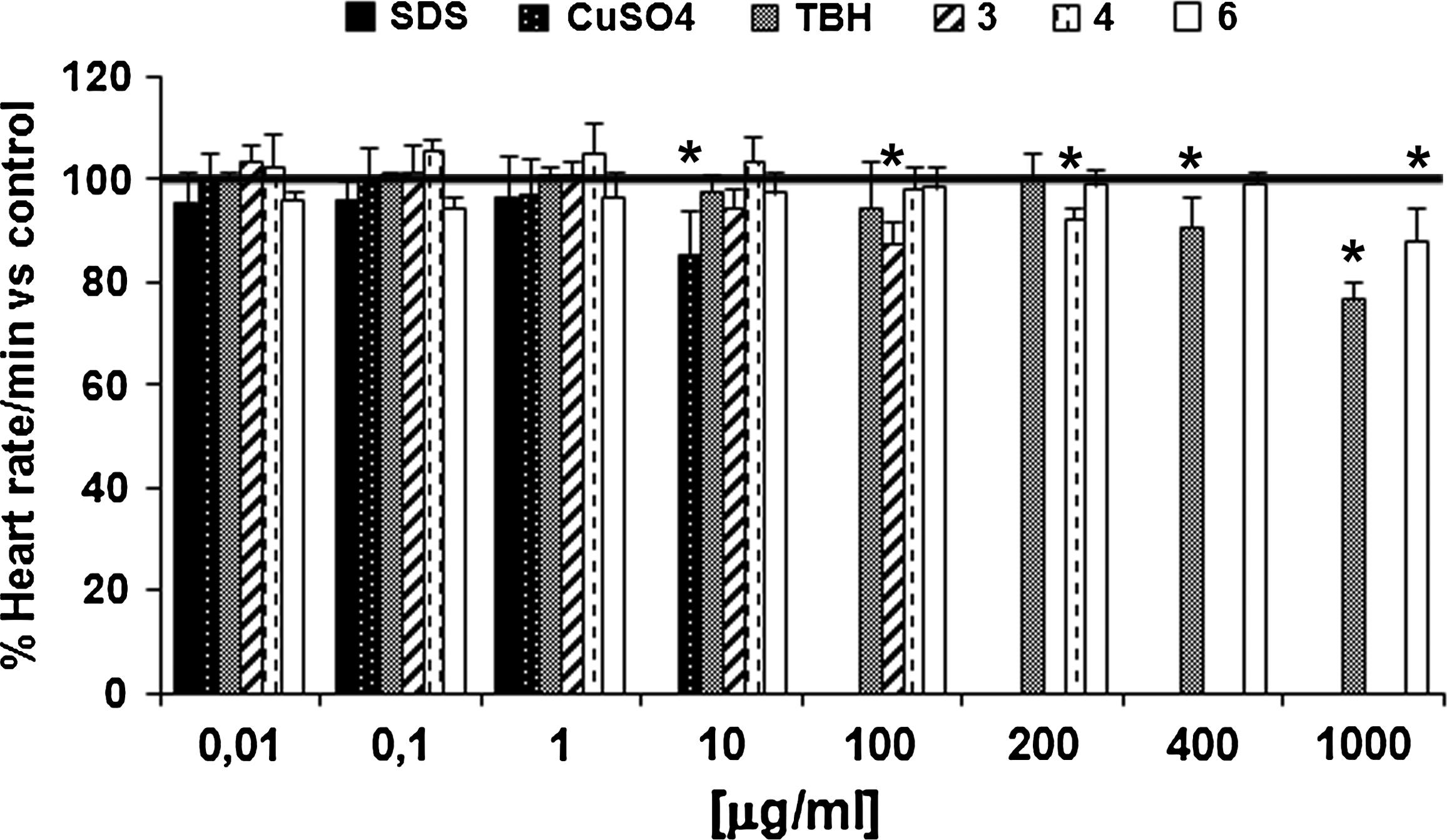
Total heart rate in zebrafish embryos treated with increasing doses of ruthenium and reference compounds. The heart rate of the embryos was recorded at 24 hpf + 48 hpt after exposure to the compounds and the average was plotted. The black horizontal line represents the average value of the heart rate of the controls. The asterisks represent the significant differences between the treated groups compared with the controls (*p < 0.01, Student's t-test). Values are the mean ± SD for two independent experiments performed in triplicate.
Toxicity of ruthenium compounds in dechorionated embryos
To discover whether the effects of the compounds were due to direct toxicity or a differential permeability through the chorion, 24 hpf dechorionated embryos were exposed to a single concentration (100 μg/mL) of the compounds and several endpoints were recorded (Table 2: 24 hpf + 48 hpt − chorion), which were compared with previous results (summarized in Table 2: 24 hpf + 48 hpt + chorion). Once again compound
Embryos were exposed for 48 h and then the compounds were removed.
n, number of embryos used; nd, not determined.
Early-stage testing to assess the teratological effects of the compounds
Essential information in toxicology studies with new chemical entities is provided by teratogenic tests, in which the drug is applied at a fairly early stage, such as around the time of gastrulation. Thus we replicated fish-embryo test assays in which embryos were treated at 30 min postfertilization (Table 2: 0 hpf + 48 hpt + chorion) to observe any differences from treatments administered at 24 hpf (Table 2: 24 hpf + 48 hpt + chorion). All the treatments were administered at 100 μg/mL. In general, the results were very similar to those found in the previous embryotoxicity studies, although compound
Discussion
We have assessed the biomedical properties and biosafety of a series of ruthenium-NHC complexes: (i) antioxidant/pro-oxidant capacity (in vitro); (ii) embryotoxicity; (iii) chorion function as a barrier for the uptake of the compound in embryotoxicity; and (iv) teratological effects (early-stage test).
Our in vitro results showed that the ruthenium complexes assayed act as either antioxidants or pro-oxidants at certain doses, suggesting their possible application as antitumoral or neuroprotective agents, for instance. In harmony with our observations, it has recently been demonstrated that a series of ruthenium compounds present a similar dual behavior showing genotoxic (pro-oxidant) effects at higher doses (200 μM), and antioxidant potential at lower doses (0.1–10 μM). 30 According to these results, the choice of a specific concentration must determine the balance between the two faces of their oxidative status. Although high doses may be useful to sensitize or kill tumoral cells, lower doses, with antioxidant power, may eventually be used to treat or prevent neuropathological conditions associated with aging.
In summary, our in vitro studies suggest that the most active complexes as far as cytotoxicity is concerned are
In morphological studies, the embryos treated with the ruthenium compounds did not present side effects, although at high doses those treated with
One of the most important parameters to explore in determining the toxicity of a compound is its capacity to induce deleterious effects to the heart, and because of its morphological characteristics the zebrafish embryo is an optimum model for this, allowing us not only to measure heart rate but also to study alterations to heart rhythm, such as tachycardia and bradycardia, or anomalies in its development, such as pericarditis, atrophy, and hypertrophy. All three compounds produced good results in the heart-rate study, showing toxicity with doses over 100 μg/mL: compound
In an attempt to clarify the role of the chorion in the embryotoxicity of ruthenium compounds (as a possible barrier for the compounds), 24 hpf dechorionated embryos were exposed to a single concentration (100 μg/mL) and several endpoints were recorded. The toxicity profile of the compounds was the same as in the previous embryotoxicity tests, compound
In summary, the results of this study clearly reveal that ruthenium complexes are safe, showing LC50 ≥ 100 μg/mL in every experimental parameter studied (according to OECD TG 212, substances responsible for LC50 > 100 μg/mL are safe and can be considered nontoxic). Moreover, the three ruthenium complexes, with different chemical structures, present different toxicity profiles. In conclusion, the oxidant activity of the ruthenium complexes studied in vitro correlates well with the profiles of the toxicological parameters studied in zebrafish embryos, which may be related to their structures.
Footnotes
Acknowledgments
This work was funded by Neuron BPh and supported by funds from the Torres Quevedo Program (J.M.A., M.C.R., and J.S.B). The authors thank Professor Fernando Valdivieso for his continuous encouragement and help. The authors from the Universitat Jaume I gratefully acknowledge financial support from the MEC of Spain (CTQ2008-04460) and Bancaixa (P1.1A2008-02), and the Juan de la Cierva program (to M.P.).
Disclosure Statement
The authors from Neuron BPh have served as paid researchers for the company.