
Abstract
Abstract
How distinct cell populations are distributed in three-dimensional space under homeostasis or following injury, neurodegeneration, or with senescence can teach us much about brain-wide patterns and signaling along the neuroaxis. Visualizing individual cell populations in the mature vertebrate central nervous system (CNS) has remained a challenge as a result of difficulty clearing adult brain tissue or limitations in imaging depth or resolution. We have developed a simple clearing and imaging pipeline optimally suited for the adult zebrafish brain to investigate changes in patterns of cell proliferation in wild-type and transgenic backgrounds that can easily be quantified and represented using FIJI and IMARIS software.
Methods
T
The small size of the adult zebrafish brain (∼6 × 3 × 3 mm; Fig. 1A) makes it ideally suited for OPT imaging as a tool to gain new insight into brain-wide cellular patterns in vertebrates. OPT allows imaging of fixed specimens at near cellular resolution (3.21 μm/pixel) with three or more fluorescent channels from UV to the IR spectrum (typically excitation at 350, 488, 555, and 647; Fig. 1B–D, K, L), as well as brightfield, which can be reconstructed to obtain a 3D view of organs. This method was originally used to study mouse embryo development, 11 but has more recently been applied to organ morphogenesis 12 and the distribution of neural stem cell niches. 13 Compared with more recent imaging technologies such as light sheet microscopy, OPT offers isotropic scanning of specimens that can be readily cleared in a simple mixture of benzyl benzoate: benzyl alcohol. In addition, reconstruction is simple and fast and datasets are compatible with FIJI or IMARIS for quantification and visualization. Below we present an inexpensive and straightforward method to examine patterns of cell proliferation in the adult brain or other adult organs, which can be performed in wild-type, transgenic, or mutant backgrounds.
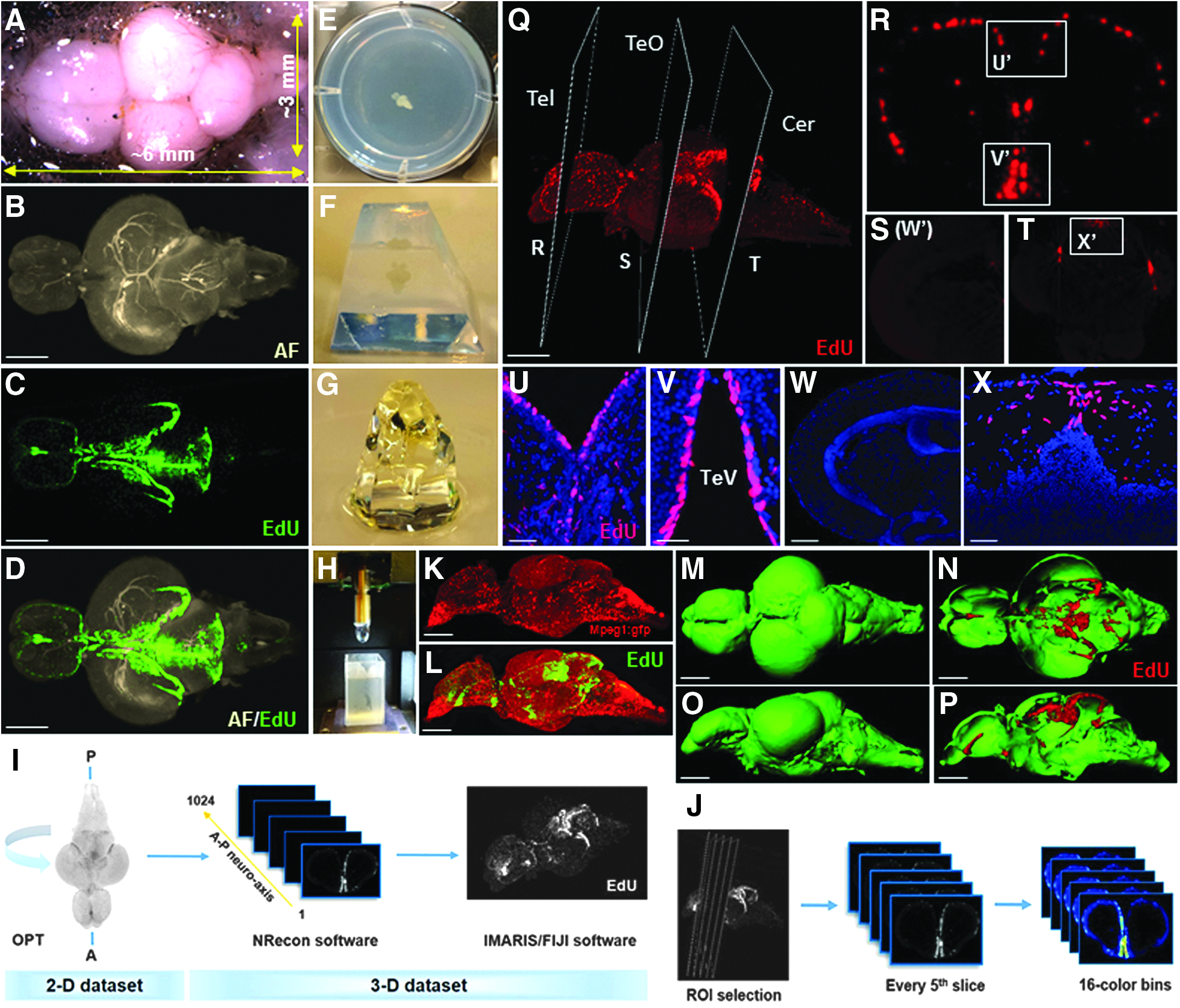
Sample preparation, output, and quantification of cleared adult zebrafish brains imaged using OPT.
Brief overview of workflow:
1. Labeling proliferating cells: Inject adult zebrafish twice intraperitoneally with 40 μL of a 10 mM solution of the S-phase marker 5-ethynyl-2′-deoxyuridine (EdU) over 4-h to label proliferating cells in the CNS. 2. Tissue processing: Sacrifice animals and dissect out the brain. Fix samples in 2% paraformaldehyde overnight. Rinse samples in phosphate-buffered saline (PBS, pH 7.4) with 0.3% Triton X-100 for 2-h, followed by 1 × -PBS with 1% Triton X-100/5% dimethyl sulfoxide for 24-h. 3. Staining proliferating cells: Commence EdU staining over 4 days in 12-well plates using EdU staining solution with Alexa Fluor Azide dyes (Supplementary Table S1; Supplementary Data available online at www.liebertpub.com/zeb). In addition, immunohistochemistry can be performed after EdU staining. 4. Embedding: Embed samples in a 1% solution of distilled water and low melting agarose in a six-well plate. Position samples in the centre of well with the brain oriented dorsally. Once agarose is set, trim brains (Fig. 1E–G). 5. Dehydration and clearing: Dehydrate samples in 100% methanol, providing three to four solution changes over 24 h. Clear samples in a 2:1 solution of benzyl benzoate: benzyl alcohol as above, over 24 h (Fig. 1G). 6. OPT scanning and reconstruction: Adhere cleared brain samples onto OPT mounts (Fig. 1H). Image brains using desired parameters on a Bioptonics 3001 OPT scanner (Bioptonics, Edinburgh, United Kingdom; Supplementary Table S2) and reconstruct using Nrecon software (Bruker microCT; Fig. 1I). 7. Quantification and visualization: Import final reconstructed datasets into FIJI or IMARIS software to analyze fluorescence by intensity, surface area, or volume (Fig. 1J), and visualize single or multichannel data as 3D maximum projections (Fig. 1K, L) or isosurface-rendered images (Fig. 1M–P).
Work in our laboratory using this imaging approach to examine adult neurogenic compartments in the zebrafish brain has shown this method to be highly consistent in EdU staining compared with our standard cryosectioning protocols, demonstrating reliable penetration of EdU through the entire adult brain (Fig. 1Q–X, Supplementary Movie SM1). The protocol is compatible with fluorescent proteins and green fluorescent protein (GFP) reporter lines, for example, microglia can be visualized in the brain using the Tg(mpeg1:gfp) line 14 (Fig. 1K, L).
We envision this method to be an excellent tool to investigate brain-wide changes in proliferation postlesion (Supplementary Fig. S1), brain structure development and senescence, or to screen for proliferative abnormalities in mutants. Unlike many of the more advanced clearing and imaging methods today, no specialized reagents or equipment are required for this method, making it accessible to most laboratories. We welcome collaborations for those interested in taking advantage of this method and provide a more detailed protocol in the Supplementary Data.
Footnotes
Acknowledgments
We thank the laboratory of Prof. I. Smyth in the Department of Anatomy and Developmental Biology at Monash University for the use of their Optical Projection Tomography scanner. Thanks to G. Lieschke for providing the Tg(mpeg1:gfp) line. This work was supported by an NHMRC project grant (GNT1068411), Monash University Faculty of Medicine and Nursing strategic grant and Operational Infrastructure Support from the Victorian Government. BL was supported by a fellowship from NSERC in Canada. We thank Monash Medical Micro Imaging Facility and Fish Core for excellent support.
Disclosure Statement
No competing financial interests exist.
References
Supplementary Material
Please find the following supplemental material available below.
For Open Access articles published under a Creative Commons License, all supplemental material carries the same license as the article it is associated with.
For non-Open Access articles published, all supplemental material carries a non-exclusive license, and permission requests for re-use of supplemental material or any part of supplemental material shall be sent directly to the copyright owner as specified in the copyright notice associated with the article.