
Abstract
Abstract
Astyanax is a species-rich polyphyletic genus distributed between the southern United States and central Argentina. The genus contains groups of cryptic species, which are difficult to distinguish, and are sometimes identified wrongly. Basic and molecular cytogenetic analyses were run on Astyanax abramis and three junior synonyms of Astyanax lacustris: Astyanax altiparanae, from the upper Paraná River basin, Astyanax asuncionensis, from the lower Paraná basin, and Astyanax jacuhiensis, from the upper Uruguay River. These species all belong to the Astyanax bimaculatus group. All species presented 2n = 50 chromosomes and single nucleolar organizing regions (NORs). In A. altiparanae, the karyotype was 6m + 28sm+4st+12a and the NORs were present in pair 20, while A. jacuhiensis was 8m + 28sm+6st+8a, with NORs in pair 22, and A. asuncionensis was 8m + 24sm+6st+12a, with NORs in pair 20. A. abramis was 4m + 30sm+8st+8a with NORs in pair 22. Fluorescence in situ hybridization revealed single 5S rDNA cistrons in A. altiparanae and A. asuncionensis, and multiple (4) cistrons in A. abramis and A. jacuhiensis. Heterochromatin had a distinct distribution in each species, but was predominantly centromeric and interstitial proximal. In A. abramis and A. asuncionensis, the first acrocentric chromosome pair presented centromeric, telomeric, and interstitial-proximal heterochromatin in the long arm, which may represent the presence of homologous chromosomes in these species. While there are some cytogenetic similarities, differences in the location of 5S rDNA, distribution of heterochromatin, and karyotype formulae contribute to the differentiation of the study species, and support the identification of phylogenetically proximate groups in the “Astyanax clade.”
Introduction
A
The Astyanax bimaculatus (Linnaeus 1758) species complex includes 22 valid species, and can be diagnosed by a horizontally oval black humeral spot (the most conspicuous feature of the group), two diffuse vertical brown bars in the humeral region (the first crosses the humeral spot and the second is located 2–3 scales posteriorly), and a black spot on the caudal peduncle, which extends as far as the tip of the median ray of the caudal fin. 14 Astyanax altiparanae was originally described from the upper Paraná River basin, with its distribution being extended subsequently to the basin of the Iguaçu River.15,16 Astyanax jacuhiensis was described originally from the Jacuí River and is known to be distributed throughout the basin of the Uruguay River and in the coastal streams of southern Brazil, 17 while Astyanax asuncionensis is distributed throughout the Paraguay and lower Paraná rivers, and Astyanax abramis (Jenyns 1842) is found in the upper Amazon basin, and in the upper Meta and La Plata basins.17,18
In a recent taxonomic review of the morphological data on the A. bimaculatus “caudal peduncle spot” subgroup from the La Plata and São Francisco basins, together with the coastal basins of southeastern Brazil and Uruguay, Lucena and Soares 14 identified A. altiparanae, A. asuncionensis, and A. jacuhiensis as junior synonyms of Astyanax lacustris (Lütken 1875), and considered only A. lacustris to be a valid species. This species is distributed throughout the La Plata and São Francisco basins, the Laguna dos Patos coastal lagoon complex, and the Tramandaí and Araguaia basins. The authors also considered Astyanax paraguayensis Fowler 1918 to be a junior synonym of A. abramis, with a distribution throughout the Paraguay River basin, the mid-lower Paraná, and lower Uruguay basins. Despite these conclusions, the authors recommend further research into the mechanisms that have isolated these lineages within the subgroup, in particular to determine whether the speciation process has occurred without morphological differentiation or if morphological similarities arose after genetic differentiation due to the adoption of adaptations to similar habitats.
The available cytogenetic data on Astyanax indicate ample karyotypic diversity among its species. Diploid numbers range from 36 chromosomes in Astyanax schubarti Britski 196419 and Astyanax correntinus (Holmberg 1891) 20 to 50 chromosomes in species such as A. altiparanae 21 and A. jacuhiensis. 22 In addition to the variation in karyotypic macrostructure and the number and location of the nucleolar organizing regions (NORs), the presence of B chromosomes, triploidy, and heterochromatin polymorphisms are also common in Astyanax.19,23,24 The A. altiparanae and A. jacuhiensis forms presented the same diploid numbers, and similar patterns of NORs and C-banding, in terms of their quantity and location.25,26 Similarly, A. abramis and A. asuncionensis also presented a number of cytogenetic similarities, although they could be distinguished by the variation in their karyotype formulae and the number/location of 5S rDNA sites. 20
In this article, we aimed to expand on the cytogenetic data available for A. abramis and A. lacustris, to contribute to the identification, characterization, and differentiation of these species, and to verify whether A. altiparanae, A. asuncionensis, and A. jacuhiensis should be considered to be junior synonyms of A. lacustris or valid species. We also hope to contribute to the understanding of the taxonomy and systematics of the A. bimaculatus “caudal peduncle spot” subgroup and the Astyanax clade, sensu Mirande,3,4 as a whole.
Materials and Methods
Despite of synonymizations proposed by Lucena and Soares 14 due to overlapping morphological characters, we considered the synonymized names valid and their geographical distribution previously known, to allow comparisons with the literature and test synonymization, referring to them in quotes from now on. Morphometric analyses usual for taxonomic studies of Astyanax were also performed for comparative purposes. Specimens analyzed were deposited in the Coleção Ictiológica do Núcleo de Pesquisas em Limnologia, Ictiologia e Aquicultura—(NUP), of the Universidade Estadual de Maringá, Maringá, Brazil: five males and four females of A. altiparanae (NUP 17156, from the Paraná River, upper Paraná River basin, 22°45′53.50′′S; 53°15′27.31′′W); two males and four females of A. jacuhiensis (NUP 14927, from the Ijuí River, upper Uruguay River basin, 28°18′06.30′′S; 53°53′33.60′′W); three males and three females of A. abramis (NUP 14581, from the Iguaçu River, downstream from the Iguaçu Falls, 25°38′18.72′′S; 54°28′4.74′′W); and 13 males and 12 females of A. asuncionensis (NUP 14584, from the Iguaçu River, lower Paraná River basin, downstream from the Iguaçu Falls, 25°38′18.72′′S; 54°28′4.74′′W).
This study was carried out in strict accordance with the recommendations of the Guide for the Care and Use of Laboratory Animals, approved by the Committee on the Ethics of Animal Experiments of the Universidade Estadual do Oeste do Paraná (License Number: Protocol 13/09–CEEAAP/Unioeste). All specimens were anesthetized and sacrificed by an overdose of clove oil. 27 Chromosome preparations were obtained from cells of anterior region of kidney. 28 AgNORs were revealed by silver impregnation. 29 Heterochromatin regions were determined following the C-banding technique, 30 with staining modifications. 31 Physical mapping of 5S rDNA and 18S rDNA was carried out by fluorescence in situ hybridization (FISH) 32 and modifications, 33 using DNA probes obtained from Megaleporinus elongatus ( = Leporinus elongatus) 34 and Prochilodus argenteus, 35 respectively. Probes were labeled by nick translation method with digoxigenin-11-dUTP (5S rDNA) and biotin-16-dUTP (18S rDNA) (Roche®). Detection of signals was performed with antidigoxigenin-rhodamine (Roche) for probe of 5S rDNA and amplified avidin-FITC with biotinylated anti-avidin (Sigma-Aldrich) for probe of 18S rDNA, with the chromosomes counterstained with 4′,6-diamidino-2-phenylindole (DAPI, 50 μg/mL). Metaphases were photographed using a BX 61 epifluorescence microscope, coupled with Olympus DP 71 digital camera (Olympus America, Inc.) with the Olympus DP Controller software 3.2.1.276. Chromosomes were classified and organized in metacentric (m), submetacentric (sm), subtelocentric (st), and acrocentric (a). 36 The fundamental number (FN) was calculated considering m, sm, and st chromosomes as having two arms, and a chromosomes as having only one chromosome arm.
Results
Astyanax altiparanae
Diploid number was 50 chromosomes (6m + 28sm+4st+12a, FN = 88) for males and females (Fig. 1a). A single pair of NORs was located in terminal position on the short arm of chromosome pair 20 (Fig. 1a in box). C-banding showed centromeric heterochromatin blocks in pairs 2, 8, and 12, interstitial-proximal heterochromatin on the long arm of pairs 6, 7, 8, 10, 11, 14, 15, 18, 22, and 25, and coincident with NORs (Fig. 1b). FISH revealed a single 5S rDNA site in centromeric position in the m pair 2 and a single 18S rDNA site in terminal position on the short arm of the a pair 20 (Fig. 2a).
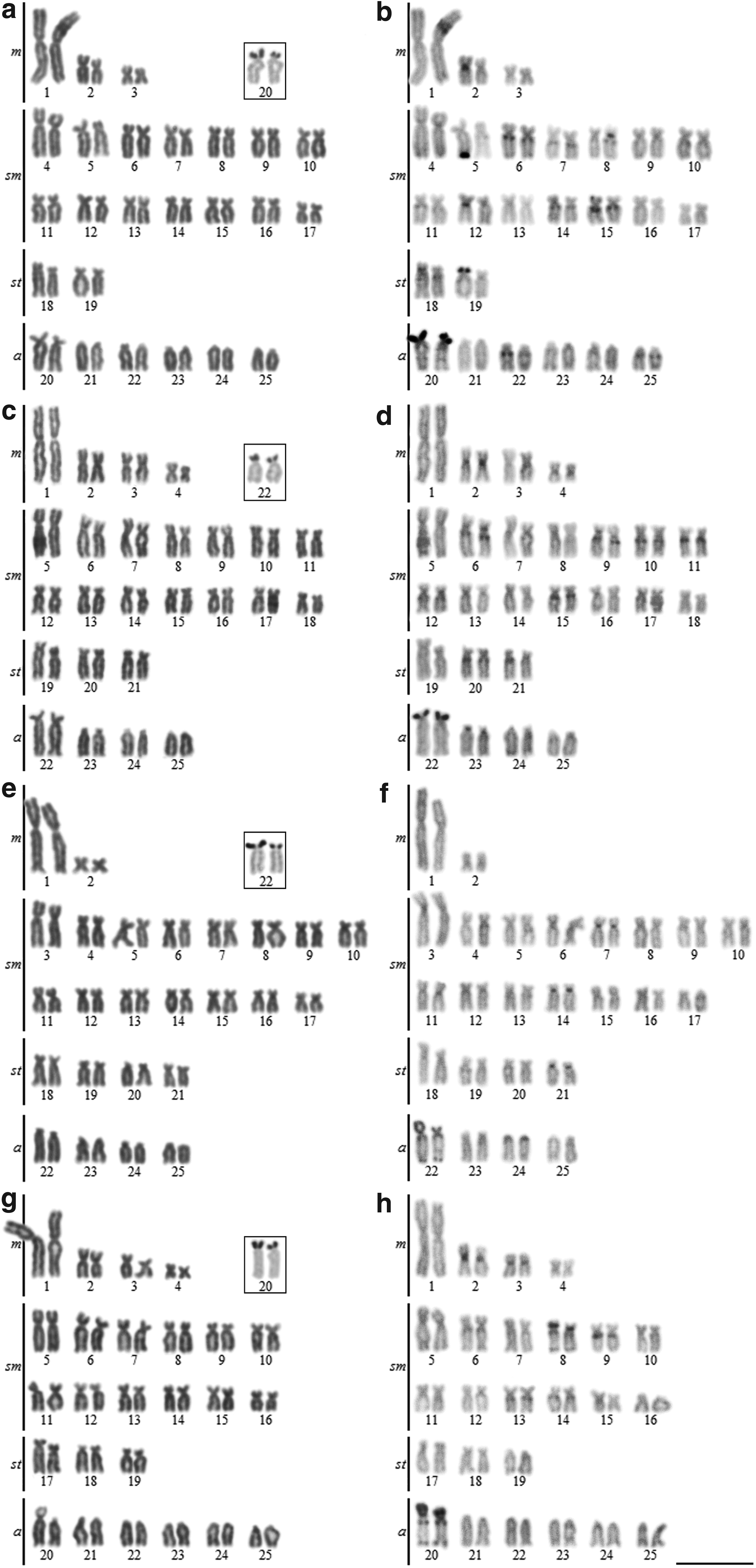
Karyotypes stained by Giemsa in
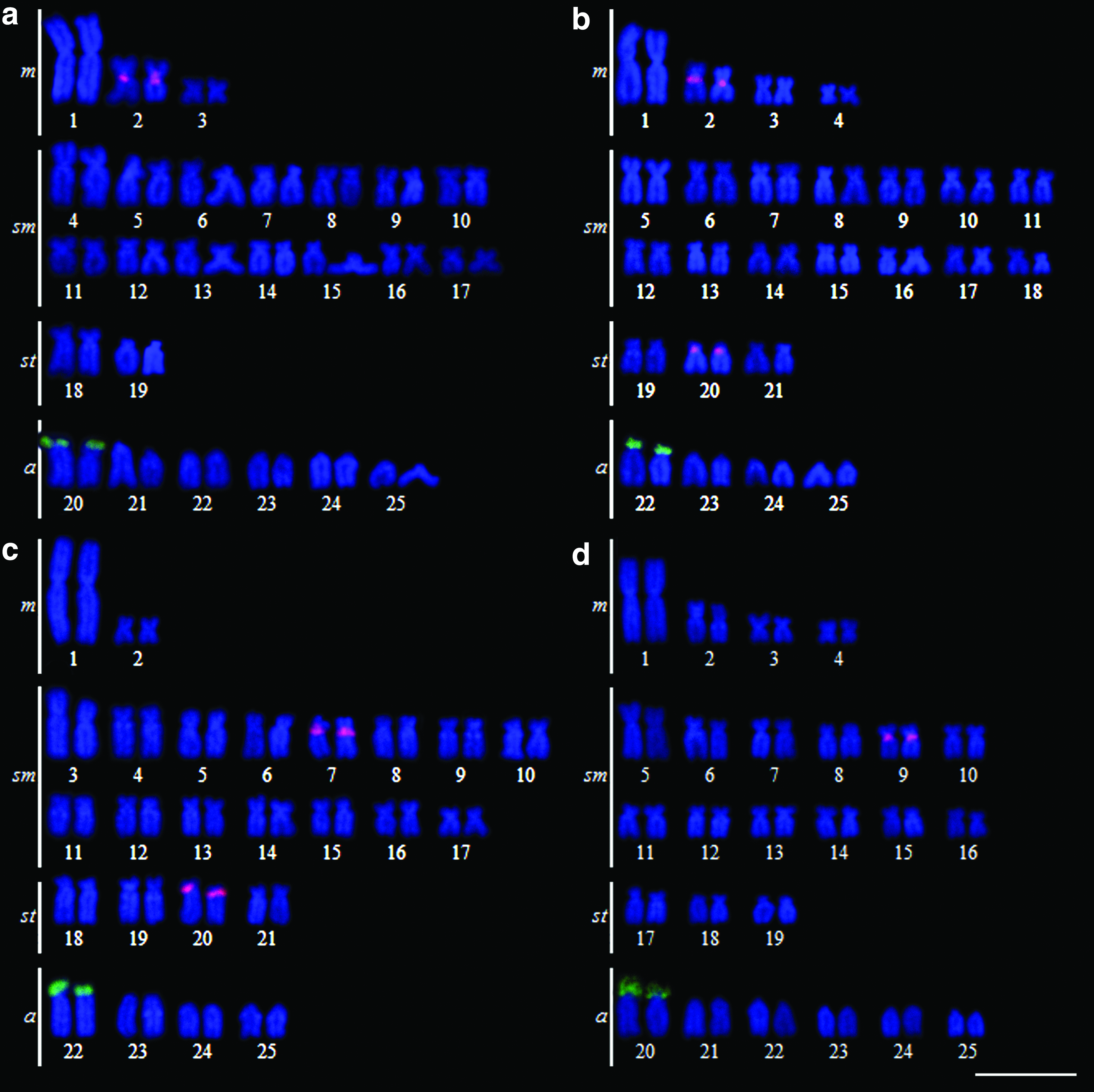
Karyotypes after FISH with 5S rDNA probe (red) and 18S rDNA probe (green) in
Astyanax jacuhiensis
Diploid number was 50 chromosomes (8m + 28sm+6st+8a, FN = 92) for males and females (Fig. 1c). A single pair of NORs was located in terminal position on the p arm of chromosome pair 22 (Fig. 1c in box). C-banding showed centromeric heterochromatin blocks in pairs 2, 6, 9, and 23, interstitial-proximal heterochromatin on the q arm of pairs 6, 9, 10, 11, 12, 15, 20, and 21, and coincident with NORs (Fig. 1d). FISH revealed multiple 5S rDNA sites in centromeric position in the m pair 2 and the st pair 20, and a single 18S rDNA site in terminal position on the p arm of the a pair 22 (Fig. 2b).
Astyanax abramis
Diploid number was 50 chromosomes (4m + 30sm+8st+8a, FN = 92) for males and females (Fig. 1e). A single pair of NORs was located in terminal position on the p arm of chromosome pair 22 (Fig. 1e in box). C-banding showed centromeric heterochromatin blocks in pairs 7, 14, and 21, interstitial-proximal heterochromatin on the q arm of pairs 22 and 24, telomeric heterochromatin on the p and q arms in pair 22, and coincident with NORs (Fig. 1f). FISH revealed multiple 5S rDNA sites in centromeric position in the sm pair 7 and the sm pair 20, and a single 18S rDNA site in terminal position on the p arm of the a pair 22 (Fig. 2c).
Astyanax asuncionensis
Diploid number was 50 chromosomes (8m + 24sm+6st+12a, FN = 88) for males and females (Fig. 1g). A single pair of NORs was located in a terminal position on the p arm of chromosome pair 20 (Fig. 1g in box). C-banding showed centromeric heterochromatin blocks in pairs 2, 3, and 20, interstitial-proximal heterochromatin on the p arm of pair 8 and on the q arm of pairs 9, 13, and 14, telomeric heterochromatin on the q arm of pair 8 and on the p and q arms in pair 20, and coincident with NORs (Fig. 1h). FISH revealed a single 5S rDNA site in centromeric position in the sm pair 9, and a single 18S rDNA site in terminal position on the p arm of the a pair 20 (Fig. 2d).
Discussion
Of the cytogenetic characters analyzed in this study (diploid number, karyotypic formula, NOR, heterochromatin, and 5S rDNA), the physical mapping of 5S rDNA cistrons proved to be the most effective for the differentiation of the morphologically similar Astyanax species. While single 5S rDNA sites were found in A. altiparanae and A. asuncionensis (2 cistrons), multiple (4 cistrons) sites were identified in A. jacuhiensis and A. abramis. In all cases, these sites were located in a centromeric position, but in different chromosome pairs. Single 5S rDNA sites have also been recorded in populations of A. altiparanae,21,37 A. asuncionensis, 20 and A. lacustris. 37
Multiple 5S rDNA-FISH sites have been reported in Astyanax scabripinnis, 38 Astyanax fasciatus, 39 A. abramis, and A. correntinus. 20 The cytogenetic data available for the genus indicate variation in the number and location of 5S rDNA cistrons, which thus represent an important marker for the diagnosis of the relationships among species.40–43 Similar results have been obtained for other genera, such as Brycon, 44 Characidium, 45 Hypostomus, 46 and Hoplias. 47 In this context, the physical mapping of the 5S rDNA sites in A. altiparanae, A. asuncionensis, and A. jacuhiensis revealed differences in comparison with A. lacustris, both in terms of the host chromosome pair and the occurrence of synteny with 18S rDNA, that is, colocation, 40 which indicates that these taxa represent four valid species.
In the case of the heterochromatin, all the species of the A. bimaculatus complex have pale heterochromatin in most of the chromosomes, primarily in centromeric and interstitial-proximal positions, in addition to associations with NORs.22,40 However, it was possible to observe two distinct patterns in the amount of heterochromatin found in the four species analyzed. In A. abramis and A. asuncionensis, the first pair of acrocentric chromosomes had centromeric, telomeric, and interstitial-proximal heterochromatin in the q arm. These features may represent corresponding chromosomes, and thus reinforce the proximity of these species in the “Astyanax clade,”3,4 as well as differentiating them from the other species of the A. bimaculatus group analyzed in this study. In addition, A. altiparanae and A. jacuhiensis have more chromosomes pairs with interstitial-proximal heterochromatin than either A. abramis or A. asuncionensis.
A diploid number of 50 chromosomes has been identified frequently in Astyanax species, including Astyanax argyrimarginatus Garutti 1999, Astyanax elachylepis Bertaco & Lucinda 2005, A. aff. bimaculatus (Dois de Agosto stream, Tocantins-Araguaia river basin), and Astyanax xavante, 48 including the species analyzed in this study. The species analyzed in this study presented a predominance of biarmed chromosomes, although the karyotype formulae permitted the differentiation of the species.
The number and location of the NORs (Ag-staining and 18S rDNA-FISH) were conserved in the four species, which presented single NORs located on the p arm of the first acrocentric chromosome pair. This may thus represent the plesiomorphic condition for the A. bimaculatus group, especially as it has also been confirmed in other species of this group, such as A. lacustris,37,40 A. argyrimarginatus, and A. aff. bimaculatus from the Dois de Agosto stream and the Tocantins-Araguaia basin. 48
Garutti 10 and Garutti and Britski 49 mentioned the presence of maxillary teeth in some species of the A. bimaculatus group (likewise the type species of the group), found in the Amazon and other basins of northern South America, but not in basins further south, such as the São Francisco River, La Plata, Paraíba do Sul, and Ribeira de Iguape, nor the Patos lagoons. Lucena and Soares 14 found only one specimen of A. lacustris with a tooth in the left maxilla bone in a sample of 237 specimens of this species, and found no maxillary teeth in A. abramis. However, maxillary teeth were found in all the species analyzed in this study. A small, unicuspidated or tricuspidated tooth is found occasionally in one of the maxillary bones in A. asuncionensis (NUP 14584, 5 of 28 specimens), A. jacuhiensis (NUP 14927, 1 of 4 specimens), and A. altiparanae (NUP 17156, 2 of 23 specimens). No tooth was found in A. abramis (NUP 14581, six specimens), although the additional analysis of six specimens (NUP 7956) from the Ariranha River in the Paraguay basin revealed one specimen with a tricuspidated maxillary tooth. Both A. altiparanae specimens collected from the Pitangui River, upper Paraná basin (NUP 6771), have the tooth. While infrequent, the occurrence of the tooth in species from the La Plata basin must be taken into account in any further reviews and species descriptions (CAMO pers. obs.). The variation in the presence of the tooth further reinforces the conclusion that some of the morphological traits used for the diagnosis of species may be unreliable for the differentiation of the species of the A. bimaculatus group, as concluded by Lucena and Soares, 14 who suggest that speciation may often occur without any apparent morphological divergence.
The analysis of mitochondrial DNA has also been ineffective for the differentiation of some Astyanax species. In a recent analysis of more than 1600 samples, covering more than 70 nominal species, Rossini et al. 13 reinforced the complexity of the genus and the difficulties of identifying its species, in particular, those of the A. bimaculatus complex. Kavalco et al. 43 suggest that mitochondrial evolution in this group is slower than the rate of chromosomal mutation, and that molecular cytogenetics provides the ideal resolution for the diagnosis of Astyanax species.
Following phylogenetic analyses in the Characidae, and the establishment of the Astyanax clade,3,4 cytogenetic and molecular studies of A. abramis and A. asuncionensis identified specific diagnostic markers, such as the karyotype formula and the location of the 5S rDNA location, 20 which are reinforced in this study. These markers can be used to separate these species from other members of the Astyanax clade. Similarly, while A. altiparanae, A. asuncionensis, and A. jacuhiensis are considered to be junior synonyms of A. lacustris due to the overlap in morphological traits, 14 our data indicate the existence of considerable differentiation in variables such as the karyotype formula, distribution of heterochromatin, and the location of 5S rDNA sites. The combination of these chromosomal variables with molecular and morphological analyses may contribute to a better diagnosis of the phylogenetic relationships within the genus Astyanax.
The diploid number and NOR-bearing chromosomes pair are shared by all the members of the A. bimaculatus species complex, confirming their morphological similarities. The presence of a maxillary tooth, while less frequent in the species from the southern basins (São Francisco, La Plata, coastal drainage) in comparison with the northern basins (Amazon, etc.), is not a diagnostic trait for the A. bimaculatus species complex. Chromosomal markers are very effective for the analysis of some fish groups, such as the Erythrinus erythrinus 50 and Hoplias malabaricus species complexes, 51 and while they cannot replace classical taxonomic approaches completely, they may represent an important complementary diagnostic tool. In particular, the results of this study indicate that the location of the 5S rDNA sites and the heterochromatin should be included in the diagnosis and phylogenetic analyses of the species of the A. bimaculatus complex.
Footnotes
Acknowledgments
The authors are grateful to the Chico Mendes Institute for Biodiversity Conservation (MMA/ICMBio) for authorizing the capture of the fish specimens (license number: SISBIO 10522-1). We also thank the Western Paraná State University (UNIOESTE), Iguaçu National Park, Macuco Safari, and Research Nucleus in Limnology, Ichthyology and Aquaculture (Núpelia) for logistical support. This study was supported by CAPES (Coordenadoria de Aperfeiçoamento de Ensino Superior), the Araucária Foundation (Fundação Araucária de Apoio e Desenvolvimento Científico e Tecnológico do Estado do Paraná), CNPq (Conselho Nacional de Desenvolvimento Científico e Tecnológico), and FPTI (Fundação Parque Tecnológico Itaipu).
Disclosure Statement
No competing financial interests exist.