
Abstract
Peripheral vascular disease (PVD) is a chronic condition caused by atheroma formation, resulting in reduced blood flow to the tissues. It presents in a wide spectrum of ways ranging from pain experienced on exercise (intermittent claudication), through pain at rest preventing sleep at night, to ulceration and gangrene (critical ischaemia). Patients with PVD also have increased risk of other cardiovascular disease and mortality, adding to the burden that this condition places on primary and secondary care.
The GP curriculum and peripheral vascular disease
Peripheral vascular disease (PVD) is listed as a common and important condition within the knowledge base of
Manage primary contact with patients who have a cardiovascular problem Demonstrate a reasoned approach to the diagnosis of cardiovascular symptoms using history, examination, incremental investigations and timely appropriate referral Demonstrate an understanding of the importance of risk factors in the diagnosis and management of cardiovascular problems Intervene urgently when patients present with a cardiovascular emergency, e.g. critical ischaemia Coordinate care with other appropriate specialists, leading to effective and appropriate acute and chronic disease management Calculate cardiovascular risk and communicate the patient's risk of cardiovascular problems clearly and effectively in a non-biased manner Identify the patient's health beliefs regarding cardiovascular problems and reinforce, modify or challenge these beliefs as appropriate Describe and be able to implement the key national guidelines that influence healthcare provision for cardiovascular problems
This statement also requires GPs to advise patients regarding lifestyle interventions according to their cardiovascular risk and level of disability and promote cardiovascular well-being by applying health promotion and disease prevention strategies appropriately. Prevention will involve management of
blood pressure lipids smoking other modifiable risk factors (including alcohol, exercise, obesity and diet) fixed factors such as age, ethnicity, sex and family history co-morbidities (especially diabetes) and combination of risk factors (requiring risk calculation and communication of risk)
GPs should recognize that non-concordance is common for many preventative cardiovascular medicines and respect the patient's autonomy when negotiating management.
In addition, GP
Epidemiology
The epidemiology of PVD has been investigated by a range of surveys that have focused on identifying different facets of the disease. Because of the inconsistencies in the methodological techniques used to investigate PVD, its incidence and prevalence are difficult to determine. Using non-invasive measurements such as the Ankle Brachial Pressure Index (ABPI), the prevalence of asymptomatic PVD has been estimated at 7–15% of middle-aged males.
The prevalence of intermittent claudication varies from 1.4 to 6.1% in individuals aged 40–79 years. Incidence increases steeply with increasing age. Critical limb ischaemia has been estimated as having an incidence of 400 cases per million population per year. One in a 100 patients with intermittent claudication will develop critical limb ischaemia per year.
Aetiology
PVD is caused by atherosclerosis (Fig. 1). This affects the large- and medium-sized vessels, particularly the aorto-iliac or infrainguinal arteries. The classic course of disease starts with the development of a fatty streak or patch; this is a yellow linear elevation of the intima of the vessel, composed of a mass of lipid-laden macrophages. While this is considered to be inconsequential in itself, in susceptible individuals with clinical risk factors, this forms the site of internal lipid deposition. This may progress to become a fibro-lipid plaque or complicated lesion, which if inflamed may rupture and thrombose. If the thrombus is large, stenosis and/or occlusion may occur, causing ischaemia, ulceration and gangrene.

Cross-section through an artery showing athersclerotic plaque.
Lower limb anatomy
Knowledge of the lower limb arterial system is essential to understand PVD (Fig. 2). This facilitates interpretation of results from investigations conducted and aids the localization of the arterial lesion.

Arterial anatomy of the lower limb.
Associations and risk factors
While increasing age, male sex, diabetes, hypertension and hypercholesterolaemia have been implicated in the development of PVD, the most important risk in its development is cigarette smoking. Although the modification of associated risk factors has received significant focus in the prevention of cardiac and cerebrovascular disease, there has been less emphasis on this for primary and secondary prevention of PVD.
Smoking
There is an abundance of evidence that smoking significantly increases the risk of PVD, with studies showing a 4-fold increase. In total, 14–53% of PVD has been shown to be directly attributable to current smoking, with up to 78% of intermittent claudication occurring in individuals who have a history of smoking. Multivariate analyses have shown that of all the factors that contribute to the development of PVD, smoking is the single most significant.
As well as increasing the risk of developing PVD, smoking also affects the clinical outcome in patients with PVD, with accelerated progression to critical ischaemia, requiring vascular intervention or amputation. Smoking increases the overall mortality in patients with claudication (odds ratio 1.5–3).
There is rapid reduction in cardiovascular risk on smoking cessation, with return to the level of a non-smoker in 2–4 years. The effect of smoking cessation on cardiovascular risk is much faster than for excess cancer risk, which is elevated for up to 10 years after smoking cessation.
When discussing smoking cessation with patients, actively promote local smoking cessation programmes that offer support to smokers motivated to stop. Aids to smoking cessation produce an average of 2-fold increase in quit rates compared to placebo. Consider prescribing nicotine replacement therapy, bupropion or varenicline as appropriate to smokers who commit to a stop date.
Glucose metabolism
After smoking, diabetes mellitus is the most important risk factor in the development of PVD. Diabetes increases the risk of intermittent claudication by two to four times and increases the lifetime risk of lower limb amputation by 10–16 times. In both type I and type II diabetes mellitus, glucose level has been identified as being an independent risk factor for PVD. The UK Prospective Diabetes Study (UKPDS) identified a strong association between glycosylated haemoglobin levels (HbA1c) and PVD. Each 1% increase in HbA1c was associated with a 28% increase in risk for developing PVD.
Lipids
There is evidence from the Heart Protection Study that cholesterol has a significant role in PVD. This demonstrated that individuals who have a cholesterol level of at least 3.5 mmol/l would benefit from statin therapy which could produce a 25% relative risk reduction (95% confidence interval: 16–33%) of a first major vascular event.
Hypertension
Elevated blood pressure has been repeatedly shown to be associated with PVD, with angiotensin converting enzyme (ACE) inhibitors having a positive effect on morbidity and mortality. In patients with PVD, tight blood pressure control reduces risk of coronary heart disease and the risk of stroke. Even a 5–10 mmHg reduction in blood pressure may reduce the mortality from stroke by 40%, coronary artery disease by 16% and all cardiovascular causes by 30%.
History
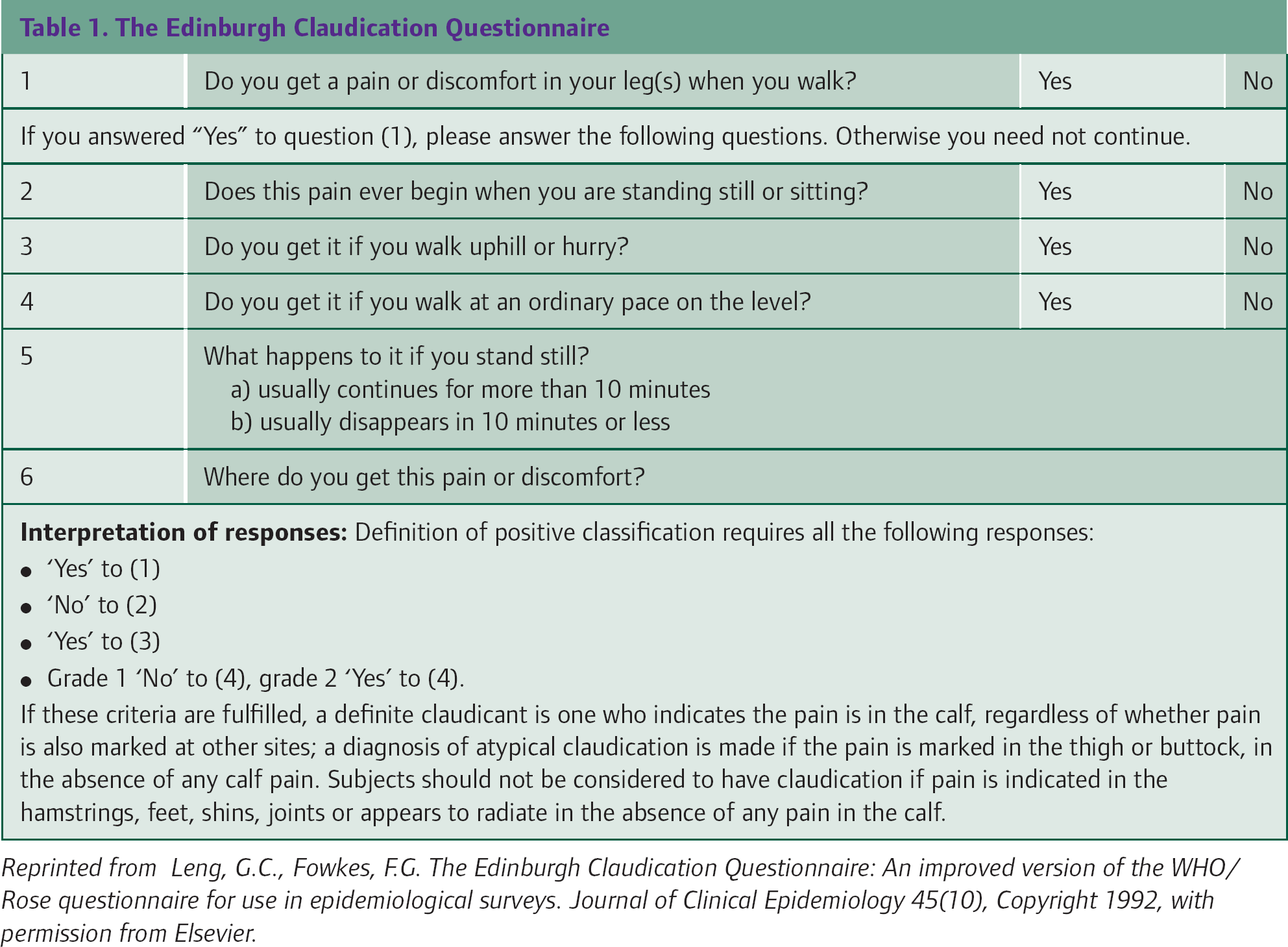
Patients who present to primary care with suspected lower limb PVD may exhibit an array of symptoms (Box 1), ranging from no discernible symptoms to critical limb ischaemia. The exact nature of the clinical presentation will depend upon the duration and severity of the disease. A useful screening tool for use in primary care is the Edinburgh Claudication Questionnaire (Table 1).
The Edinburgh Claudication Questionnaire
Features of intermittent claudication
No pain at rest or for the first few steps A relatively constant walking distance Relief on standing for 1–3 minutes Recurrence of pain on walking a similar distance Worse on walking quickly or uphill
Atherosclerosis often leads to stenosis or occlusion of one or more arterial segments supplying the lower limb. Intermittent claudication may be the only complaint a patient presents to their GP with; if the stenosis or occlusion occurs specifically at the aortic bifurcation or in the superficial femoral artery (SFA) at the adductor canal, they may have no other symptoms. However, in incidences of Leriche's syndrome, disease at the aortic bifurcation may result in patients presenting with intermittent claudication and erectile dysfunction.
In more diffuse disease, involving the proximal and distal arteries, or if the stenosis or occlusion is in a critical location where the collaterals are poor (e.g. the geniculate collaterals), patients may present with a plethora of problems, such as critical limb ischaemia with rest pain, ulceration or gangrene involving the toes or forefoot. The Trans Atlantic Inter-Society Consensus (TASC) defines critical limb ischaemia as the occurrence of ischaemic rest pain, which requires regular analgesia that persists for more than 2 weeks and/or ulceration or gangrene of the foot and toes that is attributable to objectively proven arterial occlusive disease, i.e. an ankle systolic pressure of less than 50 mmHg.
GPs must differentiate between patients who are at risk of limb loss in the absence of intervention and those who can be managed conservatively in primary care. Take a comprehensive history focusing on the cardiovascular system and its major risk factors. Document any history of stroke, myocardial infarction, angina, shortness of breath or erectile dysfunction along with the patient's claudication distance. Differentiate between patients presenting with ischaemic leg pain and those who have leg pain of an alternative aetiology (Box 2).
In many incidences, patients will present with multiple pathologies or an atypical history, and other symptoms may be impacting more significantly on their quality of life. Specific health assessment tools that are tailored for the use in patients with intermittent claudication may be appropriate for determining their health status (e.g. the Vascular Quality of Life Questionnaire). The Fontaine classification is a tool that can be used to grade patients on a symptomatic basis (Box 3).
Examination
In all patients with suspected PVD, perform a full cardiovascular examination. In addition, always examine the patient's abdomen for a pulsatile mass that may indicate the presence of an abdominal aortic aneurysm.
Look
Note the general appearance of the lower limbs. Specific points to look for include skin appearance, hair growth (this is scarce in patients with PVD) and evidence of poor tissue healing, particularly on the foot and lower leg. Look carefully at the micro-circulation of the foot. A foot that is erythematous compared to the other and associated with cyanosis demonstrates vasodilatation of the micro-circulation due to tissue ischaemia. A hyperaemic appearance should not mislead the clinician into thinking that the foot is well perfused. Look for evidence of venous guttering in the veins of the foot.
Differential diagnosis for lower limb pain
Fontaine classification

Check the toe nails as there is an increased risk of fungal infections in patients with PVD. It is also very important to examine between all the toes for signs of ulceration and infection, particularly in diabetics.
Feel
All pulses should be palpated and documented (Table 2). However, the presence of good foot pulses does not exclude PVD and patients with a classic history of claudication will require further investigation even if foot pulses are present. Any arterial bruits should be recorded.
Location of the pulses of the lower limbs

Test
Perform Buerger's test if you suspect PVD. Examine the feet with the legs elevated to at least 45°. Pallor is rapid if there is poor arterial supply. Then place the feet hanging dependent at 90° over the edge of the bed. Cyanosis occurs if the arterial supply is impaired. An adjunct to the Buerger's test, the ischaemic angle is the level at which a pedal Doppler signal disappears on elevation. This technique is only useful in severe ischaemia as it is not possible to raise the foot high enough for the Doppler signal to disappear if the leg has normal arterial pressures.
Arrange for the ABPI to be measured (Fig. 3). Using a sphygmomanometer and a hand-held Doppler, the ABPI is the ratio of the ankle to brachial systolic pressure. The systolic pressure in the ankle is normally greater than the brachial pressure, with an ABPI of 1–1.2. An ABPI of less than 0.9 is suggestive of arterial disease and an ABPI of less than 0.3 is associated with critical limb ischaemia (Box 4). A falsely high ankle arterial pressure may be measured if the calf arteries are calcified and incompressible, as in patients with diabetes. Listening to the Doppler signal over the artery can also give valuable information. Damped monophasic flow suggests vascular disease.

ABPI measurement.
Significance of ABPI results

Further primary care investigations
Primary care investigations should aim to identify cardiovascular risk factors in patients with suspected PVD. Blood tests are summarized in Box 5. Consider an electrocardiogram (ECG) as 60% of patients with intermittent claudication have ECG evidence of pre-existing ischaemic heart disease. Consider referral for an abdominal ultrasound if there is any suggestion of an abdominal aortic aneurysm on examination.
Blood tests to consider in the initial investigation of PVD

Primary care management
Secondary prevention
Patients with claudication have a 3-fold increase in risk of death from myocardial infarction or stroke. Advise patients to stop smoking and lose weight. Ensure optimum treatment of hypertension, lipids and diabetes.
Current guidance for secondary prevention of cardiovascular disease is to start a statin immediately regardless of initial cholesterol levels (National Institute for Health and Clinical Excellence (NICE), 2008). Start simvastatin 40 mg each night. Aim to reduce total cholesterol by 25% or to less than 4 mmol/l, whichever is the lower value and to decrease low-density lipoprotein (LDL) cholesterol by 30% or to less than 2.0 mmol/l, whichever is the lower value. Lowering total cholesterol and LDL cholesterol by 25% reduces cardiovascular risk by about a quarter.
Aspirin also decreases risk of cardiovascular events and is recommended at a dose of 75 mg daily for all those with symptomatic PVD. Clopidogrel is an expensive alternative for those who are intolerant of aspirin or may be used in combination with aspirin for those who continue to have cardiovascular events on aspirin alone. Antiplatelet agents reduce vascular death by around 25%.
Evidence from the Heart Outcomes Prevention Evaluation (HOPE) study (Yusuf et al., 2000) also suggests that the ACE inhibitor, ramipril, may reduce cardiovascular mortality and morbidity by about 25% for patients with PVD. This effect is greater than the effect that would be predicted by lowering of blood pressure alone.
Exercise
Training for 6 months or longer by regularly walking as far as possible before being stopped by pain increases pain-free and maximum walking distances (Gardner and Poehlman, 1995). This compares favourably with outcomes from angioplasty.
Vasodilators
For patients not being referred to secondary care, treatment with vasodilating agents may be appropriate. Naftidrofuryl has vasodilator effects, leading to improved tissue oxygenation, increased adenosine triphosphate levels and reduced lactic acid. It may increase walking distance but it is unclear whether treatment with this agent influences long-term outcome. Reassess after 3–6 months and discontinue treatment if there is no improvement.
Cilostazol is thought to have antiplatelet and vasodilator effects. Its mechanism of action is via the inhibition of phosphodiesterase III and it increases the level of cyclic adenosine monophosphate, promoting vasodilation. The proliferative effects of a range of pro-atherogenic growth factors are also reduced. Cilostazol has been demonstrated to increase the mean walking distance and quality of life in patients with intermittent claudication. It should only be used for patients without peripheral tissue necrosis who do not have pain at rest.
Referral to secondary care
Patients with acute ischaemia of a limb should be admitted as a vascular surgical emergency (Box 6). Consider urgent referral or admission for patients with critical limb ischaemia (Box 6).
Limb threatening signs and symptoms
Rest pain Ulceration Gangrene ABPI less than 0.5
Pain Pallor Pulseless Paraesthesia Paralysis Perishingly cold
In addition, the Scottish Intercollegiate Guidelines Network (SIGN, 2006) recommends that patients with suspected peripheral arterial disease should be referred to secondary care if
the primary care team is not confident of making the diagnosis, lacks the resources necessary to institute and monitor best medical treatment or is concerned that the symptoms may have an unusual cause risk factors are unable to be managed to recommended targets the patient has symptoms which limit lifestyle and objective signs of arterial disease (clinical signs and low ABPI)
SIGN (2006) also recommends that young and otherwise healthy adults presenting prematurely with claudication should be referred to exclude entrapment syndromes and other rare disorders.
Secondary care investigations
Toe pressures
When calf arteries are incompressible or there is severe arterial disease, toe pressures and the toe brachial index (toe systolic pressure divided by brachial systolic pressure) may be used as an alternative to ABPI. A special digital blood pressure cuff to fit around the great toe is required, so it is not usually possible to measure toe pressures in primary care. A toe:brachial index of 0.7 or greater is normal. A toe brachial index of less than 0.3 is often indicative of critical ischaemia. A toe pressure of greater than 30 mmHg may be an indicator of healing potential in foot ulcers.
Exercise testing
If Doppler ankle pressures are normal but the history is suspicious of claudication, repeating ABPI after exercise increases the sensitivity of the test. The demands of exercise result in an increase in the blood flow to the tissues. If significant arterial disease is present, this results in a fall in the ankle pressures. However, if symptoms are due to an alternative cause, the pressure remains stable.
Imaging
Duplex ultrasonography is the most commonly used secondary care investigation for PVD. It can be used to locate sites of disease and also shows the degree of stenosis and length of any occlusion. Magnetic resonance imaging (MRI) and computed tomography (CT) angiography are also being increasingly used in the UK. The gold standard investigation remains digital subtraction angiography (Fig. 4), but use of this investigation is now usually limited to preoperative planning.

Digital subtraction catheter angiography.
Surgical management of stable claudication
In addition to the lifestyle, secondary prevention and drug treatment options described under primary care management, vascular intervention for stable claudication is rarely required, although it may be considered when patients' symptoms are having significant negative impact on their quality of life.
Endovascular management
Angioplasty is an option in patients with short stenosis in either the common or the external iliac arteries of less than 3 cm in length. The primary success of short SFA stenoses is 90–100%, and 60–80% are patent 1–2 years later. However, outcomes following balloon angioplasty are not as good as optimal medical management (Gardner and Poehlman, 1995). Angioplasty of the popliteal vessels carries an increased risk and is not usually performed for patients with intermittent claudication.
Surgical management of claudication
There is little evidence to support bypass grafting for patients with intermittent claudication. In general, the risks of surgery (most notably from other cardiovascular events) outweigh the benefits. Therefore, bypass grafting is not usually considered unless the patient is verging on rest pain and becoming housebound and angioplasty/stenting cannot be performed.
Critical limb ischaemia
Ischaemic rest pain requires urgent referral to a specialist vascular unit. In patients suffering from critical limb ischaemia, the primary goal of treatment is limb salvage. Limb salvage is achieved by restoring adequate arterial foot perfusion. The first-line treatment for critical limb ischaemia is percutaneous balloon angioplasty. Endovascular stents can be used for significant stenoses above the inguinal ligament. If angioplasty is not possible or unsuccessful, bypass grafting should be considered. Ideally, the graft used is the patients' own saphenous or arm vein. If vein harvesting is not possible, synthetic grafts are used. Five year limb salvage rates are up to 80%.
About 10% of patients with critical limb ischaemia require amputation (usually below knee). This is more likely if the patient continues to smoke. In patients for whom limb salvage measures are impossible or have failed and if amputation is not feasible, palliative care should be instituted.
Primary care management post-hospital discharge
As for patients who have ischaemic heart disease, patients with PVD should be routinely monitored post-discharge at intervals in primary care. This provides the opportunity to
Determine if there is improvement or deterioration in the patients' clinical condition Identify if further intervention is required Monitor risk factor modification for secondary prevention Monitor compliance with secondary prevention medication
For patients who have had surgery, there is a graft surveillance programme, which monitors post-operative status. There is a high risk of graft stenosis, with 25–30% of patients requiring further interventions.
Prognosis
Coronary heart disease is the major cause of death of people with PVD. Those with PVD have a three to four times increased risk of death from stroke or myocardial infarction than age and sex matched controls and 30% will die from these causes in any 5 year period.
Over a 5 year period, of those with intermittent claudication, about half will have symptoms that improve over time. Of those who do not improve, half remain stable and half have worsening symptoms. Eventually, about 15% of those with intermittent claudication will progress to critical limb ischaemia needing intervention.
Key points
The history is the cornerstone to arriving at a diagnosis of PVD Intermittent claudication is associated with an increased risk of cardiovascular events such as stroke and myocardial infarction A comprehensive cardiovascular examination should be performed paying particular attention to peripheral pulses; the abdomen should be palpated to detect the presence of an abdominal aortic aneurysm In patients with intermittent claudication, the primary role of the GP is to assess and modify risk factors for atherosclerosis It is essential to identify patients with critical limb ischaemia and refer urgently to secondary care for further management, i.e. revascularization