
Abstract
Gout is an inflammatory arthropathy secondary to the deposition of monosodium urate crystals in synovial fluid. There are four well-described stages of the condition: asymptomatic hyperuricemia, acute gout attack, intercritical stage and chronic tophaceous gout. Management of the condition is dependent on the individual patient and the stage of disease. The principles are to terminate the acute event, prevent recurrent attacks and disease progression.
The GP curriculum and gout
Manage primary contact with patients who have a musculoskeletal problem Explain the aetiology and natural history of common and important musculoskeletal conditions Describe the indications for referral within a suitable time frame to the most appropriate health care practitioner Describe the epidemiology of musculoskeletal disorders at all ages and apply this when developing a differential diagnosis Distinguish inflammatory from non-inflammatory conditions Describe when blood tests and imaging methods are required for diagnosis of musculoskeletal problems Describe problems that can be caused by the treatment of musculoskeletal disorders and explain primary and secondary prevention of these Describe the key national guidelines that influence health care provision for musculoskeletal problems, e.g. the National Institute for Health and Clinical Excellence (NICE) guidelines, Royal College of General Practitioners (RCGP) low back pain guidelines, Scottish Intercollegiate Guidelines Network (SIGN) guidelines
Clinical presentation
Currie (1979) showed the annual UK incidence and prevalence of gout to be 0.3 and 2.6 per 1000, respectively. The classic presentation is of an acutely red, swollen and painful joint. The most commonly affected joint is the first metatarsophalangeal but during its course, gout can affect a wide range as shown in Table 1. At first presentation, the majority of cases are monoarticular but recurrent attacks become more frequent and involve more joints. On inspection, the joint is erythematous, warm, shiny and swollen. The joint line will be tender and a restricted range of movement is evident.
Distribution of acute and chronic joint involvement in patients with recurrent acute gout

History
A focused history should be taken with patients questioned on previous trauma, episodes of arthritis or surgery to the affected joint. Relevant past medical history includes chronic renal impairment, hypertension, other rheumatology conditions and patient drug history. If this is a recurrent attack, then prior investigations and success of past management may also aid diagnosis. Risk factors for gout should be assessed and any systemic symptoms noted.
Differential diagnoses are shown in Box 1. Factors that should raise the suspicion of an alternative diagnosis include polyarticular symptoms, atypical joint involvement or other associated symptoms, such as diarrhoea or dysuria. Clinical examination may not be overly helpful in differentiating the diagnoses but the presence of tophi, fever and scars are potential clues to the cause.
Differential diagnosis of gout.
Pseudogout Septic arthritis Reactive arthritis Rheumatoid arthritis
Pathophysiology
Gout is an inflammatory arthropathy secondary to hyperuricemia and the deposition of monosodium urate crystals into connective tissue, cartilage and synovial fluid. Hyperuricemia results from an imbalance in uric acid metabolism, which is a by-product of purine nucleotide catabolism and is eliminated mainly by the kidneys. If no direct cause for the hyperuricemia is found, then gout can be termed ‘primary’ and is associated with males, obesity, alcohol abuse, hypertension, renal dysfunction and hypertriglyceridemia. In contrast, a condition or medication, which results in hyperuricemia and symptoms of gout is classified as ‘secondary’. Medications can cause gout by either increasing production or decreasing excretion of urate. The most common examples of such medication are given in Box 2.
Drug causes of secondary gout.
Diuretic therapy Low-dose aspirin Pyrazinamide Nicotinic acid Losartan Cyclosporine
The condition is described in four stages: asymptomatic hyperuricemia, acute gout, intercritical gout and chronic tophaceous gout. Disease progression is poorly understood and the time period from asymptomatic hyperuricemia to chronic tophaceous gout varies between individuals.
Asymptomatic hyperuricaemia
Patients with asymptomatic hyperuricaemia experience no arthritic symptoms but are found to have a high uric acid level, which is associated with obesity, alcohol consumption and hypertension. Zhu, Pandya and Choi (2011) showed that the US A prevalence of hyperuricaemia is 21.2% in males and 21.6% in females.
Acute gout
Acute episodes of gout last days to weeks depending on the treatment obtained. Subsequent attacks typically become more frequent, with symptoms persisting for longer and eventually failing to resolve. At the first presentation, 85–90% of cases are monoarticular with the metatarsophalangeal joint most commonly affected (Fig. 1). The susceptibility of the foot has no known explanation but as the condition progresses, attacks spread to the upper limb and become polyarticular.
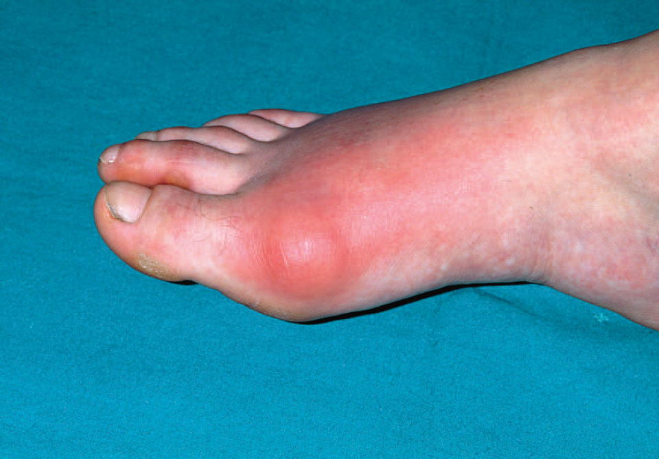
Acutely inflamed gouty joint.
Intercritical gout
The length and number of asymptomatic periods between attacks varies between patients but typically decrease as the condition progresses. During this stage, the deposition of monosodium urate crystal continues, and it can be used as an opportunity to identify crystals in the synovial fluid and instigate preventive treatment.
Chronic tophaceous gout
Typically, after 10 or more years of intermittent gout attacks, the disease can become chronic. This stage is defined by a lack of pain-free intercritical periods with joints found to be persistently uncomfortable and swollen. The signs of chronic gout tend to be more obvious in hands than feet, with clinical signs including swollen joints, articular or periarticular tophi and swan neck or boutonniere deformities. Tophaceous deposits are precipitates of urate and commonly found on the pinna, elbows, Achilles tendon, finger joints (Fig. 2) and bursae. These deposits can lead to bone erosion and occasionally the skin over these areas breaks down discharging a paste or pus-like material containing uric acid crystals.

Chronic gout with tophi.
Pseudogout
Pseudogout is an acute inflammatory arthropathy with clinical symptoms similar to gout. Typically, the knee joint is affected with the wrist, metacarpophalangeal, hips and shoulders also commonly involved. The condition occurs secondary to calcium pyrophosphate dehydrate crystal deposition and for this reason, serum urate levels may be persistently normal and the use of urate-lowering agents unsuccessful.
The condition can only definitively be differentiated from gout by aspiration and analysis of synovial fluid but this only becomes necessary if the diagnosis is unclear or if a patient has recurrent episodes with no relief from urate-lowering agents. Most acute attacks can be managed successfully in primary care but a confusing clinical picture or a polyarticular presentation should prompt rheumatology referral.
Investigation
The demonstration of serum hyperuricaemia in the presence of acute or chronic joint symptoms is insufficient to diagnose gout. The two can be simultaneously present in a variety of conditions and Snaith and Coomes (1977) showed that a third of patients suffering an acute attack are actually normouricaemic. However, hyperuricaemia remains the most common biochemical abnormality found and should be measured at presentation as high levels support the diagnosis. No other blood tests are specific for an acute gout attack with inflammatory markers, such as the white cell count, erythrocyte sedimentation rate and C-reactive protein likely to be raised in differentials. If these alternative diagnoses are suspected, then these additional tests become necessary.
Standard radiographs contribute little to the diagnosis of acute gout but in the chronic stage, they may show joint erosion with well-demarcated round- or oval-shaped deficits with sclerotic margins on the radiographs.
For a definitive diagnosis, the clinician must demonstrate the presence of monosodium urate crystals in synovial fluid, tophaceous material or synovial tissue, which requires fluid or tissue sampling. The aspirated joint fluid is usually turbid and microscopy reveals a large number of polymorphs with the presence of crystals. The use of a polarizing microscope allows differentiation of monosodium urate crystals from calcium pyrophosphate crystals, which are present in the diagnosis of pseudogout.
However, the routine aspiration of affected joints is not advised in primary care and sampling is reserved for cases when diagnosis remains unclear from the history or distribution of joints involved. More commonly, a presumptive diagnosis can be made in the presence of hyperuricaemia, acute monoarticular arthritis and resolution of symptoms with colchicine. The American College of Rheumatology proposed a series of alternative criteria for the diagnosis of gout, which are shown in Box 3.
American College of Rheumatology criteria for diagnosis of gout.
Presence of urate crystals in joint fluid Characteristic urate crystals in a tophus Six of the following 12 clinical laboratory and radiographic phenomena:
More than one attack of acute arthritis Maximal inflammation developed within 1 day Attack of monoarticular arthritis Joint redness observed First metatarsophalangeal joint painful or swollen Unilateral attack involving first metatarsophalangeal joint Unilateral attack involving tarsal joint Suspected tophus Hyperuricemia Asymmetric swelling within the joint (radiograph) Subcortical cysts without erosions (radiograph) Negative culture of joint fluid for microorganisms during attack of joint inflammation
The presentation of gout in children or premenopausal women raises the suspicion of a secondary cause for the hyperuricaemia and these cases may warrant further investigation. The first step is to measure uric acid levels, renal function and prove the presence of monosodium urate crystals in the affected joint. The suggestion of gout on initial investigation should prompt referral to secondary care for measurement of 24 hour urine uric acid excretion with high urate clearance, raising the possibility of an inborn error of purine metabolism.
Treatment
The key to understanding gout management is to separate the issues of reducing inflammation in an acute attack and managing the precipitating hyperuricaemia in the long term. Incorrect implementation of these principles will result in unnecessary recurrent attacks and progression of the condition. The therapeutic goals are highlighted in Box 4 and the British Society for Rheumatology (Jordan et al., 2007) guidelines for management in Box 5. The Arthritis Research UK and UK Gout Society websites provide an excellent source of information for patients to learn about the diagnosis and treatment of gout. Links to these sites are given in the references and further information section of this article.
Therapeutic goals.
Terminate acute attacks
Non-steroidal anti-inflammatory agents colchicine or corticosteroids Prevent recurrence of acute gouty arthritis
Correct risk factors Colchicine Permanent urate-lowering drugs Prevent or reverse complications of the disease
BSR and British Health Professionals in Rheumatology Guideline for the Management of Gout (2007).
Affected joints should be rested and analgesic and anti-inflammatory drug therapy commenced immediately and continued for 1–2 weeks Fast acting oral NSAIDS at maximum dose are drugs of choice if no contraindications In patients with increased risk of peptic ulcers, bleeds or perforations, co-prescription of gastroprotective agents should follow standard guidelines for the use of NSAIDs and coxibs Colchicine can be an effective alternative but it is slower to work the NSAIDS. In order to diminish the risks of adverse effects, it should be used in doses of 0.5 mg two to four times daily. Allopurinol should not be commenced during an acute attack but in patients already established on allopurinol, it should be continued and the acute attack should be treated conventionally Opioid analgesics can be used as adjuncts Intra-articular corticosteroids are highly effective in acute gouty monoarthritis and intra-articular, oral, intramuscular or intravenous corticosteroids can be effective in patients refractory to other treatment If diuretics are being used for hypertension, an alternative antihypertensive agent should be considered, but in patients with heart failure, diuretic therapy should not be discontinued
Managing an acute attack
Acute gout can be treated with non-steroidal anti-inflammatories (NSAIDs), colchicine or corticosteroids but timely recognition and instigation of treatment are more important than which of the three treatments are chosen. Prompt action is related to early termination of the attack and treatment should continue for 2–3 days after resolution of symptoms. If initial treatment fails and the patient represents with ongoing symptoms, then the diagnosis needs to be reconsidered. If gout remains the most likely cause of symptoms, treatment should be continued and the patient referred as an outpatient to a rheumatologist. NSAIDS are most commonly used with Arnold, Preston and Buchanan (1998) showing all NSAIDs are effective in treatment of acute gout with initial high-dose therapy recommended. Contraindications include patients with renal disease or peptic ulceration.
Colchicine has an anti-inflammatory action but is associated with gastrointestinal side effects and rarely with bone marrow suppression and myoneuropathy. The British National Formulary (2011) recommends 500 mcg two to four times daily until symptoms are relieved with a maximum of 6 mg per course.
Corticosteroids are considered if NSAIDs and colchicine are contraindicated. The oral dose is 20–40 mg that is tapered over 1–2 weeks following resolution of symptoms. Intra-articular administration provides an alternative administration method and has been shown to be particularly effective.
Preventing further attacks
A number of treatment options exist to prevent further attacks of gout:
Modification of risk factors or Drug therapy with colchicine, xanthine oxidase inhibitors or uricosuric agents
Modifying risk factors
Hyperuricemia is directly related to obesity, high alcohol intake, hypertension and dietary intake of purine. Tackling each of these factors if present will have an effect on serum uric acid levels and patients may be able to lower their urate levels to a point where no further attacks occur and they can avoid taking long-term tablets. The BSR recommends avoiding crash diets, encouraging fluid intake, restricting alcohol consumption and avoiding high purine foods, such as red meat, liver, kidneys, shellfish and yeast extracts. Box 6 summarizes dietary advice to give to patients.
Prophylactic drug therapy
Hyperuricemia may also be corrected by drug therapy with the goal of treatment to lower plasma uric acid levels to less than 0.36 mmol/l. Treatment is indicated after the second or third acute attack or in tophaceous gout. However, there is no evidence supporting the use of urate-lowering medications in asymptomatic hyperuricemia. It is paramount that at the onset, patients are educated that treatment may be for life and that cessation of medications may result in acute changes of uric acid levels with subsequent acute flares and disease progression.
Dietary advice for gout sufferers, who should avoid

It is important that anti-hyperuricaemic agents should not be started, stopped or altered during an acute attack as fluctuations in urate level can precipitate an attack or worsen the ongoing inflammatory process. Medications that affect urate levels should be delayed until more than 2 weeks after the complete resolution of all signs of inflammation.
Colchicine
Colchicine can also be used for its prophylactic effect with a dose of 0.5 mg twice daily shown to be effective. Colchicine acts by reducing the functionality of neutrophils, mast cells and macrophages with resultant reduction in inflammation. It does not have an effect on the serum urate metabolism and in the long term should be used with a serum urate-lowering agent. In combination, it should be continued until the patient has been normouricemic and had no acute attacks for 1–2 years.
Xanthine oxidase inhibitors
Xanthine oxidase is an enzyme involved in the production of uric acid. An inhibitor, such as allopurinol, functions by decreasing production. An initial dose of 100 mg daily should be increased according to response seen in the serum urate level and usually 300 mg daily is required to achieve a level of less than 0.36 mmol/l. The dose should be adjusted according to renal function with 200 mg used if glomerular filtration rate (GFR) is 50–60 ml/minute and 100 mg when GFR is 20–30 ml/minute. About 1 in 20 patients are unable to tolerate allopurinol due to minor gastrointestinal disturbance, bone marrow suppression, an erythematous skin rash or acute interstitial nephritis. Practical points to consider when prescribing are listed in Box 7.
Practical prescribing points for allopurinol.
Initiate after resolution of acute attack Increase dose slowly over 1 month Initiate with concurrent colchicine or NSAID prophylaxis Treatment is long term
Uricosuric agents
The most common uricosuric agents used are probenecid and sulfinpyrazone. They are competitive agonists for urate reabsorption in the renal tubules and result in an increased renal excretion of uric acid. They are contraindicated in renal impairment, patients with a history of previous renal calculi or in patients with poor urine output. Practical prescribing points are given in Box 8.
Practical prescribing points for uricosuric agents.
Initiate with concurrent colchicine or NSAID prophylaxis Commence an initial low dose Ensure treatment is long term
Referral
Gout is a commonly seen and managed condition in primary care but occasionally, patients need to be referred to secondary care. Indications for referral to secondary care are given in Box 9. Surgical intervention is rarely required but if the tophi press on important neurovascular structures or have become infected, then surgical removal or debridement may become necessary.
When to refer?
Uncertain diagnosis Paediatric cases Difficulty controlling attacks Difficulty maintaining low urate levels Contraindication to commonly used preventative medications
Follow-up
Patients should be recalled at around 6 weeks after an acute attack to discuss outstanding issues. Their urate level should be reviewed and lifestyle advice can be given to attempt to reduce it. Consideration can be given to urate-lowering agents if the patient has suffered more than one attack or the condition is chronic. Advice can also be given at this stage for a plan of action in the event of further attacks with advanced prescriptions, allowing patients to commence prompt treatment and avoid prolonged attacks. The opportunity should be taken to investigate the at-risk patient for diabetes, hypertension and cardiovascular risk before offering lifestyle advice in the form of dieting and exercise.
Key points
There are four stages of gout: asymptomatic hyperuricemia, acute gout attack, intercritical gout and chronic tophaceous gout Diagnosis can be confirmed by identifying urate crystals in synovial fluid when appropriate Use NSAIDs, colchicine and corticosteroids to terminate acute attacks For recurrent attacks, a urate-lowering agent should be prescribed; urate-lowering therapy should be lifelong and patient education is key Low-dose colchicine or NSAIDs should be used in prophylactic manner prior to initiating urate-lowering therapy
Footnotes
Acknowledgements
We would like to thank Dr Paul Wainman for his help with the writing of this article under the InnovAiT ‘buddy’ scheme.