
Abstract
Background
Increased aerobic exercise capacity appears to reduce both all-cause mortality and cardiovascular disease mortality. Physical exercise to improve peak oxygen uptake (VO2peak) is thus strongly recommended, however evidence regarding the most efficient training intensity for patients with coronary artery disease (CAD) is still lacking. The purpose of this randomized study was therefore to assess the effects of high intensity aerobic interval exercise compared to moderate intensity exercise, representing the same total training load, for increasing VO2peak in stable CAD-patients.
Methods
Twenty-one stable CAD-patients were randomized to supervised treadmill walking at either high intensity (80–90% of VO2peak) or moderate intensity (50–60% of VO2peak) three times a week for 10 weeks.
Results
After training VO2peak increased by 17.9% (P = 0.012) in the high intensity group and 7.9% (P = 0.038) in the moderate intensity group. The training-induced adaptation was significantly higher in the high intensity group (P = 0.011).
Conclusions
High intensity aerobic interval exercise is superior to moderate exercise for increasing VO2peak in stable CAD-patients. As VO2peak seems to reflect a continuum between health and cardiovascular disease and death, the present data may be useful in designing effective training programmes for improved health in the future.
Introduction
Higher levels of physical activity and fitness appear to reduce all-cause mortality and cardiovascular disease (CVD) mortality [1–6]. Physical exercise is thus strongly recommended in both primary and secondary prevention of CVD [7–11]. Peak aerobic exercise capacity is found to be the strongest independent predictor of mortality compared with other established risk factors among both healthy individuals and those with CVD [12]. More specific, peak aerobic exercise capacity directly measured as peak oxygen uptake (VO2peak) was recently found to be the single best predictor of both cardiac and all-cause deaths among patients with established CVD [13]. These findings indicate that exercise-induced gain in VO2peak should thus make a difference not only in functional capacity but also in survival prospects.
For health promotions, patients with coronary artery disease (CAD) are recommended to regularly exercise at intensities ranging from 40–90% of VO2peak [7–9]. However, aerobic exercise training programmes are most often carried out at low-to-moderate intensities [14]. Several studies have shown a significant inverse relationship between participation in rehabilitation programmes involving exercise and reduced progression of CAD [15–19], but data regarding effects of exercise intensity are scarce. Both walking and vigorous exercise were found to be equally effective in increasing aerobic capacity and reduce cardiovascular risks [20, 21]. However, two large cohort studies found that higher intensity of physical activity was related to reduced risk, as reflected by an inverse association between exercise intensity and coronary heart disease incidence in men [22, 23].
As aerobic exercise capacity seems to reflect a continuum between health and cardiovascular disease and death, it is important to design effective programmes for exercise-induced gains of VO2peak for patients with cardiovascular risk. Previous studies investigating the influence of different exercise intensities for improvements of VO2peak have met criticism because of insufficient equation between intensity and duration [14]. Thus, consensus about the most efficient exercise intensity to increase VO2peak is still lacking. The aim of the present study was therefore to assess the effects of two different aerobic exercise programmes of treadmill walking where training intensity and duration were carefully matched. The hypothesis was that interval exercise at high intensity (80–90% of VO2peak) is superior to continuous exercise at moderate intensity (50–60% of VO2peak), representing the same total training load, for increasing aerobic capacity in stable CAD-patients.
Methods
Subjects
Twenty-one eligible participants were enrolled in the study in August 2002 and the study was accomplished in December 2002. A flow diagram of the progress of the study is presented in Figure 1. All patients had undergone a medical investigation for CAD at St. Olavs University Hospital of Trondheim, within one year prior to the study. The patients were recruited by mail and resided within 40 km of the hospital. Physical characteristics of the subjects at inclusion are presented in Table 1. Inclusion criteria were angiographically documented CAD in at least one major epicardial vessel. In addition, subjects had clinical evidence of CAD in the form of previous myocardial infarction, significant stenosis treated with coronary artery bypass surgery (CABG) or percutaneous coronary intervention (PCI), or ischaemia in exercise-electrocardiogram (ECG). Exclusion criteria were left main coronary artery disease, unstable angina pectoris, claudicatio intermittens, myocardial infarction within the last 3 months, CABG or PCI performed within the last 12 months, complex ventricular arrhythmias, left ventricular ejection fraction below 40%, orthopedic or neurological limitations to exercise, or regular exercise for the past 3 months. Medications received by the patients included beta-blockers (41%), antiplatelet agents (88%), statins (94%), angiotensin-converting enzyme-inhibitors (35%), calcium antagonists (6%), long-acting nitrates (12%), and diuretics (12%). Medication was not different between groups and no changes in medication status were made during the study period.
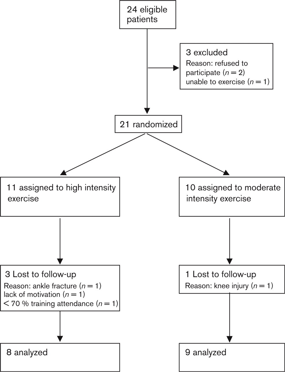
Flow diagram of the progress of the study.
Study protocol
The study was accomplished according to the Declaration of Helsinki. The regional committee for medical research ethics approved the study protocol. Written and informed consent was obtained from all subjects at the beginning of the study. Before measurements of VO2peak the subjects were informed about the test, and instructed to exercise to their maximum limit. A standard 12-lead ECG was recorded at rest and at the end of each work level, and patients were stopped if any indication for terminating testing according to current guidelines took place [9]. To familiarize with treadmill walking, the test started on a flat treadmill where participants learned to walk without grasping the handrails. As soon as they could walk properly, the speed and inclination was individually adjusted (3–6km·h–1 and 0–5%) for a 10-min warm-up. After the warm-up period a facemask was placed on the subject's face for metabolic measurements using Meta-Max II (Cortex, Leipzig, Germany). The VO2peak-test was performed using a ramp protocol where the speed was constant and the incline was increased with 2% every second minute until VO2peak was reached. Both before and after training, the intention of the individually adjusted start-level was to bring the subjects to VO2peak after approximately 8–12 min, as recommended by Froelicher and Myers [24]. All patients were walking during the two test occasions, except one person before training and four persons after training who had to run due to the limited inclination capacity of the treadmill. The average of the three highest 10-s measurements determined VO2peak. Heart rate was continuously recorded using a Polar Sport Tester (Polar Electro OY, Finland), and maximum attainable heart rate (HRpeak) was determined. After 1 week an instructor blinded for the values of the initial test carried out a second VO2peak-test on 10 of the subjects to ensure that learning to walk properly on the treadmill did not affect VO2peak measured during the initial test. The mean difference from test two to test one was only 0.011 ± 0.11l/min, or 0.46% of the two measurements.
Physical characteristics of the subjects at inclusion
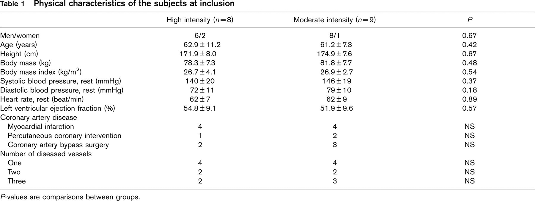
P-values are comparisons between groups.
Exercise training
The patients met for training three times per week for 10 weeks under supervision of an exercise physiologist. The patients were instructed not to add any leisure exercise during the study period. All training consisted of uphill treadmill walking. The high intensity aerobic exercise group carried out a 5-min warm-up period at an intensity corresponding to 50–60% of VO2peak (65–75% of HRpeak) before walking four intervals of 4 min at 80–90% of VO2peak (85–95% of HRpeak). The patients exercised at the lower intensity border for the first 2 weeks of the training period before increasing the intensity towards the upper border. Between the intervals 3 min of walking at 50–60% of VO2peak was conducted. The training session was terminated by a 3-min cool-down period at 50–60% of VO2peak [25]. This gave a total exercise time for the high intensity group of 33 min. To equate the total work performed by the two groups the following calculation was used: the average VO2peak for all subjects before training was 2.55 l/min. The high intensity group performed 4 × 4min exercise at 2.29l/min (90% of VO2peak) and 3 × 3min exercise at 1.53l/min (60% of VO2peak). The total VO2-time relationship for the high intensity group was then divided by the intensity for the moderate intensity group (50.49/1.53 = 33 min). Finally the warm-up and cool-down exercise time for the high intensity group (5 + 3 min) at 1.53 l/min was added to this value. The moderate intensity exercise training thus consisted of 41 min continuous exercise at an intensity of 50–60% of VO2peak, representing the same total training load as the high intensity aerobic exercise group. All subjects exercised using a heart rate monitoring device during every training session. The subjects could thus control their corresponding exercise heart rate relative to VO2peak, and were encouraged by the instructor to exercise as close to the upper intensity border as possible (after 2 weeks for the high intensity group). The speed and inclination of the treadmill was continually adjusted along as training adaptations occurred, to ensure that all training sessions were carried out at the desired heart rate throughout the 10-week training period. The BORG 6–20 scale [26] was used to measure the rate of perceived exertion after each training session. A compliance with the training programme of 70% was set as criteria for completing the study.
Randomization procedure
Subjects were randomly assigned to either high intensity exercise or moderate intensity exercise. The randomization code was developed using a computer random number generator to select random permuted blocks. The unit of Applied Clinical Research at the university carried out all randomization procedures to secure complete blinded randomization.
Statistical analysis
All values are expressed as mean ± standard deviation (SD). Non-parametric statistics were used due to the small groups and to avoid assumptions of normal distributions. Changes within each group were assessed using the Wilcoxon signed ranks test. Differences between the groups were calculated using the Mann-Whitney U-test. A two-tailed P<0.05 was accepted as statistically significant for all tests.
Metabolic data for the two groups before and after training
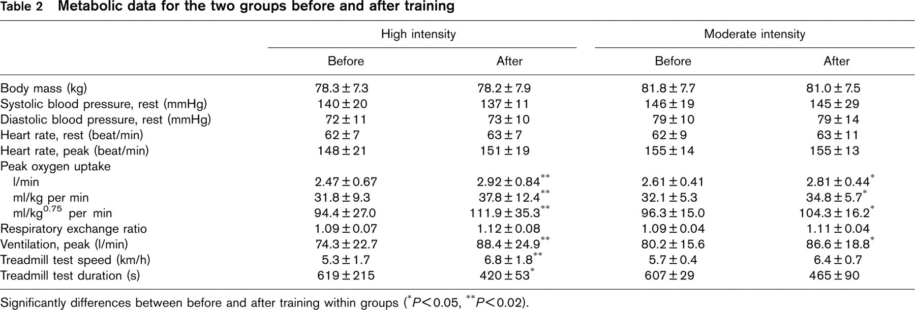
Significantly differences between before and after training within groups (∗P<0.05, ∗∗P<0.02).
Results
The VO2peak increased significantly between the before and after training within both the high intensity and the moderate intensity group (Table 2). The 17.9% improvement in the high intensity group was significantly greater compared to the 7.9% improvement in the moderate intensity group (Fig. 2). The high intensity group carried out 28.3 (range 25–30) of the prescribed 30 training sessions while the moderate intensity group carried out 25.4 (range 21–30) sessions (P = 0.074). The high intensity group also showed an improvement of 0.63% when VO2peak was divided by the number of training sessions attended, which was higher compared to the 0.29% improvement in the moderate intensity group (Fig. 3). There were no detectable changes in resting blood pressure, resting heart rate, or body mass in neither group after the training period. The BORG rate of perceived exertion after each training session was 14.4 for the high intensity group and 13.5 for the low intensity group (P = 0.093). There were no episodes of cardiac events during the study.
Discussion
The results of this randomized controlled study demonstrate that high intensity aerobic exercise is superior compared to moderate intensity exercise for increasing VO2peak in stable CAD-patients. Although VO2peak increased in both groups after 10 weeks of training, the improvement was significantly larger in the high intensity group. Because the two groups were equated with regard to the total amount of work performed, this study solely points out high aerobic intensity as a key factor for increasing aerobic capacity in this patient group. In view of the prognostic importance of increasing VO2peak for this patient group, high intensity exercise may be considered in future rehabilitation programmes.

Peak oxygen uptake (VO2peak) before and after aerobic exercise training. Significant differences within groups after training. Increment significantly larger in high intensity group.
The present study is one of few where CAD-patients are performing aerobic interval exercise at 80–90% of VO2peak throughout the whole training period, while continuous exercise around 50–60% of VO2peak is more frequently used. Since all subjects exercised with a heart rate monitoring device, the load of the treadmill could progressively be adjusted to keep the relative exercise intensity constant as training adaptations occurred. The improvement of 17.9% in the high intensity group compared to 7.9% in the moderate intensity group reflects the importance of intensity when determining the increase of VO2peak. Two earlier studies involving CAD-patients have employed aerobic interval exercise with elements of the same high intensity as in the present study, both with a tremendous increase of VO2peak [27, 28]. The results of these studies showed that 12 months exercise at an intensity of 50–95% of VO2peak carried out three to five times per week produced an improvement of 37–42%. The longer training period, along with the large dispersion in intensity and various numbers of training sessions per week, makes these results difficult to compare to the present study. These studies however demonstrate that high aerobic exercise intensity is associated with a large improvement of VO2peak.
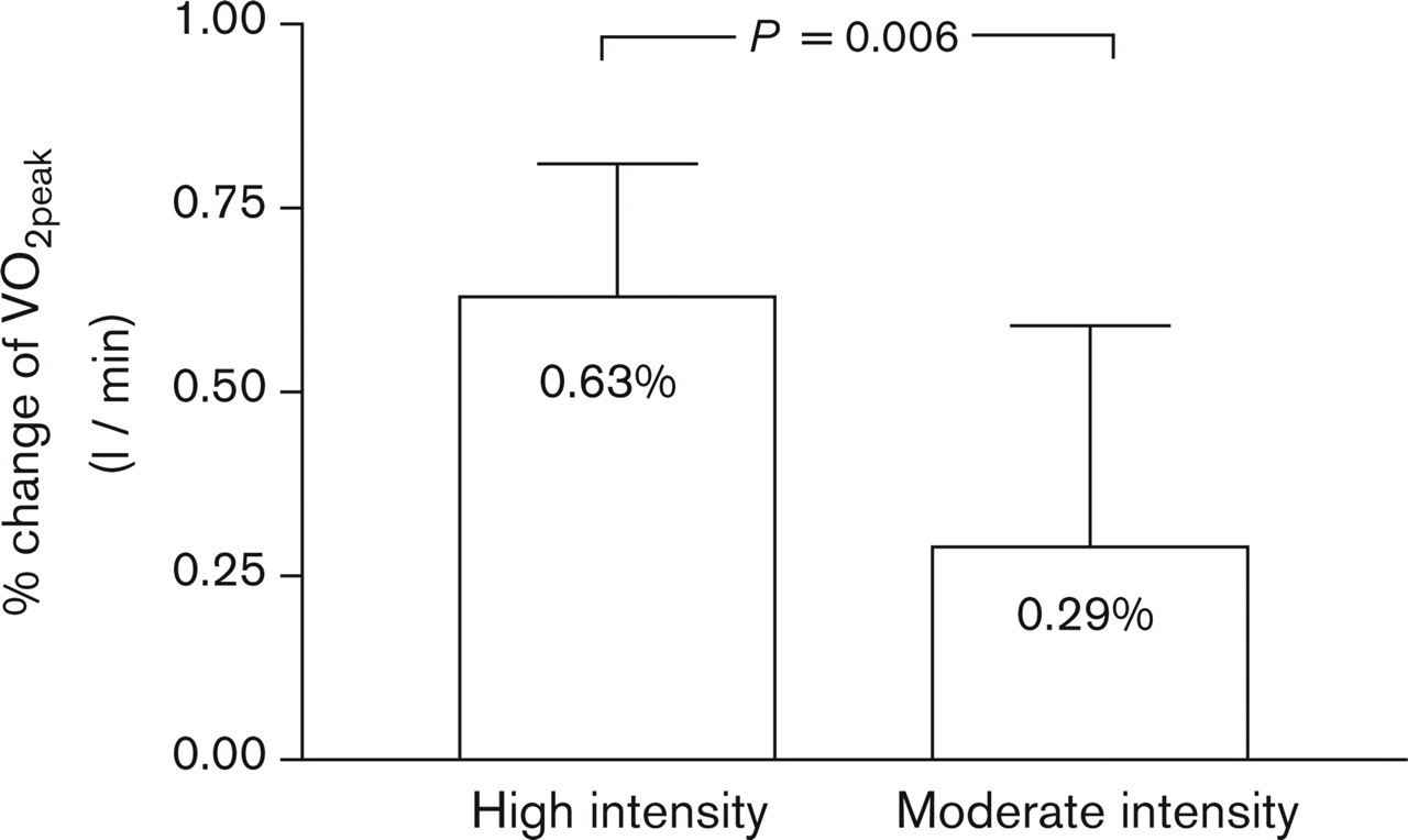
Percentage change of peak oxygen uptake (VO2peak) divided by the number of training session attended. High intensity group significantly different from moderate intensity group.
The main goal of the present study was to evaluate the effect of exercise when intensity is the only parameter being manipulated. The high intensity exercise was chosen to be aerobic interval exercise at 80–90% of VO2peak because this training method has been employed by our research group in healthy individuals, yielding great improvements of VO2peak in a relative short time period [25]. The moderate intensity exercise at 50–60% of VO2peak was selected because it is typically used in training studies involving CAD patients [14]. The effects of higher versus lower exercise intensities with regard to increasing VO2peak have been evaluated in earlier studies. Jensen et al. [29] had two groups of CAD patients training for 12 months, involving high-intensity exercise within the range of the present study. One hundred and eighty-six subjects with a documented CAD event within the previous 24 months were randomized to 45min of walking or jogging at either 85 or 50% of VO2peak. The results showed that VO2peak in the high intensity group increased by 13%, which was significantly higher compared to the 9% increase in the lower intensity group. Adachi et al. [30] compared 29 patients with previous myocardial infarction performing walking exercise at 70% of VO2peak with exercise at 55% of VO2peak over 8 weeks. The VO2peak increased by 17% in the high intensity group and by 9% in the moderate intensity group. These findings support our study with regard to the fact that higher intensity exercise being more suitable for increasing VO2peak compared to lower intensity exercise. Contrary to these studies, Blumenthal et al. [31] did not detect differences between moderate intensity (75% of VO2peak) and low intensity exercise (45% of VO2peak) after 12 weeks of training among 45 patients with myocardial infarction. The VO2peak increased by 11% within the high intensity group and 14% in the low intensity group, but the differences were not statistically significant. In sum, however, these studies indicate that high intensity exercise being more suitable for increasing VO2peak compared to moderate or low intensity. But, in each of these studies the two groups used the same exercise duration and the higher intensity group therefore performed a greater total amount of training. It is therefore questionable whether intensity or total amount of work determined the increase of VO2peak. The equation of exercise duration between the groups in the present study was emphasized to control whether intensity is a capital factor for increasing VO2peak.
The initial VO2peak of the patients in our study was 32.0 ml/kg per min, which is higher compared to the other studies evaluating exercise intensity in cardiac patients (18.7–25.3 ml/kg per min) [29–31]. Thus, the 17.9% improvement of VO2peak in the high intensity group is therefore considerable when calculating percentage improvement from such a higher baseline value. In fact, VO2peak increased by 0.63 and 0.29% per training session accomplished for the high- and moderate exercise group, respectively (Fig. 3). To ensure that exercise is conducted at the proper intensity, it is important that patients are exercising close to their maximal effort on the initial VO2peak-test. If not, the reported exercise intensities are likely overestimates of the actual ranges. Even if the peak values of respiratory exchange ratio were close to 1.10 in our study, indicating effort close to the maximal oxygen uptake (VO2max), subjects within both groups attained values below 1.10 both before and after training (Table 2). Together with the observation that only about half of the subjects in each group terminated the VO2peak-test because of a plateau of the O2 curve, this indicates that a true VO2max was not uniformly reached [32]. Hence, the term VO2peak was used instead of VO2max to describe exercise capacity throughout the study. The high intensity group carried out 4-min interval exercise at 80–90% of VO2peak before a relaxation phase was required due to dyspnea. This is in contrast to the study of Jensen et al. [29], where the high intensity group was able to exercise continuously at the same intensity for 45 min. Exercise at 85% of VO2max for a prolonged time period is described to be possible only for highly endurance-trained individuals, and for an untrained person the critical point may be at 50% of VO2max [32]. This may indicate that the individuals in the study by Jensen and colleagues were not able to exercise near their true maximal effort during the initial VO2peak-test, and consequently, the intensity reported may thus have been over-estimated. The researchers state that use of the Bruce protocol with relatively large and uneven work increments, made several subjects exceeding their anaerobic threshold in the first 3-min stage [29]. Work rate increments that are too rapid may result in reduced exercise capacity and it is suggested that individualized protocols with estimated test duration of 8–12 min are optimal [24]. Adachi et al. [30] carried out the VO2peak-test on bicycle ergometers, which are reported to produce VO2peak-values that are 6–25% lower compared to treadmill exercise [24]. Finally, in our study, myocardial infarction was present in only half of the subjects in each group. This may have increased the initial VO2peak compared to the studies of Adachi et al. [30] and Blumenthal et al. [31] investigating the effects of exercise intensity in patients early after myocardial infarction.
Although the high intensity group in the present study performed bouts of intervals with high aerobic intensity, the BORG rate of perceived exertion reported after every training session was not significantly higher (14.4) compared to the moderate intensity group (13.5). A critical question is still whether it is safe to exercise up to 90% of VO2peak. Individuals with cardiac disease seem to be at an increased risk for sudden cardiac arrest during vigorous physical exercise compared to healthy individuals [9]. However, aerobic exercise is clearly beneficial in lowering mortality compared to a sedentary lifestyle, and current guidelines suggest that the incidence of sudden cardiac arrest across a variety of activities, except jogging, is similar to that expected by chance alone [9]. Exercise is also shown to be a potent trigger of myocardial infarction [33–35]. The adjusted relative risk of myocardial infarction during or soon after exertion have been found greater in persons who do not regularly participate in physical activity [34, 35] and it is thus of great importance to get this group more active. However, selected exercise testing should be performed at the discretion of a physician before vigorous exercise in patients with known cardiovascular problems [36]. For stable CAD-patients in particular, Hauer et al. [37] demonstrated that adherence to prescribed target heart rate up to 95% of HRpeak (90% VO2peak) reached during symptom-limited exercise testing is associated with very few ischaemic episodes even during high intensity exercise training. More studies are however needed to evaluate the safety of possible detrimental effects of high intensity exercise in a non-selected population of CAD-patients.
In current statements, CAD-patients are recommended to perform aerobic exercise at a minimum frequency of three times per week, for at least 20 min at a minimum intensity of 40% of VO2peak to improve exercise capacity [7–9]. The results of the present study may bring new thoughts into the area of cardiac rehabilitation with respect to the superiority of high intensity aerobic exercise for increasing aerobic capacity. The study manifests the previous indications that high intensity aerobic exercise will elicit greater improvements of VO2peak not only in trained subjects, but also in cardiac patients when the total amount of work is equated against intensity [14, 38]. As increasing VO2peak is found to be a major determinant of increasing functional capacity and thereby survival [12–13], this type of exercise may thus be employed to optimize the exercise component of rehabilitation programmes for stable CAD patients in the future.
Footnotes
Acknowledgements
We gratefully acknowledge the assistance of research fellows Aud Hiller and Ragna Elise Støre Govatsmark. The study was supported by grants from the Department of Cardiology, St. Olavs Hospital, Trondheim University Hospital, Norway.