297 Previous exercise training is associated with lower local levels of apoptosis and inflammatory markers after myocardial infarction in rats
M Santos1; ECA Veiga2; EL Antonio1; DS Bocalini1; AA Santos1; SA Palomino3; PJF Tucci1; ML Higuchi2; RC Maranhao2
1INCOR-HCFMUSP Pathology, Sao Paulo, Brazil; 2EPM-UNIFESP Cardiac Physiology Lab, Sao Paulo, Brazil; 3InCor-HC - FMUSP Pathology Lab, Sao Paulo, Brazil
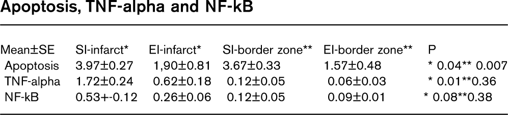
Apoptosis, and inflammatory modulators, such as NF-kB and TNF-alpha participate in cardiovascular remodeling after myocardial infarction (MI) and are associated with heart failure. Physical exercise reduces cardiovascular morbidity and mortality. The cardioprotective effects of exercise training include higher myocardial tolerance to ischemia-reperfusion, improved cardiac performance and higher cell defense capacity against stress. Here we evaluated the effects of previous exercise training on myocardial levels of apoptosis, NF-kB and TNF-alpha 4 weeks after MI.
Methods: 20 Wistar rats were randomly distributed in groups: Sedentary (S); Exercise (E); Sedentary plus MI (SI); Exercise plus MI (EI). Exercise training consisted in 8 wks of swimming; 1h/day, 5d/wk. MI was performed through surgical ligation of the left coronary artery. 4 wks after MI animals were submitted to echocardiographic study and sacrificed and the heart was excised and prepared for histological studies. Apoptosis was detected by TUNEL. Rabbit polyclonal antibody anti-NFkB p65 and anti-TNF alpha were used for immunohistochemistry. Quantification of % positive area for each antigen was performed through an Automatic Color Detector Analysis System.
Results: The number of apoptotic cells/20x field was higher in SI comparing to EI, in both infarcted myocardium and infarct border zone (3.97±0.27 vs 1.9±0.81, P=0.04 and 3.67±0.33 vs1.56±0.48, P=0.007, respectively). Comparing to EI, SI group had higher % positive area for NF-kB and TNF-alpha antigen at myocardium infarcted area (0.53±0.12 vs 0.26±0.06, P=0.08 and 1.72±0.24 vs 0.62±0.18, P=0.01).
Conclusion: MI in previously trained animals was associated with lower levels of apoptosis, TNF-alpha and NF-kB. Physical exercise training promotes a better immune response against myocardial infarction that can lead to better functional outcomes. These results reinforce the need to develop cardiovascular prevention programs with exercise.
298 Gene expression profiling in left ventricles of rats after 10 week of mild exercise training
F Sofi1; B Giusti1; L Rossi1; I Lapini1; A Capalbo1; M Marini2; M Samaja3; R Abbate2; GF Gensini4; A Veicsteinas5
1University of Florence Medical and Surgical Critical Care, Florence, Italy
2University of Bologna Histology, Embriology and Applied Biology, Bologna, Italy
3University of Milan Medicine, Surgery and Dentistry, Milan, Italy
4Don Carlo Gnocchi Foundation Onlus IRCCS, Florence, Italy
5University of Milan Institute of Physical Exercise, Health, Florence, Italy
Purpose: Whereas physical exercise is a known protective factor against cardiovascular morbidity and mortality, the effects of mild exercise (as practiced by most adult humans for cardiovascular fitness) are less clear and the underlying molecular mechanisms still remain explored. To identify the gene expression changes potentially involved in the induction of this phenotype, a genomic approach was used in an animal model that induces cardioprotection.
Methods: Rats were trained on a treadmill with a mild protocol: 25 m/min, 10% incline, 1 h/day, 3 days/week, 10 weeks; 60% VO⊂2max. By using the Affymetrix technology, we investigated the gene expression profile (30.000 transcripts) induced by exercise training in left ventricle (LV) of trained (n=10) and control (n=10) rats. Validation of the results obtained by the Affimetrix technology was obtained by real time PCR. Rats were sacrificed 48 hours after the last training session, in order to identify long-lasting changes in gene expression.
Results: No statistically significant differences in heart weight, heart-to-body weight ratio and thickness of the left ventricular wall were observed, by indicating that training did not induce cardiac hypertrophy. However, the soleus to body weight ratio was higher in trained than in control rats, showing that animals had been efficiently trained. We observed 12 transcripts (10 genes) differentially expressed in LV of exercised animals with respect to controls, and we validated the upregulation of three genes: caveolin 3, beta enolase, and hypoxia in-ducible factor 1 alpha. Moreover, caveolin 3 protein levels resulted higher in exercised rats in comparison to control rats by Western Blot analysis. Finally, in order to evaluate the cardioprotective effect of the mild chronic exercise training protocol, ischemia/reperfusion experiment was conducted in a similar pattern of 10 control and 10 exercised rats. A decreased infarct size (p=0.02) was observed in trained with respect to control rats.
Conclusion: The gene expression profile analysis indicates that Cav3, Eno3, Cyp27a1, Egln1, Cst3 and Tnfaip1 genes show long-lasting expression changes in rat LV as a consequence of mild exercise training associated to cardioprotection without induction of hypertrophy.
299 In chronic heart failure excercise training reduces myostatin in heart and skeletal muscle
K Lenk; V Adams; A Linke; S Gielen; S Erbs; G Schuler
University of Leipzig, Heart Center Cardiology, Leipzig, Germany
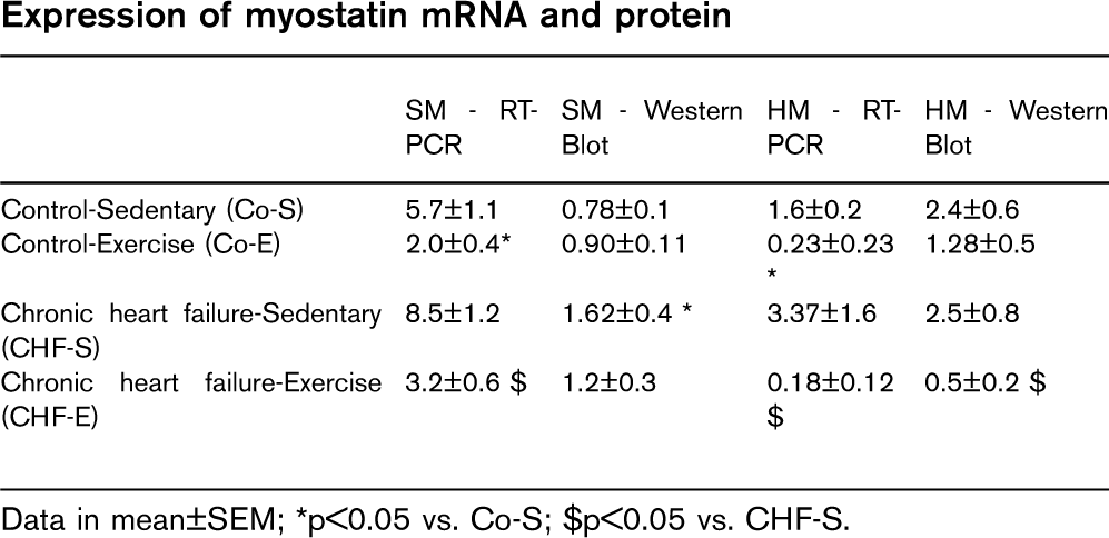
Objectives: In terminal chronic heart failure (CHF) cachexia is often observed and is associated with a severely reduced quality of life and a poor prognosis. Several studies documented that exercise training exerts beneficial effects on skeletal muscle in this setting. It has been shown that the expression of myostatin, a key regulator of skeletal muscle mass is increased in a variety of cachectic states. Therefore, the aim of the study was to investigate the role of myostatin in CHF and the influence of exercise training.
Methods: Sprague-Dawley rats underwent ligation of the left anterior descending coronary artery to induce CHF or were sham-operated (Co). CHF was verified by echocardiography one month after operation. After induction of CHF the two groups were divided into either an exercise training group (CHF-E/Co-E) or a sedentary group (CHF-S/Co-S). Each group consisted of 10 animals. Rats of the exercise groups were trained for 4 weeks on a treadmill (2 sessions/day, each 30 min). The expression of myostatin was analyzed in skeletal (SM) and heart muscle (HM) by RT-PCR and Western blot.
Results: See table
Conclusion: In CHF the expression of myostatin tends to be increased in heart and skeletal muscle. Exercise training reduces its expression significantly. This could be one possible mechanism to explain the beneficial effects of exercise training in CHF.
Data in mean±SEM; ∗p>0.05 vs. Co-S; $p>0.05 vs. CHF-S.
300 Skeletal muscle alterations in chronic heart failure: differential effects of left ventricular dysfunction on catabolic activation in the quadriceps and the diaphragm
N Mangner1; S Gielen1; B Weikert1; M Sandri1; R Hoellriegel1; V Adams1; R Hambrecht2; G Schuler2
1 Universitaet Leipzig - Herzzentrum Department for Cardiology, Leipzig Germany
2Klinik Links der Weser - Herzzentrum Bremen, Bremen, Germany
Progressive muscle wasting is increasingly recognized as major cause of exercise intolerance in chronic heart failure (CHF). The catabolic process is in part mediated by the muscle specific E3-Ligase Murf-1 via activation of the ubiquitin proteasome system. It is, however, unclear if respiratory muscles are equally affected by muscle catabolism as compared to peripheral skeletal muscles. Aim of this study was therefore to examine the differential effects of left ventricular dysfunction on expression of Murf-1 and the activity of manganese superoxiddismutase (Mn-SOD) in quadriceps muscle (Qua) and diaphragm (Dia) in LAD-ligated (MI) rats.
Methods: Twelve weeks after operation left ventricular function was assessed in MI (n=12) and sham operated (C, n=9) rats using echocardiography and Millar-catheter. Murf-1 protein expression was quantified by Western Blot, and Mn-SOD activity was measured in Qua and Dia.
Results: Compared to C, LAD-Ligation resulted in a severe left ventricular dysfunction (EF: 33.4±3.3 vs. 64.8±2.6% (p>0.01); LVEDD: 9.6±0.3 vs. 6.8±0.22mm (p>0.01), LVESD: 8.1 ±0.3 vs. 3.7±0.22 mm (p>0.01), dp/dt max: 4.4±0.3 vs. 5.8±0.5 (p=0.02), dp/dt min: −3.1±0.2 vs. −4.6±0.5, (p>0.01)).
MI resulted in a 4.5-fold increase in Murf-1 expression in Qua (3.7±0.7 vs. 0.8±0.1 arbitrary units, p>0.001). Negative correlations were observed with EF (r= −0.66, p=0.01) and dp/dt min (r= −0.57, p=0.03). Positive correlations were found with LVEDD (r=0.59, p=0.03) and LVESD (r=0.62, p=0.02). Mn-SOD activity decreased significantly in MI (0.15±0.1 vs. 1.4 ±0.4, p>0.01). This was negatively correlated with Murf-1 protein expression (r= −0.53, p=0.04).
MI also resulted in an increase of Murf-1 protein expression in Dia. Induction was with 2.2 fold lower than in Qua (1.3±0.3 vs. 2.8±0.4 arbitrary units, p>0.05). In contrast to Qua Mn-SOD activity in Dia increased with heart failure development (2.4±0.5 vs. 0.7±0.3, p=0.01). All observed correlations in Qua could not be detected in Dia.
Conclusion: LAD-ligation resulted in left ventricular dysfunction accompanied by an increase of catabolic active Murf-1 in peripheral muscle. This is significantly correlated with left ventricular dysfunction and decreased antioxidative capacity. The increase is more distinct in Qua than in Dia. One reason for the attenuation of catabolic activation and the increase of antioxidative Mn-SOD actitivity may be a dyspnea related training effect. These findings indicate differential effects of CHI on peripheral skeletal muscle and diaphragm and support the rationale of training interventions as an anti-catabolic stimulus in heart failure.
301 Cardiac SERCA2 function is essential for aerobic capacity and response to exercise training
M Ericsson1; KB Andersson2; BH Amundsen1; SH Torp3; I Sjaastad3; G Christensen3; OM Sejersted3; O Ellingsen1
1Department of Circulation and Medical Imaging Medical Faculty, Trondheim, Norway
2Institute for Experimental Medical Research Ullevaal University Hospital, Oslo, Norway
3Department of Laboratory Medicine Medical faculty, NTNU, Trondheim, Norway
Purpose: Aerobic fitness is closely linked to function of sarco-endoplasmic reticulum Ca2+ ATPase (SERCA2) and cardiomyocyte contractile function, in both healthy and failing hearts. In a new mouse model of progressive heart failure, deletion of the Serca2 gene in cardiac myocytes is induced by tamoxifen administration (SERCA2 KO). SERCA2 KO mice develop severe congestive heart failure within 7 weeks after Serca2 deletion. Control mice with floxed alleles (SERCA2 FF) are unaffected. Our hypothesis was that aerobic capacity, measured as maximal oxygen uptake (VO2max) is reduced in both sedentary and trained SERCA2 KO mice.
Methods: Two studies were performed. To assess aerobic capacity, sedentary groups of either control or SERCA2 KO animals underwent daily testing of VO2max from day 5 before knockout through day 14 after knockout. To determine whether the training response is altered in the SERCA2 KO mice, a four week training program, starting two weeks after knockout, was set up. Animals were either subjected to exercise training or remained sedentary. Exercise training was performed as treadmill running in intervals, 8 min at 85-90% of VO2max, interspersed with 2 min at 40–50%, for 1 hour, five days per week.
Results: In the first study, aerobic capacity in sedentary SERCA2 KO mice transiently decreased to a nadir 50%–70% below baseline 3–5 days after knockout, before reverting to baseline values. In the second study, VO⊂2max fell by 50% after 4 weeks of exercise training in the SERCA2 KO mice, although running speed was maintained. Echocardiography showed dilation of the left atrium in SERCA2 KO. Increased heart and lung weights as well as pulmonary congestion were consistent with congestive heart failure in both training and sedentary SERCA2 KO mice.
Conclusion: After ablation of the Serca2 gene, aerobic capacity declines in parallel with the reduction of SERCA2 protein in the myocardium of sedentary and exercising mice. Response to exercise training is altered in SERCA2 KO animals compared to control mice. Thus, normal SERCA2 function in the heart seems essential to sustain cardiac function and aerobic capacity.