






Abstract
Background
Methods and results
Conclusion
Keywords
Introduction
In patients with chronic heart failure (CHF), the poor relationship between resting left ventricular dysfunction and exercise capacity has been ascribed to peripheral abnormalities [1]. Besides endothelial dysfunction, muscular deficiency is considered to be a key mechanism. Exercise training in these patients seems to be an effective way to tackle both peripheral vascular and muscular dysfunction, and has been shown to significantly improve exercise tolerance [2, 3].
Skeletal muscle alterations, covering the entire range of macroscopic (i.e. loss of muscle bulk) [4] to histological [5] and ultrastructural changes [6, 7], as well as functional abnormalities related to fiber shift (from slow oxidative type I to glycolytic type II fibers) [5–8], are an integral part of the heart failure syndrome. A profound anabolic/catabolic imbalance secondary to overactivated neurohormonal pathways, finally leads to muscle atrophy [9]. Recently, an important role in the process of muscle wasting has been attributed to the ubiquitin proteasome pathway [10, 11]. Another attractive explanation is the reduction of muscle mass through programmed cell death. More in particular, apoptotic nuclei have been detected in skeletal muscle of CHF patients. Their presence correlates with structural changes, (i.e. cross-sectional area of fibers) [12] and reduced exercise capacity [12, 13], and the suppression of apoptosis might explain the benefits of exercise training [14]. Although apoptosis has been addressed in severely debilitated CHF patients, the process has hitherto not been investigated in patients with mild-to-moderate disease severity. Nevertheless, it has been shown that also in these subgroups of patients, loss of muscle bulk determines exercise capacity.
The hallmark of apoptosis is DNA fragmentation, which characterizes the final execution phase of the process and is also the basis for the frequently used DNA in-situ end-labeling technique (terminal deoxynucleotidyl transferase end labeling, TUNEL). Concerns about TUNEL specificity have led to the strong recommendation to add confirmatory tests. For example, nonapoptotic nuclei, showing high levels of RNA synthesis and splicing, can be labeled by the TUNEL technique [15].
The aim of this study was to evaluate the presence of apoptotic nuclei and of active gene transcription in skeletal muscle biopsies of CHF patients with mild-to-moderate exercise intolerance and to relate these to exercise parameters. We used TUNEL and immunostaining techniques as apoptosis markers, in addition to detection of SC-35 splicing factor to investigate the level of active gene transcription/splicing before and after 4 months endurance-resistance exercise training.
Materials and methods
Study population
Sixteen symptomatic CHF patients (New York Heart Association class > I, left ventricular ejection fraction < 40%, ischemic or dilated cardiomyopathy of at least 6 months duration) referred to the Cardiac Rehabilitation Centre of the University of Antwerp Hospital participated in this study. They had to be stable (1 month) with regard to symptoms and medical treatment. Eight age-matched nontrained participants served as a healthy control group (no chronic underlying disease or cardiac history, no medication, no inflammatory disease). The study was approved by the Institutions' local Ethics Committees and all patients gave their written informed consent.
Cardiopulmonary exercise testing
Each patient performed a symptom-limited cardiopulmonary exercise test (CPET) on a treadmill at baseline and at the end of the 4 months training program. Twelve-lead ECG and heart rate were recorded continuously, whereas automatic cuff blood pressure was measured every 2 min and at peak exercise. Breath-by-breath gas exchange measurements were performed using a metabolic cart. Ventilation (VE), oxygen uptake (VO2), and carbon dioxide production (VCO2) were determined online every 15 s. Peak oxygen consumption (VO2peak) was determined as the highest attained VO2 during the final 30 s of exercise and was also expressed as a percentage of the predicted value (VO2peak%) using the equation proposed by Wassermann et al. [16]. Patients performed a maximal test according to the identification of the anaerobic threshold (the V-slope method). The slope of the relation between VE and VCO2 was calculated by linear regression, excluding the nonlinear part after reaching the ventilatory threshold.
Training program
Patients attended the exercise program three times/week for 1 h. The training session commenced and concluded with a 5 min warming-up and cooling-down and stretching period, respectively. During the first 2 months of the program, focus was on resistance training (40 vs. 10 min endurance training), with a progressive increase in aerobic exercise duration. After 2 months, both training modalities were equivalent in time (each 20 min). Resistance training intensity was set at 50% of the pretraining 1-repetitive maximum tests and was increased to 60% after 2 months. The resistance circuit consisted of nine predetermined resistance exercises, involving muscle groups of the lower and the upper limbs and torso. Per station, one set, consisting of 10 repetitions, was performed with a gradual increase (15 repetitions after 1 month, two sets of 10 repetitions after 2 months, two sets of 15 repetitions after 3months).
Endurance training (cycling and jogging) intensity was aimed at a target heart rate, defined as 90% of the heart rate achieved at the anaerobic threshold during CPET. Exercise intensity was adjusted on a monthly basis (repeated CPET). In addition to individual pulse rate measurements [Polar Heart Rate Transmitters, Polar T31, Polar Electro Oy, Kempele, Finland], a central monitoring system permitted arrhythmia detection. Patients were unable to alter training intensity as they were individually permanently controlled by an automated adjusting system (TGS, TechnoGym System, Gambetola, Italy).
Measurement of peripheral skeletal muscle strength and linear isokinetics
Maximal strength of the upper limbs was defined as the combination or three movement patterns (1-repetitive maximum of pull down forward, chest press, and butterfly using Unica; Technogym). To evaluate maximal force (Newton) and force at 0.25 s, we used a Linear Isokinetic Dynamometer (Aristokin, Lode BV, Groningen, The Netherlands). Patients performed a rowing movement to evaluate their ability to carry out complex movements of both arms and legs. Linear isokinetic speed was set at 100 cm/s and minimal force at 5 N. Patients were allowed to perform five maximal repetitions. From these five maximal performances a mean performance was calculated.
Skeletal muscle biopsy
Skeletal muscle biopsies were taken percutaneously from the middle part of the m. vastus lateralis under local anesthesia (10 ml lidocaine) with the Bergström needle at baseline in CHF patients and controls, and after 4 months exercise training in the CHF group only. Care was taken to assure that tissue samples were obtained at least 24 h after strenuous exercise. One section was fixed in formalin (24 h) and paraffin embedded. The second section was snap-frozen in liquid nitrogen and stored at −80°C.
Immunohistochemistry and terminal deoxynucleotidyl transferase end labeling
Immunohistochemical analyses were completed by an indirect antibody conjugate method using the following primary antibodies: mouse monoclonal antisplicing factor SC-35 (clone SC-35, Sigma, St Louis, Missouri, USA), rabbit polyclonal anti-cleaved caspase-3 (Cell Signaling Technology, Beverly, Massachusetts, USA), and rabbit polyclonal anti-cleaved poly(ADP-ribose) polymerase — PARP — (Promega, Southampton, UK). Antigen retrieval was done with trypsin digestion and citrate buffer treatment. To prevent unspecific binding, sections were blocked with goat serum for 20 min. After this blocking step, the respective primary antibody was applied diluted in PBS (1:500 for cleaved caspase-3, 1:200 for cleaved PARP, and 1:1000 for SC-35). Reactions were carried out overnight at 37°C in a humidified chamber and visualized by a goat-anti-mouse or a goat-anti-rabbit biotinylated antibody and a subsequent layer of avidin-biotin peroxidase. 3-Amino-9-ethylcarbazole was used as a chromogen. The specificity of the antibodies was checked by omitting the primary antibody and substituting an unrelated antibody of the same isotype. For the detection of oligonucleosomal DNA cleavage, the TUNEL technique was performed on deparaffinized 5-μm thick sections with a ApopTag kit (Chemicon, Temecula, California, USA) according to the manufacturer's instructions. In addition, we conducted the stringent TUNEL assay (by leaving out proteinase K digestion) as previously described by Kockx et al. [15], which avoids nonspecific labeling because of active RNA synthesis. Negative controls were generated by omitting terminal deoxynucleotidyl transferase from the labeling mixture. In this study, human tonsils were used as positive controls because this type of tissue shows a high apoptosis frequency under physiological conditions. Apoptosis in germinal centers of tonsil is a key regulatory event to eliminate nonautoreactive and autoreactive lymphocytes. Human noninflamed hyperplastic tonsils were obtained from patients undergoing tonsillectomies.
Cell counting (see Results for detailed information) was performed twice by two independent observers with no knowledge of the patients' clinical characteristics. Mean value of both counts is reported.
Proinflammatory cytokines
Fasting blood samples were collected between 08:00 and 09:00 h into ethylenediaminetetraacetic acid tubes (Vacutainer, Becton and Dickinson, Meylan, France). Plasma concentrations of interleukin (IL)-6, tumor necrosis factor (TNF)-α, soluble TNF-receptor 1 (sTNFRl), and soluble TNF-receptor 2 (sTNFR2) were measured using an enzyme-linked immunosorbent assay according to the manufacturer's specifications Quantikine (R&D Systems, Minneapolis, Minnesota, USA) sensitivity: 0.7 pg/ml for IL-6, 1.5 pg/ml for sTNFR1, and 1 pg/ml for sTNFR2). A high sensitivity kit (Quantikine HS, R&D Systems, sensitivity 0.18 pg/ml) was used to measure TNF-α. All samples were run-in duplicate.
Statistical analyses
All data are expressed as mean value ± SEM. Comparisons of numerical data between groups were carried out with the nonparametric Mann-Whitney U test. Pairwise comparisons were carried out using the Wilcoxon's matched pair signed rank test. Correlations were determined using Spearman's rank correlation test. All statistical analyses were performed using the software package SPSS, version 15.0 (SPSS Inc., Chicago, Illinois, USA).
Results
Patient characteristics and exercise data
Table 1 summarizes demographic characteristics of the patients, as well as exercise data, information on left ventricular function and dimensions, cytokine levels, and medical treatment. The healthy control group (n = 8) consisted of five women and three men (P = 0.2 compared with patients), with a mean age of 57.4 ± 8.5 years (P = 0.7 compared with patients). Cytokine levels of CHF patients were higher than those measured in healthy participants (P < 0.05 for IL-6, TNF-α, sTNR1, and sTNFR2). Exercise training in CHF patients effectively increased the percentage of predicted VO2 (VO2%), maximal workload, work economy (maximal workload/VO2peak), upper limb and quadriceps strength, and linear isokinetic measurements (Table 2).
Characteristics of chronic heart failure patients (n = 16)
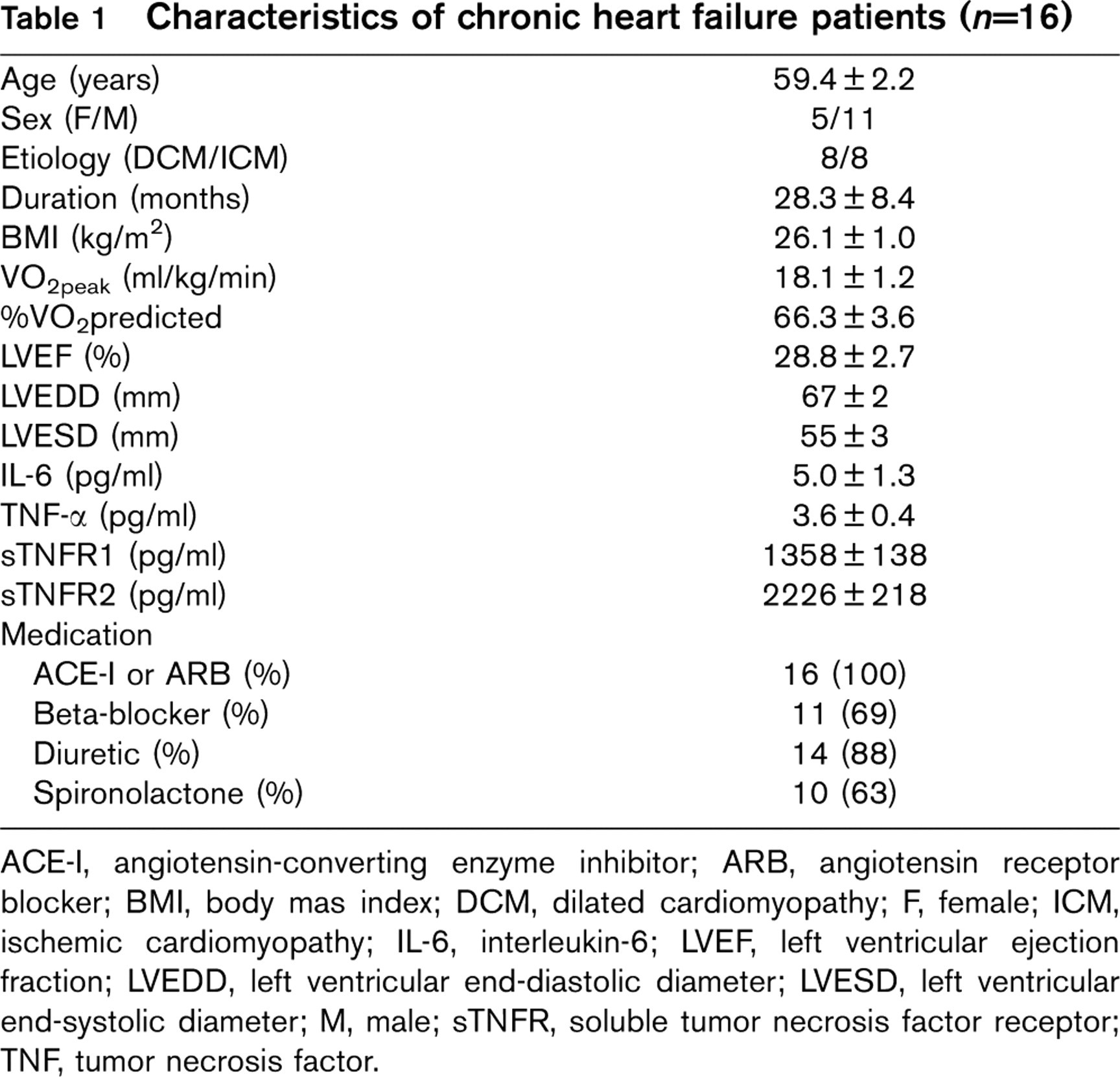
ACE-I, angiotensin-converting enzyme inhibitor; ARB, angiotensin receptor blocker; BMI, body mas index; DCM, dilated cardiomyopathy; F, female; ICM, ischemic cardiomyopathy; IL-6, interleukin-6; LVEF, left ventricular ejection fraction; LVEDD, left ventricular end-diastolic diameter; LVESD, left ventricular end-systolic diameter; M, male; sTNFR, soluble tumor necrosis factor receptor; TNF, tumor necrosis factor.
Assessment of apoptosis and correlation with exercise data
Terminal deoxynucleotidyl transferase end labeling
Ten serial sections of skeletal muscle biopsies per patient at baseline and after 4 months were screened on an Olympus BX50 microscope using a semiautomated computer-assisted stereological tool (CAST-grid V1.10, Olympus, Ballerup, Denmark). TUNEL positivity was expressed as the mean number of TUNEL-positive nuclei counted per square millimeter tissue measured in these 10 sections (Fig. 1a). There was a significant variation between the patients studied, ranging from 0.3 to 10.0 positive nuclei/mm2 tissue area with a mean count of 3.2 ± 0.7 positive nuclei/mm2 tissue area. Comparison of the healthy control group (3.1 ± 1.7 positive nuclei/mm2 tissue area) with CHF patients showed no difference (P = 0.2)(Table3). There were no correlations between the number of TUNEL-positive nuclei and VO2peak (r =0.2, P =0.4), VO2peak% (r =0.2, P =0.4), Wattmax (r =0.09, P =0.8), or VE/VCO2 slope (r = −0.3, P = 0.3). However, in contrast to tonsil tissue, used here as a positive control (Fig. 1b), positive nuclei in skeletal muscles of CHF patients could not be detected using the stringent TUNEL technique (data not shown). After 4 months training, the number of TUNEL-positive nuclei significantly decreased (3.2 ± 0.7 before vs. 2.1 ± 1.5 positive nuclei/mm2 tissue area after training, P = 0.04), but values were still comparable with the control group (P = 0.1) (Table 3).
Effects of 4 months combined endurance-resistance training
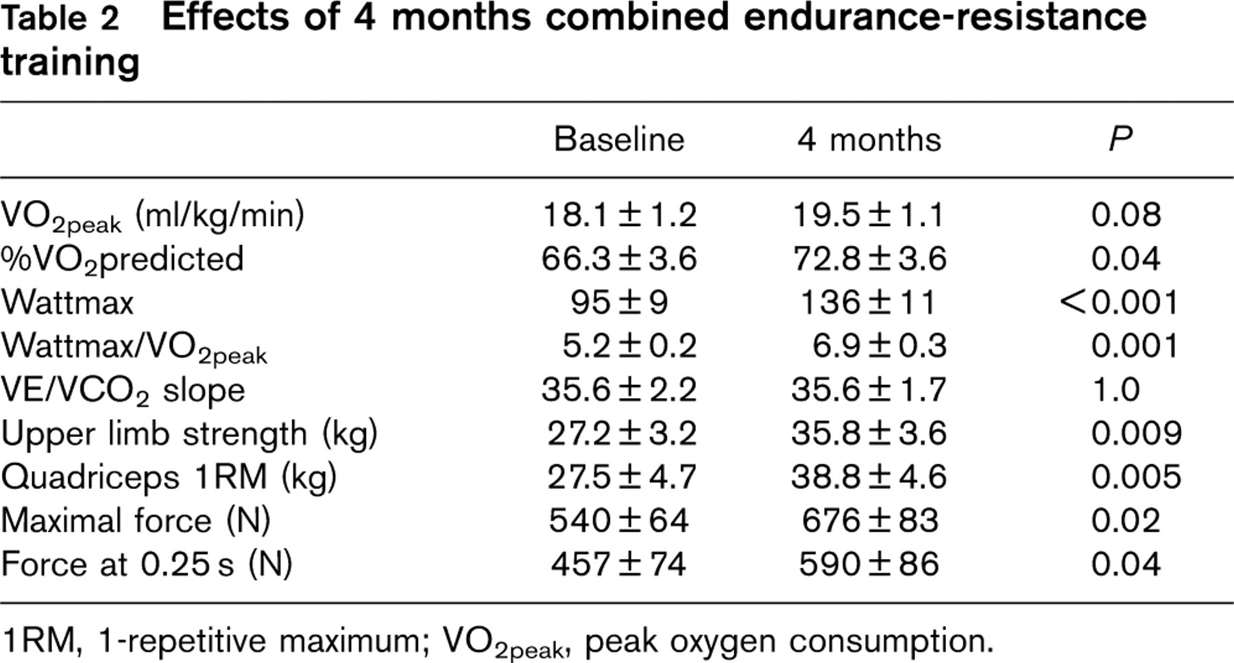
1RM, 1-repetitive maximum; VO2peak, peak oxygen consumption.

Detection of oligonucleosomal DNA fragmentation in skeletal muscle of patients with chronic heart failure (CHF) by the terminal deoxynucleotidyl transferase end-labeling (TUNEL) technique. (a) Nonstringent TUNEL reveals aspecific labeling in skeletal muscle (arrow). (b) Stringent TUNEL stains apoptotic cells in tonsils (arrows) but not in skeletal muscle of CHF patients (not shown). Scale bar = 50 μm (a) or 20 μm (b).
Cleaved caspase-3 and cleaved poly(ADP-ribose) polymerase
Full-length PARP is a 116-kDa protein involved in the repair of DNA, in differentiation, and in chromatin structure formation. During apoptosis, this protein is cleaved by active (cleaved) caspase-3, and possibly other caspases, into an 85-kDa fragment. Antibodies against both cleaved protein markers were applied to skeletal muscle tissue that showed the highest number of TUNEL-positive nuclei using the nonstringent TUNEL method. Although lymphoid tissue showed clear activity for both cleaved caspase-3 (Fig. 2a) and cleaved PARP (Fig. 2b), none of the studied skeletal muscle biopsies was positive.
TUNEL-positive nuclei and SC-35 immunoreactive area
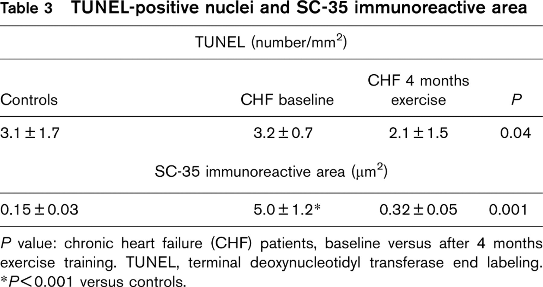
P value: chronic heart failure (CHF) patients, baseline versus after 4 months exercise training. TUNEL, terminal deoxynucleotidyl transferase end labeling.
P < 0.001 versus controls.
SC-35
Ten fields (150 × 100 μm per field) per patient were screened (40X) at baseline and after 4 months and quantification was done using a color image analysis system, which allows measurement of the nucleoimmunoreactive area by interactive selection of the specific color used (Fig. 3). The immunoreactive area varied from 0.3 to 18.6 μm2 with a mean value of 5.0 ± 1.2 μm2. Values at baseline were significantly higher in CHF patients versus the control group (0.15 ± 0.03 μm2, P < 0.001) (Table 3). There was a significant correlation with VO2peak% (r =0.6, P =0.02), Wattmax (r = 0.7, P = 0.005), and VE/VCO2 slope (r = −0.6, P = 0.03) (Fig 4). The correlation between VO2peak and SC-35 immunoreactive area was of borderline statistical significance (r = 0.5, P = 0.05). After 4 months training, the immunoreactive area was significantly smaller (5.0 ± 1.2μm2 before vs. 0.32 ± 0.05μm2 after training, P = 0.001). Although at baseline there was a significant difference with the control group, after 4 months training, values were comparable (P = 0.07) (Table 3). Figure 5 shows several TUNEL-labeled nuclei (Fig. 5a) that were also positive when stained with the SC-35 antibody (Fig. 5b). The presence of splicing factor indicates a high rate of transcription in these cells. As such, these cells do not seem to be apoptotic.
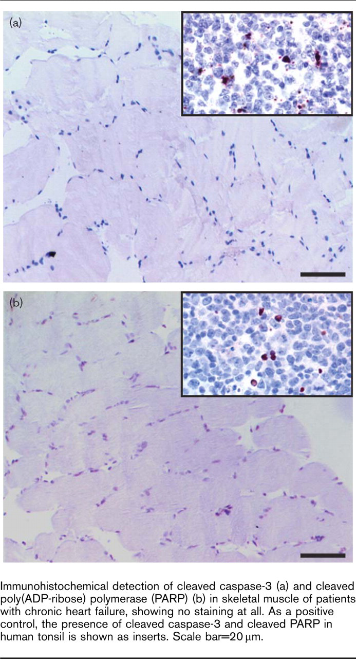
Immunohistochemical detection of cleaved caspase-3 (a) and cleaved poly(ADP-ribose) polymerase (PARP) (b) in skeletal muscle of patients with chronic heart failure, showing no staining at all. As a positive control, the presence of cleaved caspase-3 and cleaved PARP in human tonsil is shown as inserts. Scale bar = 20 μm.
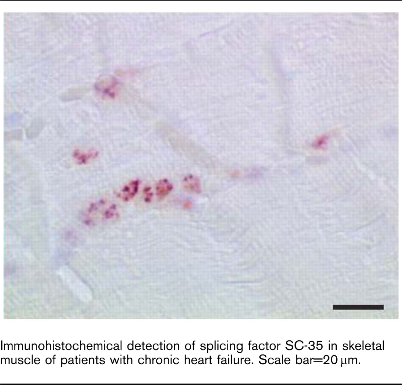
Immunohistochemical detection of splicing factor SC-35 in skeletal muscle of patients with chronic heart failure. Scale bar=20 μm.

Relationship between SC-35 nucleoimmunoreactive area in skeletal muscle of chronic heart failure patients and ventilation (VE)/carbon dioxide production (VCO2) slope.

Colocalization of nonstringent terminal deoxynucleotidyl transferase end labeling (TUNEL) and SC-35 immunoreactivity in skeletal muscle of patients with chronic heart failure (CHF). Serial sections of skeletal muscle of CHF patients were stained with the nonstringent TUNEL method (a) or probed with an antibody against SC-35 (b). TUNEL-positive muscle cells are also SC-35 positive (arrows), suggesting false-positive TUNEL. Scale bar=50 μm.
Discussion
Investigation of mechanisms that explain muscle wasting in patients with moderate-to-severe CHF has pointed at the possible relevance of apoptosis. From this study, performed in patients with mild-to-moderate disease severity, the following findings emerge:
In skeletal muscles of CHF patients with mild-to-moderate exercise intolerance, apoptosis could not be confirmed. Despite TUNEL-positive nuclei, other apoptotic markers (i.e. stringent TUNEL, active caspase-3, cleaved PARP) remained negative. The number of TUNEL-positive nuclei in these biopsies was not different from healthy participants and was not related to exercise capacity.
Active gene transcription, as indicated by immunostaining for RNA splicing factor, was strongly correlated with several exercise parameters. At baseline, SC-35 immunoreactive area was significantly higher compared with the control group, but the difference was no longer present after 4 months exercise training.
Skeletal muscle tissue that shows signs of RNA transcription and splicing might stain false positive for apoptotic nuclei with TUNEL.
The role of apoptosis in skeletal muscle wasting in chronic heart failure
Apoptotic nuclei in myocytes and in interstitial cells were first described in an animal model of monocrotaline-induced right ventricular heart failure [17]. Later, in a selective group of nine patients with CHF who were scheduled for surgical revascularization, skeletal muscle showed a higher number of TUNEL-positive nuclei compared with controls [12]. Tissue concentrations of caspase-3 and ubiquitin were increased, whereas the antiapoptotic protein bcl-2 was decreased. There was an inverse relationship between TUNEL-positive nuclei and both fiber cross-sectional area and VO2peak. Data from Adams et al. [13] confirm these findings. A larger group of CHF patients (n = 34) was divided according to the presence of apoptosis or not. Besides a significantly lower exercise capacity, the apoptosis-positive subgroup was characterized by increased inducible NO synthase and a lower bcl-2 expression.
Patient selection might account for the apparent contradictory results observed in this study. Patients in the apoptosis-positive group in the study by Adams et al. [13] were more debilitated with lower VO2peak and left ventricular ejection fraction as compared with our studied participants. The absence of apoptosis in this study could indicate that this process plays no role in the initial stages of the developing heart failure syndrome, despite the fact that circulating proinflammatory cytokines (i.e. IL-6, TNF-α, and their receptors i.e. sTNFR1, sTNFR2) were significantly upregulated. Early findings with respect to the loss of muscle mass, as described by Harrington et al. [18], might therefore be related to other processes, such as activation of the ubiquitin proteasome pathway [10, 11].
In this study, the search for apoptotic nuclei was conducted in a rigorous way; the classical TUNEL technique was additionally refined by using the so-called stringent approach. TUNEL detects not only DNA fragmentation but also single-stranded DNA breaks with free 3′-OH terminals. Hence, a positive TUNEL reaction can be seen in living cells during active gene transcription [15] and with increasing activity of DNA repair [19, 20]. The lack of specificity of the classical TUNEL led us to apply confirmatory tests. By omitting proteinase K in the TUNEL protocol (stringent TUNEL), Kockx et al. [15] proposed the means to exclude false TUNEL positives resulting from the endogenous release of endonucleases. In addition, the quantification of apoptosis-regulating proteins (i.e. bcl-2, Bax), of downstream-activated caspases (i.e. cleaved caspase-3 and caspase-9; regulators of execution phase) and the specific cleavage of cellular target proteins, PARP (DNA repair), can be used for this purpose. In-situ labeling, neither for cleaved caspase-3 nor for cleaved PARP was positive in our hands. Interestingly, and in accordance with the finding of Kockx et al. [15], some of the TUNEL-positive nuclei in the skeletal muscle of our patients showed immunereactivity against the splicing factor SC-35. SC-35 is a member of the group of nonsmall nuclear ribonucleoprotein particle factors and is required for the first step of splicing and spliceosome assembly. As apoptotic cells in the execution phase of cell death lose their ability for RNA synthesis/splicing, this cooccurrence precludes apoptosis and is a strong argument for viability.
Exercise training and the skeletal muscle in chronic heart failure
Exercise training in CHF patients is recommended as an effective means to counteract muscle wasting and to preserve physical performance. Proposed mechanisms include neurohormonal modulation [21, 22], anti-inflammatory [23], and antioxidative effects [14]. Linke et al. [14] recently demonstrated that 6 months endurance training downregulated muscular expression of proinflammatory cytokines (i.e. IL-1 β and TNF-α) and reduced local oxidative stress by augmenting radical scavenger enzyme activity (i.e. catalase, glutathione peroxidase). In addition, a 24% lower number of TUNEL-positive nuclei after the training period were found. In this study, we applied a combined endurance-resistance training protocol, of which previous evaluations confirmed both clinical efficiency, neuromodulatory [22] and anti-inflammatory adaptations [23]. The significant reduction in TUNEL positivity found by Linke et al. [14] was confirmed. However, the fact that in this study skeletal muscle tissue from healthy controls was also TUNEL positive, the lack of confirmatory evidence for apoptosis with additional tests, and the absence of a relation with exercise capacity, question whether these are truly apoptotic nuclei. As mentioned before, the cooccurrence of TUNEL and of SC-35 splicing factor suggests that at least part of TUNEL-positive nuclei are in fact undergoing DNA repair and/or active gene transcription instead of entering the apoptotic execution phase. The observed larger immunoreactive areas for SC-35 in skeletal muscle biopsies of CHF patients versus controls and the fact that after the training program this difference is minimized, support the notion that regular physical exercise partially restores skeletal muscle integrity. In a study on the progression from cardiac compensatory hypertrophy to failure, increased myocyte DNA synthesis and repair, assessed through SC-35 immuneractivity, went hand in hand [24]. These mechanisms were considered essential to maintain home-ostasis in the setting of persisting and excessive afterload.
Conclusion
There is circumstantial evidence to support an association of neurohormonal maladaptations and apoptosis in skeletal muscle in heart failure animal experiments, but human data are scarce and limited to severe CHF. On the basis of present results, the presence of apoptosis in the skeletal muscle of patients with mild-to-moderate CHF cannot be confirmed. As apoptosis is a transient process, we acknowledge that the short window of opportunity to detect apoptosis might have lead to underestimation of the phenomenon. In an attempt to overcome this drawback, 10 sections per muscle specimen were counted. The co-occurrence of TUNEL with an immune reaction against the splicing factor SC-35 supports the cautionary interpretation of the TUNEL technique. Finally, the level of active gene transcription and splicing, reflecting metabolic activity, seemed to be significantly related to the global exercise performance of the patients studied.
Footnotes
Acknowledgements
Grants: Viviane Conraads is a Senior Clinical Investigator of the Fund for Scientific Research, Flanders (Belgium). Wim Martinet is a postdoctoral fellow of the Fund for Scientific Research, Flanders (Belgium).