






Abstract
Aim
To show that subclinical atherosclerosis (subclinical-ATS) of carotid arteries [intima–media thickness (IMT) or asymptomatic carotid plaque (ACP)], may provide additional information for risk stratification, in asymptomatic patients, aged greater than 45 years, with a cluster of risk factors (RFs).
Methods and results
We studied 558 asymptomatic patients (235 males). RFs for atherosclerosis were assessed and the 10-year-risk was calculated according to the Italian risk score. Doppler ultrasound of carotid arteries identified the presence of IMT greater than 0.9 mm in 183 patients and ACP in 147 patients. One hundred and fifty-three patients developed cerebrovascular or cardiovascular (CV) events in the follow-up: 67 developed acute myocardial infarction, 39 developed angina, 25 had a stroke or transient ischemic attack, six died for CV events, and 16 underwent percutaneous or surgical revascularization. The incidence reflected the different risk profiles (4,14, and 20%, respectively). However, in patients with baseline subclinical-ATS the incidence of events increased to 35, 46, and 63%, respectively. In the multivariate analysis the incidence of events was significantly influenced by the presence of asymptomatic carotid lesions in each risk category.
Conclusion
In our experience, the incidence of CV events is enhanced in patients with subclinical-ATS. Increased IMT and ACP predict CV events and improve the risk stratification of asymptomatic patients aged greater than 45 years and with a cluster of RFs, in a long-term follow-up.
Keywords
Introduction
Global cardiovascular risk (GCVR) identifies patients with a high risk of developing cerebrovascular or cardiovascular (CV) events. It is calculated by mathematical algorithms obtained from large epidemiological studies and represents a simple and objective method to assess the probability of the first CV event during 10-years follow-up, only by considering common risk factors (RFs) [1]. Nowadays, different risk scores are used in different countries: each one is specific for the type and number of analyzed RFs and characteristics of population in which epidemiological study has been conducted. The model from the Framingham Heart Study, updated over the years, allows calculating the risk of CV events in 10 years in the American population [2].
In 2003, the European Score Group introduced the ‘European Chart of CV risk’, to stratify CV risk in the European population [3].
The Italian ‘Progetto Cuore’ was realized to estimate the probability of a cerebrovascular or CV event in 10 years in the Italian population. The risk chart was created by building a database of CV RFs from longitudinal studies performed on the Italian population. End points were mortality and morbidity (as the first cerebral or coronary event) with differences between men and women [4].
On the other hand, epidemiological studies [5, 6], including a very large meta-analysis [7], have shown that subclinical atherosclerosis (subclinical-ATS) may predict the development of cerebrovascular and CV events; an intima–media thickening (IMT) of carotid artery greater than 0.9 mm is strongly related with cerebrovascular and CV events [5–7].
The purpose of our study was to evaluate whether data on subclinical-ATS, that is, increased IMT or asymptomatic carotid plaque (ACP), obtained by Doppler ultrasonography imaging, may increase the accuracy of risk stratification of patients aged greater than 45 years and with a cluster of RFs, in a 10-year follow-up period.
Materials and methods
We evaluated 558 asymptomatic white patients (235 males), aged greater than 45 years who were recruited in the research project on CV aging, MURST-GESCO-CIFTI-4 [8].
The average age was 58 ± 9.84 years at first examination; patients with a personal history of cerebrovascular or CV events, with blood pressure (BP) values greater than 200/110 mmHg, with total cholesterol less than 130 mg/dl (3.4 mmol/l) or greater than 320 mg/dl (8.3 mmol/l) and pregnant women were excluded from the study.
All patients were asked about their clinical history, and underwent physical examination including the evaluation of height, body weight, and BP.
We evaluated, in each patient the family history of cerebrovascular or CV disease and the presence of RFs for atherosclerotic disease, such as hypertension, dyslipidemia, diabetes mellitus, smoking habit, and obesity.
A blood sample was drawn from each patient after a 12–14-hour overnight fast. Glycaemia, total cholesterol, triglycerides, and high-density lipoprotein cholesterol were quantified by standard enzymatic-colorimetric methods; low-density lipoprotein cholesterol was calculated by the Friedewald formula.
Identification of diabetic patients was obtained by detecting, in two or more measurements over a week, fasting plasmatic concentration of glucose ≥ 126 mg/dl (7 mmol/l) or a blood glucose level, during the second hour after oral glucose load, ≥ 200 mg/dl (11 mmol/l) or undergoing treatment with oral hypoglycaemic drugs or insulin or a clinical history of diabetes [9].
Systolic BP was the average value of two consecutive assessments according to the standard methodology. We considered those patients hypertensive who had systolic BP greater than 140/90 mmHg or were undergoing antihypertensive therapy [10].
Patients were considered as smokers if they smoked regularly, including also those who smoked one cigarette per day, or if they had stopped smoking for less than 12 months [11].
General characteristics of the population were used to calculate the GCVR according to the Italian risk score.
B-mode real-time ultrasonography of the common carotid arteries was performed with an Esaote Caris Plus machine (Caris Plus, Esaote SpA, Genoa, Italy) using a probe of 7.5–10.0 MHz. Patients were examined in the supine position and each carotid segment was examined to identify the thickest intima–media site. Three segments were identified and measured in anterior and posterior planes on each side: the distal 1 cm of the common carotid arteries proximal to the bifurcation, the bifurcation itself, and the proximal 1 cm of the internal carotid artery. At each of these sites, we determined the IMT and detected any possible plaque. We primarily used the maximum carotid IMT value, which was determined as the mean of the maximum IMT of near and far-wall measurements of both the left and right side arteries for each of the three arterial segments. If data of one of the walls or one of the sides were missing, maximum thickness of the available wall and side was used. Carotid ultrasonography was carried out by one sonographer to limit the risk of an interobserver variability. According to the ESC/ESH guidelines on hypertension, we considered as normal an IMT less than 0.9 mm, increased IMT when greater than 0.9 and less than 1.5 mm, and ACPs if IMT was greater than 1.5 mm [10]. If patients contemporaneously had IMT and ACP, they were included only in the ACP group.
All patients were evaluated over a 10-year follow-up to assess the occurrence of cerebrovascular and CV events such as CV death, nonfatal acute myocardial infarction (AMI), angina pectoris (AP), percutaneous or surgical revascularization, and ischemic stroke or transient ischemic attack (TIA). The true incidence of fatal and nonfatal events was compared with the prevision deriving from the GCVR, obtained by using the Italian score and baseline data of patients.
Statistical analysis
Univariate and multivariate analysis were performed according to Cox's linear regression model: a P value of less than 0.05 was considered as significant.
This is an observational study, because it does not include any pharmacological or diagnostic intervention in addition to the usual clinical activity; so it is not subject to the directive 2001/20/CE of the European Parliament regulating the application of a good clinical practice during clinical experimentation (Art 1 comma 1; Art 2/c).
All patients enrolled were asked to sign an informed consent to use their personal data and to also read an informative report about the tutelage of the personal data according to law 675/696.
Results
Table 1 summarizes the general features of the studied population at the first examination. Three hundred and sixty-four patients (62%) were on antihypertensive drugs, 151 (27%) were on statins, and 95 (17%) were on hypoglycaemic drugs or insulin.
Calculating the risk profile according to the Italian risk score, 435 patients (78%) fell within the low-risk category, 35 (6%) belonged in the intermediate risk, and 88 (16%) in the high-risk category.
In the low-risk group there was a considerable preponderance of women (M = 128, W = 307), whereas the intermediate (M = 26, W = 9) and high-risk groups were composed mostly of men (M = 81, W = 7).
Univariate analysis performed according to Cox's regression model allowed us to show that CV risk is linked to some RFs. The occurrence of a cerebrovascular or CV event was significantly linked to age (P < 0.001), hypertension (P < 0.001), type 2 diabetes mellitus (P < 0.003), and hypercholesterolemia (P < 0.001).
Moreover, statistical analysis proved a strong association between the risk of CV events and IMT or ACP, in comparison with patients who did not show any evidence of subclinical-ATS (P < 0.001). Univariate analysis also allowed to assess a significant correlation between cerebrovascular or CV events and the intermediate and high-risk classes (P < 0.001), in comparison with patients at low risk.
General features of the population studied
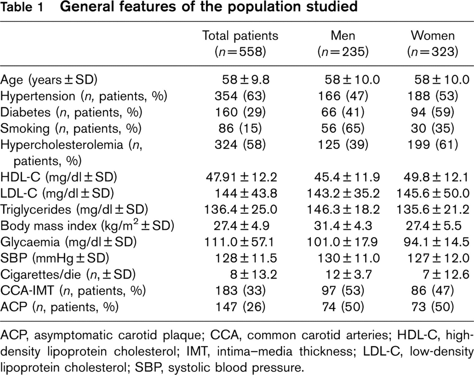
ACP, asymptomatic carotid plaque; CCA, common carotid arteries; HDL-C, high-density lipoprotein cholesterol; IMT, intima–media thickness; LDL-C, low-density lipoprotein cholesterol; SBP, systolic blood pressure.
Doppler ultrasound results in each risk class obtained through the Italian risk score
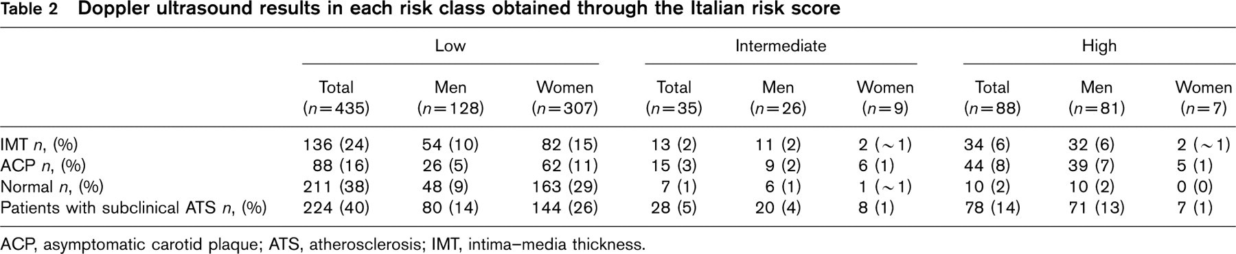
ACP, asymptomatic carotid plaque; ATS, atherosclerosis; IMT, intima–media thickness.
Matching the results from the carotid Doppler examination with risk profiles derived from the Italian risk score, we obtained the number of patients with subclinical-ATS for each risk class. Seventy-eight of 88 patients with a high-risk score showed the evidence of subclinical-ATS, 28 of 35 patients with an intermediate risk and 224 of 435 patients in the low-risk group showed subclinical-ATS (Table 2).
During the follow-up period 153 of 558 patients developed cerebrovascular or CV events: 67 developed AMI, 39 developed AP, 25 had a stroke or TIA, and 16 patients had to undergo percutaneous or surgical coronary revascularization. Among all the enrolled patients, six individuals died of CV causes. The death was verified by the hospital reports, if the patients were hospitalized, or through their death certificates.
Among 153 patients who developed events, 142 showed an ultrasonography evidence of subclinical-ATS: 23 of 64 patients who suffered from AMI had IMT, while 41 of 64 had ACP; among 36 patients with AP, 20 had an increased IMT, while 16 had ACP. An IMTwas also present in seven of 23 patients who developed stroke whereas, in the same group an ACP was detected in 16 cases. Sixteen patients underwent coronary revascularization procedures, and 13 of them showed subclinical-ATS carotid lesions. Finally, all patients who died from CV events had an ultrasonography detection of subclinical-ATS.
The incidence of events among patients with low, intermediate, and high risk but without subclinical-ATS, resulted 4,14, and 20% respectively, whereas it was equal to 35, 46 and 63% respectively, in those patients in the same category of risk but with baseline IMT or ACP; that is a remarkably increased risk (Table 3 and Fig. 1).
Our study shows a significant statistical correlation between risk categories, assessed in accordance with the Italian risk score and the occurrence of events [odds ratio (OR) = 3.21 for intermediate-risk patients and OR = 4.87 for high-risk patients]. Multivariate analysis also showed that the variable that correlated the strongest with the occurrence of events is IMT or ACP (OR = 4.59 in patients with IMT and OR = 8.11 in patients with ACP). This finding confirms that the detection of subclinical-ATS of the carotid arteries by Doppler ultrasonography improves the risk stratification in a population of patients aged greater than 45 years and with a cluster of RFs.
Number of events for each risk category according to the presence or absence of carotid lesions (IMT or ACP)

ACP, asymptomatic carotid plaque; AMI, acute myocardial infarction; CV, cardiovascular; IMT, intima-media thickness; TIA, transient ischemic attack.
Discussion
The long-term GCVR according to the risk's charts is calculated by mathematical algorithms, which analyze simultaneously all the main traditional RFs for atherosclerotic disease. The Framingham risk score represents the main system for risk assessment in the United States [2]. In 2003, the European Score Group [3] elaborated the ‘European Chart of CV risk’, to realize a specific stratification of risk for the European population.
The Italian algorithm of risk is a model to assess the risk of a first cerebrovascular or CV event in the Italian population. It represents a method to estimate the probability of a first event, only on the basis of some RFs for atherosclerosis (sex, age, smoking, diabetes, hyperch-olesterolemia, systolic hypertension, as well as high-density lipoprotein cholesterol and antihypertensive therapy by using not the chart but a computed system) in the Italian population [4].
Research has shown that subclinical-ATS may offer improved risk stratification for patients with a low or intermediate risk profile [5–7]. These patients have a routinely underestimated CV risk and generally do not undergo appropriate therapeutic preventive strategies. Thus, prognostic information in a long-term follow-up, obtained only by the risks charts, should be integrated through the evaluation of the carotid vessels by Doppler ultrasound or ankle brachial index for peripheral vessels [12].
The evidence of IMT or ACP of the carotid wall is predictive of cerebrovascular and CV events and guarantees a further support in assigning patients to the most appropriate CV risk group [13–15]. According to studies in literature, in our group of patients, the carotid Doppler ultrasound examination represents a tool for early identification of subclinical-ATS, which has been associated with almost all major modifiable and not RFs, and with higher occurrence of AMI and CV death.
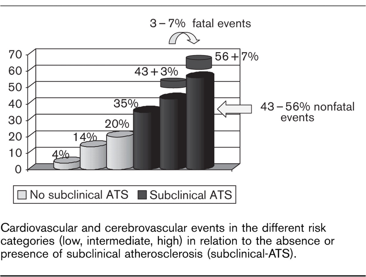
Cardiovascular and cerebrovascular events in the different risk categories (low, intermediate, high) in relation to the absence or presence of subclinical atherosclerosis (subclinical-ATS).
In accordance to the Italian risk score, the majority of patients in our sample had essentially a low risk of developing cerebrovascular or CV disease. This finding suggests that during a 10-year follow-up, in the presence of a subclinical arterial damage, the risk concerning the number of events that really occurred is underestimated by this algorithm alone. Matching the results of the Italian score and data obtained by carotid ultrasonography, with those concerning the number of events that occurred, it is clear that in our study the incidence of cerebrovascular and/or CV events agrees with the risk score prediction in patients without baseline subclinical-ATS (4, 14, 20%); however, in the presence of baseline subclinical-ATS it is really much higher (35, 46 and 63%, respectively for each risk category) (Fig. 1).
Table 3 shows the incidence of fatal and nonfatal events, according to the risk profile and the ultrasonography results: all patients who died of CV in our sample belonged to the intermediate and high-risk categories and contemporaneously had subclinical-ATS.
Our results confirm what was mentioned in the 2009 ESC/ESH Guidelines for hypertension, according to which IMT doubles the risk of acute coronary events, whereas the presence of ACP is associated with a four-fold risk and the presence of a significant carotid stenosis is associated with a six-fold increased risk compared with a person free from arterial atherosclerosis [10]. So, the ultrasonography detection of subclinical-ATS represents an important tool for prevention of cerebrovascular or CV events because it suggests a more aggressive preventive strategy, as recommended by the ESC guidelines on CV prevention [11] and by the TASC II [12].
A review presented by Cheng affirmed that an increased carotid or femoral IMT was a condition found in patients with the presence of RFs for atherosclerosis and at a higher risk of cerebrovascular or CV events; the treatment of RFs is able to provide both a reduction of IMT and of cerebrovascular or CV events in the long run [16].
Important recent studies in the literature have reached similar results to the ones we obtained: a meta-analysis by Lorenz et al. [7] showed that a 0.1 mm-progression in IMT is associated with an increased risk of coronary events equal to 10–15% and of cerebral events equal to 13–18%. Similar results, have been recently reported by Steg et al. [17].
Further consideration derives from the assessment of cerebrovascular or CV risk in patients depending on the sex; according to other studies, the risk of events in our population was underestimated more in women than in men, especially for the categories of low and intermediate risk.
Conclusion
In conclusion, RFs detection is currently considered important for the primary prevention of cerebrovascular or CV events and for the calculation of the 10-year risk both with the Framingham and the Score risk charts [18]. In the patients we examined, the baseline probability of a cerebrovascular or CV event, according to the Italian chart of risk [4], is lower than the true number of events developed over time.
On the basis of our results it is clear that the detection of subclinical-ATS by a noninvasive diagnostic method, such as carotid Doppler ultrasonography, could improve the assessment of subjective risk obtained from risk chart in a selected group of patients aged greater than 45 years and with a cluster of RFs.
Footnotes
Acknowledgements
There are no conflicts of interest.