1 Neurofibromin Regulates Monocyte/Macrophage Function
Stansfield B1, Ingram D2. 1Georgia Regents University, Augusta, GA and 2Indiana University, Indianapolis, IN.
Purpose of Study: Neurofibromin results from mutations in the NF1 gene and functions as a negative regulator of Ras activity. Loss of neurofibromin, as observed in persons with neurofibromatosis type 1 (NF1), sensitizes cells to growth factor stimulation and aberrant Ras activation. Some NF1 patients develop arterial stenosis in adolescence and early adulthood. We recently showed that loss of a single Nf1 gene copy in myeloid cells is sufficient to induce arterial stenosis after arterial injury and enhance the mobilization of pro-inflammatory Ly6Chi CCR2+ monocytes. Therefore, we tested the hypothesis that activation of the MCP-1/CCR2 signaling cascade mediates the recruitment of neurofibromin-deficient myeloid cells to induce arterial stenosis.
Methods Used: We utilized a carotid artery ligation model to induce neointima formation. The left common carotid artery was ligated proximal to the bifurcation and mice were allowed to recover for 28 days. Control and ligated arteries were analyzed for neointima formation. Nf1+/- mice with genetic deletion of MCP-1 or CCR2 were subjected to arterial injury
Separately, chimeric Nf1+/- were generated with specific deletion of CCR2 in bone marrow cells or vascular wall cells and carotid artery ligation was performed. Nf1+/- and WT macrophages and smooth muscle cells were isolated from the long bones and aorta, respectively. Functional assays were performed on Nf1+/- and WT macrophages and SMC. Finally, a competitive inhibitor of CCR2 signaling was administered daily following arterial injury and arteries were analyzed for neointima formation.
Summary of Results: Nf1+/- mice develop a robust neointima after arterial injury. Genetic deletion of either MCP-1 or CCR2 completely abrogates neointima formation in Nf1+/- mice. Loss of CCR2 in bone marrow cells of Nf1+/- mice abolishes neointima formation in Nf1+/- conditioned mice, while CCR2 deficient vascular wall cells partially reduces Nf1+/- arterial stenosis. Nf1+/- macrophages exhibit a marked sensitivity to MCP-1 and MCP-1 is secreted in the growth media of Nf1+/- macrophages. Finally, daily administration of a CCR2 antagonist after arterial injury significantly reduced Nf1+/- neointima formation.
Conclusions: Nf1+/- monocytes/macrophages exhibit increased sensitivity to MCP-1 and are recruited to sites of vascular injury via the MCP-1/CCR2 signaling axis.
2 Impact of Cation Dyshomeostasis on Corrected Qt Interval and Arrhythmogenicity
Bomb R, Bolorunduro OB, Flatt DM, King BJ, Heckle MR, Valasareddy P, Can A, Weber KT. University of Tennessee Health Science Center, Memphis, TN.
Purpose of Study: The electrical activity of the heart depends on transmembrane ionic gradients and electrolyte abnormalities may facilitate arrhythmias. Hypokalemia is the most common electrolyte abnormality encountered in clinical practice. It can cause electrocardiographic (ECG) changes, prominent amongst which are its effects on action potential duration and prolongation of corrected QT interval (QTc). Hypomagnesemia has been known to also prolong QTc. Herein, we addressed serum K+ and Mg2+ concentrations in patients having prolonged QTc on standard ECG.
Methods Used: A retrospective chart analysis of 1400 patients who presented to Regional One Center Memphis between July 1, 2013 and June 30, 2014 having an ECG with prolonged QTc (>440 msec). Patients who were on medications that could prolong QTc were excluded. Serum K+ and Mg2+ concentrations obtained at the time of ECG were recorded for evaluation. Statistical analysis was done using IBM SPSS v20.
Summary of Results: A direct correlation was noted between K+ concentration and QTc. 18% (n=176) of patients with QT prolongation had K+ <3.5 meq/L, whereas 52% (n=512) with QT prolongation had K+ <4.0 meq/L. 34% (n=336) with K+ between 3.6 and 4.0 meq/L had prolonged QTc interval. Similarly 8% (n=80) of patients with prolonged QTc had hypomagnesemia when normal Mg2+ was considered to be above 1.5 meq/L as compared to 60% (n=596) when normal Mg2+ was considered above 2.0 meq/L. 52% (n=516) of patients with QTc prolongation had Mg2+ values between 1.5 and 2.0 meq/L. Very few patients with higher K+ and Mg2+ values had QTc prolongation (8 and 3% respectively).
Conclusions: Both hypokalemia and hypomagnesemia can prolong QTc and thereby raise arrhythmogenic potential. This includes a broader definition of reduced K+ concentrations of 3.6-4.0 meq/L and Mg2+ concentrations of 1.5-2.0 meq/L. Hence, to minimize the risk of arrhythmias K+ and Mg2+ concentrations should be kept above 4.0 and 2.0 meq/L respectively, which we refer to as the 4/2 rule.
3 Glucocorticoid Levels in Resistant Hypertensive Patients with and without Aldosterone Excess
Ghazi L, Dudenbostel T, Calhoun D, Oparil S. University of Alabama Birmingham, Birmingham, AL.
Purpose of Study: Resistant hypertension (RHTN) is a prevalent and growing clinical problem. Aldosterone excess is common in patients with RHTN. Recently, cortisol (C), cortisone (Cn) levels and the Cn-C ratio have been described as potential additional factors that may contribute to RHTN. Our aim in this study was to evaluate cortisol, cortisone levels and the urinary cortisone to urinary cortisol ratio in resistant hypertension patients with and without aldosterone excess.
Methods Used: We retrospectively analyzed 77 patients seen at the referral Clinic at the University of Birmingham Alabama who were evaluated for RHTN. Tests included blood pressure measurement, physical exam, complete metabolic profile, plasma aldosterone, plasma renin activity, and 24 hour urinary aldosterone (UAldo, ug/24 hr), sodium (U-Na+, mEq/24 hr), potassium (U-K+, mEq/24 hr), urinary cortisole (U-C, ug/24 hr), and urinary cortisone (U-Cn, ug/24 hr) levels.
Summary of Results: In this study, 30 patients had aldosterone excess and 47 had no biochemical evidence of aldosteronism. Patients with aldosterone excess were significantly younger (51.3±11.5 vs 58.6 ± 14.4 yrs, p=0.018) and had more males (63.33 vs 34.05 %, p=0.012). There was no difference for race, BMI, or duration of hypertension. The biochemical evaluation revealed that for patients with aldosterone excess; Ualdo (23±12.6 vs. 6.96±3.17, p<0.001), U-C (17.94 ±14.8 vs 11.52 ±7.65, p=0.037), U-Cn (88.4±47.4 vs 58.5±29.0, p= 0.0038), U-Na+ (223.7±121.4 vs 164.2±76.5, p=0.025), and U-K+ (85±45.3 vs 49.1±24.2, p=0.00033) values were higher than in patients without aldosteronism. The U-Cn- to U-C ratio was not significantly different in patients with aldosterone excess and without aldosteronism. However, there was a wide range of U-Cn to U-C ratios in patients with aldosterone excess (2.0-18.2) and in patients without aldosteronism (2.25- 15.3). Similarly, preliminary analysis revealed that Cn-C ratio was not associated with U-Na+ and U-Aldo levels.
Conclusions: Further analysis is needed to characterize the phenotypes of resistant hypertension patients with and without aldosterone excess and different U-Cn-U-C ratios and its effects on blood pressure and its control.
4 Measures of Obesity and Oxidative Stress: The Bogalusa Heart Study
Barshop RP1, Fernandez-Alonso C1,2,3, Chen W1, Srinivasan SR1, Berenson GS1. 1Tulane University, New Orleans, LA; 2Tulane University, New Orleans, LA and 3Tulane University, New Orleans, LA.
Purpose of Study: Oxidative stress is considered to be associated with obesity, and more specifically the accumulation of adipose tissue. F2-Isoprostanes are synthesized through the peroxidation of fatty acids, a process which also creates reactive oxygen species. As such, F2-Isoprostanes serve as useful biomarkers of oxidative stress originating from adipose tissue and lipid metabolism. There is currently a number of obesity indices employed within scientific literature, each with its own utility. However, the relationship between measures of obesity and oxidative stress has not been fully examined.
Methods Used: Urinary F2-Isoprostane levels were collected from 898 adults, mean age of 43 years (29-50 years); 42.2% male 68.2% white, as part of the Bogalusa Heart Study. Sex and race specific independent associations were tested through multivariable-adjusted linear regression analyses. Because the different obesity indices have different scales, standardized Z-scores were used in the regression analyses.
Summary of Results: When using standardized Body Mass Index (BMI) as the measure of obesity, it was found to be independently associated with isoprostane (β=32. 6, p<0.0001) after controlling for sex, race, gamma-glutamyl transferase, and triglycerides. Standardized waist-height ratio has a similar association with oxidative stress; an independent association was found (β=33.1, p<0.0001) after adjusting for the same covariates. Similar associations were found using triceps and subscapular skinfolds. However, the Z-standardized measurement of A Body Shape Index (ABSI) was not found to be associated with isoprostane through linear regression.
Conclusions: These findings indicate that the relationship between obesity and oxidative stress is dependent on the metric used for measuring obesity. It is of note that ABSI, which uses waist circumference and is considered to be more associated with mortality events than other obesity metrics, is not associated with oxidative stress. This calls for a need to understand better the effect of body fatness and obesity related oxidative damage to the risk of morbidity and mortality.
Adult Clinical Case Symposium
1:00 PM
Thursday, February 26, 2015
5 When An Anxious Young Man Cannot Stand: Think Thyrotoxic Periodic Paralysis
Dev R, Ashfaq M, Kollipara V, Werner H, Smalligan RD. Texas Tech University HSC, Amarillo, TX.
Case Report: A 35yo Hispanic male, recently emigrated from Mexico, presented with the sudden onset of generalized muscle weakness and four extremity paralysis. He had a 6mo h/o intermittent anxiety, diaphoresis, palpitations, weakness and a 20lb weight loss. His three previous episodes of weakness were confined to the lower extremities, each resolving within 2hr. PMH, FH, SH and drug history were all negative. PE: profoundly weak, HR 100, BP 130/69, RR 17, T 37.4, a 2X2cm mass palpable on R lobe of thyroid with an audible bruit. Lungs, heart and abd were nl. Neuro:CN intact, no lid lag, proptosis, or exophthalmos. Strength 2/5 in UE and LE, DTRs nl. Lab: Na 136, K 1.2, Gluc 156, Ca 9.6, Mg 1.8, Phos 6.0, TSH 0.02, Free T4 5.4, and Free T3 23.6. Hospital Course: IV fluids, potassium and propranolol given. Muscle strength returned over 36h. Anti-TPO and anti-TSH elevated at 187 and 594. Patient lost to follow up.
Discussion: Thyrotoxic periodic paralysis (TPP) is a rare and potentially lethal complication of hyperthyroidism characterized by muscle paralysis and hypokalemia. More often seen in patients of Asian descent with a 20:1 male to female ratio, it tends to be overlooked in non-Asian patients. Symptoms of hyperthyroidism are often absent, although our patient did have such symptoms. Attacks usually occur after meals or in the morning upon awakening. The mechanism posed is that thyrotoxicosis increases the Na/K ATPase causing K to shift intracellularly leading to hypokalemia. One-third of cases have gene mutations with decreased number of K rectifying channels (Kir), which further inhibits flow of K extracellularly. Hyperadrenergic, hyperinsulinemic, and hypertestosterone states inhibit the Kir channels further exacerbating the situation. Symptomatic patients develop weakness and paralysis due to severe hypokalemia. Therapy with KCl and β blockers can prevent cardiopulmonary complications and hasten recovery as seen in our case. Effective control of hyperthyroidism will prevent future attacks. Due to population and genetic admixture, TPP as the presenting feature of hyperthyroidism has become somewhat more common in Western countries. TPP should be included in the differential diagnosis of acute paralytic syndromes since early diagnosis and treatment can prevent lethal complications.
6 Pituitary Apoplexy Induced by Gonadotropin Releasing Hormone Agonist Leuprolide
Pourmorteza M1, Stuart CA2. 1East Tennessee State University, Johnson City, TN and 2East Tennessee State University, Johnson City, TN.
Case Report: This is a 85-year-old male with history of adenocarcinoma of prostate presented with chief complaint of sudden onset “worse headache of my life” one hour after first dose of leuprolide injection. This was associated with light sensitivity, nausea and vomiting. On physical examination, pupils were equal, round, and reactive to light. Extra-ocular motion, peripheral vision, and cranial nerves II-XII were intact without any sensory or motor deficits. Laboratory analysis showed thyroid stimulating hormone (TSH) of 0.13 mcIU/ml (0.35-5.5 mcIU/ml), random cortisol 2.5 µg/dl (4.5-22.7 µg/dl), adrenocorticotropic hormone 6.2 pg/ml (7.2-63.3 pg/ml), Prolactin 2.7 ng/ml (4.0-15.2 ng/ml), with normal levels of follicle stimulating hormone, luteinizing hormone, and growth hormone. Magnetic resonance imaging showed pituitary hemorrhage within an adenoma measuring 19 x 16 x 12 mm concerning for pituitary apoplexy. Patient was given intravenous hydrocortisone with improvement of his symptoms over 24 hours. He was discharged on po hydrocortisone 20 mg in the morning and 10 mg at night and continued to stay asymptomatic after 1 month follow up.
Pituitary apoplexy is a rare but serious life-threatening condition. Patients usually present with sudden onset of severe headache followed by rapidly worsening visual field defects. Although the pathophysiology is ill defined at this time, several factors associated with this phenomenon have been suggested-pituitary vasculature abnormalities, size of the adenoma, and elevated intrasellar pressure. Pituitary apoplexy after gonadotropin releasing hormone (GnRH) agonist administration is very rare. This may be because most incidental pituitary adenomas are microadenomas rather than macroadenomas which would be less likely to develop into a symptomatic pituitary apoplexy even if hemorrhagic necrosis did occur. However, given the seriousness of this condition and frequency of pituitary adenomas in the general population of 10-20%, physicians should be cautious and pay close attention if patients present with sign and symptoms associated with pituitary apoplexy. Screening patients with pituitary adenoma prior to treatment would likely not be cost effective given its rare occurrence.
7 Idiopathic Chronic Eosinophilic Pneumonia - a Diagnostic Conundrum
Ali RA1, Baldeo C1, Stemboroski L1, Cury J1, Sidiqqi A2. 1UF Health, Jacksonville, Jacksonville, FL and 2UF Health, Jacksonville, FL.
Case Report: A 63 year old African American female with Hypertension, Diabetes Mellitus and end stage renal disease was admitted for evaluation of recurrent pneumonia after being treated for a prior episode one month earlier. She had bilateral rales and an eosinophil predominant leukocytosis. Chest xray (CXR) showed venous congestion and patchy opacity at the left base. She was started on Vancomycin/Zosyn. CT angiogram (CTA) chest was suggestive of early interstitial lung disease, and mediastinal and hilar adenopathy. Infectious work-up was negative. Despite nine days of antibiotics, the eosinophilia worsened and respiratory symptoms progressed. Prednisone was started with favorable clinical and serologic responses. Bronchoscopy revealed white secretions and lavage (BAL) was unremarkable. Bronchial biopsy revealed interstitial fibrosis. During the steroid taper, eosinophilia and respiratory symptoms recurred. Repeat CT revealed a large pericardial effusion, bilateral pleural effusions and worsening interstitial lung opacities. Echocardiogram confirmed tamponade and urgent pericaridal window was performed. Thoracentesis revealed an eosinophilic effusion.
She was readmitted 2 months later for possible unresolved pneumonia. She had bilateral crackles. CXR suggested pulmonary edema. Eosinophilia was again noted, peaking at 46%. Repeat bronchoscopy was unchanged. BAL and paratracheal lymph node and bronchial biopsies revealed eosinophil predominance. Bone marrow biopsy showed myeloid hyperplasia and normal flow cytometry and cytogenetics.
She was started on prednisone 40mg daily on a prolonged taper with dramatic improvement in eosinophilia and symptoms.
Idiopathic chronic eosinophilic pneumonia (ICEP) is a rare disorder associated with intense infiltration of the lungs with eosinophils. Diagnosis is based on subacute respiratory symptoms, alveolar and/or peripheral blood eosinophilia and peripheral pulmonary infilitrates, after eliminating other causes.
Treatment of ICEP hinges mainly on steroids with a prolonged taper. Response is impressive, however premature discontinuation can precipitate relapses, as demonstrated in our case. Long term sequelae include asthma and peribronchial fibrosis with more than 50% of patients requiring long term corticosteroid therapy.
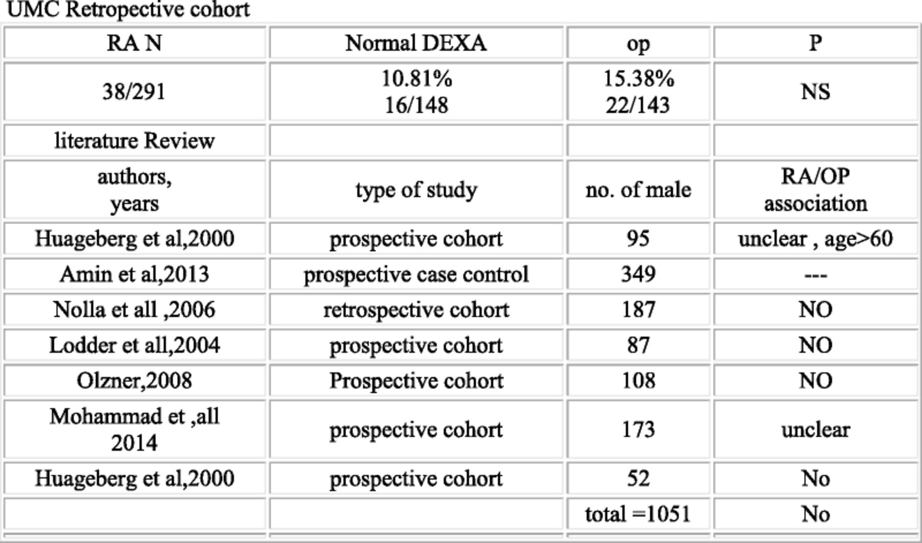
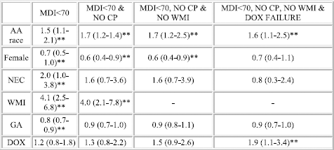
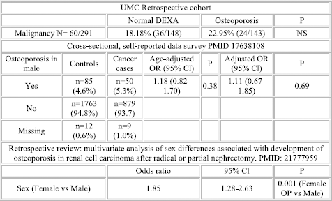
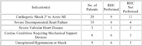
8 Ra May Not Confer Addition Risk of Osteoporosis in Male Ra Patient as Compared to Female
Bhawal J, Majithia V. UMMC, Jackson MS, MS.
Purpose of Study: Osteoporosis (OP) is a common disease and is increasingly being recognized in males. A number of underlying diseases including rheumatoid arthritis (RA) have been associated with it and this relationship has been well proven in females but unclear in males with RA. This study is done to evaluate association between RA and OP in male population.
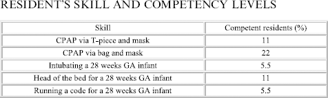
Methods Used: 585 men who underwent DEXA scan performed at UMC from 2005-2012 were included in the analysis of retrospective cohort with documented RA. PubMed literature search with was performed using keywords- Male, OP and RA yielding 775 articles (limiting data to English and publication year > 1960), 663 articles were excluded with abstract review, 112 underwent full text review with 7 having relevant data and were included in analysis.
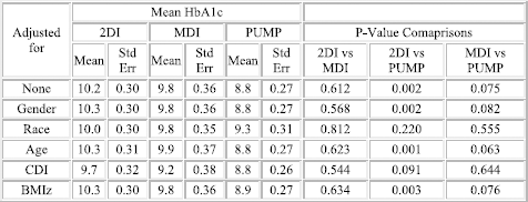
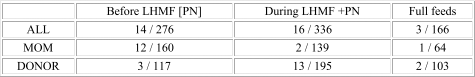
Summary of Results: as below
Conclusions: This analysis suggests that relationship of RA and OP in male patients remains confusing. Although no firm conclusions can be drawn, the results of cohort analysis and review of literature suggest that RA diagnosis and disease activity may not add any increase in the risk of developing OP above a low BMI, older age and steroid use. There was only one study out of 8 where RA itself seemed to add additional risk of developing OP while other studies suggested that this was not the case or possibly was only contributory in older age
FIGURE 1.
9 Benadryl Overdose Masked as Positive Tca in An Altered Mental Status Case
Smith MM, Engel LS, Guillory SG. LSU Health Sciences Center, New Oreans, LA.
Case Report: A 20 Year old woman presented was brought to the Emergency Room after falling unconscious in a cab. The patient had no identification available. The patient was very lethargic, only opening her eyes to sternal rub which did not improve following administration of naloxone. She was also tachycardia and received a 2 Liter normal saline bolus. CT scan of her head revealed no abnormalities. CBC and CMP laboratory data was unremarkable. Her EKG demonstrated QT prolongation of 503 and sinus tachycardia. A urine drug screen revealed a positive TCA level and her blood alcohol level was elevated as well. Benzodiazepines were ordered for patient on a prn basis and patient was sent to ICU for closer monitoring. The patient's mental status improved enough, so that she could provide a contact name. Her contact disclosed that early in the she took an undetermined amount of Benadryl tablets earlier that day after an argument with a significant other. Poison control was contacted, and we were able to discern that diphenhydramine can produce false positives on Urine drug screens. During her hospital course, she had multiple visual hallucinations, dry mouth, QT prolongation, and urinary retention. These symptoms resolved after nearly 48 hours of therapy with Benzodiazepines and IVF's.
DISCUSSION: According to the American Association of Poison Control there are over 600 compounds that have anticholinergic properties, including prescription, over the counter drugs, and plant products. The features of an overdose are usually anhidrosis, anhidrotic hyperthermia, hallucinations, nonreactive mydriasis, urinary retention, and cutaneous vasodilation. Benzodiazepines can be used to treat the agitation in these patients. Sodium bicarbonate should be used to treat prolonged QT syndrome. Sodium Bicarbonate was withheld, as this patient did not have persistent QT prolongation. Physostigmine, may be superior to benzodiazepines to treat the agitation. However, administration of physostigmine to a patient who has taken another toxin, may produce cholinergic toxicity. Therefore, it is recommended to give this medication in conjunction with a toxicologist consult.
10 Isolated Diplopia Caused by Calcineurin Inhibitor Therapy in a Patient with Idiopathic Calcineurin Membranous Nephropathy
Bahri NS, Adam-Eldien R, Gupta A. University of Florida, Jacksonville, FL.
Case Report: Both Cyclosporine (CyA) and tacrolimus (FK506) are widely used immunosuppressive drugs used to treat transplant recipients, autoimmune diseases and nephrotic syndrome. CyA binds to cyclophilin and Tacrolimus to the FK binding protein and the resulting complex inhibits calcineurin. Besides being predominantly present in lymphocytes, calcineurin is also found in abundance in the nervous tissues and diverse neurotoxicities ranging from tremors, headache, altered mental status, hallucinations and psychosis, peripheral neuropathy, seizures, cerebellar ataxia and leukoencephalopathy have been reported in the literature.
Our case is a 42 year-old female with biopsy proven idiopathic membranous nephropathy (MGN) who was being treated with FK506 and prednisone. Her Tacrolimus levels were maintained between 6-8 ng/mL. Her presenting urine protein/creatinine ratio of 8 gm/gm was successfully reduced to less than 1 gm/gm after 3 months of therapy when she presented with diplopia. The diplopia was binocular and vertical. The patient reported improvement in symptoms before the next dose was due-but she remained compliant with her medications despite the side effects. The symptoms persisted even when her tacrolimus dose was reduced and repeat levels were between 4-5 ng/mL. At this time she was switched to low dose CyA in anticipation that similar side effects may not be observed. Trough CyA levels were 44 ng/mL and 59 ng/mL on two occasions but her symptoms did not resolve. A consultation with neuro-ophthalmology was sought and the patient was instructed to discontinue CyA. The symptoms completely resolved 6 days after stopping CyA and the ophthalmology evaluation was never done. Her nephrotic syndrome remains in remission till date.
This is the first reported case of isolated diplopia secondary to FK506 and CyA in recommended therapeutic doses for treatment of Idiopathic MGN. Previously reported cases have occurred in the setting of much higher levels of CNIs, in patients with solid organ or bone marrow transplants. This case highlights the variable and unpredictable nature of calcineurin inhibitor neurotoxicity even at levels not considered to be toxic. Early detection and discontinuation of therapy led to resolution of the symptoms with no sequel in this case.
11 Hemophagocytic Lymphohistiocytosis: Pediatric Disease in an Adult
Provo J1, Kiefer AC1, Vasquez R2, Warrier R2. 1Tulane University School of Medicine, New Orleans, LA and 2Ochsner Medical Center, New Orleans, LA.
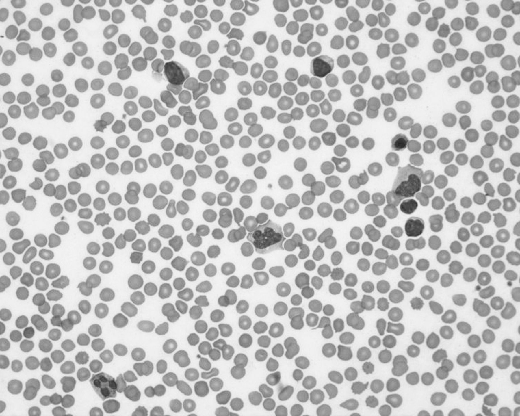
Case Report: Hemophagocytic Lymphohisticytosis (HLH) is a rare hyper-inflammatory state more common in children. We report a 21 year old Caucasian female in the Adult ICU with a fulminate illness following a Streptococcus A infection. The course of her illness proves that a high index of suspicion for HLH needs to be considered even in adults with multi-system and multi-organ failure.
Case: A 21 year old Caucasian female with morbid obesity presented to the emergency department with a fever of 40.1°C, sore throat, vomiting, rigors, cough, arthralgia, myalgia, rash, decreased oral intake, and lethargy. Her primary physician had diagnosed her to have strep throat with a positive culture 4 days earlier. She had fever, sore throat, and rash and started on Amoxicillin. She had no history of autoimmune disorders, sick contacts, pets, other medications or drug allergies. Physical Exam was remarkable for erythema and diffuse exudate of the tonsils bilaterally. Bimanual exam did not reveal the presence of a retained foreign body. Tender, violaceous macules were noted on the dorsum of the hands, feet, and chest wall. Labs demonstrated thrombocytopenia and leukocytosis. She quickly decompensated, developing respiratory distress requiring ventilator support and vasopressors. Her course was further complicated by kidney injury requiring dialysis, and DIC requiring blood products. Pediatric hematologists came to the rescue and suggested work up for HLH. Ferritin was > 10,000 ng/mL with a soluble IL-2 of >20,000 U/mL confirming an HLH diagnosis. She was treated with supportive care, dialysis, ventilation, steroids, Cyclophosphamide and Etoposide. She responded poorly and went on to develop toxic epidermal necrolysis and expired.
Pediatricians should teach internists to maintain a high index of suspicion for HLH in cases presenting as sepsis or multi-organ dysfunction.
12 An Unusual Time When Wernicke—s Makes Some Sense
Lemley RJ, Jenkins M, Smalligan RD. Texas Tech University HSC, Amarillo, TX.
Case Report: A 58yo right handed man with a history of bipolar disorder with psychosis and multiple CVAs presented with abnormal speech. Other PMH, FH, P/S were unremarkable. On physical exam he had a nonfocal detailed neurologic exam but his speech was abnormal. He spoke fluently with normal articulation and form, yet the speech contained no meaning. He repeated words and phrases in an attempt to convey his ideas. He had poor auditory processing and could not answer verbal questions. However, the patient understood written queries and provided short, appropriate verbal responses. The first few words of his response were meaningful but then he shifted into senseless speech. He did not have any recurrent theme to his answers. Head CT showed old left temporoparietal as well as multifocal right sided strokes.
Discussion: Hospitalists admit patients with stroke on a daily basis and more than 25% have an associated aphasia. Traditionally, aphasia is categorized as one of the following: Broca's, Wernicke's, global, transcortical or amnestic. The area of the brain affected typically correlates quite closely with the type of aphasia seen. Wernicke's aphasia, also called fluent or receptive aphasia, occurs when the temporoparietal cortex is damaged in the dominant hemisphere. In right handed patients, the left hemisphere is dominant 95% of the time (60% of left handed). Our patient's large left sided defect correlates with destruction of Wernicke's area, however, patients with Wernicke's aphasia usually have difficulty with both spoken and written word comprehension. It is important to note that some patients with schizophrenia or psychosis can have speech that resembles Wernicke's aphasia, however, their speech often involves fixation on a theme and their neologisms are more contextually appropriate. Rarely, as in our patient, one can still understand written language. Recent studies suggest that anterior temporal regions sustain semantic processing of visually represented language when posterior language areas are injured. This challenges the conventional neurobiological model of language, and new models are taking its place. It is important for physicians to be aware of both typical and atypical presentations of Wernicke's aphasia. Attempts to communicate with written, rather than strictly verbal cues, can be rewarding in certain instances.
13 Primary Cardiac Sarcoma Presenting as Heart Failure
Baldeo C, Ali RA, Chaudhry S, Khawaja F. UF Health-Jacksonville, Jacksonville, FL.
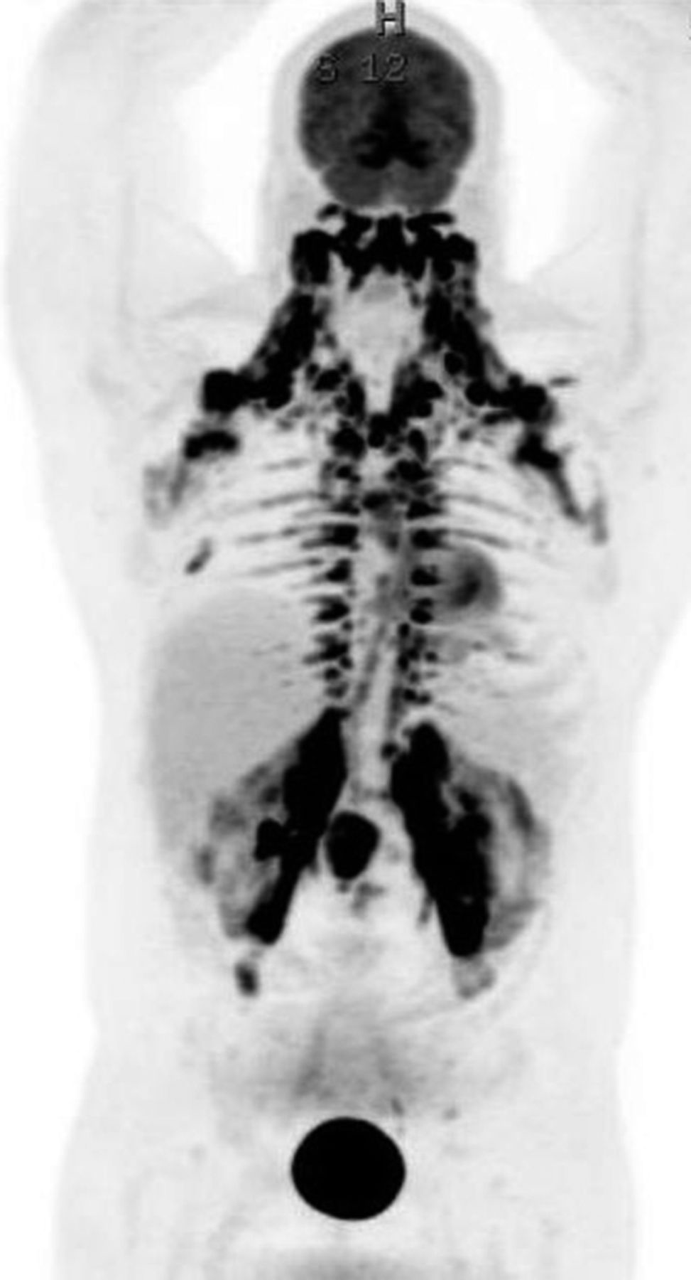
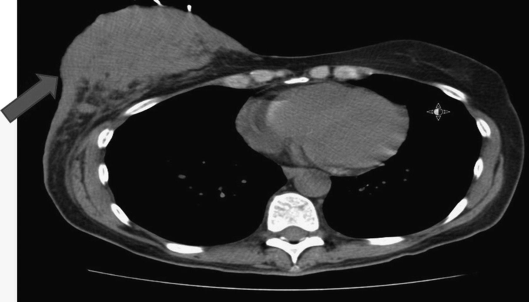
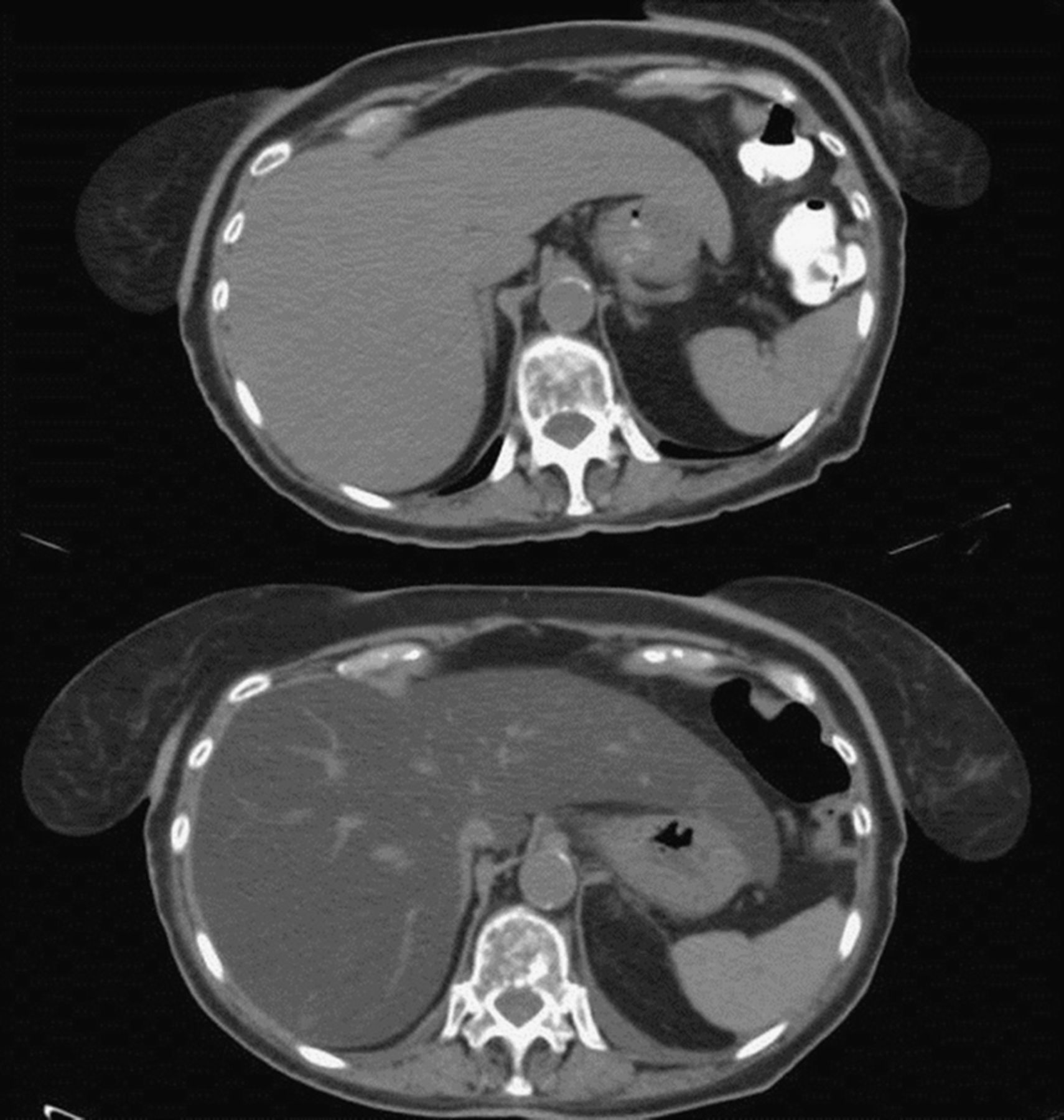
Case Report: A 64 year old female with hypertension and hyperlipidemia presented with worsening dyspnea on exertion, bilateral lower extremity edema and dry cough for 3 months. She lost 12lbs within 2 months. She never smoked.
Examination noted bilateral pitting pedal edema and right basilar crackles. CXR showed cardiomegaly and interstitial pulmonary edema. She had a low voltage EKG. CT chest showed 9.9x11.5x14.2cm mass near the anterior pericardium displacing the heart superiorly. Transthoracic echocardiogram showed a preserved ejection fraction but revealed a large extracardiac mediastinal mass compressing the right ventricle. Cardiac MRI showed a cystic-solid mass in the anterior pericardium. CT guided core needle biopsy resulted in spindle and epithelioid sarcoma. Left heart catheterization was normal.
Surgery showed an anterior mediastinal mass. Attempts to debulk the mass revealed it was also involving myocardium. After partial resection, the right ventricle was shown to be infiltrated and so the mass was considered unresectable. Pathology showed high-grade sarcoma with features favoring malignant perivascular epithelioid cell tumor. Postoperatively, she refused chemotherapy or radiotherapy and opted for hospice care.
The prevalence of primary cardiac tumors is 0.001-0.03%. Malignant tumors account for 25% of primary cardiac tumors and can occur in any chamber of the heart. Presentation of cardiac sarcomas are non-specific, ranging from arrhythmias to thromboemboli to heart failure, depending upon the location. To our knowledge this malignant perivascular epithelioid cell tumor is the fourth reported case found in the heart. Though complete surgical resection remains the gold standard, this particular case raises the possible benefit of neoadjuvant therapy.
FIGURE 1.
14 Immune System Gone Wild
Fidone EJ, Mirkes C. Baylor Scott and White Healthcare, Temple, TX.
Case Report: Hemophagocytic lymphohistiocytosis (HLH) is a rare hematologic disorder, with an estimated incidence of 1.2 cases per million per year, characterized by an exaggerated immune response leading to marked proliferation of reactive lymphohistiocytes, excessive release of inflammatory cytokines and eventual cytokine-induced, multi-organ failure. HLH presents a diagnostic challenge to physicians due to its ability to mimic more common hematologic, infectious and rheumatologic etiologies. However, prompt diagnosis of HLH is crucial, as this disorder progresses quickly and is invariably fatal without treatment.
A 28-year-old African-American male presents to the ED with a 3-day history of spiking fevers, and severe abdominal pain. He reports having a three month history of abdominal pain, night sweats, and a 100-pound weight loss. Vitals on presentation were normal except for a heart rate of 126 and a blood pressure of 104/66. Physical exam revealed a diaphoretic male with scleral icterus, axillary and inguinal lymphadenopathy, abdominal tenderness, and hepatosplenomegaly. Laboratory evaluation demonstrated pancytopenia, a ferritin level of 11,884 ng/mL and a fibrinogen level of <60mg/dL. Over the next few days, the patient continued to decline clinically, requiring multiple units of blood and cryoprecipate. An axillary lymph node core biopsy and a bone marrow aspirate and biopsy demonstrated hemophagocytic lymphohistiocytosis. After ruling out inciting infectious etiologies, the patient was initiated on high-dose corticosteroids. His clinical picture improved dramatically in the following days.
This case emphasizes the importance of early detection when confronted with HLH. The average time to diagnosis for HLH can range from 2 weeks to 3 months. However, patients suffering from HLH do not have the luxury of time. This disease is almost uniformly fatal within 2 months if left untreated yet, dramatic clinical response can occur once treatment is initiated. Thus, prompt consideration of HLH is paramount. This patient illustrates that a dramatically elevated ferritin strongly suggests an autoimmune hemolytic process. When coupled with hypofibrinogenemia, pancytopenia, generalized lymphadenopathy and hepatosplenomegaly, one must first consider a consumptive, rather than an infiltrative, process.
15 Isolated Cns Histoplasmosis in Immunocompetent Host; Mimicking Brain Metastasis
Mohamed A1, Edriss H1, Fenire M2, Ali E1, Mazek H1, Nugent K1. 1Texas Tech university health science center, Lubbock, TX and 2East Tennessee state University, East Tennessee, TN.
Purpose of Study: Histoplasmosis is a disease caused by the dimorphic fungus Histoplasma capsulatum. Most patients with histoplasmosis have no symptoms; however when symptomatic, it usually manifests with acute or chronic lung disease. Progressive disseminated disease is rare and almost always occurs in immunosuppressed hosts.
Methods Used: Case analysis and literature review.
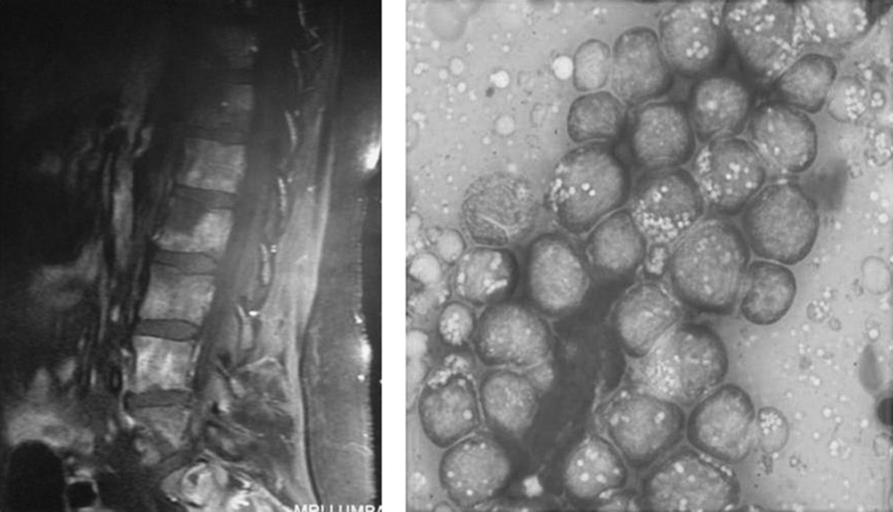
Summary of Results: We report a 56-year-old man from New Mexico with a 6 month history of weight loss, night sweats and personality changes. He also reported new onset headache and progressive right-sided weakness. His medical history is significant for hypertension. Physical exam demonstrated a well-nourished man in no acute distress; his right upper extremity muscle power was 1/5. Initial labs showed WBC 6.4x10³μL, Hb 11.6 g/dl and PLT 50 x 03μL. Head MRI with contrast showed multiple ring enhancing lesions with vasogenic edema. HIV, Hepatitis panel, toxoplasma, and leptospira serology were negative. Malignancy work up, including chest, abdomen/pelvis CT, bone scans, protein electrophoresis, tumor markers, bone marrow biopsy, were all normal. Non-specific lymphadenopathy was noted on the chest CT scan. Lumbar puncture shows lymphocyte pleocytosis with increased RBCs. CSF Gram stain, bacterial antigens, cryptococcal antigen, and fungal cultures were negative. A stereotactic right frontal craniotomy was performed; the tissue pathology showed granulomatous inflammation with multiple yeasts. Urine test was positive for Histoplasma capsulatum antigen. The patient was treated with one month of amphotericin and then voriconazole for 12 months. Repeated MRI after one month of amphotericin treatment showed improvement in the lesions.
Conclusions: This patient had Histoplasma capsulatum infection by tissue biopsy. Central nervous system involvement is rare as the sole presentation of histoplasmosis; it is almost exclusively presents as part of disseminated disease and yet occurs in only 10% of those patients. Our HIV negative patient had only CNS involvement and had no other evidence of systemic active infection. Physicians should maintain a broad differential in patients with ring enhanced brain lesions on CT head and include histoplasmosis even in low risk patients.
16 a Case of Hyponatremia Potentiated by Nsaids
Pourmorteza M1, Pourmorteza M1, Patel B1, Peiris A2, Patel P1. 1ETSU Quillen College of Medicine, Johnson City, TN and 2ETSU Quillen College of Medicine, Johnson City, TN.
Case Report: Introduction:Non-steroid anti-inflammatory agents (NSAIDs) as a sole source of hyponatremia is uncommon. They do so by inhibiting prostaglandin synthesis leading to a potentiation of vasopressin, water reabsorption, enhanced fluid retention and ultimately hyponatremia. We report an unusual cause of hyponatremia due to enhancement of desmopressin effect by NSAIDS.
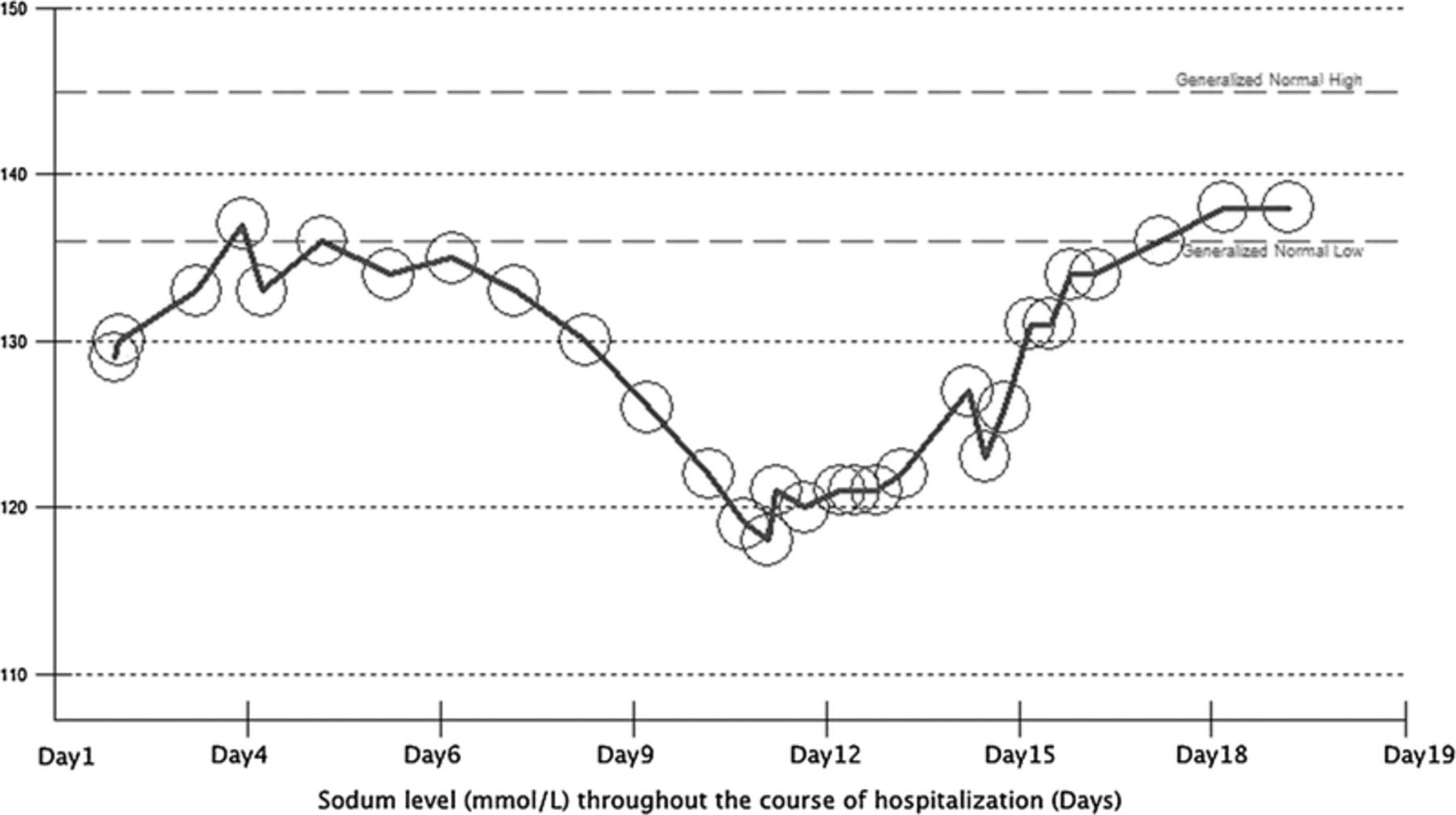
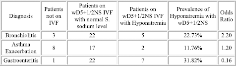
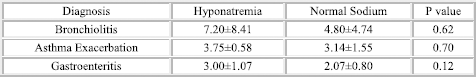
Case Report: A 47 year old male admitted to the hospital for 2 weeks of back pain, headache, nausea, acral edema. Past medical history include resected pituitary macroadenoma, hypocortisolism, hypogonadism, Diabetes insipidus. He was on desmopressin, Hydrocortisone and Testosterone Injections. Vitals were within normal limits. Physical exam demonstrated 10 lb weight gain in two weeks, mild facial and extremity swelling. Laboratory analysis showed sodium 117 mmol/L, plasma osmolality 253 mOsm/Kg, sodium excretion 63 meq/L, urine osmolality 406 mOsm/Kg. Patient began taking Ibuprofen 600 mg every 4 hours two weeks ago for back pain. After excluding other causes of hyponatremia, enhanced desmopressin effect by Ibuprofen was the likely cause of hyponatremia. Ibuprofen was discontinued as desmopressin regiment was reduced to twice daily with free water restriction. Sodium increased gradually to 137 on outpatient follow up. Subsequently the desmopressin was changed to previous TID dose and patient has done well.
Discussion: Hyponatremia usually develops during the first two weeks after drug initiation. NSAIDs as a sole source of hyponatremia is uncommon and are more commonly observed in the setting of altered renal function. NSAIDS like ibuprofen cause a reduction in renal prostaglandin synthesis which normally antagonize antidiuretic hormone (ADH). Such inhibition potentiates the effects of ADH which ultimately leads to decreased water excretion. This case represents a unique cause of NSAID-induced hyponatremia in the setting of Diabetes insipidus treated with desmopressin. Mortality rates tend to increase as the serum Na falls from 134 to 120 mEq/L. Therefore, clinicians should be aware of patients with altered fluid status, since common over the counter medication such as NSAIDs impact fluid balance and pose a great risk for induction of hyponatremia.
Pediatric Clinical Case Symposium
1:00 PM
Thursday, February 26, 2015
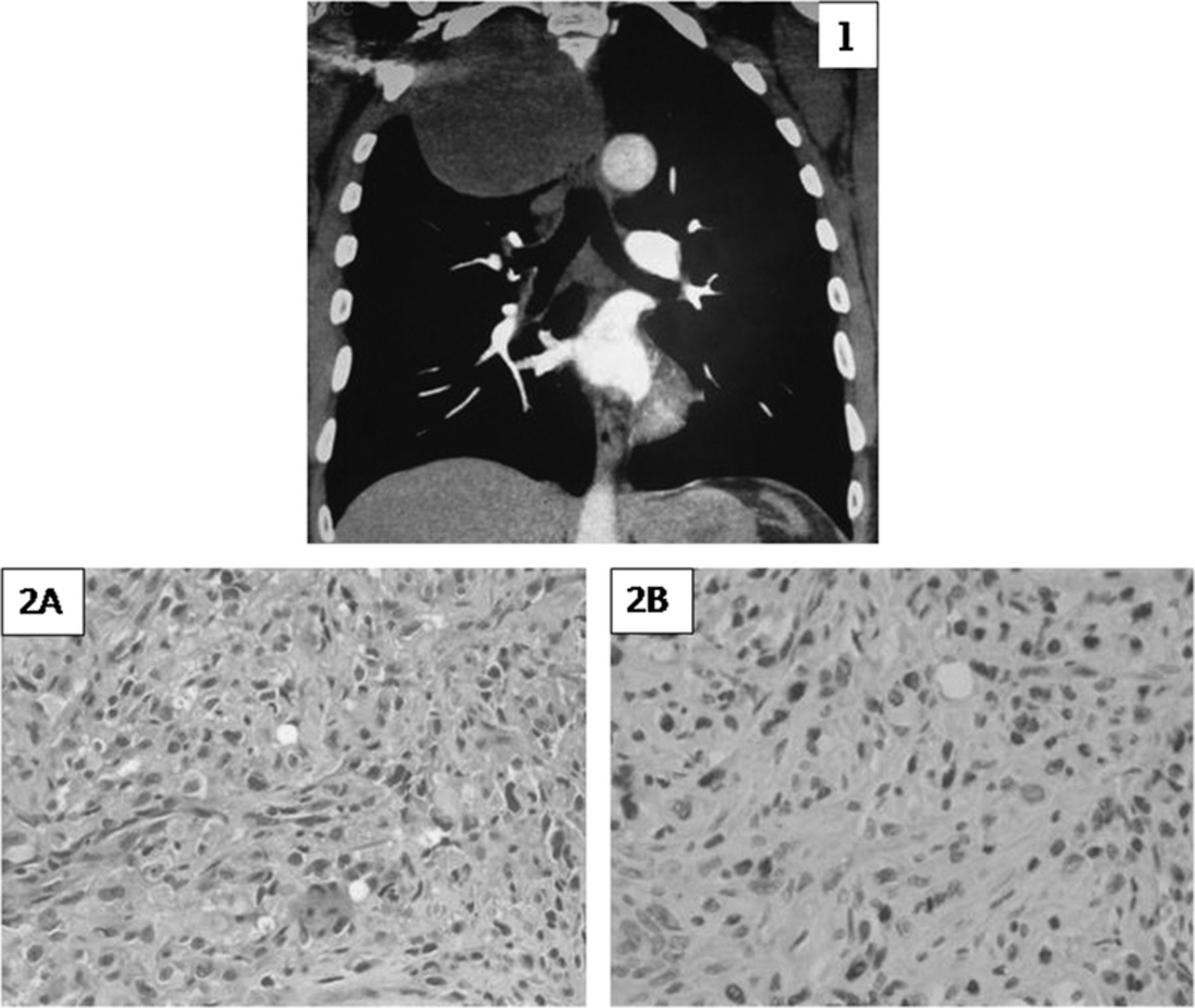
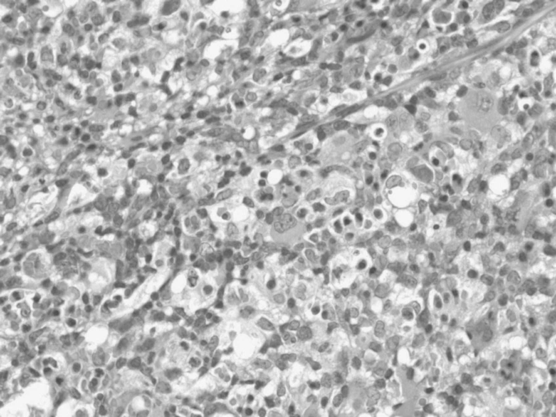
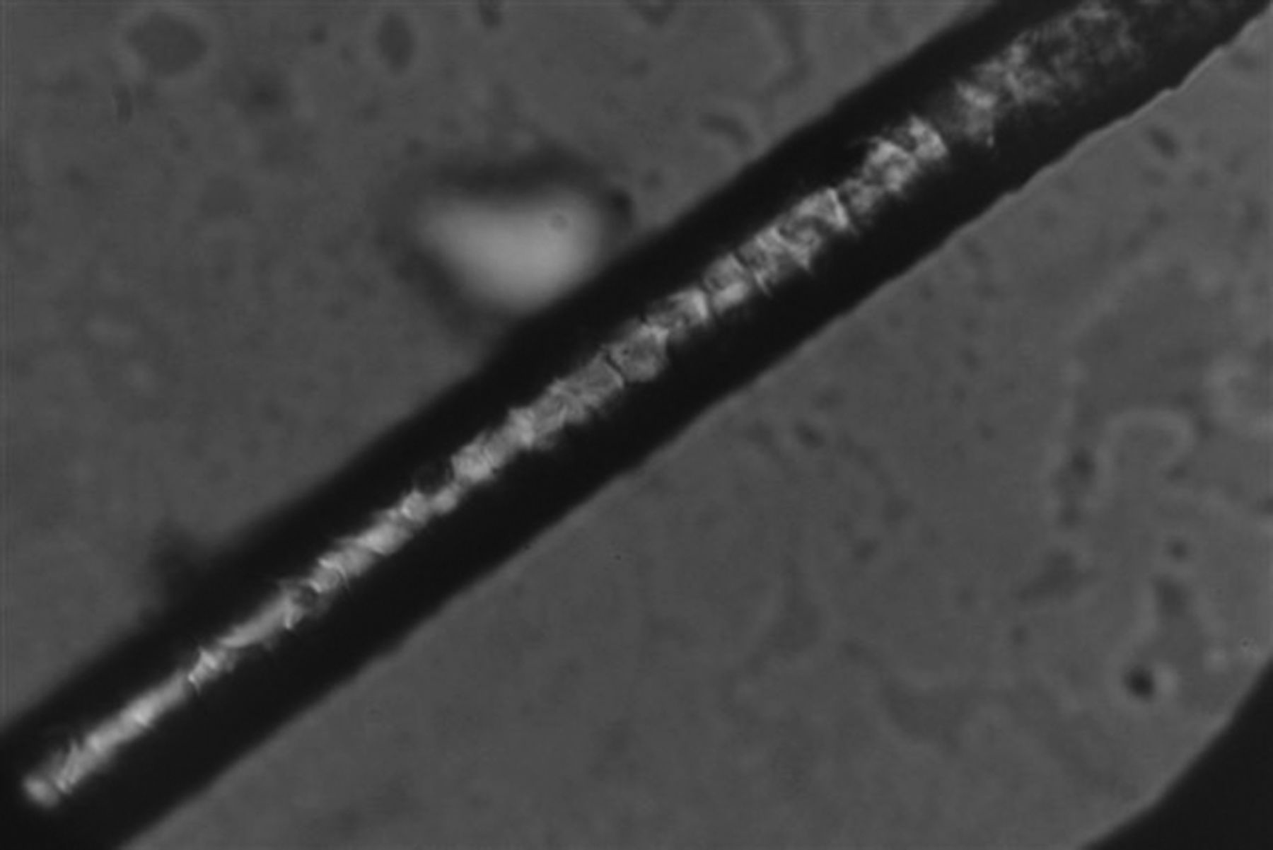
17 a Lung Mass in a Teenage Male with Asthma
Mirani G1, Valerie E2, Smith RK3, Greer DL4, Craver R3, Begue R5. 1Tulane University Health Sciences Center, New Orleans, LA; 2Children's Hospital, New Orleans, LA; 3Children's Hospital, New Orleans, LA; 4Louisiana Health Sciences Center, New Orleans, LA and 5Children's Hospital, New Orleans, LA.
Case Report: A 17-year-old male with a history of asthma and allergic rhinitis presented with cough, chest pain, and shortness of breath for several weeks. An outpatient chest x-ray revealed a right middle lobe lung mass. The patient underwent an open thoracotomy with a wedge resection of the 4 x 2 cm lesion, extending from right middle lobe into the hilum. The pathology of the resected mass was reported as necrotizing granulomatous inflammation with fungal hyphae, marked eosinophilia, and bronchiectasis. The fungal culture eventually grew Bipolaris species (Figure 1). The susceptibility testing showed following MICs: AMB 0.25, ITRA 0.25, and VORI 0.25 (μg/mL, University of Texas Health Science Center at San Antonio). The patient tolerated a three-month course of itraconazole with resolution of symptoms. Significant laboratory values included an eosinophilia of 1510 (normal 0-390/UL), elevated IgE level of 4920 (normal 0-158 IU/mL), and Bipolaris australiensis IgE of 54.80 kU/L (normal < 0.35, ARUP Laboratories). Subsequent skin scratch tests to various mold spores including Bipolaris showed marked reactivity. This clinical picture would fit the diagnosis of allergic bronchopulmonary mycosis with Pulmonary Phaeohyphomycosis due to Bipolaris spp.
FIGURE 1.
18 Persistent Fevers in An 18-Year-Old Renal Transplant Recipient
Mirani G1, McNaughton J2, Yosypiv IV1, Schmieg JJ2, Robinson J1. 1Tulane University Health Sciences Center, New Orleans, LA and 2Tulane University Health Sciences Center, New Orleans, LA.
Case Report: An 18-year-old male, with a history of kidney transplant in 2006, presented with fevers upto 104 °F for the 2-3 days prior to hospital admission in January 2014. The patient also reported a mild cough and headaches. The cough was non-productive. His headaches were non-focal and non-persistent and were present upon waking. His sister at home had a recent upper respiratory infection. There was no vomiting, diarrhea, dysuria, urinary frequency or urgency, or hematuria. The patient did not report intake of unusual foods or recent travel. His immuno-suppressive medications included cyclosporine, mycophenolate mofetil, and prednisone. CT scan of the chest revealed multiple lymph nodes including a large right-sided axillary lymph node. Excisional axillary lymph node biopsy performed on the 7th day of hospitalization, revealed necrotizing granulomatous lymphadenitis with features most consistent with cat scratch disease. Warthin-Starry stains highlighted rare possible bacilli (Figure 1). Bartonella DNA PCR (qualitative) from plasma was positive. Bartonella Henslae IgM was negative, and IgG was 1:640 (negative < 1:320 titer). The patient was treated with azithromycin 250 mg orally daily for 6-8 weeks. He became afebrile within 24 hours of starting antibiotic. Repeat CT scan a month later showed improved lymphadenopathy.
FIGURE 1.
19 Cutaneous Lymphoma Mimicking Benign Skin Rash
Sharma A, Radulescu VC. University of Kentucky, Lexington, KY.
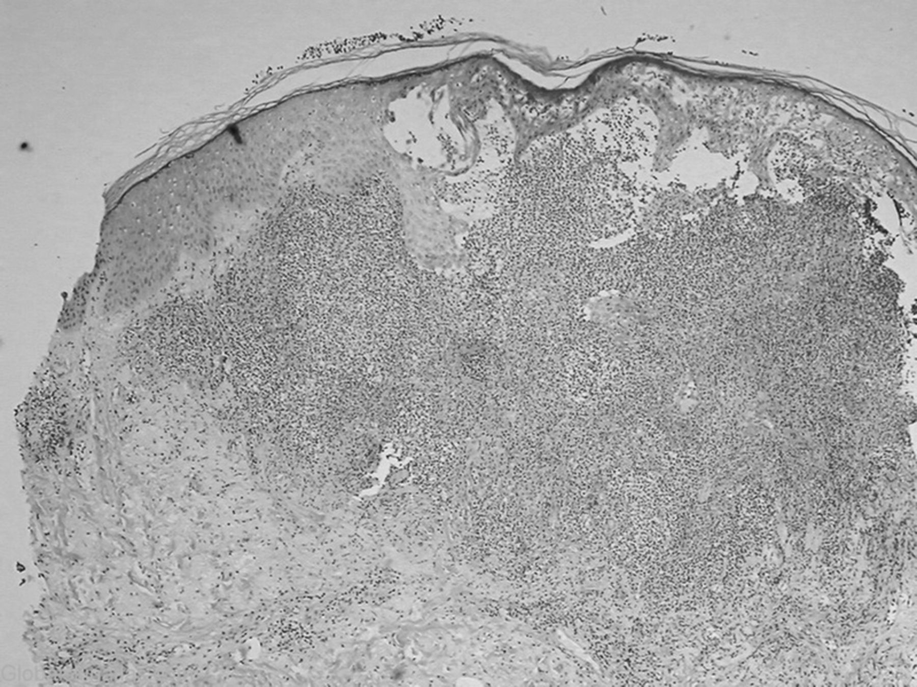
Case Report: A previously healthy 10-year-old female presented with low-grade intermittent fever and multiple areas of red tender rash since last 3 months. On examination, she had multiple erythematous, well circumscribed indurated plaques and nodules, 3 to 5 cm in diameter present on both arms, right leg, right flank and back.
CBC was normal except for a low WBC count (1900/uL). Systemic markers of inflammation (ESR, CRP, procalcitonin) were within normal limits. Serum electrolytes were normal but liver enzymes were mildly elevated (AST 187U/L, ALT 172U/L, ALP 155U/L). Extensive infectious disease and immunological work up remained negative. Imaging studies of chest, abdomen and pelvis did not show any abnormality or lymph node enlargement. Bone marrow microscopy was normal as well.
Skin biopsy of the lesions showed an epidermal perivascular infiltrate consisting of atypical lymphocytes and histiocytes surrounding individual fat cells in a necklace-like pattern along with fat necrosis and karyorrhexis. Immunohistochemical staining led to the diagnosis of subcutaneous panniculitis-like T-cell lymphoma (SPTCL). She was treated with cyclophosphamide, doxorubicin, vincristine and prednisone (CHOP) with good results.
Discussion: SPTCL is a T-cell lymphoma localized primarily to the subcutaneous adipose tissue without lymph node involvement. In SPTCL, T cells have a CD4-CD8+CD56- TCRαβ phenotype. Cutaneous lymphoma with CD4-CD8-CD56+ TCR γδ phenotype, previously included under SPTCL, is now classified as cutaneous γδ T-cell lymphoma as it has an aggressive course with worse prognosis. The incidence of SPTCL is <1% of the non-Hodgkin lymphomas. It is exceedingly rare in children and has a female predominance. Patients usually have leukopenia with mildly elevated liver enzymes. Biopsy histology as described above is pathognomonic of SPTCL. It has a good prognosis with complete resolution of the lymphoid infiltrate either spontaneously or after chemotherapy.
FIGURE 1.
20 Acute Kidney Injury and Hypertensive Emergency in a Patient with Class V Lupus Nephritis
Cantu MS1, Kidd L2, Singh D1, Yosypiv IV1. 1Tulane School of Medicine, New Orleans, LA and 2Tulane School of Medicine, New Orleans, LA.
Case Report: A 13 year old African American female with a past medical history of class V lupus nephritis (LN) with normal kidney function well-controlled with mycophenolate, hydroxychloroquine and enalapril, presented with recent facial abscess, progressive body swelling and right-sided flank pain. Physical examination showed elevated blood pressure of 171/117 mmHg, periorbital and lower extremity edema. Laboratory studies showed elevated serum creatinine of 2.9 mg/dL, reduced albumin of 2.5 gm/dL, reduced C3 complement of 58 mg/dL, elevated antibody titers: ANA (1:640), RNP (5.7), Smith (>8), and SSA (>8), reduced hemoglobin of 8.4 mg/dL and hematocrit of 26%, increased urine protein to creatinine ratio of 5.6, with 2 RBCs and no identifiable casts. After achieving control of hypertensive emergency, renal biopsy was performed to determine the cause of acute kidney injury (AKI). Light microscopy of the biopsy revealed diffuse segmental proliferation, presence of active lesions in 59% of glomeruli, cellular crescents in 14% glomeruli and global sclerosis in 20% of glomeruli. IF demonstrated a “full house” immunofluorescence with IgG, IgA and c1q. EM showed numerous subepithelial, mesangial and occasional subendothelial electron-dense deposits. The patient received high-dose pulse steroids intravenously for 3 consecutive days followed by intravenous infusion of cyclophosphamide. This case illustrates superimposition of class IV LN on stable class V LN presenting as AKI and hypertensive emergency. This case indicates the need for careful life-long monitoring of patients with LN.
21 Neonate with Bilateral Pneumothoraces
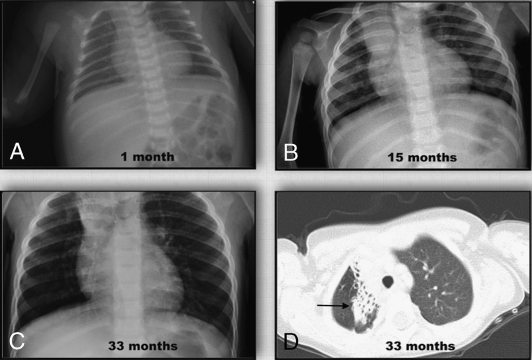
Connor EE1, Craver R1,2, McGoey R1. 1Louisiana State University Health Sciences Center, New Orleans, LA and 2Children's Hospital of New Orleans, New Orleans, LA.
Case Report: A female neonate delivered via caesarian section at 26 weeks gestation experienced immediate respiratory distress after birth requiring transfer to the neonatal intensive care unit and intubation. Imaging showed bilateral pneumothoraces, and chest tubes were placed. Despite these interventions and aggressive supportive care, the infant expired within 24 hours of birth. An autopsy was authorized by the family and later performed by an attending and resident pathologist. Immunohistochemical stains were subsequently selected and performed.
The autopsy was unremarkable with the exception of the lung findings. The lungs were hyperinflated and had a combined weight of 34gm [expected 18±6gm]. The pleural surfaces were finely nodular with visible faint white streaks. Sectioning revealed a firm tan-pink parenchyma with a diffuse, subtle cystic pattern that extended to the pleural surface. Microscopic analysis of all five lung lobes showed dilated cystic spaces in three contiguous sites: subpleural, interlobar, and periarterial. In most spaces, the lining was disrupted or fragmented. Immunohistochemical staining showed these lining cells to be positive for CD31 and D2-40. These findings are consistent with the diagnosis of primary pulmonary lymphangiectasia.
Congenital pulmonary lymphangiectasia (CPL) is a rare disorder and is often not diagnosed until autopsy. Literature reports over the past roughly 150 years cite less than 100 total cases. Thought of as a uniformly fatal disorder, CPL is thought to account for 0.5 to 1% of all stillborn and neonatal deaths. The cause is unknown but postulated to be due to an inherent developmental abnormality of the lymphatic system leading to dilated lymphatic spaces and lymphatic dysfunction. Primary CPL is limited to the lungs and does not affect in utero development, but presentation immediately after birth includes severe respiratory distress, pneumothoraces and pleural effusions. Greater awareness of CPL is needed and further reports in the literature would undoubtedly enhance our understanding of the condition and focus efforts towards the development of new therapeutic models.
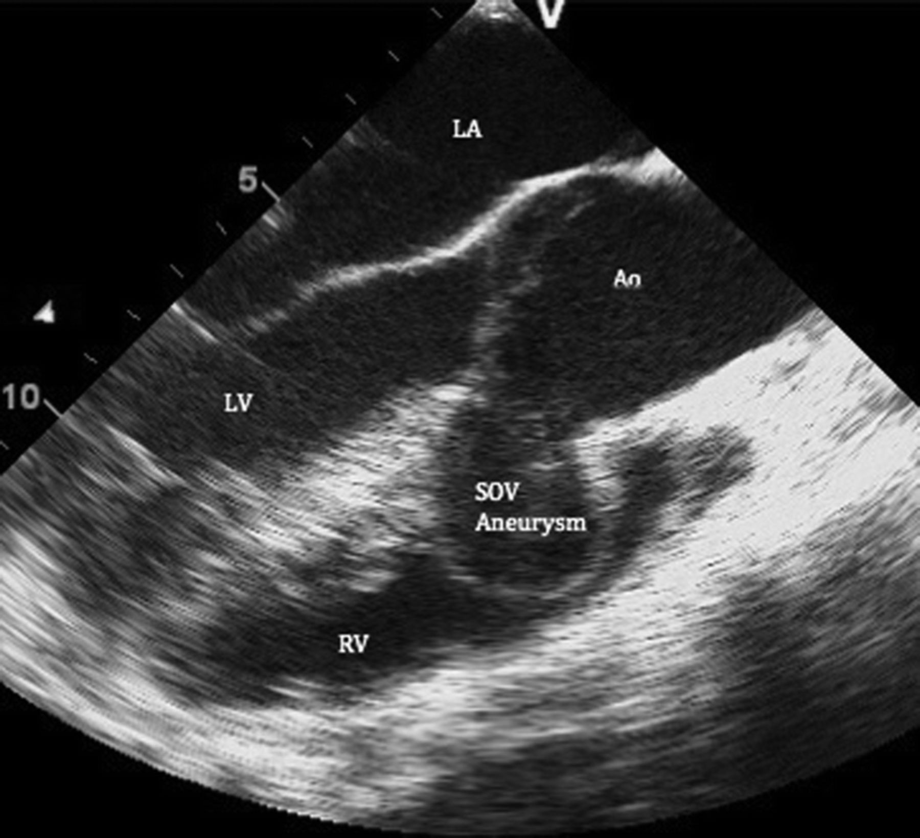
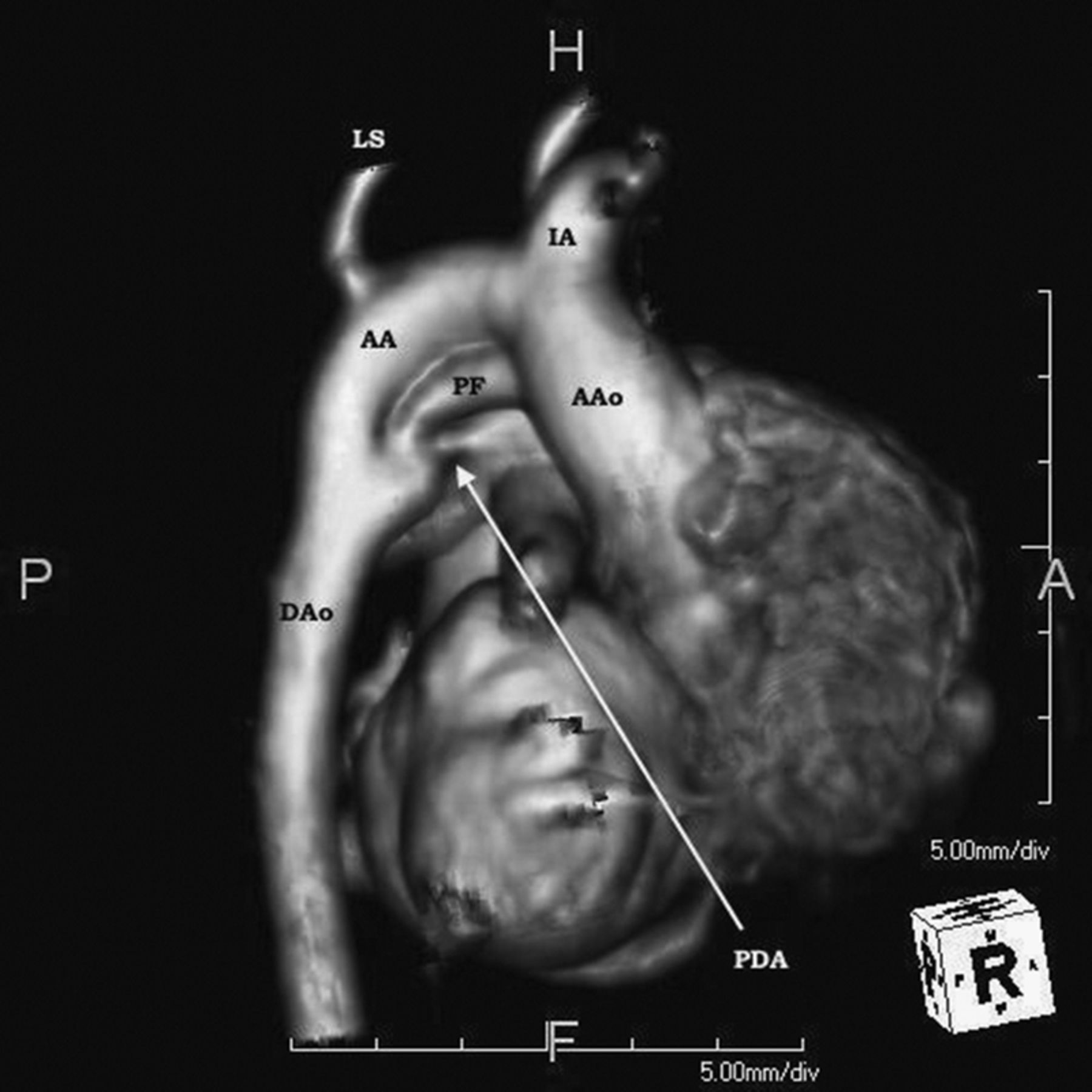
22 Self-Resolving Ortner—s Syndrome in a Term Neonate
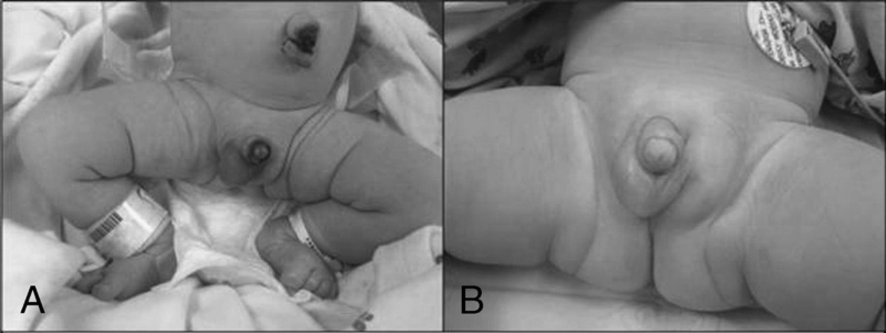
Pierce E, Bhatia J, Chan A. GRU, Augusta, GA.
Case Report: Ortner's syndrome, defined as left recurrent laryngeal nerve dysfunction resulting from cardiovascular pathology, is a rarely reported phenomenon in neonates. This case report describes a term infant who presented with hoarse cry and stridor at 33 hours of life accompanied by desaturations and respiratory distress. Work up included an echocardiogram which demonstrated the presence of a moderately large ductus arteriosus aneurysm; MRI confirmed that the mass was exerting an effect on the left recurrent laryngeal nerve. Bedside nasolaryngoscopy confirmed hypomobility of the left vocal cord. The infant was observed for several days since feedings were tolerated and desaturation episodes improved. After seven days of observation and serial echocardiograms, the aneurysm showed signs of regression without surgical intervention. This case emphasizes the importance of considering alternative pathologies for otherwise narrow differential diagnosis of hoarse cry in the neonatal population. Furthermore, unlike some previously reported cases, no surgical intervention was necessary confirming the current recommendations for several days of observation prior to pursuing surgical repair.
23 a CASE OF GRANULICATELLA ADIACENS INFECTIVE ENDOCARDITIS IN a SIX YEAR OLD
Neemuchwala F1, Struk M2, Burns J1, Whittingham E1. 1Florida State University, Pensacola, FL and 2Highland Regional Medical Center, Sebring, FL.
Case Report: Timely diagnosis of infective endocarditis (IE) in children can be a difficult clinical challenge. Herein we report a case of IE caused by an uncommon organism.
A six year old female was admitted with a history of persistent fevers and fatigue of one month duration. She had a past medical history of congenital aortic atresia, ventricular septal defect, status-post Norwood and Damus-Kaye-Stansel procedure as an infant and 6 months prior to admission had a bovine conduit placed for pulmonic stenosis.
Four weeks prior to admission she was seen at the primary care clinic for fever 103.2°F diagnosed as a viral syndrome. Three days later she had a dental procedure and although prophylaxis for endocarditis was prescribed, the patient did not take the medication. Five days later she was evaluated in the ED for persistent fever for 8 days where a blood culture obtained grew Granulicatella adiacens (GA). This was felt to be a contaminant and no antibiotics were started. Patient had repeat blood culture in ED 5 days later which again grew GA but was not treated because at that time child was clinically well. Finally, an additional 2 weeks passed and child was admitted due to recurrent fever.
On admission, child had fever 101.8, and there was a harsh systolic murmur which was documented previously. Labs revealed elevated ESR and CRP with anemia and thrombocytopenia. The patient was started on Ceftriaxone and Vancomycin. Echocardiogram showed an echogenic density in the conduit. Blood cultures obtained on admission grew GA which was Vancomycin sensitive but Penicillin and Cephalosporin resistant. Hence decision was made to discontinue Ceftriaxone and complete therapy with intravenous Vancomycin.
GA is a genus of nutritionally variant streptococci. It is often difficult to isolate in clinical laboratories and is reported to be resistant to standard treatments for streptococci. Previous reports of combined treatment with Vancomycin and Meropenem have been documented but our patient had successful treatment with single agent therapy. In conclusion, GA should not be considered a contaminant as it can cause endocarditis in pediatric patients and prophylaxis for endocarditis must be witnessed in high risk patients.
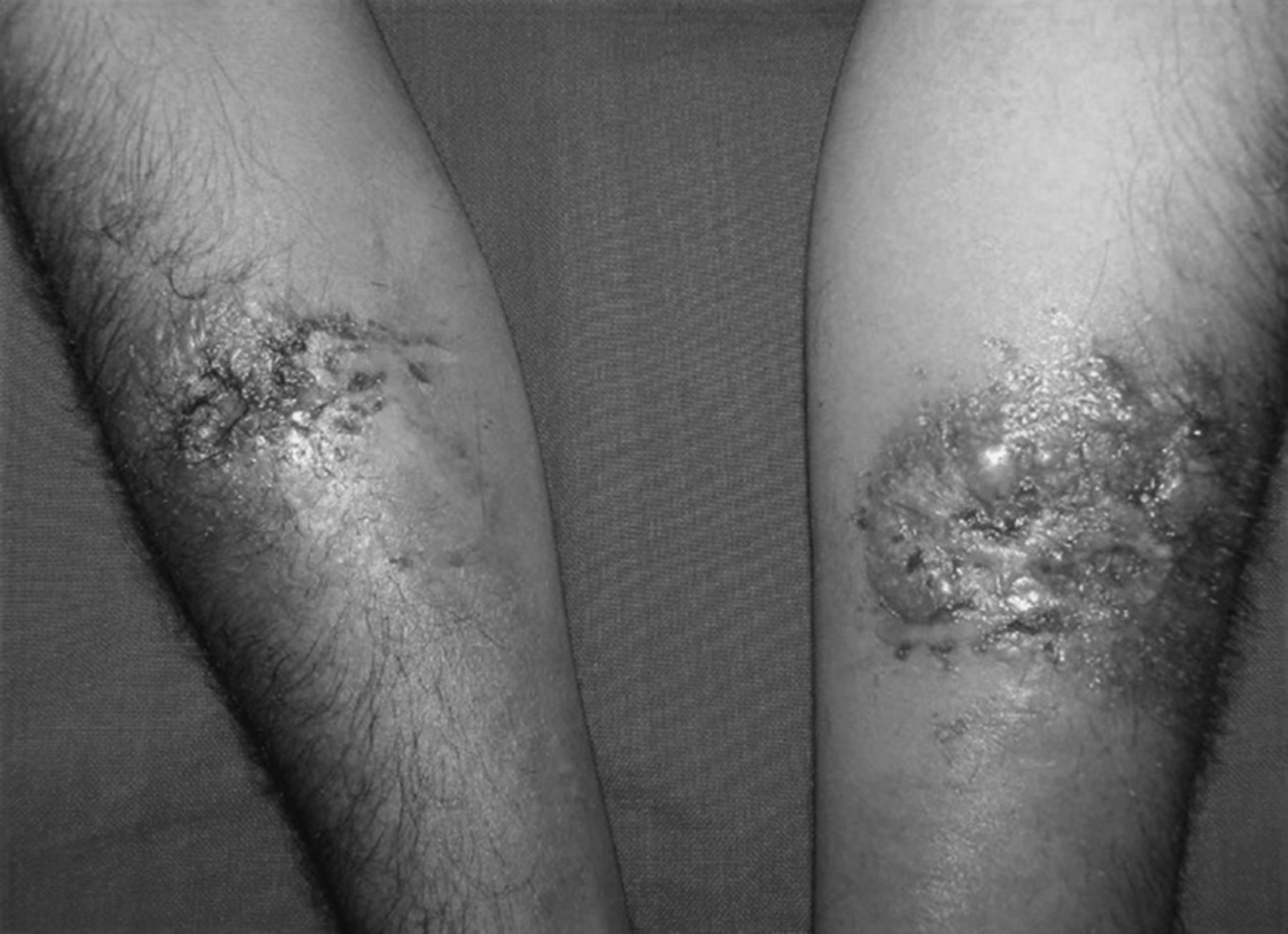
24 Acute Generalized Exanthematous Pustulosis: A Rare Pediatric Case Due to Clindamycin
Edgar-Zarate C1, Kaliki V1, Shalin S2. 1University of Arkansas Medical Sciences, Little Rock, AR and 2University of Arkansas for Medical Sciences, Little Rock, AR.
Case Report: Acute generalized exanthematous pustulosis (AGEP) is an acute reaction characterized by the sudden eruption of hundreds of sterile, non-follicular pinhead sized pustules predominantly in the main folds of the skin on a background of erythema. The incidence of AGEP is estimated to be around 1 to 5 cases per million per year with a slight female predominance. Previous studies have shown that the majority of cases are caused by adverse drug reactions and infections. AGEP has been reported only in a handful of pediatric cases, with drugs such as beta-lactams and cephalexin as the suspected causative agent. Clindamycin has been suspected to be the etiology of AGEP only in adults. We here report the first case of AGEP associated with clindamycin exposure in a child.
A previously healthy 9-year-old boy presents after taking 1 dose of clindamycin prescribed for cellulitis to his left lower leg. The following day, he developed diffuse erythema and fever. The rash progressed the next day to a pustular appearance. Once admitted to the hospital he developed hypotension, and was treated for toxic shock syndrome. A skin biopsy was performed and showed subcorneal pustules, intraepidermal neutrophils and adjacent epidermal edema which is consistent with AGEP. His hospitalization was complicated by anemia and hypoalbuminemia, for which he did received an albumin infusion. He made a full recovery with spontaneous resolution of the rash in about 9 days after cessation of the clindamycin.
The diagnostic criteria of AGEP are an acute pustular eruption, fever, leukocytosis, subcorneal or intra- dermal pustules on skin biopsy, and spontaneous resolution in less than 15 days. Our patient met all of these criteria. No treatment is necessary with AGEP as it was evidenced in the presented case. While systemic manifestations are rare in AGEP, abnormal liver function tests, renal insufficiency, respiratory distress, and agranulocytosis have been reported in adults. We suspect the hypotension, anemia and hypoalbuminemia in this case were secondary to AGEP, although this remains unclear. We believe AGEP is a rare but potentially severe adverse reaction to clindamycin and prompt discontinuation should be considered if highly suspected.
25 Porcelain Gallbladder in a Child with Nephrotic Syndrome Presenting with Spontaneous Bacterial Peritonitis
Richard KR, CaJacob NJ, Askenazi DJ, McCall DC. University of Alabama at Birmingham, Birmingham, AL.
Purpose of Study: To Report the First Case of Porcelain Gallbladder in A Patient with Nephrotic Syndrome Presenting with Spontaneous Bacterial Peritonitis.
Methods Used: PUBMED Literature search, single case report.
Summary of Results: Porcelain gallbladder, or gallbladder wall calcification, is a rare condition, the etiology of which is not well understood. It has been hypothesized that it results from inflammation or irritation of the gallbladder from causes such as disordered calcium metabolism, chronic cholecystitis, cholelithiasis, abdominal trauma, or surgery. The clinical significance of porcelain gallbladder has recently been debated secondary to recent literature revealing a lesser but still significant association with gallbladder cancer than was previously recognized in older studies. This is significant because gallbladder cancer carries a poor prognosis. We report the first case of a child presenting with porcelain gallbladder without a prior episode of cholecystitis, cholelithiasis, abdominal surgery, trauma, or a known disorder of calcium metabolism and demonstrate relevant imaging findings.
Conclusions: The pathogenesis of porcelain gallbladder is not yet fully understood, but the role of gallbladder wall inflammation appears to be a common factor most often thought to be from direct gallstone irritation. This pediatric case of nephrotic syndrome with spontaneous bacterial peritonitis demonstrates our hypothesis that, perhaps, any process leading to inflammation in the peritoneal cavity or generalized edema may be a risk factor for gallbladder wall inflammation. Furthermore, the recurrence of edema, such as from hypoalbuminemia associated with nephrotic flares, may be the catalyst for gallbladder wall calcification, even without overt symptoms of gallbladder disease.
26 Iliopsoas Abscess Presenting as Atypical Kawasaki Disease
Hall JE, Mitchell H, Pruitt CM. UAB, Birmingham, AL.
Case Report: A 13-month female presented to the emergency department (ED) for her fourth visit for recurrent fever. She initially presented to the ED 3 weeks prior due to fever for 7 days. At that time, she had a normal examination, a white blood cell count (WBC) of 15,600/µL, C-reactive protein (CRP) of 4 mg/L, and erythrocyte sedimentation rate (ESR) of 67 mm/hr. Blood cultures were obtained and she was asked to follow up as an outpatient. She returned 2 later (day 9 of illness) to the ED with continued fever and had developed nasal congestion, dry, cracked lips, mild right inguinal adenopathy, and diaper rash. Labs were significant for WBC 11,260/µL, CRP 5 mg/L, and ESR 88 mm/hr. Blood cultures had no growth to date. She was diagnosed with atypical Kawasaki Disease (KD) and treated with intravenous immunoglobulin (IVIG) and high-dose aspirin. An echocardiogram revealed coronary artery ectasia without dilation. Fever subsided with therapy and she was discharged home on day 11 from onset of fever. On day 12, fever recurred, and she was readmitted for treatment of atypical KD with IVIG and solumedrol. At this time, labs were significant for WBC 18,000/ µL, CRP of 6 mg/L, and ESR of 94 mm/hr. Repeat echocardiogram was unchanged. She improved and was discharged home on day 14. On day 29 of illness, she saw her pediatrician for recurrent fever, a new limp, and right groin swelling. An ultrasound of the right hip revealed right inguinal lymphadenopathy and no hip effusion. She was referred back to the ED, where her examination revealed an antalgic gait and decreased passive range of motion of the right hip. Her WBC was now 32,900/µL, hematocrit 20%, platelets >1,000,000/µL, CRP 21mg/L, and ESR 107mm/hr. Computerized tomography of the abdomen and pelvis demonstrated a large right iliopsoas abscess. Interventional Radiology drained the abscess and the child was started on antibiotics. The abscess culture grew methicillin sensitive Staphylococcus aureus requiring nafcillin for 18 days and then transitioned to cephalexin for two weeks upon outpatient follow up with Infectious Disease. An immunodeficiency evaluation and repeat echocardiogram were normal, all blood cultures were negative for growth, inflammatory markers normalized and she returned to baseline.
27 PRIMARY HIV MANIFESTING AS ACUTE PANCREATITIS IN AN ADOLESCENT, SYMPTOM OF a LARGER PROBLEM?
Lamb G, Dietz S, Graham R. University of Texas Southwestern, Dallas, TX.
Case Report: AF is a 16 year old African American male who presented to the hospital in August for acute abdominal pain in the setting of 3 months abdominal pain, anorexia, fever and weight loss. Symptoms began in May, but were considered insignificant and treated with analgesics and antipyretics until June 12 when the patient's mother noted that his pain and fatigue became significant enough to keep him bed-ridden, he continued to have intermittent fevers and began to lose weight. Between June 23 and July 2 he saw his PCP twice and each time was found to have mildly elevated liver enzymes and lipase trended from 821 to 1079. Symptoms improved until 8/1 when he presented to the ED with worsening abdominal pain. At this lipase was 980, abdominal US showed no abnormalities, he denied alcohol use and a lipid profile was normal. On 8/9 he had fever of 104.5 and developed emesis and watery diarrhea. He was unable to tolerate PO and urine output decreased. He developed fatigue and dizziness and presented to the ED on 8/13. Labs showed ALT: 521, AST: 257, T.Bili: 1.3, D.Bili 0.6, GGT: 92 and lipase: 490, CBC was unremarkable. He reported a 13kg weight loss over the past month. CT showed inflammation of the pancreas, but no necrosis and no tumors. Pt denied illicit drug use or sexual activity, but HIV screen was positive. Quantitative PCR returned with >10,000,000 copies. The patient's symptoms improved and HAART was deferred to outpatient treatment in order to prevent recurrence of pancreatitis.
Pancreatitis in the pediatric population is most commonly caused by trauma, medications, biliary tract disease and infections. Acute pancreatitis due to primary infection with HIV-1 has been rarely reported (7 adults, 1 adolescent). Although it has been well described as a complication of HIV secondary to medications and opportunistic infections, case reports now suggest that acute pancreatitis can be due to direct invasion of the pancreas by HIV. Dallas County has the highest rate of new HIV diagnoses in the state of Texas. The highest increase in new diagnoses is in persons aged 15-24. This extremely rare presentation has focused attention on the rapid growth of HIV + adolescents in the region and the need for a high level of suspicion in adolescent patients presenting to the ED.
Case Reports in Cardiovascular Medicine
2:00 PM
Thursday, February 26, 2015
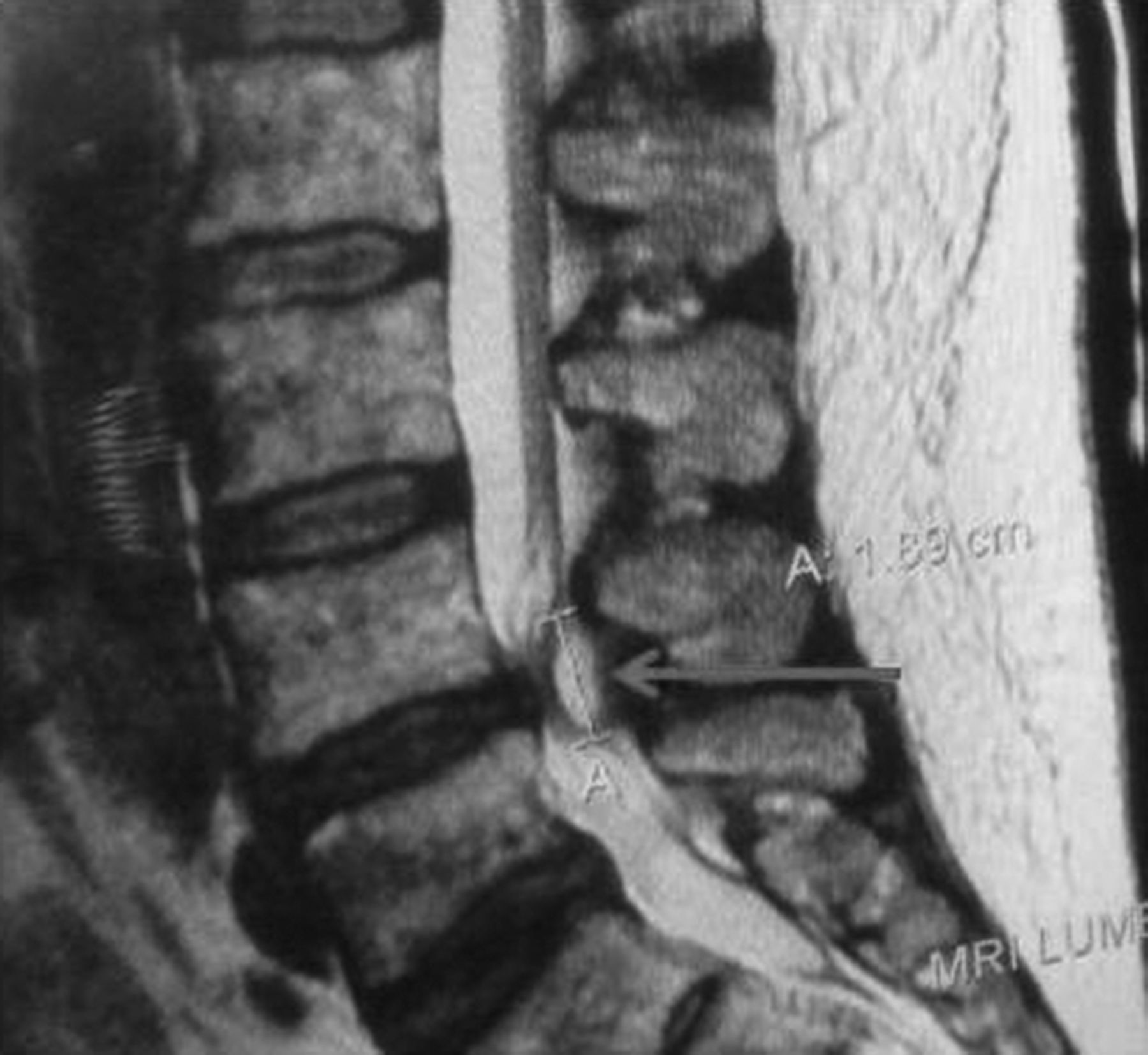
28 Acute Abdominal Pain Due to Spontaneous Renal Artery Thrombus
Ababneh B, Ali M. LSU Health Science Center, New Orleans, LA.
Background: Acute abdominal pain is commonly encountered in the emergency department (ED); spontaneous renal artery thrombosis is a very rare cause of acute abdominal pain.
Case Report: A 56 year-old male with no past medical history presented to the ED with sudden onset sharp and constant abdominal pain, starting an hour prior without associated symptoms or trauma. His vital signs were within normal limits and physical exam revealed right lower quadrant abdominal tenderness. Blood work showed elevated lactate dehydrogenase (LDH), and normal complete blood count, renal function, liver function, urinalysis, lipase, amylase, and toxicology. Computed tomography angiogram of the abdomen showed thrombus in the right renal artery branch. He was treated with intravenous heparin; invasive angiography confirmed occlusion of the right renal artery. Attempts were done to recannalize the vessel, but were unsuccessful. Due to the small amount of kidney in jeopardy, the procedure was terminated.
Transthoracic and transesophageal echocardiography were negative for right-to-left intracardiac shunts or evidence of intraccardiac thrombus. His aorta was not suggestive of an atheroembolic source. Hypercoagulability work up was negative. Patient was switched to rivaroxaban and discharged home in stable condition. 30-day event monitor showed sinus rhythm with no atrial or ventricular arrhythmias.
Discussion: Symptoms of acute renal infarction (ARI) due to renal artery occlusion are nonspecific, such as abdominal pain, flank pain, nausea or vomiting. It is usually underdiagnosed, so a high suspicion of this diagnosis is always warranted in high risk patients (1). The source of renal artery thrombosis is usually a thromboembolic event (2-4) or trauma (5). Spontaneous thrombosis of the renal artery can also be attributed to idiopathic dissection of the renal artery (6), underlying hypercoagulable state (7), and other conditions that have been associated with renal artery thrombosis and ARI such as nephrotic syndrome (8). However, renal artery thrombosis without any obvious underlying cause in an otherwise healthy patient is extremely rare (9-11). Revascularization of the occluded artery should be considered if possible, although was unsuccessful in our case (12, 13).
29 An Unusual Case of Trigeminal Neuralgia and the Heart
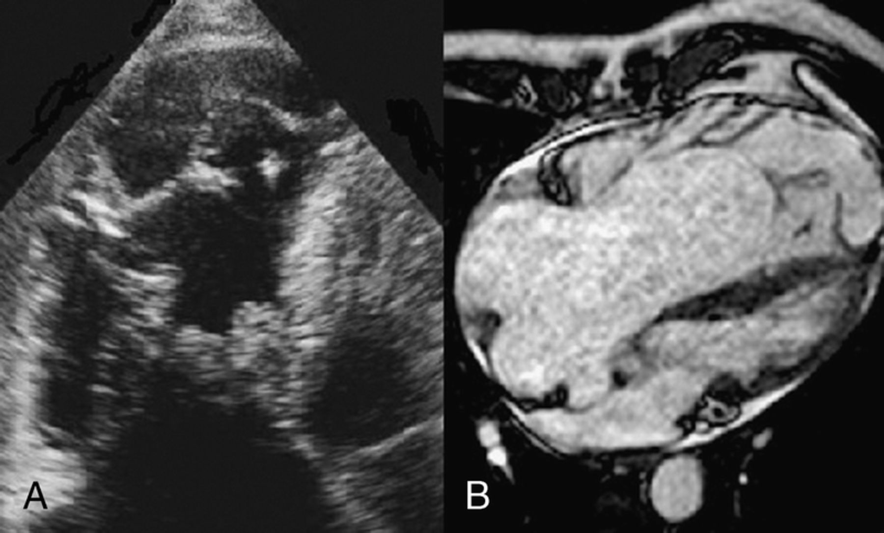
Dhakal P, Carhart R, Villarreal D. SUNY Upstate Medical University, Syracuse, NY.
Case Report: A 33-year-old Caucasian male presented to his primary care doctor with pain involving left side his face for three months, precipitated by chewing and talking. This was associated with 15 pounds weight loss. Trigeminal neuralgia was diagnosed and a subsequent CT of the neck revealed no pathology in the affected site but showed bilateral upper lobe pulmonary infiltrates. A follow up CT chest revealed a left pleural effusion along with the incidental finding of a large lesion occupying nearly entire left atrium (LA) with projection across mitral valve. The patient was referred to SUNY Upstate Medical University for further workup.
Physical examination revealed normal findings except for Atrial Fibrillation. Transthoracic Echocardiogram showed a LA mass with a portion prolapsing across the mitral valve. A CT thorax with contrast further defined the mass extending into the LA appendage. Cardiac MRI could not be performed due to patient's Claustrophobia. The patient was referred to Cardiothoracic Surgery and intraoperative assessment revealed that the mass was adherent to the posterior medisatinal structures and thus complete resection of the mass was not possible. Histopathology revealed findings consistent with high-grade sarcoma.
Primary tumors of the heart are rare and the frequency is estimated to be approximately 0.02%. Furthermore, primary heart sarcomas are exceptionally rare. They have no specific age or gender predominance. Reported mean age at clinical presentation is 41 years. Patients most commonly present with symptoms of heart failure and two-third in NYHA class III/IV. Left atrium is the most common site of the tumor. Immunohistochemical staining is the cornerstone for diagnosis with sarcomas being negative for markers for epithelial, neural, or endothelial elements.
The gold standard therapy for cardiac sarcoma without metastasis is complete surgical removal of the tumor. But unfortunately; this is often not possible because of the diffuse invasion of cardiac and mediastinal structures. Reports from large case series of 34 patients reveal median overall survival of 17 months for patients undergoing complete surgical excision and 6 months for those with partial resection. Evidence for post-surgical chemo and/or radiation therapy is limited to small series .Its benefit has not been established.
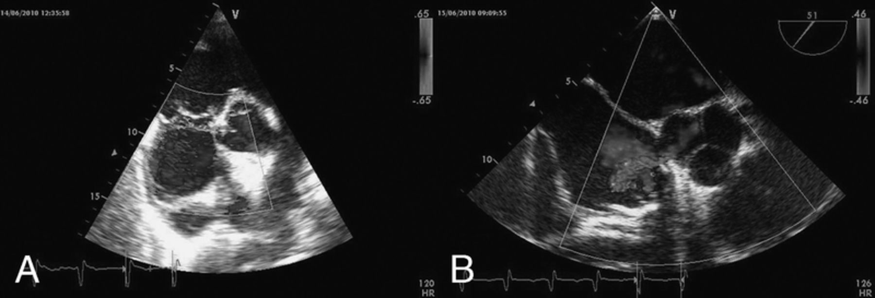
30 SHOULD WE REVISIT ANTICOAGULATION GUIDELINES DURING THYROID STORM?
Puig GD1,2, Lopez-Candales A2,1, Petersen AW3. 1University of Puerto Rico School of Medicine, San Juan; 2Internal Medicine Department, University of Puerto Rico School of Medicine, San Juan and 3University of Cincinati College of Medicine, Cincinatti, OH.
Case Report: 49 year old female with history of alcohol and illicit substance abuse evaluated due to palpitations, shortness of breath and lower extremity edema. On physical exam, heart rate irregular 174 bpm, BP 126/98 mmHg, RR 36 per minute, and T 96.9 F. Was anxious and diaphoretic with slight elevation in her jugular venous pressure and bibasilar rales. Point of maximal impulse was hyperkinetic and apically displaced. 2+ bilateral pitting edema noted as well as horizontal nystagmus and fine resting tremor. Electrocardiogram showed atrial fibrillation with 184bpm. Initial blood workup remarkable for BNP 1277 pg/ml, TSH 0.02 mIU/L, Free T4 above 7.0 ng/dl. Thyrotropin receptor antibodies elevated at 18.91 IU/L confirming diagnosis of Grave's disease. CXR with mild pulmonary edema, dilatation of the cardiac silhouette and bilateral pleural effusions. Initial echocardiogram with mildly dilated left ventricle and severe reduction in left ventricular systolic function of 30%. Ventricular response remained difficult to control for which high doses of propranolol, metoprolol and digoxin were unsuccessfully attempted. CHADS2 score of 1 and aspirin started. Methimazole, potassium iodide and hydrocortisone Initiated which controlled her symptoms and decreased free T4 level. As her ventricular response remained poorly controlled, transesophageal echocardiogram done confirming left ventricular dilation with global hypokinesis and severely reduced ejection fraction, also 7 mm thrombus identified with significant spontaneous echo contrast within the left atrial appendage. Heparin and Warfarin were immediately initiated. She responded to standard heart failure therapy with Lisinopril, Toprol XL and Furosemide. On six month follow-up, she was euthyroid with normal sinus rhythm. Repeat transesophageal echocardiogram showed normal left ventricular cavity size and ejection fraction 60% with complete dissolution of her left atrial appendage clot. In Thyroid Storm, heart failure occurs in approximately 6% of cases and less than 1% develop dilated cardiomyopathy with Left Ventricular Systolic Dysfunction.
31 Metastatic Melanoma Presenting as An Obstructing Right Atrial Tumor
Gordon S1, Poklepovic A1, Hess M2. 1Virginia Commonwealth University, Richmond, VA and 2Virginia Commonwealth University, Richmond, VA.
Case Report: Mr. D. is a 54 year old male with past medical history of rheumatoid arthritis, who presented with fatigue and shortness of breath for 2-3 months. He had a trans-thoracic echocardiogram, which demonstrated a large right atrial mass, filling the entire atrium and prolapsing into the right ventricle through the tricuspid orifice. He was found to have liver lesions and underwent liver biopsy, which revealed melanoma.
His symptoms of cardiac dysfunction increased and the patient showed hemodynamically significant signs and symptoms of venous hypertension. Coronary angiograms demonstrated a large right atrial tumor being supplied from an RCA branch. He underwent surgical intervention with excision of the right atrial mass, radical resection the right atrium and reconstruction of the right atrium with pericardial patch. Surgery revealed a large mass growing into the trabeculated area, not involving the tricuspid valve, but attached to the myocardium close to the coronary artery. The tumor was growing down the inferior vena cava, but no attachments to the hepatic tumors. Pathology confirmed melanoma.
Post-operative course was uneventful. He was agreeable to starting treatment with CTLA4 Therapy and is currently being treated with ipilimumab. He had improvement in his liver lesions after one cycle. Cycle 3 was delayed due to rash and he re-started ipilimumab after a three week delay. He is doing well.
This case highlights a presentation of metastatic melanoma found due to symptoms from a large right atrial mass, rapid cardiac evaluation, initial surgical management, followed by immunotherapy for systemic disease in a patient with known autoimmune disease. This case illustrates the close cooperation of a cardio-oncology program and the paradigm change in metastatic melanoma, converting it from a lethal disease to one that can be controlled long term and allow improved survival by manipulation of the immune system. Cardiothoracic surgery in a patient with metastatic melanoma previously may not been considered given the poor outcome of the disease. Because of cardiology, surgery and immunotherapy, his current quality of life is excellent and he has no signs or symptoms of CHF or advanced cancer.
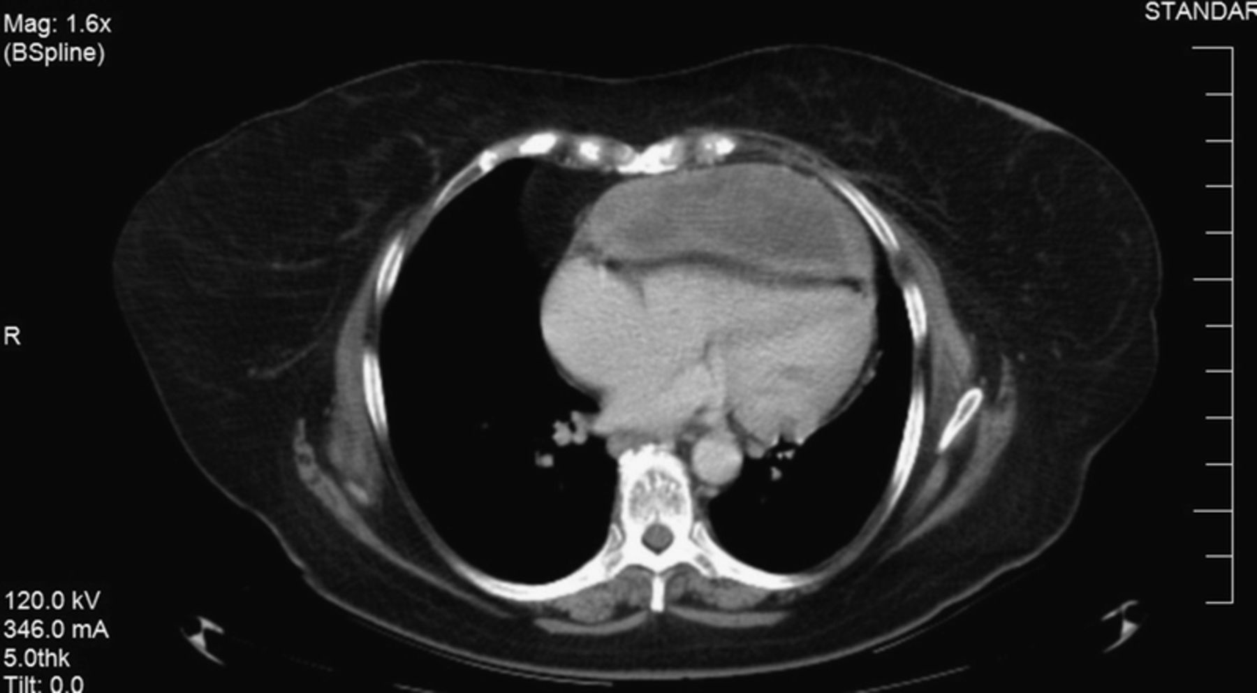
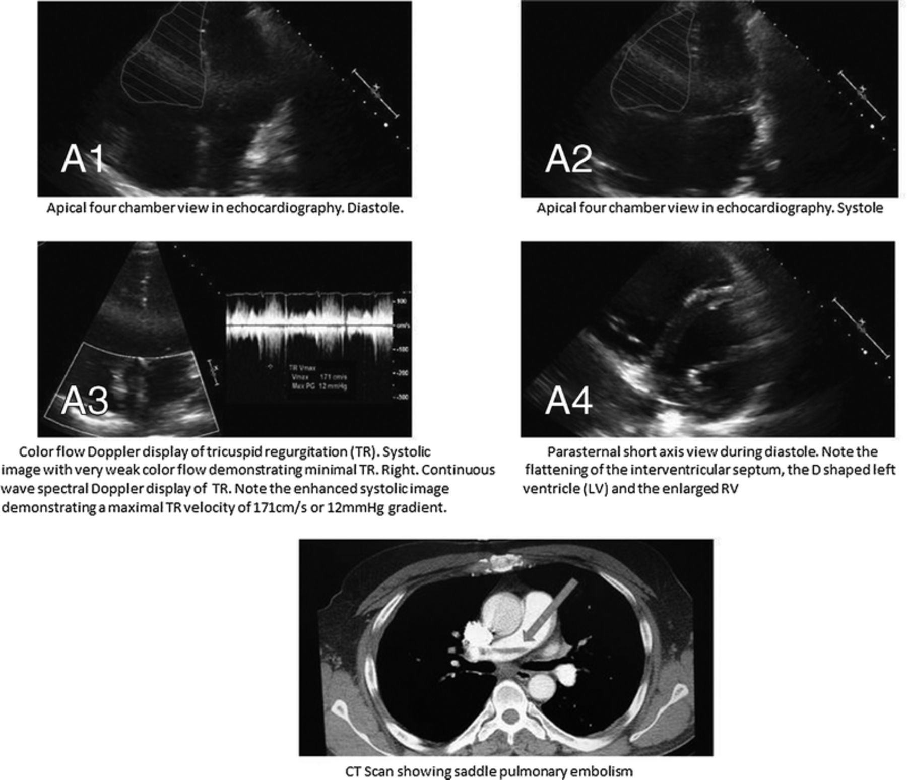
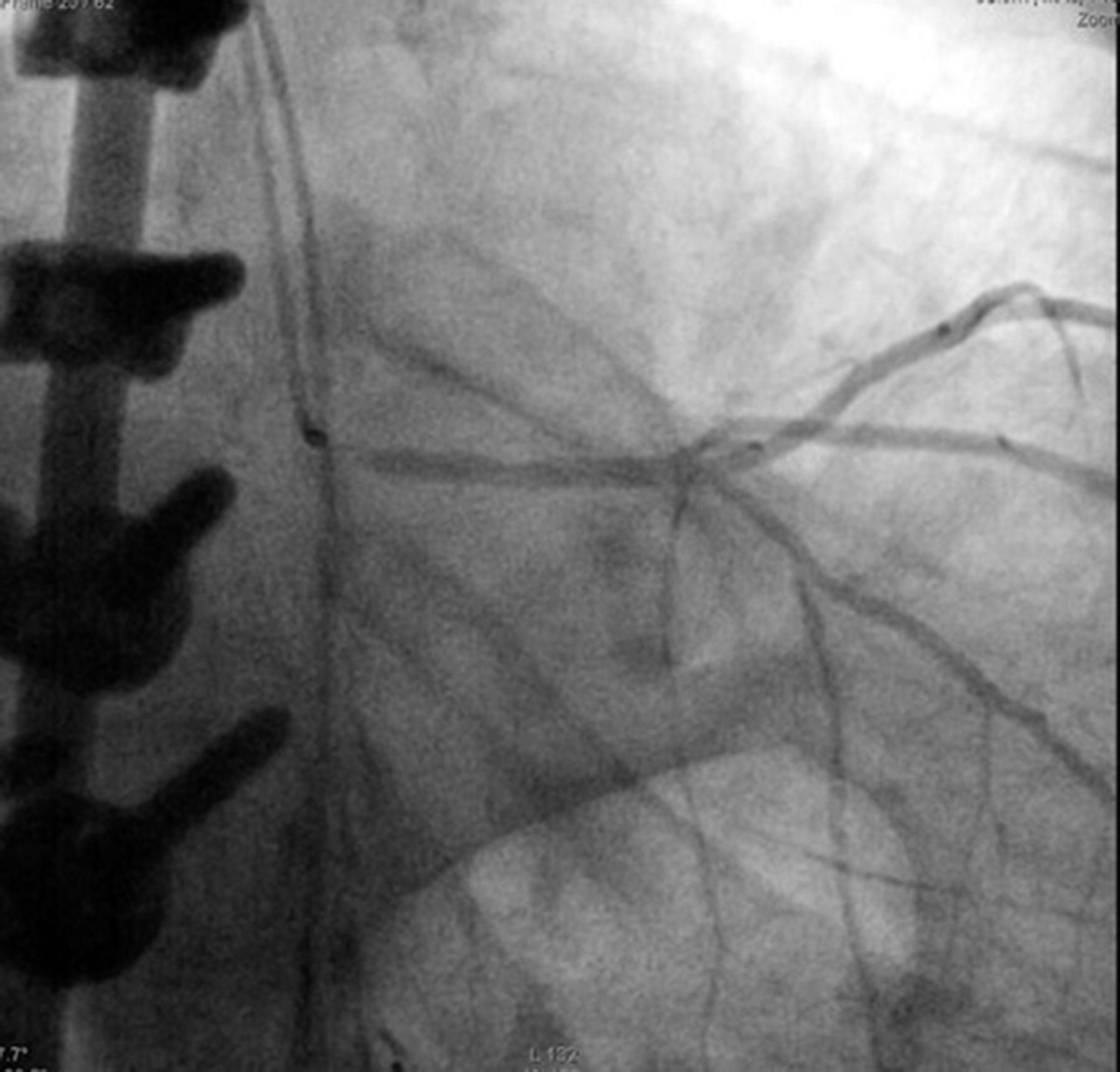
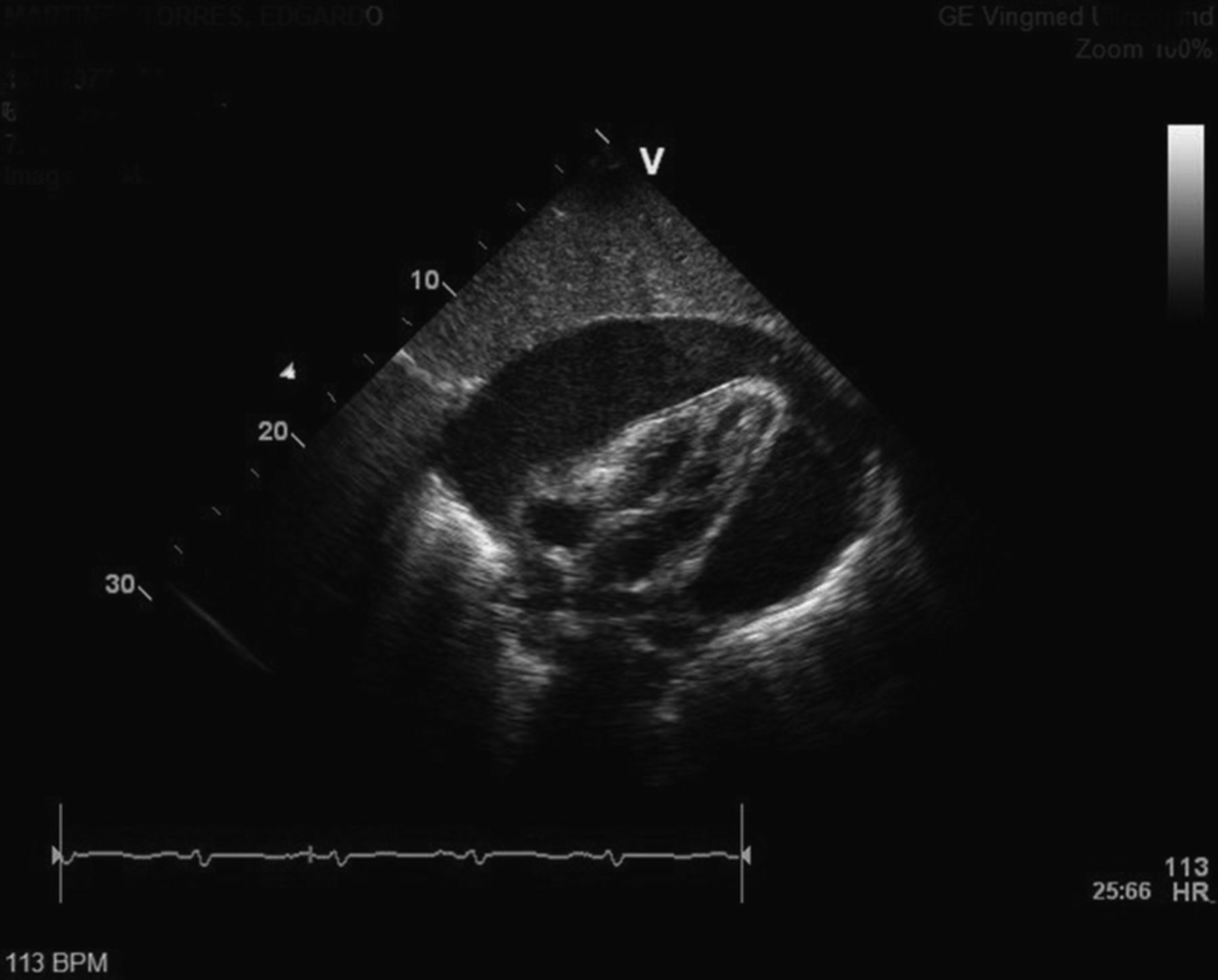
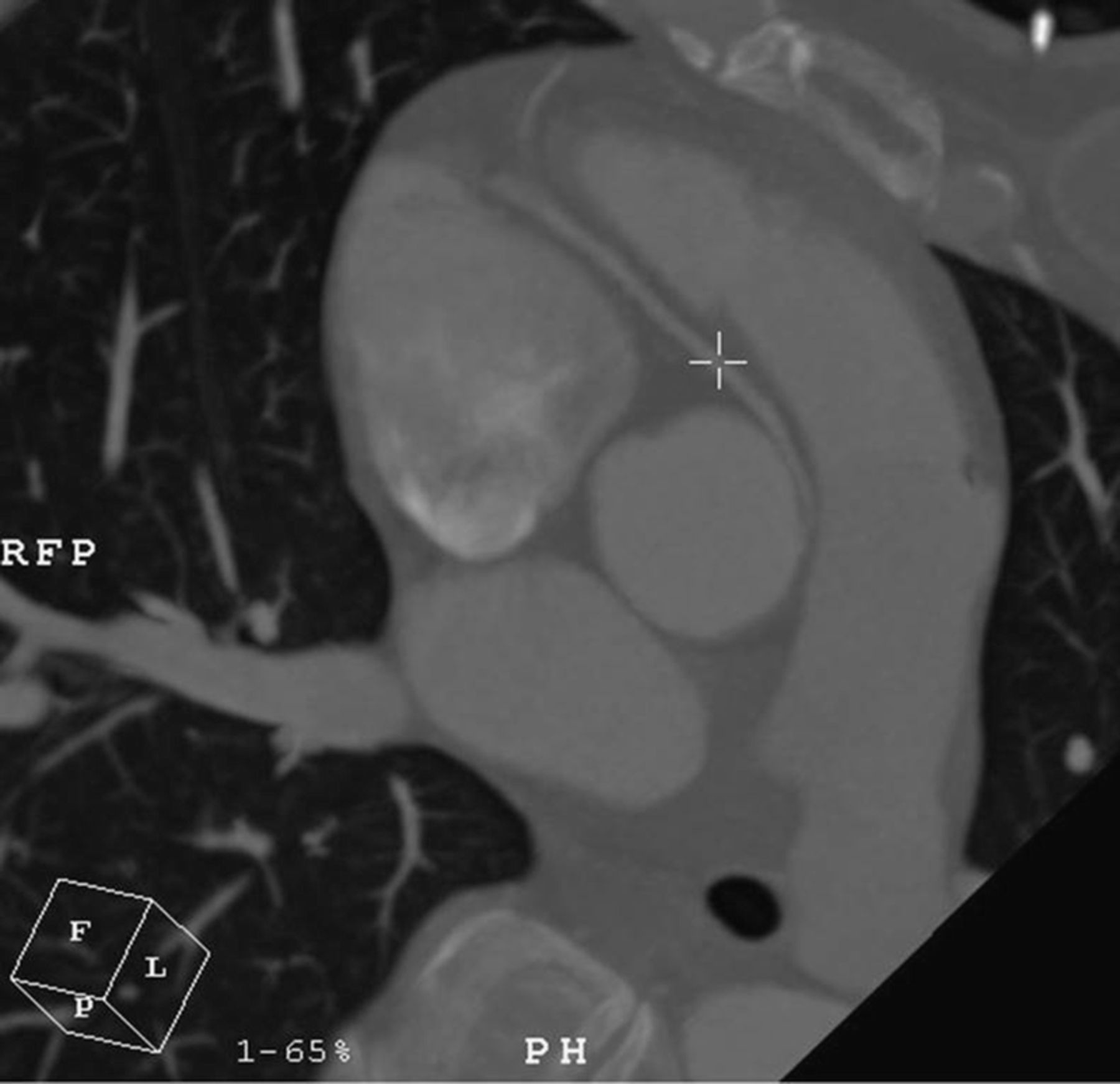
32 MCCONNELL'S SIGN: CAN WE IGNORE IT?
Robichaud FN1, Phemister J1, Bhatheja S1, Sitwala P1, Ladia V1, Halama J2. 1Quillen College of Medicine, Johnson City, TN and 2VA Medical Center, Johnson City, TN.
Case Report: McConnell's sign is an echocardiographic finding described in patients with acute pulmonary embolism (PE). Transthoracic doppler echocardiography (TTE) shows right ventricular (RV) dysfunction with akinesia of the mid free wall, but normal motion at the apex. TTE is useful in cases of massive PE, in which a rapid presumptive diagnosis is required to justify the use of thrombolytic therapy.
A 58 year old male presented with a syncopal episode reporting prior weakness, nausea, and sweats. He also reported hematuria and right flank pain. He denied any chest pain or shortness of breath. Vital signs were remarkable for blood pressure 134/90 mm Hg and pulse 104 beats/min. Physical exam was remarkable for tachycardia, with tenderness to palpation RUQ and right flank region. TTE showed mildly dilated right ventricle, mild pulmonic valve regurgitation, and mild tricuspid regurgitation (Fig. A1-4). Labs were significant for elevated troponin 0.371 ng/ml, PT 15.6, INR 1.23, RBC 3.3, H/H 9.6/30.2, AP 182. Arterial Blood Gas revealed pH 7.45, pC02 35mmHg, pO2 67 mmHg. Computed tomography angiography of chest revealed acute saddle PE with thrombus extending into lobar and segmental pulmonary arteries of all lobes (Fig. 2). He was subsequently treated with anti-coagulation. Etiology of PE was an underlying malignancy resulting in a hypercoaguable state.
Acute massive PE is defined as more than 30% obstruction of the pulmonary arterial bed. TTE reveals RV dysfunction due to dilatation or hypokinesis with paradoxical septal motion, known as McConnell's sign, which strongly increases the clinical probability of PE.
33 Disseminated Intravascular Coagulopathy and St Elevation Myocardial Infarction
Klomjit S1, Nantsupawat T2,1, Hosiriluck N1, Orellana-Barrios M1, Shurmur S2,1. 1Texas Tech Health Sciences Center, Lubbock, TX and 2Texas Tech Health Sciences Center, Lubbock, TX.
Background: Disseminated intravascular coagulopathy (DIC) can cause detrimental thrombosis in many essential parts of the vital organ. Our case is the first case report that DIC cause significant blockade of coronary artery resulting in ST elevation myocardial infarction (STEMI).
Case: This is a 27 year-old male with no known past medical history came to the hospital with severe abdominal pain and nausea and vomiting. He was hypotensive and developed respiratory distress. He was later intubated and placed on broad spectrum antibiotics and intravenous fluid. His laboratory showed increased lipase level of 1,682 units/L. CT abdomen revealed acute pancreatitis with peripancreatic fluid without masses or pseudocyst. Severe pancreatitis was diagnosed. His clinical status was complicated by septic shock, acute respiratory distress syndrome, acute kidney injury and disseminated intravascular coagulopathy with platelet count of 48,000/µL, INR of 1.26, d-dimer of 16,552 ng/mL, and fibrinogen level of 447mg/dL.
On day 4 of admission, the patient became restless and twelve leads electrocardiography showed ST elevation in V3-V6. Cardiology was consulted for emergent coronary angiogram. There was a total occlusion at the mid left anterior descending artery (LAD). Percutaneous coronary intervention of LAD with aspiration thrombectomy and three bare metal stents placement were performed successfully. Patient was transferred to the medical intensive care unit for ongoing critical care management with the continuation of ticagrelor and aspirin. There was no significant major bleeding. He improved slowly from his severe condition and was finally discharged from hospital after 44 days of hospital stay. He presented to 4-week follow up visit with significant recovery.
Discussion: There is a small study showing that silent acute myocardial infarction was more frequent in DIC patient. In our case, STEMI in young patient was a rare severe complication of DIC which required emergent intervention. This was early detected and the intervention was performed in timely fashion which resulted in good outcomes.
34 Life after Cancer: Severe Coronary Artery Stenosis in a Young Patient with History of Radiotherapy
Mesa M1, Claudio H1, Banchs-Pieretti H1, Gonzalez Cancel I2, Altieri PI1. 1University of Puerto Rico School of Medicine, Guaynabo and 2Centro Cardiovascular de Puerto Rico y el Caribe, San Juan.
Case Report: Cardiovascular disease is now the most common non-malignancy cause of death in radiation-treated cancer survivors, most often occurring decades after treatment.
We present the case of a 25 year-old man who was admitted to out institution due to a one-year history of progressive fatigue with moderate exertion. He had a past medical history of an upper back “tumor” which was treated with multiple cycles of radiotherapy ten years prior to evaluation. He had no family history of premature heart disease. He did not smoke, consume alcohol or have any history of drug use. Physical exam was remarkable for a soft II/IV diastolic murmur at the left upper sternal border. The patient underwent a diagnostic heart catheterization, which revealed evidence of a critical left main ostial coronary artery stenosis, with evidence of moderate mitral and aortic regurgitation. The likely underlying etiology was the past history of radiation therapy to the back.
As therapeutic modalities for the treatment of cancer improve, so must our efforts to give appropriate follow-up to cancer survivors. Radiation-induced coronary artery disease and cardiac complications are common and potentially lethal if not diagnosed early.
Adolescent Medicine and Pediatrics
Joint Plenary Poster Session and Reception
5:00 PM
Thursday, February 26, 2015
35 Blockade of Gli 1/2 Diminish Human Rhabdomyosarcoma Growth in Xenograft Model
Edrees N1, srivastava R2, Pressey J1, Athar M2. 1Children's Of Alabama at Birmingham, Birmingham, AL and 2University of Alabama at Birmingham, Birmingham, AL.
Purpose of Study: Rhabdomyosarcoma (RMS) typically arises from skeletal muscle. Currently RMS in patients with recurrent and metastatic disease have no successful treatment. The molecular pathogenesis of RMS varies based on cancer sub-types. Among them a small percentage of embryonal RMS are driven by the sonic hedgehog (Shh) signaling pathway. However, inhibitors of this signaling pathway particularly those which inhibit smoothened have not found to be highly effective in animal testing.
Methods Used: In this study we have investigated the effect of Gant-61, a Gli-1 & 2 inhibitor on established RMS cell lines RD and RH30 both in vivo and in vitro.
Summary of Results: We found that Shh pathway effectors GLI1 and/or GLI2 are over-expressed in the majority of RMS cells and that GANT-61, a specific GLI1/2 inhibitor dampens the proliferation of both embryonal RMS and alveolar RMS cells-derived xenograft tumors thereby blocking their growth. As compared to vehicle-treated control, about 50% tumor growth inhibition occurs in mice receiving GANT-61 treatment. The proliferation inhibition was associated with slowing of cell cycle progression which was mediated by the reduced expression of cyclins D1/2/3 & E and the concomitant induction of p21. GANT-61 not only reduced expression of GLI1/2 in these RMS but also significantly diminished AKT/mTOR signaling. The chemotherapeutic action of GANT-61 was significantly augmented when combined with temsirolimus. Finally, reduced expression of proteins driving epithelial mesenchymal transition (EMT) characterized the residual tumors.
Conclusions: Targeting Gliomas-associated oncogene transcription factors represent a novel target in both alveolar and embryonal rhabdomyosarcoma and their blockade diminish the growth of these tumors in xenograft model. Gant-61 represents a novel molecule with possible indications to be tested in combination with other known chemotherapeutic agents to improve cure and long term survival.
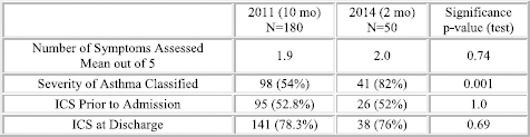
36 Evaluating The Effectiveness Of A Trauma Center-Based Driving Class For Teenagers: What Happens When The Rubber Hits The Road?
Frascogna MN, Johnson A, Foster E. University of Mississippi Medical Center, Jackson, MS.
Purpose of Study: The IMPACT (Improving Motor Vehicle Practices and Awareness in Community Teens) program is a trauma center-based class for teenage drivers who have received traffic violations that incorporates defensive driving curriculum with a demonstration of the medical consequences of reckless driving. A previous evaluation of the program showed a significant change in planned driving practices of participants based on pre- and post-class surveys. The purpose of this study was to evaluate for changes in actual driving behaviors after participation in the class.
Methods Used: The demographic information and public driving records of IMPACT participants and a control group of similar age drivers who had taken a traditional driving class were reviewed. The number of baseline traffic violations (those on record at the time of the class), and the number of violations at 6 and 12 months post-class were recorded. Data was analyzed with chi-square test.
Summary of Results: The records of 247 IMPACT class participants and 245 traditional class participants from August 2011- April 2013 were reviewed. Age was similar between the groups (Controls: N=245, 17.6 ± 1.0 versus IMPACT: N=247, 17.5 ± 1.2, p-value=0.3233). Gender was also similar between groups (Controls: 57.3% male versus IMPACT: 57.6% male, p-value=0.9431). There was no significant difference in the proportion of students receiving subsequent violations after the IMPACT class compared to the traditional driving class. From 0-6 months post-class, 11.5% of the IMPACT group and 13.1% of the control group had received violations (p=0.6277). From 6-12 months post-class, 11.0% of the IMPACT group and 9.4% of the control group had received violations (p=0.6136).
Conclusions: Despite previous data that suggested a significant change in planned driving practices of teenagers participating in the IMPACT program, there was no change found in their actual driving practices as determined by number of traffic violations received when compared to teenagers taking traditional driving classes.
37 Impact of Race and Gender in the Diagnosis of Attention Deficit Hyperactvity Disorder
Holmes J, Preudhomme D, Connelly R, Higginbottom L, Fox N. University of South Alabama, Mobile, AL.
Purpose of Study: Attention deficit hyperactivity disorder(ADHD) is the most commonly diagnosed behavioral disorder in children. Recent assessments suggest a prevalence from 4%-10% in the U.S. The prevalence of ADHD in females or African Americans (AAs) is reported at a lower rate. There is a paucity of research of ADHD symptoms, diagnosis, and treatment in females and AAs. We hypothesize that AA children and females may be under diagnosed compared with other cohorts.
Methods Used: Between July 1st, 2011 and February 28th, 2014, we performed a cross sectional analysis of patients aged 5 to 18 years old in a pediatric outpatient department using ICD9 diagnosis code of 314.01. Patient characteristics such as race, gender, co-morbidities, treatment types, and number of visits were collected. Descriptive statistics and odds ratio were calculated as appropriate.
Summary of Results: Out of a total of 9,423 patients, 1,046 (11%) were diagnosed with ADHD. Amongst patients with ADHD, 580 (55%) were AA. Of the ADHD population, 293 (28%) were females. The average age was 10(+/- 3.3) years old. The number of visits was on average 4(+/- 3.1) times over 31 months. Long-acting medications were used in 883 patients, 79% were prescribed Vyvanse. Patients receiving behavioral therapy and medication totaled 275(26%). Secondary analysis shows that the prevalence of ADHD diagnosis in AA population is 8% compared to 17% in our Caucasian population. The prevalence of ADHD in females was 6% compared to 13% in males. The likelihood of being diagnosed with ADHD as Caucasian was twice that of the AA population (OR 2.2, 95% CI 1.9-2.6). The possibility of males to be diagnosed with ADHD was more than twice that of females (OR 2.4, 95% CI 2.1-2.8).
Conclusions: In the past decade, the research evaluating ADHD across gender and race is limited. Our review indicates that AA and female children are significantly less likely to be diagnosed with ADHD. These disparities may be the consequence of rating scale bias, difference in symptom presentation, or a cultural difference in the approach to mental illness. An understanding of which etiological and cultural mechanisms account for the observed racial and gender differences would allow for more appropriate diagnosis and care for these children.
38 IS MICROARRAY CGH a USEFUL DIAGNOSTIC TEST IN AUTISM SPECTRUM DISORDERS?
Jensen K2, Janssen A2, Settles L1, Chen T2, Morava E2. 1Tulane University School of Medicine, New Orleans, LA and 2Tulane University School of Medicine, New Orleans, LA.
Purpose of Study: Autism spectrum disorder (ASD) has a heterogeneous clinical presentation that involves problems with social understanding, language acquisition, and repetitive behaviors. ASD is a multifactorial disease caused by a combination of genetic and epigenetic factors and environmental triggers. Chromosomal abnormalities, rearrangements, deletions, and duplications are associated with ASD; however information on phenotypic presentations of genomic variants is limited. The purpose of this study is to identify phenotypes associated with chromosomal alterations in children with ASD.
Methods Used: ArrayCGH, Fragile X, and chromosome analysis were performed on 28 children between the ages of 2 -18 seen at Tulane University Hospital for ASD or autistic features. Metabolic studies were requested (amino acids, ammonia, lactate, uric acid and urine organic acids), and information was collected on dysmorphic features, speech delay, learning problems, developmental delay, behavior problems, and aggressive behavior.
Summary of Results: Of the 28 children in the study 18 had an additional speech delay, 20 had a developmental delay, 20 had behavior problems, 9 had aggressive behavior, 8 had seizures or epilepsy, and 4 had decreased muscle tone. Dysmorphic features included 8 children with a high forehead and 9 children with macrocephaly. Eight children had chromosome alterations; two had a 16p13.11 microdeletion associated with autistic features including developmental, speech, and learning delays. However, the two patients did not share other phenotypic features. Other chromosome alterations in the cohort included deletion of 21q22.3 and duplications at 1q25, 9q34.3, 12q23, 15q13, and 6p22.
Conclusions: Our cohort had a high number of patients with chromosomal abnormalities, 28%, compared to the 10% reported in the literature. This may be due to selection bias as the patients in our cohort were referred for genetic consultation by Tulane's psychiatry department possibly for suspected genetic abnormalities. The small sample size in the study limited our ability to create subgroups with common chromosome alterations. Future work will involve increasing the sample size to allow classification of subgroups based on phenotypic similarities.
39 LYMPHOPROLIFERATIVE DISORDER IN a CHILD WITH CROHN'S DISEASE AND THIOPURINE USE
Messer A, Raulji C, Fletcher M, Keith B, Velez M. LSU School of Medicine - Health Sciences Center, New Orleans, LA.
Case Report: Thiopurines are widely used in the treatment of patients with inflammatory bowel disease (IBD). Concern for associated lymphoproliferative disorders (LPD) must be addressed when deciding appropriate treatment. Multiple large studies have showed a strong association of increased risk of LPD in IBD patients receiving thiopurines for treatment. However, this association is not frequently described in the pediatric population and pediatric data are limited.
An 11-year-old African-American female with one-year history of Crohn's Disease (CD) presented with fever, respiratory distress and seizure activity. Prior to admission, she had URI symptoms for one month and intermittent fevers for two weeks. Her CD treatment consisted of 6-mercaptopurine; however, it was discontinued ten days earlier due to neutropenia.
On admission, CBC showed pancytopenia with WBC 1.66 x 10³ with ANC 460, hemoglobin 7.7 g/dL, and platelets 36,000. Ferritin level was elevated at 1823ng/ml. EBV PCR was positive in plasma (24,700 copies), bone marrow (19,900 copies) and CSF (400 copies). A head CT and MRI showed multifocal, subcortical white matter lesions, although neurological exam remained normal. Bone marrow aspirate and biopsy revealed hypocellular marrow with focal hemophagocytosis. Upon further evaluation CT scans showed multiple pulmonary nodules, an 8x5cm anterior mediastinal mass, and enlarged adenoids and tonsils. Biopsy of the anterior mediastinal mass was consistent with LPD.
The patient's treatment included high dose methylprednisolone, which was later changed to dexamethasone for better CNS penetrance, and rituximab. End of treatment demonstrated normal CBC and ferritin, negative EBV PCR with improvement of brain lesions.
This patient is among the youngest documented to have EBV-associated LPD due to thiopurine use for CD. Her new-onset seizures and CNS lesions made her case at first puzzling. The development of LPD in children with CD can occur as a complication of the treatment, due to the immunosuppression associated with thiopurine.
40 Critical Congenital Heart Defects among Infants Born in Arkansas Hospitals: Implications for Newborn Screening
Onukwube J1,4, Mosley B1,4, Bowman C5, Schaefer G1,2,4, Collins R1,3,4, Robbins J1,4. 1University of Arkansas for Medical Sciences, Little Rock, AR; 2University of Arkansas for Medical Sciences, Little Rock, AR; 3University of Arkansas for Medical Sciences, Little Rock, AR; 4Arkansas Children's Hospital Research Institute, Little Rock, AR and 5Yale Univerisity, New Haven, CT.
Purpose of Study: The U.S. Department of Health and Human Services recommends the addition of pulse oximetry (PO) screening for critical congenital heart defects (CCHD) to the newborn screening panel. We determined the number of infants born in Arkansas with CCHDs and identified the number of those infants recognized beyond 3 days of life who could potentially benefit from earlier PO screening.
Methods Used: The prevalence of CCHDs among Arkansas’ livebirth population was computed for birth years 2000-2010 using data obtained from the Arkansas Reproductive Health Monitoring System. Timing of initial diagnosis of CCHD was evaluated for 7 CCHD phenotypes among term infants. Infant mortality rates were computed and stratified by gestational age and timing of initial diagnosis. Delays in initial diagnosis, defined as diagnosis made after day 3 of life, were compared across birth hospital neonatal level of care.
Summary of Results: There were 559 livebirths in Arkansas having at least one CCHD diagnosis. The birth prevalence of CCHD was 13 per 10,000 livebirths. Approximately 20% of term neonates with CCHD experienced a delay in initial diagnosis. Mortality among infants with CCHD was 24.9%. Infants with CCHD born prematurely had a higher infant mortality (41.6%) than term infants (21.5%). A disproportionate number of late diagnoses occurred in neonatal care level 1 hospitals.
Conclusions: These results characterize the CCHD prevalence in Arkansas prior to implementation of universal PO screening. Universal screening may improve outcomes for CCHD term infants, where 1 in 5 experience delays in initial diagnosis. Many infants experiencing delays in diagnosis are born at level 1 neonatal facilities; thus supporting the need for universal screening, particularly in the smaller, birthing hospitals found in most rural parts of Arkansas. These results establish a pre-screening baseline that may be used to assess the effectiveness of PO screening in reducing infant mortality and severe morbidity.
41 Still Searching: Underlying Genetic Cause of Cutis Laxa and Multiple Aneurysms
Preston GJ1, Scott K1, Settoun C2, Marble M2, Baskind H1, Yosypiv IV1, Andersson H1, Morava E1. 1Tulane University, New Orleans, LA and 2LSU, New Orleans, LA.
Purpose of Study: Vascular aneurisms associated with Cutis Laxa can cause life-threatening complications in young patients. Several genes including SLC2A10, CBS, ACTA2. FBN1, FBN2, COL3A1, TGFBR1, TGFBR2, SMAD3, GLUT10, LTBP4 and MYH11 have been implicated. We evaluated a patient with CL, cardiovascular aneurisms and severe hypertension, with suspected connective tissue disorder. DNA analysis revealed a heterozygous mutation in MYH11, a gene coding for myosin heavy chain, a contractile protein in smooth muscle.
Methods Used: Mutations of MYH11 are associated with thoracic aortic aneurysm, aortic dissection, and abnormal elastin formation. The patient's mutation (c.3651+5_+11del7insG) results in the deletion of seven nucleotides and the insertion of a single G nucleotide five base pairs into intron 27. To evaluate pathogenicity we performed RT-PCR for abnormal MYH11 splice variants, cDNA sequencing for cryptic splice sites, long range PCR for intron retention and additional sequencing for ABCC6 and GGCX. Patient expired at the age of 5 and underwent autopsy. We also performed immune histology for elastin structure and Golgi secretion studies.
Summary of Results: Autopsy confirmed severe atherosclerotic saccular aneurysms of the distal abdominal aorta, bilateral renal arteries, and bilateral internal carotids. Pathology identified fragmented elastin fibers in skin biopsy, and extreme atherosclerosis of vascular aneurysms, characterized by ossification and bone marrow formation in the calcified plaques. Molecular analysis of MYH11 cDNA revealed only normally spliced, mature mRNA transcripts. Sequencing of ABCC6 and GGCX revealed no sequence anomalies in the protein-coding region. Histology in fibroblast revealed delayed elastin secretion and Golgi retention.
Conclusions: Based on functional data the MYH11 mutation is non-pathogenic. Histology confirmed the diagnosis of Cutis Laxa. The underlying genetic cause is still unknown. This patient represents an ideal candidate for next-generation sequencing analysis, and whole-exome sequencing is our next goal. The case highlights the inherent limitations in single-gene sequencing approach and the ability of next-generation techniques to provide crucial leads in perplexing cases, tapping previously hidden genomic data toward new genetic diagnoses.
42 Comparing Teen Vaccine Uptake and Clinician Recommendations between South Carolina and the United States
Roberts JR1, Naifeh M2, Jacobson R3, Thompson D2, Rogacki B2, Margolis B4, Darden P2. 1Medical University of South Carolina, Charleston, SC; 2University of Oklahoma Health Sciences Center, Oklahoma, OK; 3Mayo Clinic, Rochester, MN and 4Maternal Child Health Bureau, Rockville, MD.
Purpose of Study: To examine trends over 3 years for teen vaccine uptake and their corresponding clinician recommendations in South Carolina (SC) and the US.
Methods Used: Using data from the publicly available National Immunization Survey-Teen (NIS-Teen), we performed multiple logistic regression to assess 3-year trends (2010-2012), among teens in SC and the US with provider verified vaccination rates, in up-to-date (UTD) status and provider recommendation for each of 3 vaccines: meningococcal conjugate quadrivalent (MCV4), tetanus toxoid, reduced-dose diphtheria and acellular pertussis (Tdap), and human papillomavirus (HPV). We assessed yearly differences between SC and the US using the z-statistic.
Summary of Results: Approximately 20,000 US and 310-362 SC respondents between 2010 and 2012 were weighted to generate population estimates. UTD rates increased from 2010 to 2012 for MCV (OR 1.31, 95% CI 1.07, 1.61) and Tdap (OR = 1.42, 95% CI 1.15, 1.74). The yearly increase for HPV UTD in SC females was not significant. We could not assess male UTD for HPV since the vaccine was first recommended in 2011. The odds of SC males initiating the 1st HPV dose increased (OR = 3.65, 95% CI 1.29, 10.35). UTD rates in SC were < the corresponding US rates for MCV (45.3%, 56.8%, and 58.7% for 2010-2012 in SC, compared to 63.5%, 71.4%, and 76.0% in the US, p < .05) and Tdap (48.1%, 59.4%, and 64.9% in SC compared to 68.7%, 78.2%, and 84.6% in the US, p < .05). HPV vaccine coverage was low nationally and in SC. SC differed from the US for 2011 in females (HPV dose 1 = 38.7% in SC v. 53.0% in US; UTD = 23.3% in SC v. 34.8% in the US) and males (UTD = 3.8% in SC v. 8.3% in US, p < .05). Parents recalled provider recommendations <50% of the time. Notable differences were for MCV4 in 2010 (28.7% (SC) v. 37.9 (US)) and for Tdap in 2012 (44.8% (SC) v. 54.0% (US), p < .05). About half of SC and US providers recommended HPV for females and <30% recommended it for boys (p >.05).
Conclusions: Vaccination rates improved yearly in SC and US but SC still lagged the US. HPV is particularly low in SC and the US. Parental recall for clinician recommendation is very low.
43 Cancelled
44 Missing the Mark? Intestinal Parasite Prevalence in Informal Settlements in Lima, Peru
Searing R2, Naifeh M1, Cooper MT1, Gonzales C3, Thompson D4, Johnston SE4, Zavala C5, Woodson K2. 1University of Oklahoma Health Sciences Center, Oklahoma City, OK; 2University of Oklahoma Health Sciences Center, Oklahoma City, OK; 3Instituto Nacional de Salud, Lima, Peru; 4University of Oklahoma Health Sciences Center, Oklahoma City, OK and 5Health Bridges International, Portland, OR.
Purpose of Study: The WHO suggests large scale preventive chemotherapy for soil-transmitted helminthiasis (STH) for communities in which prevalence is estimated to be greater than 20%. WHO lists Peru as a country potentially needing STH prevention. Prevalence varies widely regionally and remains understudied in the newest urban informal settlements in Lima.
Objective: (a) Determine the STH prevalence in the new informal settlements in Lima, Peru
(b) Estimate seasonal (summer/winter) variation in the prevalence of STH in 3 informal settlements in Lima, Peru.
Methods Used: Children aged 1 to 10 were recruited from 3 new informal settlements in Lima Peru. After informed consent, participants were interviewed, weighed and measured. Stool samples were examined using macroscopic examination, direct microscopic examination, technique of spontaneous sedimentation in tube, and Baermann technique. We attempted to obtain 2 stool samples from each participant during 2 seasons (summer/winter).
Summary of Results: The prevalence of STH and pathogenic protozoa did not differ between summer and winter (p>.05). Only 3% (summer) to 9% (winter) of study participants had STH. The prevalence of pathogenic protozoa was 25% during the summer and 17% during the winter. Approximately 1% (summer) and 5% (winter) of participants had both pathogenic protozoa and STH.
Conclusions: Even using the broadest definition of STH, the prevalence in this study is considerably lower than the WHO recommended threshold (20%) for large scale preventive chemotherapy for STH. Although pathogenic protozoa prevalence was higher than STH prevalence, recommended empiric treatment for STH with mebendazole/ albendazole is inadequate in the treatment of these pathogenic protozoa. The practice of blanket empiric treatment for STH by groups active in these newest invasion settlements is not supported by the data and could contribute to unnecessary medication exposures and poor resource allocation.
45 a CURIOUS CASE OF CUTIS LAXA WITH HOLOPROSENCEPHALY: COULD a SEEMINGLY INNOCUOUS INTRONIC DELETION BE THE CULPRIT?
Wong S1, Sullivan L2, Singh D3, Frieberg E3, Janssen A1, Kozicz T1, Chen T1, Morava E1,3. 1Tulane University, New Orleans, LA; 2Tulane University, New Orleans, LA and 3Tulane University, New Orleans, LA.
Purpose of Study: We report a new syndrome of cutis laxa, alobar holoprosencephaly, and cerebellar agenesis observed in a baby girl born at term. Microarray CGH analysis revealed a small deletion in intron 3 of Neuregulin 3 gene (NRG3). The deletion was confirmed to be present in the patient but not the mother. Mutations in NRG3 gene have been associated with cognitive impairment. Additionally, transgenic mice overexpressing NRG3 in the skin exhibited a cutis laxa phenotype. While there is evidence supporting the pathogenicity of the intronic deletion, the mechanism is unknown. The purpose of the study is to establish the clinical significance of the intronic deletion, with the end goal of providing informed genetic consultation for the mother.
Methods Used: Using immunohistochemistry, we conducted skin pathohistological studies to confirm the clinically observed cutis laxa. To investigate whether the deleted intronic DNA has a role in modulating NRG3 expression, we examined the known transcription factor binding sites within the deleted intron and designed in vitro experiments with luciferase reporter assay. We also directly reprogrammed the patient's skin fibroblasts to neuroblasts to investigate the expression level of endogenous NRG3.
Summary of Results: Abnormal wrinkling and altered elastin and beta-integrin was found in the patient's skin. Remarkably, the findings resemble the histology of the adult skin. Our preliminary in vitro data suggested that 3 regions within the deleted intron exhibited moderate repressive property and 1 region, potent repressive property. Preliminary expression study in the reprogrammed neuroblasts revealed abnormal NRG3 expression.
Conclusions: Our pathohistological study confirmed skin developmental anomaly in our patient and suggested premature skin aging. Our in vitro data demonstrated that the deleted intronic DNA may have a role in regulating NRG3 expression. Interestingly, MYC may have a repressor function in NRG3 expression. Additional studies will help elucidate the mechanisms which led to the remarkable anomaly in our patient, and even more importantly, offer closure to the patient's mother.
Adult Clinical Case
Joint Plenary Poster Session and Reception
5:00 PM
Thursday, February 26, 2015
46 Promethazine Toxicity Misdiagnosed as Meningoencephalitis
Ali E, Mazek H, Mohamed A, Baser K, Mulkey Z. Texas Tech University Health Science Center/Lubbock, Lubbock, TX.
Introduction: Promethazine is a commonly used medication in practice, but toxicity is rare with only a few cases of intoxication reported in the literature. We report a misdiagnosed case initially treated as meningoencephalitis due to overlap of findings.
Case description: A 62-year-old man was transferred from outside facility with confusion, fever, and jerking movements. A lumbar puncture had been done, and antibiotics had been started. Upon arrival he was agitated and had dysarthria and visual hallucinations. His blood pressure was 89/72 mmHg, pulse 98 beats/minute, respiratory rate 22/minute, and temperature 1010F. The examination demonstrated disorientation, dilated pupils, dysmetria, trunk ataxia, positive Babinski's sign, and nystagmus. Lab work revealed an elevated creatine kinase and leukocytosis while glucose, creatinine, liver enzymes, and electrolytes were all normal. Drug screen was negative; as was a head CT. Acyclovir was added. Agitation was treated with haloperidol. Within twelve hours he dramatically improved. Confusion and dysarthria resolved and he was fully oriented. Truncal ataxia and nystagmus improved but were still present. Detailed history revealed nonspecific illness beginning two weeks prior to admission consisting of nausea, vomiting, and a mosquito bite. He visited an emergency room twice, received fluids, and intravenous promethazine and was sent home. He returned to the emergency center after developing fever and again received fluids, antipyretics, promethazine, and was sent home with a promethazine prescription. Two days later he started having jerky movements and confusion. Patient was admitted locally, had a head CT and lumbar puncture and was started on antibiotics before transfer to our facility. Cerebrospinal fluid was negative for Gram stain, cultures, HSV, VZV, West Nile virus, CMV, and had normal glucose, protein, and cell counts. Given the rapid resolution of his symptoms and exclusion of infection we concluded that the diagnosis was promethazine overdose.
Discussion: Promethazine intoxication may result in wide range of symptoms, including CNS depression, agitation, or delirium, and anticholinergic findings, such as dilated pupils, dry mouth, fever, seizures, and coma. Treatment is supportive.
47 Cryoglobulinemia as An Idiosyncratic Drug Reaction of Moringa Oleifera
Ali RA, Baldeo C, Chaudhry S, Landa C. UF Health, Jacksonville, Jacksonville, FL.
Case Report: A 43 year old Caucasian male with treatment-naive Hepatitis C presented for evaluation of a rash of four days duration. Two days prior to onset he started an herbal medication, Moringa oleifera, for headache. The rash was a palpable purpura with scant pus-filled blisters that densely populated the ankles. The trunk, face and genitals were spared. There was no mucosal involvement. He denied constitutional symptoms, arthralgias and tick bites. Previous drug screen was positive for oxycodone.
He had a mild peripheral eosinophilia and elevated inflammatory markers. Biochemistry, urinalysis, coagulation profile and porphyrin screen were all unremarkable. Infectious work-up for HIV, CMV, syphilis and Lyme disease were negative. ANA, ANCA's and Rheumatoid factor (RF) titers were negative, and C3/C4 were normal. Hepatitis C antibody was positive but viral load was undetectable. Cryoglobulins were elevated at 8%, with immunofixation electrophoresis revealing a type III cryoglobulinemia. Skin biopsy showed leukocytoclastic vasculitis.
He was treated with clindamycin for the cellulitis. No specific treatment was instituted for the cryoglobulinemia and the rash resolved independently.
Cryoglobulinemia refers to the presence in the serum of single or mixed immunoglobulins, which precipitate at temperatures below 37C and re-dissolve on re-warming, with types II and III representing mixed cryoglobulinemia (MC).
The triad of MC is purpura, weakness and arthralgia. Orthostatic purpura ranges from petechiae to severe vaculitic lesions which are attributed to the deposition of circulating immune complexes on vessel walls. Leukocytoclastic vasculitis is the histopathological hallmark of MC. Hepatitis C virus (HCV) exerts a chronic stimulus on the immune system kindling the production of monoclonal IgM RF which is implicated in the development of cryoprecipitate immune complexes, along with complement.
Considering the undetectable viral load, negative RF titers and normal complement levels, it is unlikely that HCV was responsible for the precipitation of MC in this case. As the introduction of Moringa oleifera coincided with disease onset, it postulates that this may be an idiosyncratic drug reaction and that alternate immune stimuli may influence the structure of circulating immune-complexes responsible for MC.
48 Hemolytic Uremic Syndrome and Thrombotic Thrombocytopenic Purpura Associated with Advanced Prostate Cancer
Bahaa Aldeen MA, Talibmamury N, Nadham O, Garrido JP, Ahmed M. Texas Tech Univ Health Sciences Center, Amarillo, TX.
Case Report: An 85 year-old gentleman who was recently diagnosed with metastatic prostate cancer with multiple bones involvement presented initially with confusion. Patient denied any recent diarrhea, headache, trauma to head or other symptoms. His physical examination was unremarkable except for confusion, multiple skin bruises throughout the body and trace pedal edema bilaterally. His initial blood workup shows hemoglobin 9.8 gm/dL, platelet count 73000/cml, bicarbonate 20 meq/L, creatinine 3.8 mg/dL, total billirubin 5.5 mg/dl with direct portion of 0.8 mg/dL, LDH 553 IU/L and heptoglobin 5 mg/dL. His CT scan abdomen, U/S, and HIDA scan showed no gallbladder stone or infection. His creatinine increased gradually to 7 mg/dL and was started on hemodialysis. Peripheral blood smear showed anemia with numerous schistocytes, burr cells, acanthocytes and moderate thrombocytopenia. Considering multiple comorbid conditions, the patient adopted hospice care at the end.
Discussion: Thrombotic thrombocytopenic purpura (TTP) and hemolytic uremic syndrome (HUS) are both acute hematological disorder involving multiple organ systems. The main manifestations are microangiopathic hemolytic anemia and thrombocytopenia. There are association between solid tumors and TTP/HUS. Prostate cancer is one of malignancies that can manifest with acute TTP/HUS. Our case reminds clinician about the association between advanced prostate cancer and TTP/ HUS. The diagnosis of thrombotic thrombocytopenic purpura and hemolytic uremic syndrome (TTP/HUS) is made clinically and requires only thrombocytopenia and microangiopathic hemolytic anemia without other clinically apparent etiology. Idiopathic TTP/HUS is a medical emergency that is almost always fatal if appropriate treatment is not initiated promptly. Treatment should involve plasma pharesis and may add steroid and immunotherapy.
49 Lung Cancer Presenting as a Soft Tissue Mass
Baldeo C, Ali RA, House J. UF Health-Jacksonville, Jacksonville, FL.
Case Report: A 60 year old female presented with left hip pain for 5 months. She had a previous stroke and left renal artery stenosis. She had a 45 pack year cigarette smoking history and an unintentional 10lb weight loss 1 month prior. Examination revealed a cachectic female with an incidental right scapula soft tissue mass, measuring about 4x4cm. This was previously dismissed as a lipoma.
Routine CXR showed a right hilar mass. Whole body CT scan revealed several bilateral pulmonary nodules, right suprahilar mass, left renal mass, right adrenal mass, hepatic hypodensity and vertebral lytic lesions.
Endobronchial mass biopsy revealed non-small cell carcinoma, NOS. The tissue sample was inadequate to differentiate subtype, but biopsy of the scapula soft tissue mass showed poorly differentiated large cell carcinoma, favoring metastatic disease likely from lung. This was further corroborated with specialized stains. PET scan showed multiple hypermetabolic lesions correlating with the abnormal CT findings, and consistent with neoplasm. She developed left sided hemiparesis. MRI brain showed metastatic brain masses and vasogenic edema. She also developed a post obstructive pneumonia and right upper lobe atelectasis secondary to the endobronchial lesion. The obstruction was not amenable to bronchial stenting. She had palliative radiation to left hip, right lung and brain. She expired one month after diagnosis.
Soft tissue metastasis may occur after diagnosis of a primary internal malignancy or later in the course of the disease. In our case, the soft tissue metastasis and primary cancer were diagnosed simultaneously.
Lung cancer can metastasize to any organ. About 50% of cases are metastatic at the time of diagnosis. Soft tissue metastases from lung cancer are uncommon with a reported overall prevalence of 2.3%. Soft tissue metastases are perceived as a sign of advanced disease and are regarded as a grave prognostic indicator. Despite combination radiotherapy and chemotherapy these patients have a poor outcome with a median survival of 3-6 months.
Soft tissue metastasis from lung cancer is unusual and ominous. Our case highlights how even a single subcutaneous lesion can represent serious occult pathology warranting a low threshold for biopsy and further investigative work-up. It also shows the importance of good physical examination and clinical acumen.
50 a Case of Rheumatoid Neutrophilic Dermatosis
Bhawal J. UMMC, Jackson MS, MS.
Purpose of Study: Rheumatoid Neutrophilic dermatitis (RND) is cutaneous manifestation of RA, It can see in patients who are both positive and negative for RF and common in middle age females, it usually Present as vesiculopustular, plaques and nodules.
Methods Used: 37 y/o AAF with PMH of RA diagnosed in 2007 severe arthralgia and synovitis..In 2012 she developed a vesicular rash on her right elbow; hydrocortisone and Benadryl were prescribed which did not help. Later it worsened and she developed severe itching with a burning sensation which disseminated to her b/l arms and legs.
On physical examination, firm nodules 3 to 5 mm in size were observed over her both elbows; these lesions were erythematous papules and plaques, some in an annular configuration, with central umbilication over extensor arms and knees. In addition she had many of hyperpigmented lesions over her entire body. MCPS showed synovitis and she was unable to make fist. The nails and mouth were clear of lesions.
The ESR was elevated at (60-80 mm/hr), ANA, Dsdna, SSA/SSB were strongly positive and RF, CCP were strongly positive.
A 3 mm punch biopsy was done on her elbow and knee which showed lymphocytes and neutrophils cuffing the blood vessel in mid and upper dermis; no frank leukocytoclastic vasculitis was seen.
The patient was started on oral prednisone 40 mg daily with tapering to 10 mg daily with almost complete resolution of her skin lesions. Because of persistent synovitis, she was switched from methotrexate to azathioprine.
Summary of Results: as below
Conclusions: Rheumatoid arthritis may affect skin and its common manifestations are rheumatoid nodules, Bywater lesions, pyoderna gangrenosum, the rash of adult onset Still disease, rheumatoid vasculitis including cryoglobulinemia, as well as rheumatoid neutrophilic dermatitis.
Rheumatoid neutrophilic dermatitis is a prototype of sweet syndrome (neutrophilic vascular reaction). Histology shows dense dermal neutrophilic infiltrate with or without vasculitis, there may be dermal papillae with microabscess; the infiltrate may extend to the panniculus.
Therapy consist of topical or systemic glucocorticoids, sulfamethoxazole and trimethoprim (100 to 200 mg/day, and hydroxychloroquine. In resistance case steroid sparing agents azathioprine, mycophenolate or cyclophosphamide may be tried.
51 My Heart Was Fixed but My Tummy Hurts! Contrast Induced Pancreatitis following Cardiac Catheterization
Garrido JP, Thakore S, Zohra F, Islam A. Texas Tech University HSC, Amarillo, TX.
Case Report: Acute Coronary Syndrome is the leading cause of death in the United States. Angiography and percutaneous coronary interventions are the gold standard in the diagnosis and management. Complications have been described: anaphylaxis, vascular injury, contrast induced kidney injury and others. Contrast Induced Pancreatitis has been reported as one of the rare complications. A 68 year-old female with history of HTN and DMII, presented to the ER with crushing 10/10 chest pain, and shortness of breath. Inferior leads STEMI was the diagnosis, LHC was done and 98% stenosis of the RCA was found. Angioplasty and stenting was done using Isovue contrast. Nine days later, she was readmitted complaining of LUQ abdominal pain, dull, constant, 9/10. Troponins were 0.06 ng/ml, and no new EKG changes were found. Patient was evaluated for abdominal pain and Lipase level was elevated at 1300 units/L. CT scan of the abdomen showed acute pancreatitis findings and cholecystectomy stigma. The management was conservative. For determining the cause of pancreatitis further investigation was done and Triglycerides were 220mg/dl, Ca++ 8.7 mg/dl, USG of HBS was normal. CPK and LFT's were WNL, showing no evidence of statin induced toxicity. Patient denied alcohol consumption. IgG Subclass 3 was elevated at 173mg/dl. Discussion: Contrast Induced Pancreatitis after PCI has not been studied well, few case reports have been reported. Although relatively more common in patients with ESKD, few cases have been reported in patient with normal renal function as our patient. The pathophysiology is poorly understood. One possible theory is the hyper viscosity of the drug, which increases plasma viscosity and decreases blood flow. This decrease in blood flow potentially causes pancreatic ischemia leading to necrosis. The use of low viscosity contrast media such as “Iohexol” instead of “Isovue or Iodixanol” showed promising results in preventing contrast induced acute pancreatitis. IgG subclass 3 is involved in Opsonization, and elevation of IgG3 might be significant in these cases. Our case reminds internists to be aware of contrast induced acute pancreatitis. Future research is needed to find out if causal relationship exists between hyperviscosity of contrast or IgG Subclass 3 and acute contrast induced pancreatitis.
52 Chronic Eosinophilic Pneumonia in the Setting of Undifferentiated Connective Tissue Disease
Googe B1, Weeks AQ2. 1University of Mississippi Medical Center, Jackson, MS and 2University of Mississippi Medical Center, Jackson, MS.
Case Report: Chronic eosinophilic pneumonia (CEP) is an idiopathic condition that results in the abnormal accumulation of eosinophils in the lung. Among causes of pneumonia, CEP must be included in the differential diagnosis, particularly when there is a history of autoimmune disease. We present a case of CEP diagnosed in the setting of undifferentiated connective tissue disease (UCTD).
A 71-year-old male with a history of UCTD and common variable immunodeficiency on monthly intravenous-immunoglobulin (IVIG) infusions, presented to the emergency department with episodic fever and dyspnea over the previous three months. Of note, patient missed past month's IVIG treatment due to travel to California. He was hospitalized at an outside medical center the previous week, where he was treated with IV antibiotics for community acquired pneumonia (CAP) and discharged with mild symptomatic improvement. In the following days, his symptoms acutely worsened. On exam, he had labored breathing, rhonchi in the right middle lobe of lung, and 2+ pitting edema in legs bilaterally. Vancomycin and piperacillin-tazobctam were started prophylactically for broad spectrum antibacterial coverage. Spiral CT ruled out pulmonary embolism. Bronchoscopy revealed an elevated eosinophil count (28%). Patient had recent negative work up for Churg-Strauss. Drug-induced and fungal causes were excluded with appropriate tests. Despite the lack of peripheral eosinophilia, CEP was diagnosed given duration of illness. Labs for serum IgG to Strongyloides, ELISA to Giardia antigen, and stool parasites came back negative. Following initiation of steroid treatment, patient showed drastic improvement in presenting symptoms.
While the exact pathophysiologic mechanism is not definitively established, it has been shown the production of proinflammatory cytokines in autoimmune disease can induce infiltration of eosinophils into various tissues. Absence of peripheral eosinophilia is unusual in CEP, however it can be present in up to 10-20% of cases. Steroid treatment should not begin for CEP until parasitic infection is unequivocally excluded to avoid potentially life-threatening steroid induced hyperparasitic infection.
53 Global Stress-Induced Cardiomyopathy with Left and Right Ventricular Dysfunction in the Setting of Severe Chronic Obstructive Pulmonary Disease (Copd) Exacerbation
Hritani A, Dakkak M, Baxi K, Nakshabendi R, Paterick T. University of Florida-Jacksonville, Jacksonville, FL.
Introduction: Stress-induced Cardiomyopathy (SIC) is defined as a reversible, stress-induced left ventricular (LV) regional hypokinesis, or akinesis, in the absence of coronary artery disease (CAD). An interesting and rarely reported phenomenon is global SIC with right ventricular (RV) involvement. Here, we present a case of global LV and RV SIC in the setting of acute hypercapnec respiratory failure from COPD exacerbation.
Case description: A 70 year old male with a PMH of COPD on home oxygen presented after he was found in tripod position with empty nebulizer. Patient was intubated and brought to emergency department, where he was found to be hypotensive with bilateral diffuse inspiratory and expiratory wheezes on exam. Initial Electrocardiogram revealed 2 mm ST-elevation in V3-V4, which subsequently resolved. Troponin I was elevated. The patient received intravenous fluids, norepinephrine, and antibiotics. Transthoracic echocardiogram revealed severe global LV and RV dysfunction. The left ventricular ejection fraction (LVEF) was 15% and there was an akinetic RV.
He was treated for COPD exacerbation, as well as type II non ST-segment elevation myocardial infarction with heparin. The patient was weaned off pressers and extubated on day 3. Cardiac catheterization revealed non-obstructive CAD. Repeat echo on day 5 revealed LVEF of 65% and normal RV function. The patient was discharged with the diagnosis of global SIC in the setting of a COPD exacerbation.
Discussion: This case represents global biventricular SIC secondary to COPD exacerbation. The pathophysiology of SIC is thought to be due to coronary vasospasm secondary to catecholamine excess, or coronary microvascular dysfunction. RV involvement has been reported in patients with lower LVEF. Since SIC is regarded as a myocardial stunning-like phenomenon, patients with biventricular involvement appear to have had a more severe inciting event. The connection of COPD exacerbation and SIC may be due to exaggerated sympathetic activation, and the use of sympathomimetic bronchodilators. Additionally, the increased adrenergic activity from severe hypoxia, hypercapnea, use of beta agonists, and acidosis all increase the risk of SIC.
54 Multiple Bilateral Kidney Infarcts as Initial Presentation of Paroxysmal Atrial Fibrillation
Hritani A, Baidoun F, Poenariu A. University of Florida-Jacksonville, Jacksonville, FL.
Introduction: Acute renal infarction is an uncommon cause of abdominal pain and is under-diagnosed as a cause of kidney dysfunction. Early diagnosis is important, since renal function can be restored with revascularization of an occluded vessel. Bilateral renal infarctions secondary to atrial fibrillation is a rare thromboembolic complication and only one reported case in the literature was found.
Case description: A-71 year old caucasian female with a past medical history of hypertension and peripheral arterial disease presented to the hospital complaining of right-sided flank pain and hematuria.
On presentation her blood pressure was 165/77 mmHg and her cardiac exam revealed a regular rate and rhythm; she had bilateral costovertebral angle tenderness. The rest of her exam was not significant. Her urinalysis showed microscopic hematuria and a serum creatinine of 1.26 mg/dl up from her baseline of 0.72 mg/dl. Her initial Electrocardiogram (EKG) showed unchanged left bundle branch bundle block with normal sinus rhythm.
A CT angiogram of the chest and abdomen showed no aortic pathology and bilateral kidney infarcts.
On the second day of hospitalization her telemetry monitor showed Atrial fibrillation confirmed by a stat EKG. A diagnosis of paroxysmal Atrial fibrillation was made and the patient was started on anticoagulation in addition to beta blockers for heart rate control.
Discussion: The renal infarction is a rare disease, difficult to diagnose because the symptoms are often misleading. Mechanisms of renal infarction are multiple and varied. The embolic pathogenesis involves the occlusion of renal artery or branch vessels by cholesterol or blood clots. Imaging is the key to diagnose renal artery occlusion. The CT scan has a good sensitivity about 85% mainly with reconstruction thickness of 5 mm. Treatment guidelines for renal infarction are yet to be established but anticoagulation is being employed so far.
Conclusion: The renal circulation is considered the final resting place for up to 2.3% of systemic arterial embolizations making bilateral renal infarctions a rare thromboembolic complication. But because of the necessity for prompt treatment interventions it should always be on the differential diagnosis list for patients presenting with flank pain and hematuria.
55 Primary Ewing—s Sarcoma/ Primitive Neuroectodermal Tumor of the Penis: An Unusual Presentation
Hsu J, Hamilton R. University of Mississippi, Jackson, MS.
Case Report: 22 year old white male with history of anxiety and seizure disorder presented with a one week history of left inguinal and testicular pain. He was treated for epididymitis after a negative scrotal ultrasound. He presented one month later with fatigue, low-grade fevers, malaise, and shortness of breath without weight loss or night sweats.
CT chest/abdomen/pelvis showed a right-sided pleural effusion with multiple bilateral nodules and a 7 cm heterogeneously enhancing inguinal canal mass indistinguishable from the penis with normal appearing testicles.
A needle core biopsy was obtained which showed a small round blue cell tumor that was CD3, CD30, LCA, Cam 5.2, Desmin, S-100, CD20, NSE, CD56, WT-1, Myogenin, and Cytokeratin AE1/AE3 negative. Immunostains were CD99 and synaptophysin positive. EWSR1-FLT1 fusion was detected by reverse transcriptase-polymerase chain reaction which confirmed the diagnosis of Ewing's Sarcoma.
Patient was treated with vincristine 1.4 mg/m2 on Day 1, doxorubicin 25 mg/m2 on days 1 and 2, cyclophosphamide 1200 mg/m2 on day 1, and mesna 500mg IV q6h x 48 hours in 21 day cycles. Patient completed the first cycle but left against medical advice after Day 1 of the second and third cycles and was subsequently lost to follow-up.
Discussion: Ewing's sarcoma/Primitive Neuroectodermal tumor (ES/PNET) is a rare malignancy that mainly affects the skeletal systems of the adolescent young adult (AYA) population. There have been 7 reported cases of primary penile ES/PNET in the literature that presented as painless masses progressing over weeks to years on the shaft or glans of the penis. The lungs are the most common site of metastatic disease with multi-agent chemotherapy, radiation, and surgery previously described as treatment options. Prognosis is poor for metastatic disease even with therapy. Difficulties in treatment are related to delayed diagnosis and adherence with treatment as demonstrated by our case.
We report the first case of penile ES/PNET that presented as inguinal and testicular pain involving the base of the penis with aggressively metastatic disease to the lung. ES/PNET of the penis represents a rare diagnostic and therapeutic challenge secondary to its presentation and patient population.
56 Hemobilia: A Rare Cause of Melena
Joglekar KP, Lovelace C. Georgia Regents University, Augusta, GA.
Case Report: A 63-year-old African American male with past medical history of hypertension, ESRD and esophageal adenocarcinoma with liver metastasis presented with 2 episodes of large bloody bowel movements. He had undergone a CT guided percutaneous liver biopsy of the metastatic liver lesions 4 days prior to presentation without any complications.
His physical exam revealed right upper quadrant tenderness. There were no signs of portal venous hypertension. Digital rectal exam revealed melena in the rectal vault. His hemoglobin (Hb) on presentation was 11g/dL, which quickly dropped to 7g/dL within a few hours. His coagulation studies were within normal limits and platelet count was 61,000/uL. Due to concerns for gastrointestinal bleed, an upper GI endoscopy was performed, which was inconclusive. He received several transfusions of packed red blood cells but his Hb still continued to decline. He underwent another upper GI endoscopy the following day, which revealed active oozing of blood from the major duodenal papilla suggestive of hemobilia. A CT angiogram confirmed an arterio-biliary fistula at the biopsy site arising from the distal left hepatic artery. Arterial embolization of the fistula resulted in resolution of symptoms.
With increasing interventions on the liver and the biliary tree, the incidence of hemobilia is increasing. Percutaneous liver procedures are the most common cause of hemobilia in the western world. Laparoscopic cholecystectomy, exploration of the bile duct and other surgical biliary procedures are other causes of the same.
Diagnosing hemobilia can be challenging since the presentation can be of upper or lower GI bleed. Upper GI endoscopy can demonstrate blood coming from the ampulla of Vater, but only 12% of these endoscopies may be diagnostic. Angiography is the investigation of choice after GI endoscopy and can also demonstrate arterio-biliary and arterio-portal fistulas in over 90% of cases. Scintigraphy studies with 99mTc labeled red blood cells are very helpful in the detection of intermittent hemobilia.
Conservative therapy and correction of any coagulopathy usually results in symptom resolution. Trans-arterial embolization is successful in 80-100% of cases of persistent bleeding. Surgery is indicated if embolization has failed or in the case of hepatic sepsis.
57 When Apparent Metastases Are Not Disturbing
Lemley RJ, Singh M, Smalligan RD. Texas Tech University HSC, Amarillo, TX.
Case Report: A 35yo previously healthy African American male presented with a 2-month hx of progressive back pain. Two weeks prior he had noticed tingling in his right leg that progressed to weakness in the leg. He had also noticed a nontender right clavicular mass for one week. He denied fever, sweats, chills, night sweats and cough though he had noticed loss of appetite and an 11 pound weight loss. The day of admission he suddenly could not walk due to profound weakness in both legs. PMH, FH, P/S unremarkable except for hx of incarceration and no recent drug use. Physical exam: nontender mass palpable on the medial aspect of R clavicle, clear lungs, heart regular, abd normal and neuro exam revealed increased patellar DTRs and bilateral positive Babinski signs. CT of the spine showed a 3x3cm mass anterior to T8 with bony destruction and spinal canal invasion. CT chest showed osteolytic lesions in the R clavicle, multiple ribs, hilar adenopathy, and some small lung nodules. Hospital course: Emergent surgery for excision of the spinal mass showed caseous necrosis without malignancy; AFB and fungal stains were negative as was a serum HIV test. Three sputum samples were negative for AFB. The clavicular mass was then biopsied and showed similar results to the paraspinous mass. The patient was dx'd with skeletal TB and was started on rifampin, isoniazid, pyrazinamide and ethambutol and his symptoms gradually improved.
Discussion: Although global TB incidence has decreased steadily by 2% per year for the past decade, it remains a significant clinical burden and has a prevalence of 169/100k persons. The USA has a low incidence rate of 3/100k and one-fifth of these cases are extra-pulmonary. Extrapulmonary TB can affect many tissues but 30% of the time it will be a skeletal site. Of these, more than half will be Pott disease—a singular caseous TB lesion in the spinal column. Extraspinal tuberculous osteomyelitis involving multiple bones is the most rare presentation of extrapulmonary tuberculosis and is often mistaken for malignancy as it may resemble bony metastases prior to dx. Other factors complicating dx are the frequent lack of associated pulmonary dz and only 80% sensitivity in bone bx cultures. As in this case, patients who are TB suspect should be started immediately on standard TB therapy to prevent further debilitating neurologic consequences.
58 Native Valve Endocarditis with Candida Glabrata
Mazek H1, Mohamed A1, Edlaiw M2, Fenire M3, Omar S1, Nugent K1, Arvandi A1. 1Texas Tech University Health Science Center, Lubbock, TX; 2Rosalind Franklin University, Chicago, TX and 3East Tennessee, Nashville, TN.
Purpose of Study: Fungal endocarditis is an uncommon infection associated with a high mortality. The incidence of fungal endocarditis has risen in the past 2 decades. Candida albicans remains the most frequent fungal species isolated from blood, followed by C. parapsilosis and C. glabrata. We report a case of native valve endocarditis with C. glabrata.
Methods Used: Case analysis and literature review.
Summary of Results: A 48-year-old woman with history of DM, chronic pancreatitis, an ileal anastomosis due to gut obstruction, and chronic diarrhea for one year presented with generalized weakness, nausea, vomiting, and loss of appetite. She denied fever or abdominal pain. Her physical examination was normal. Laboratory testing revealed leukocytosis and an erythrocyte sedimentation rate of 73 mm/h. Three blood cultures grew out yeast. The infectious disease consultant recommended treatment with micafungin. Transesophogeal echo showed a small vegetation (0.38 cm x 0.12 cm) on mitral valve leaflet with mild mitral regurgitation. Follow-up cultures grew C. glabrata. Cardiothoracic surgery didn't recommend valve replacement since the vegetation was small. The patient continued on antifungal therapy, and she was discharged to complete therapy.
Conclusions: C glabrata has been considered a relatively nonpathogenic saprophyte in the normal flora of healthy individuals, rarely causing serious infection in humans. The prevalence of C. glabrata has increased over time with widespread use of immunosuppressive drugs and broad-spectrum antibiotics. Central venous catheters use, total parenteral nutrition, and GI surgery are frequent factors associated with C. glabrata fungemia. Isolated, non-rheumatic, native valve endocarditis in non-addicts is rare in patients with C. glabrata fungemia. Patients with fungal endocarditis are usually treated with combined medical and surgical therapy, but these recommendations primarily represent expert opinion. C. glabrata is highly resistant to azoles but is sensitive to echinocandins, including caspofungin, and micafungin. Physicians need to rethink the empiric use of fluconazole in candidemia cases.
59 Splenules Masquerader of Metastatic Disease
Mazek H, Verma R, Mohamed A, Ali E, Hosford S, Cobos E. Texas Tech University Health Science Center, Lubbock, TX.
Purpose of Study: Splenosis is heterotopic autotransplantation of splenic tissue usually following splenectomy and traumatic rupture of spleen. They result from seeding of the peritoneal cavity with splenic tissue which recruits local blood supply.
Methods Used: Case analysis.
Summary of Results: A 52 year old female with past medical history of splenectomy in 1980 due to MVA, presented to our clinic with history of abdominal bloating last 5 months, decrease appetite and weight loss about 12lb for 6 months and constipation. She had guaiac positive stool, was admitted to the hospital and had colonoscopy that was suggestive of inflammation of sigmoid colon. CT scan abdomen was suggestive of diverticulosis with minimal inflammation. She was treated with antibiotics but her symptoms did not improve completely. A repeat CT was done which showed pelvic lymphadenopathy, peritoneal nodules and severe sigmoid inflammatory changes more suggestive of extrinsic compression and peritoneal disease. Patient had positive family history of breast and ovarian cancers. CEA, CA 19-9 and CA 125 were unremarkable. Based on her clinical presentation and family history ovarian cancer was suspected and gyne oncologist was consulted and thought that inflammation of sigmoid secondary to peritoneal disease. Surgery was consulted for possible pelvic malignancy with possible peritoneal metastasis. Patient underwent surgery, peritoneal nodule with frozen section showed splenules (remnant of spleen) with calcification, biopsy of sigmoid showed inflammatory bowel disease.
Conclusions: Splenules are incidental findings of little clinical significance in most patients. However, they must be properly identified in various situations, such as: differentiating them from metastatic lesions, torsion, infarction, endometriosis, mesenchymal tumors and peritoneal mesothelioma. These differential diagnoses can be differentiated using clinical history and radiological appearance. On noncontract CT, the attenuation of splenules is comparable to that of the spleen. However, small splenules (<1 cm) may be hypodense and if calcified as in our patient can be mistaken for metastatic disease. Fused SPECT/CT imaging with Tc-99m labeled denatured red blood cells facilitates the definitive diagnosis of a splenule and may also helpful in identifying splenules that have relocated within the abdominal cavity.
60 My Patient Can—t Talk after the Cath
Mazek H1, Edlaiw M2, Mohamed A1, Ali E1, Nugent K1, Meyerrose G1. 1Texas Tech University Health Science Center, Lubbock, TX and 2Rosalind Franklin University, Chicago, IL.
Purpose of Study: Coronary angiography is generally considered safe procedure. However, it can cause complications. One uncommon complication is a stroke. Periprocedural strokes often occur during or immediately after the procedure when the femoral artery sheath is still in place, but the diagnosis can be delayed up to 36 hours in some cases. Periprocedural stroke occurs in 0.03% to 0.3% of diagnostic procedures and 0.3-0.4% of percutaneous coronary interventions.
Methods Used: Case analysis and literature review.
Summary of Results: A 69-year old man underwent coronary angiography for evaluation of chest pain. During the procedure, he received 2 mg of midazolam and 1 mg of hydromorphone. The patient did not wake up at the end of the procedure and remained minimally responsive. The patient withdrew from painful stimulation but had no spontaneous movements. One dose of naloxone 0.4 mg intravenous did not change his clinical examination. Immediate neurology consultant was done, his pupils (left-3 mm, right-2 mm) were minimally reactive to light and that he had left medial rectus palsy. His reflexes were normal and there were no other focal deficits. His immediate CT scan of brain showed no acute hemorrhage or infarction. MRI of the brain confirmed bilateral medial thalamic and left midbrain infarctions consistent with an artery of Percheron infarction. The patient didn‘t receive TPA because of an arterial puncture at a non-compressible site within the last 7 days. The patient had slow improvement in his cognitive function.
Conclusions: The Artery of Percheron (AOP) is a small perforating artery supplying paramedian thalamus and mid-brain. AOP infarction is rare and typically has a triad of altered mental status, vertical gaze palsy, and memory impairment. In the case of intraprocedural stroke, the major advantages of performing a cerebral angiogram is that the diagnosis of embolic stroke can be confirmed and treatment can be given immediately. The occurrence of postprocedural stroke requires urgent cerebral imaging (well before 4.5 hours have elapsed) to confirm an ischemic cause and to plan subsequent treatment. One of exclusion criteria for use of TPA is arterial puncture at a non-compressible site within 7 days. Although stroke is uncommon, we should all monitor patients closely throughout the procedure.
61 Newer Drugs, Better Treatment Options Come with a Price
Mazek H, Verma R, Mohamed A, Ali E, Figueroa J. Texas Tech University Health Science Center, Lubbock, TX.
Case Report: Introduction:
Pericardial effusion is an excessive accumulation of fluid in the pericardial space that results as a complication of malignancy and/or anticancer therapy. We report a case of a patient who developed a non-malignant pericardial effusion after receiving treatment with the combination of pertuzumab with trastuzumab.
Case: A 36-year-old female with metastatic, triple positive, invasive ductal carcinoma of the right breast was admitted to our Hospital in June 2014. She was originally diagnosed 18 months back with lymph node and osseous metastasis and four cycles of weekly gemcitabine, paclitaxel, and trastuzumab, along with monthly denosumab treatment, resulting in a complete response. Patient continued on maintenance tamoxifen, trastuzumab and denosumab after confirming a normal left ventricular ejection fraction (LEVEF). In March 2014 she was found with generalized lymphadenopathy indicative of progressive disease. Patient refused cytotoxic therapy and pertuzumab was added to her trastuzumab regimen. In June 2014 she developed severe dyspnea and an echocardiogram confirmed the presence of a large pericardial effusion with a LVEF of 40%. Patient underwent pericardiotomy with marked clinical improvement. Pericardial fluid was negative for malignant cells.
Discussion: Pericardial effusion is a consequence of inflammation of the pericardium antineoplastic agents such as doxorubicin, cyclophosphamide and trastuzumab. Pertuzumab is a monoclonal antibody that selectively binds to the human epidermal growth factor receptor-2 protein (HER-2) inhibiting its dimerization and subsequent activation. Pertuzumab has been approved by the FDA in combination with trastuzumab (a monoclonal antibody that also inhibits HER-2 activation), and docetaxel as first-line treatment for metastatic HER-2-expressing breast cancer. Since HER-2 is involved in the growth and survival of adult cardiomyocytes treatment with trastuzumab may result in a reversible decrease in LVEF in up to 20% of patients. Our patient's new-onset pericardial effusion developed only after pertuzumab was added to trastuzumab. Thus, our case report illustrates the importance of close follow-up and monitoring of patients receiving dual anti-HER-2 therapy for early detection of this life threatening complication.
62 Approaching Patient with Chronic Total Occlusion of Coronary Artery Using a 5 French Catheter Sheath Radially
Mazek H1, Sherif K1, Ali E1, Mohamed A1, Fenire M2, Suarez J1. 1Texas Tech University Health Science Center, Lubbock, TX and 2East Tennessee University, Nashville, TN.
Case Report: Introduction:
Chronic total occlusion (CTO) intervention of coronary artery is both complex and time consuming but has been associated with better long term outcomes. Limited techniques are available for CTO's of coronaries including LASER atherectomy, rotational atherectomy devices and re-entery catheters. Most of these approaches utilize femoral approach. We present a case of CTO approached with LASER endovascular intervention (EVI) by radial artery approach using a 5 French sheath.
Case: A 57 year old, man presented with retrosternal chest pain for two days. Patient has history of coronary angiography two years before this presentation that showed diffuse right coronary disease at that time. Examination was normal at time of presentation. EKG showed no acute change. Cardiac enzymes within normal limit. Patient had myocardial perfusion stress test that revealed inferior wall ischemia. Decision was made to perform coronary angiography. Access in right radial artery was achieved by modified Seldinger technique and a 5-French vascular sheath was placed. Coronary artery angiography showed right coronary artery (RCA) CTO. 5-French JR4 guide catheter successfully engaged RCA. We were unable to advance balloon for angioplasty and at that time decision to perform Eximer Laser Coronary Angioplasty was made. Laser angioplasty was performed across the CTO into the distal RCA; a marked improvement of flow was evident thereafter and demonstrated significantly excellent results with TIMI III flow in RCA.
Discussion: To best of our knowledge this is the first case report describing successful LASER EVI of a coronary CTO with a 5 French radial sheath. This approach shows that smaller sheath sizes can be utilized despite the procedural complexities that come with LASER EVI in coronary arterial system. There are two rationales to use 5 Fr system guiding catheter. First, is to reduce amount of contrast used during the procedure. This is an important strategy in patients with chronic kidney disease to reduce risk of acute kidney injury in those patients. Second reason is the favorable impact of using 5 Fr guiding catheter on vascular complication.
63 Dysphagia in Patient with Bullous Pemphigoid
Mohamed A, Mazek H, Panikkath D, Rakvit A, Klomjit S. Texas Tech University Health Science Center, Lubbock, TX.
Purpose of Study: Bullous pemphigoid is an acquired autoimmune skin disease that predominantly affects elderly. It is characterized by the presence of blisters resulting from defect of the adhesion of the basal epidermal layer with the basement membrane. It affects any part of the skin. Mucus membranes are rarely affected. Esophageal involvement has only been reported in a few cases.
Methods Used: case analysis and literature review.
Summary of Results: 56 Years old farmer male with medical history of Bullous Pemphigoid diagnosed three years ago presented with progressive dysphagia to solid food and odynophagia for one year. The bullous lesions involved his upper and lower extremities and were worsen with friction from work in his farm. He also had mouth ulcers, but denies any genital or anorectal lesions. No weight loss. His medications were Prednisone, Cellcept and Methotrexate. Skin exam revealed multiple widespread, symmetrically distributed Bullous lesions involving his upper and lower extremities. Complete blood count, and complete metabolic panel were unremarkable. Esophagram showed esophageal stricture. Upper endoscopy showed esophageal stricture extending from 25-40 cm from the incisors. Ballon dilation was performed. Pathology showed evidence of acute and chronic inflammation. Patient was started on Proton Pump inhibitor and reported improvement of symptoms.
Conclusions: Bullous pemphigoid is an autoimmune disease associated with circulating antibodies against hemidesmosomes that leads to disruption of the adhesive material which leads to intraepithelial bullous formation. It is predominantly affects the skin. In contrast to Pemphigus vulgaris, mucosal involvement is rare and extra oral involvement is exceptional. Our case is interesting as the disease presents with severe esophageal involvement with subsequent dysphagia. Esophageal involvement has been only reported in few cases. Our patient has severe esophageal disease with very long stricture. This case highlights the importance of considering esophageal disease in those patients with suggestive symptoms. Early diagnosis could reduce the risk of esophageal strictures development.
64 is it Really a Malignancy? a Case of Chronic Pancreatitis with Vitamin D Deficiency Masquerading as Malignancy
Mohamed A, Verma R, Mazek H, Tijani L, Rakvit A. Texas Tech University Health Science Center, Lubbock, TX.
Purpose of Study: Clinicians should know the causes of incidental bone lesions. The most common differential for patients with bone lesions is malignancy. Other possible causes include vitamin D deficiency.
Methods Used: case analysis and literature review.
Summary of Results: 49-year-old African-American inmate with no significant medical history presented to our hospital with the diagnosis of possible metastatic cancer after bone scan showed osteoblastic lesions in the humeri and ribs. The patient had significant weight loss, night sweats, generalized weakness, and bone pain for one year. He also had rib fractures secondary to simple falls. He reports a history of heavy alcohol abuse before he was incarcerated in prison in 1995. On physical exam he looked cachectic and anemic. He had chest wall tenderness with palpation over the ribs. He had no abdominal organomegaly. Rectal exam showed a normal prostate with no nodules. His admission labs were remarkable for calcium 5.9 mg/dl and vitamin D level < 5 ng/ml. His HIV serology was negative. Repeated bone scan shows pathologic radiotracer uptake in the ribs and pubic bones consistent with healing fractures. Malignancy work up, including CT scans of the chest, abdomen and pelvis, was unremarkable. Tumor markers including PSA were negative. The pancreas was atrophic in the abdominal CT scan. On further questioning, patient gave history of chronic abdominal pain and diarrheal episodes. His PTH was 1590 pg/ml; a parathyroid sestamibi scan was negative. It was concluded that his clinical presentation was secondary to severe pancreatitis with malabsorption and severe vitamin D deficiency leading to brown tumors of the bone with pathological fractures. The patient then was started on pancreatic enzymes replacement, vitamin D, and calcium supplementation.
Conclusions: We are reporting a rare presentation of chronic pancreatitis. Clinical manifestation, such bone pain, fractures, weight loss, and bone lesions in bone scan, could suggest malignancy. Clinician should be alert to the late presentations of chronic pancreatitis causing vitamin D deficiency with subsequent secondary hyperparathyroidism leading to brown tumors. These patients need simple lab tests, such as vitamin D levels, and, if low, then PTH levels.
65 Dyspnea in a Copd Patient - Not Always An Exacerbation
Panikkath D, Panikkath R, Gadwala S, Edriss H, Nugent K. Texas Tech University of Health Sciences, LUBBOCK, TX.
Case Report: Inhaled B2 agonists is the first line therapy in COPD exacerbation. Repetitive or continuous administration of these agents reverse airflow obstruction. Below we describe a patient who presented with acute exacerbation of COPD and had worsening dyspnea despite appropriate treatment with beta agonists. A 53-year-old woman with COPD presented in respiratory distress of 4 days duration. She had taken multiple albuterol inhaler and nebulization treatments at home with no relief. She was afebrile, dyspneic, tachycardic, and wheezy. Initial lab work was unremarkable except for significant lactate of 7.2 mmol/l. She was given multiple albuterol treatments with fluid boluses. Her dyspnea however did not improve even though her wheezing resolved and she did not need increased oxygen requirement. Lactate levels rechecked 4 hours later was 8.5 mmol/l. ABG drawn showed mixed respiratory alkalosis and metabolic acidosis. This was inconsistent with the possible explanation of worsening of COPD as the cause of dyspnes in which case she would have shown primarily respiratory acidosis. Beta agonists were then suspected as the possible cause of elevated lactate levels. We believe that increase in lactate level was due to inhaled β-adrenergic agonists. There are several case reports that suggest high lactate concentrations can develop during the inhaled β agonist treatment. Several mechanisms might explain the lactic acidemia, stimulation of β-adrenergic receptors causes increased adenylate cyclase activity; increased lipolysis; increased free fatty acids, which inhibit conversion of pyruvate to acetyl-coenzyme A with consequent increases in lactic acid; and increased glycogenolysis and gluconeogenesis. It is unclear as to why only few patients treated with β-agonists develop lactic acidosis. Lactic acidosis due to its metabolic consequences can increase the sensation of dyspnea and compensatory hyperventilation. This could be easily misinterpreted as a sign of treatment failure and lead to inappropriate intensification of treatment. Any discrepancy between exacerbation of dyspnea and improvement of bronchospasm when using beta agonists should alert us to possible lactic acidosis. Serial blood gas analyses should detect evolving lactic acidosis.
66 Ruptured Sinus of Valsalva Aneurysm: An Unusual Cause of Acute Heart Failure
Perez R, Calderon R. University of Puerto Rico, San Juan.
Case Report: 45 y/o male with no past medical history who developed epigastric discomfort and progressive shortness of breath. At ER, patient was tachypneic and had no evidence of JVD or cyanosis. Auscultation revealed clear lung fields and a new non-radiating IV/VI holosystolic murmur throughout the left sternal border. A low pitch I/IV diastolic murmur and S3 gallop were also audible. Bedside echocardiogram showed an unspecified left to right shunt.
Transesophageal echocardiogram revealed a large ruptured right sinus of valsalva aneurysm (7.4 cm) with a significant left to right shunt from the aortic root into the right ventricle. Left heart volume overload and a preserved left ventricular ejection fraction were present. Moderate aortic valve regurgitation noted. Mild tubular aortic root dilation (3.9cm) and a small atrial septal defect (ASD) with bi-directional flow were also identified. Patient immediately underwent Bentall procedure and patch closure of the ASD with no complications at 6 months.
Sinus of valsalva aneurysms are very uncommon, with an incidence of 0.1 to 3.5% of all congenital heart defects. Such aneurysms account for only 0.14% of all open-heart surgical procedures. It is frequently associated with other degenerative conditions such as aortic valve regurgitation and ascending aortic aneurysm, which must be ruled out before time of surgery. Rupture is the mayor cause of death and should be clinically suspected in all patients with subacute onset of atypical chest pain and shortness of breath in association with a new heart murmur.
67 Benign Lumbar Epidural Cyst Presenting as Cauda Equina Syndrome
Phemister J, Devani K, Ginn D. East Tennessee State University, Johnson City, TN.
Case Report: Cauda equina syndrome is neurologic impairment caused by lumbosacral root compression. It is commonly due to tumor, infection, or herniated disk. It has been reported to occur secondary to an epidural cyst formed after spinal anesthesia. However, we report a patient with cauda equina syndrome secondary to an benign epidural cyst.
A 54 year old female presented with lower back pain, bilateral lower extremity weakness, and bladder incontinence. Patient denied any recent trauma, illness, or lumbar injections. Vitals were unremarkable. Physical exam was significant for motor strength 2/5 left lower extremity, 1/5 right lower extremity, and absent bilateral patella tendon reflexes. Labs were unremarkable. Magnetic resonance imaging (Fig. 1) of lumbar spine revealed L4-L5 posterior extradural cystic lesion causing severe thecal sac stenosis and compression of the cauda equine. Neurosurgery performed a hemilaminotomy, foraminotomy, and drainage of epidural fluid collection, which was visually purulent, but gram stain and culture were negative for organisms. After surgery there was marked improvement in strength and sensation of lower extremities, with complete resolution of bladder incontinence. Patient was discharged to a rehabilitation facility where she continued physical therapy.
Benign lumbar epidural cyst is an uncommon cause of acute cauda equina syndrome. Prompt diagnosis and neurosurgical intervention may reduce long term sequela associated with this condition.
68 Expected and Unexpected Electrolyte Disturbances with Bath Salt Ingestion
Pourmorteza M1, Al Shathir M2, Roy T2, Byrd R2. 1East Tennessee State University, Johnson City, TN and 2East Tennessee State University, Johnson City, TN.
Case Report: A 61-year-old male with history of drug abuse was admitted to the intensive care unit with mental status changes. His family reported that the patient had ingested “bath salts”. His blood pressure was 210/112mmHg, heart rate 107 bpm and respiratory rate 24 bpm. He was confused and combative. Laboratory analysis showed: potassium 2.3 meq/L, corrected calcium 16.9 mg/dl, magnesium 1.4 mg/dl, creatinine 2.0 mg/dl, blood urea nitrogen 10 mg/dl, and creatinine phosphokinase 189 U/L. His serum bicarbonate was 34 mEq/L, pH 7.56 and partial pressure of carbon dioxide 37 mmHg. His blood alcohol, salicylate, acetaminophen, ammonia and liver function test were normal. Urine drug screen was positive for benzodiazepine. Parathyroid hormone, vitamin D and Lithium levels were normal. Intravenous infusion of lorazepam, labetolol, and normal saline were initiated. The hypercalcemiaand alkalemia resolved slowly over three days. An analysis of the substance he ingested could not be accomplished.
Synthetic cathinones are sold under the name “bath salts”. Electrolyte disturbances due to the use of these agents have been documented. Hypokalemia was reported in 10 out of 236 patients in one study. Hyponatremia is also rarely reported. Hypercalcemia associated with synthetic cathinone use has not been demonstrated. Bupropion is the only cathinone derivative that has a medical indication. Despite widespread clinical use of this medication, hypercalcemia has not been observed. Causes of hypercalcemia were investigated and ruled out in our patient. It is doubtful that our patient's hypercalcemia was due to direct cellular toxicity. Given the extensive experience with bupropion, it seems unlikely that his hypercalcemia was due to a metabolic effect of the synthetic cathinone. His biochemical profile is consistent with calcium-alkali syndrome. We suspect that the bath salt our patient ingested had been adulterated with a calcium-containing antacid and resulted in his metabolic and electrolyte disturbances.
69 Metronidazole-Induced Encephalopathy
Randall G. Kansas City University of Medicine and Biosciences, Kansas City, MO.
Case Report:
INTRODUCTION: Encephalopathy is an uncommon but serious side-effect of prolonged metronidazole use. It is important for clinicians to be aware of metronidazole-induced encephalopathy and recognize its clinical findings.
CASE DESCRIPTION: A 55-year-old male was admitted to the hospital for vertigo and weakness. He had a recent history of a liver abscess for which he was taking oral metronidazole. He complained of dizziness described as the room spinning that worsened with change in position, which had become progressively worse. He also had nausea, vomiting, and headache. He denied fever, neck pain, and tinnitus.
Vital signs were within normal limits, and physical exam was remarkable only for globally mild strength reduction. He had no nystagmus or nuchal rigidity. Laboratory data was non-contributory. During his hospital stay his dizziness worsened. His weakness increased to the point that he could not walk or grip objects. He began to have slurred speech, dysphagia, and trismus. He developed dysmetria with abnormal heel-to-shin and finger-to-nose tests, Babinski sign, and ataxia. Reflexes were absent in a stocking and glove distribution. Cranial nerves remained intact.
Computerized tomography of the head without contrast was unremarkable. Lumbar puncture revealed mildly elevated protein.
Magnetic resonance imaging (MRI) of the brain with contrast revealed bilateral abnormal signals in the dentate nuclei and punctate foci in the posterior pons including diffusion abnormality. This pattern is consistent with documented metronidazole encephalopathy. The metronidazole was immediately discontinued. His symptoms began to dissipate over the next few days.
On reconciliation of his drug regimen, he had been given metronidazole 500 mg three times daily for 33 days when his symptoms abruptly started. The metronidazole was discontinued after 42 days of use.
CONCLUSION: Metronidazole-induced encephalopathy is an uncommon but serious side-effect of prolonged use of metronidazole. Clinicians that encounter a patient with new-onset encephalopathy (particularly with cerebellar signs and symptoms) as well as a current metronidazole use should consider obtaining an MRI of the brain, followed by discontinuation of metronidazole if deemed contributory.
70 Takotsubo Cardiomyopathy in Subclinical Hyperthyroidism: A Case Report
Schlactus L1, Jbara M1, Panchal H1, Bhatheja S1, Ladia V1, Patel B2, Paul T1. 1ETSU Quillen College of Medicine, Johnson City, TN and 2ETSU Internal Medicine, Johnson City, TN.
Case Report: Takotsubo cardiomyopathy is a rare nonischemic cardiomyopathy characterized by self-limited left ventricular wall dysmotility in the setting of normal coronary vasculature and is typically preceded by severe physical or psychological stress. Our report details a case of takotsubo cardiomyopathy in a 68 year-old woman with rheumatoid arthritis. She presented to the Emergency Department with a three-day history of nausea, vomiting, and non-bloody diarrhea and developed chest pain and shortness of breath while in triage. Evaluation demonstrated hyperthyroidism, as well as the presence of hematuria, proteinuria, and elevated troponin levels. Cardiac catheterization established a diagnosis of takotsubo cardiomyopathy. The patient was treated with clopidogrel and aspirin with complete resolution of her cardiomyopathy over several weeks. This case illustrates the possible association of takotsubo cardiomyopathy with subclinical hyperthyroidism.
71 PERITONEAL DIALYSIS-RELATED PERITONITIS CAUSED BY STREPTOCCUS BOVIS BIOTYPE-II/1
Bansal S, Manickam H, Auger T, Kessler R. Sultan G. UTHSCSA, San Antonio, TX.
Case Report: Streptococcal species have been responsible for up to 10% of all peritoneal dialysis (PD) related peritonitis; however, S. bovis subspecies are rarely reported to cause PD related peritonitis. Classically, S. bovis biotype-I infection has been associated with endocarditis or intestinal pathologies. Contrary, S. bovis biotype-II associates with chronic liver disease. To our knowledge, we present the first case of S. bovis biotype-II/1 (S.infantarium) related peritonitis.
A 57-year-old man presented to PD clinic with diffuse abdominal pain and hazy peritoneal fluid for 1 day. Patient was recently diagnosed with end stage renal disease due to diabetic nephropathy and had undergone manual PD training. He was discharged from the clinic 48 hours prior to presentation to perform manual PD independently at home. Physical examination showed temperature 99.3F, BP 92/50 mmHg and HR of 85/minute. His abdomen was soft but tender on palpation. Peritoneal catheter exit site had no erythema or discharge. Dialysis fluid was extremely hazy with visible sediment. After collection of blood and peritoneal effluent, patient was given empirical treatment with intraperitoneal (IP) vancomycin, cefepime and heparin, and admitted to the hospital. Examination of peritoneal fluid showed white blood cell count (WBC) of 26,630/ mm3 (90% neutrophils). Culture of the peritoneal effluent yielded gram-positive cocci, later confirmed as streptococcus bovis biotype II (streptococcus infantarius), susceptible to penicillin, vancomycin and ceftriaxone. He improved significantly within 48 Hr with resolution of fever and abdominal pain. Peritoneal effluent became clear and cell count decreased to 161/mm3 by 72 hours. Patient was treated with 1 gram IP vancomycin every 4 days for 2 weeks with complete recovery. Patient was not found to have evidence of chronic liver disease.
We report a first case of PD peritonitis caused by S. bovis biotype II (S. infantarium). This biotype is unlikely to cause infective endocarditis or colonic neoplasm; therefore it is important for the clinical microbiology laboratory to identify the biotype of S. bovis as this can determine the necessity of performing particular investigations to rule out any underlying disease.
72 Successful Treatment of An Elderly Patient with Psychosis via Video Teleconferencing
Tan SY, Wells J. LSUHSC New Orleans, New Orleans, LA.
Case Report: Patients in medically underserved areas experience significant barriers to mental health care. In the management of an acutely psychotic patient, the quality and frequency of interaction with the mental health provider is of the utmost importance. By leveraging advanced telecommunications technology, in concert with a patient's primary care provider, a remotely located psychiatrist may effectively provide care for patients in psychiatric extremis.
We present the case of an elderly patient who presented to a rural primary care center with complaint of depression and PTSD as a consequence of spousal abuse. At the time of initial presentation, the patient was tearful, malnourished, and complained of anxiety and insomnia. She had a remote history of psychiatric hospitalization.
We were consulted by the primary care team to provide psychiatric care via video teleconferencing, from our academic center in a nearby metropolitan area. We performed initial assessment, medication management, and counseling via teleconferencing. The patient was thereby initially stabilized. During routine telephone follow-up to assess medication efficacy and adherence, the patient's family reported waning adherence to the medication regimen, and overt psychotic symptoms including auditory and visual hallucinations, anorexia, and extreme paranoia.
Through video teleconferencing with the primary care team and patient, we were able to effectively assess this patient's need for hospitalization, while simultaneously managing her psychotic symptoms. Weekly telemedicine follow up visits were scheduled to monitor symptoms. Frequent weight checks and care management team phone calls were made to ensure compliance and safety.
The patient was ultimately stabilized with aripiprazole, mirtazapine, and chlorazepate. Her paranoia and hallucinations resolved, and patient was subsequently able to attend various social functions and resume care for herself and other family members.
High fidelity video teleconferencing, while still a relatively novel technology, is a very effective modality for the delivery of psychiatric care to medically underserved areas. Current research suggests that this promising technology decreases hospitalization, reduces costs, and improves patient adherence and satisfaction.
73 Cephalosporin Side Chain Idiosyncrasies: Ceftriaxone Induced Agranulocytosis
Uy N1, Musher D1,2. 1Baylor College of Medicine, Houston, TX and 2Michael E. DeBakey Veterans Affairs Medical Center, Houston, TX.
Case Report: Drug-induced agranulocytosis is an adverse event characterized by a sharp drop of absolute neutrophil count (ANC) to <500/mm3 due to immunologic or cytotoxic mechanisms. Although associated with a large variety of drugs, these adverse effects remain rare, occurring in 2.4-15.4 cases per million courses of therapy. We describe a patient with ceftriaxone-induced agranulocytosis whose ANC fully recovered after treatment was switched to cefepime.
A 49 year old male with polysubstance abuse was admitted with altered mental status after taking multiple Vicodin pills for severe lower back pain. Imaging showed osteomyelitis (L4-L5), phlegmon (L3-S1), and epidural abscess (L4). Four blood cultures revealed Streptococcus intermedius. A 6-week course of ceftriaxone, 2 gm intravenously (IV) every 12 hours, was prescribed. On day 25 of his therapy, a routine complete blood count documented an ANC of 480 per mm3, down from 4435 per mm3 12 days prior. The following day he developed a fever of 103.3F with tachycardia to the 130s; antibiotic therapy was changed to IV vancomycin 2 gm every 12 hours and cefepime 2gm every 8 hours. Neutropenia reached its lowest point on day 28, with an ANC of 0. On day 32, one week after ceftriaxone discontinuation and while receiving cefepime, his ANC recovered to 2024 per mm3. MRI of the spine revealed markedly improved L4-L5 osteomyelitis. The patient was discharged with oral clindamycin 450 mg 3 times daily for 2 weeks to complete 6 weeks of therapy.
This patient developed neutropenia and fever during a prolonged ceftriaxone course and improved shortly after cessation. There is limited literature on ceftriaxone-induced agranulocytosis, especially within the last decade; most reports follow a similar time course of development and recovery. A unique feature of our patient is his neutrophil recovery during treatment with cefepime; to our knowledge, this is the only such reported case. Cefepime and ceftriaxone have remarkably similar structures with an identical core and R1 side chain; thus, his neutropenia was likely associated with ceftriaxone's unique R2 side chain. Overall, neutropenia during treatment with a beta lactam antibiotic is uncommon but remains an important consideration in any patient whose ANC falls during an extended course.
74 Psoriasis and Coronary Artery Disease: A Hispanic Population Study
Altieri PI1,2, Nieves J1,2, Sulia C1, Figueroa L1, Banchs HL1,2, Crespo M1, Escobales N1. 1University of Puerto Rico, Medical Sciences Campus, San Juan and 2Cardiovascular Center of Puerto Rico and the Caribbean, San Juan.
Purpose of Study: Psoriasis is an immune mediated disorder characterized by chronic elevated levels of systemic inflammation. Recent studies have found that this chronic inflammatory state may contribute to the development of coronary artery disease (C.A.D.). There is a suggestion that psoriasis itself increases the risk of myocardial infarction (M.I.) and cerebrovascular disease (C.V.D.) compared to the general population. Psoriasis is relatively common among Hispanics, who are known to have a less aggressive C.A.D. compared to Caucasians in the United States. We intend to find the incidence of C.A.D. in Puerto Ricans with psoriasis.
Methods Used: Retrospective study analyzing the coronary angiography reports of 46 patients with moderate-to-severe psoriasis. Findings were recorded and statistically analyzed using the student's T test.
Summary of Results: The male to female ratio found was 7:3 and the median age was 56 years. Of the 46 patients, 27 had C.A.D. by angiographic criteria (59%). Twelve patients were referred for bypass graft surgery, 8 underwent coronary stent placement and 7 were treated medically. None showed electrocardiographic changes suggesting a M.I. or had history or physical findings suggesting of C.V.D.
Conclusions: Whether psoriasis can be considered a risk factor for developing C.A.D. or strokes (S.) is still on debate. At least 50% of our patients showed angiographic C.A.D., but no clinical M.I. or C.V.D. This is particularly important because the most frequent cause of death in the Island is C.V.D., followed by M.I. The HDL-C and LDL levels were normal. Half of patients had Diabetes Mellitus Type 2 (D.M.), when in Puerto Rico is 16% of the total population. We believe inflammation is an important factor.
75 Withdrew
76 Relationship between the Ecg Pattern of Left Ventricular Hypertrophy and Prolonged Qt Interval
Heckle MR, Bomb R, Flatt DM, King BJ, Can A, Valasareddy P, Weber KT. University of Tennessee Health Science Center, Memphis, TN.
Purpose of Study: Many patients with electrocardiographic evidence of LVH will have concurrent prolongation of corrected QT interval (QTc) of the electrocardiogram. Myocardial hypertrophy may lead to electrical remodeling and delayed repolarization with prolonged QTc. The objective of this study is to determine whether one ECG pattern of left ventricular hypertrophy (LVH) is more susceptible than another to QTc prolongation. ECG-LVH pattern was defined as limb lead, concentric (RI+SIII>25 mm and RaVL>13 mm) or chest lead, eccentric (SVI+RV5>35 mm). We sought to determine if one ECG pattern of LVH would be more likely associated with prolonged QTc than the other and hence may have an increased propensity for supra- and ventricular arrhythmogenicity.
Methods Used: A retrospective analysis of 989 patients who presented to Regional One Medical Center, Memphis between July 1, 2013 and June 30, 2014. ECG pattern of LVH was recognized as concentric, eccentric or combined. An independent sample t-test was performed to compare the average length of QTc with each pattern. Patients taking QT prolonging medications were excluded from the study. Statistical analysis was performed using IBM SPSS v20.0.
Summary of Results: Of the 869 patients with prolonged QTc, 496 had LVH with a mean QTc of 477±1 msec; 214 (43%) had concentric LVH with a mean QTc of 474±1 msec while 234 (47%) patients had eccentric LVH with a mean QTc of 482±2 msec. 48 (10%) patients met both criteria with a mean QTc of 469±2 msec. A t-test showed that the difference in QTc interval between the two groups were statistically significant (p<0.05).
Conclusions: The length of the QTc interval was statistically different in favor of eccentric LVH being more prolonged. Thus, there is an association between electrocardiographic pattern of LVH and length of QTc interval. Further studies will be needed to address ECG and echocardiographic patterns of LVH and their relation to QTc interval and propensity for cardiac arrhythmias.
77 Risk of Atrial Arrhythmias in Patients with Prolonged Qtc Interval and Left Atrial Enlargement
Can A, Flatt DM, Bomb R, King BJ, Heckle MR, Valasareddy P, Weber KT. University of Tennessee Health Science Center, Memphis, TN.
Purpose of Study: The association between prolongation of the electrocardiographic QTc interval and ventricular arrhythmias is well established. A growing body of work suggests a link between prolonged QTc interval and atrial arrhythmias. Left atrial enlargement is considered an additional risk factor implicated in the occurrence of atrial arrhythmias. This study examined the association between prolonged QTc interval, ECG-diagnosed left atrial enlargement (LAE) and atrial arrhythmias.
Methods Used: Data was collected from 12-lead standard ECGs obtained from an inpatient and ambulatory pool of adult patients followed at an urban medical center over a one-year period from July 2013 to June 2014. A prolonged QTc interval was defined as greater than 440 ms. LAE was defined as P wave duration in any lead greater than 0.11 s or notched P wave with interpeak duration greater than 0.04 s.
Summary of Results: In patients with prolonged QTc interval, chi-square analysis showed a statistically significant negative association between the presence of left atrial enlargement and incidence of atrial arrhythmias (p<0.01). Among patients with prolonged QTc, those with no evidence of LAE on ECG were 8.3 times more likely to develop atrial arrhythmias than patients with evidence of LAE on ECG (p<0.05).
Conclusions: Both prolonged QTc interval and electrocardiographic evidence of left atrial enlargement are implicated as risk factors associated with atrial arrhythmias. In this study, patients with both prolonged QTc and LAE were significantly less likely to demonstrate atrial arrhythmias than patients with prolonged QTc interval alone.
78 Controlling Atrial Fibrillation by Atrial Flutter Ablation in the Presence of La Enlargement
Ibrahim S, Al-Saffar F. University of Florida-Jacksonville, Jacksonville, Iraq.
Purpose of Study: Typical isthmus dependent atrial flutter (AFL) ablation targeting the isthmus between the inferior vena cava and the tricuspid annulus is an established therapy for the treatment of AFL. AFL is often complicated by new coexistent atrial fibrillation or new incident arrhythmias with reported incidence of up to 50%. Our study investigated the benefit of AFL ablation on atrial fibrillation (AF) burden after isthmus dependent AFL radiofrequency ablation with Left Atrium (LA) dilatation taken into account.
Methods Used: Records were reviewed for the last 10 years on adult patients who had isthmus dependent AFL ablation and implanted cardiac device. The general characteristics and comorbidities of the patient population were summarized. LA diameter was assessed at the time of ablation and at 12 months. Daily AF frequency before AFL ablation was extracted though device interrogation. AF delta (daily frequency before minus frequency after ablation was the primary outcome). The correlation between these predictors and the outcome was tested using t-test and generalized linear models.
Summary of Results: 80 patients were included. Mean AF per day before ablation was 23.15 and after ablation was 4.74 (p value 0.204). 71.42% of patients had LA diameter (> 4 cm). Patients with normal LA diameter had stable AF delta (mean 0.01, standard deviation 0.156), while patients with dilated LA had a great increase in AF delta (mean +30.19, standard deviation 73.82) (p = 0.425). It was surprising to see that hemoglobin levels lower than 12.5mg/dl had beneficial effect in reducing AF recurrence with odds ratio (0.18, 95% CI 0.036 - 0.89, p value 0.03).
Conclusions: AFL ablation showed a trend toward reducing AF burden in this sample of patients. This effect; however, did not achieve statistical significance. Most of those patients had concomitant LA dilation, which may have reduced the efficacy of AFL ablation as dilated LA can increase the odds and the severity of AF. The protective effects of lower hemoglobin levels seen in our study can be attributed to the fact that patient with more comorbidities (like COPD, obesity hypoventilation, or heart failure) have higher probability of being hypoxemic and develop secondary polycythemia.
79 An Unusual Case of Coinciding Anomalous Coronary Artery and Papillary Fibroelastoma
Ibrahim S, Al-Saffar F. University of Florida - Jacksonville, Jacksonville, FL.
Case Report: Inroduction: The primary cardiac tumors are extremely rare tumors that arise from the normal tissues that make up the heart. Those tumor can be divided into Benign, mostly myxoma or malignant tumors. Fibroelastoma are rare benign tumors of the endocardium. They are usually left sided, with few cases reported of pulmonary valve papillary fibroelastoma. No case of fibroelastoma and anamolous coronary artery has been reported before.
The Case: 61 yo female with PMH of DM was complaining of chronic stable angina. Admitted after an outpatient positive treadmill EKG test (ST segment depression inferiolaterally). Left heart cath demonstrated angiographically normal coronaries with an anomalous left main coronary artery originating from the right coronary cusp with no significant disease. CT angiogram similarly demonstrated an anomalous origin of the left main coronary artery from a common ostium with the right coronary artery originating from the right sinus of Valsalva. The left main coronary courses between the aorta and right pulmonary outflow tract consistent with an intra-arterial course. Echo showed an incidental finding of a mobile structures attached to the pulmonic valve, 0.7x0.7 cm in size consistent with papillary fibroelastoma.
Management: Cardio-Thoracic surgery was consulted for patient continuous angina symptoms not responsive to medical therapy and the above coronary artery findings, it was believed that she would not benefit from relocation of the left main coronary artery due to the common ostia with the right and its intra-arterial course. Patient had 2 vessel coronary artery revascularization (LIMA to LAD, reverse saphenous vein graft to ramus and excision of pulmonary valve mass). The pathology report confirmed the diagnosis of papillary fibroelastoma.
Discussion: Our case combined two rare pathologies that cause the patient to be admitted with angina, a rare form of valve fibroelastoma and anomalous coronary artery requiring a unique diagnosis and management. The fibroelastoma was facing the pulmonary side and was on the anterior pulmonary valve leaflet. Given that the pulmonary valve fibroelastoma was mobile and tumor mobility was the only independent predictor of papillary fibroelastoma related death or nonfatal embolization, surgical removal appeared to be logical.
80 Relationship between Left Ventricular Hypertrophy and Corrected Qt Interval of the Electrocardiogram
King BJ, Bomb R, Bolorunduro OB, Flatt DM, Heckle MR, Valasareddy P, Can A, Weber KT. University of Tennessee Health Science Center, Memphis, TN.
Purpose of Study: Prolongation of the corrected QT interval of the electrocardiogram increases the risk of ventricular arrhythmias. Whether left ventricular hypertrophy raises the potential for QTc prolongation remains uncertain. Herein, we examined electrocardiograms of patients both with and without ECG evidence of left ventricular hypertrophy to determine if its presence was associated with QTc prolongation.
Methods Used: A retrospective chart analysis of 1400 consecutive patients who presented to the Regional One Health Medical Center between July 1, 2013 and June 30, 2014 and received an electrocardiogram. Patients who were taking medications known to prolong the QTc interval were excluded. After exclusion, 988 patients (53±0.41 yrs; 46% male) qualified for review. The corrected QT interval of each electrocardiogram was recorded as well as the presence or absence of left ventricular hypertrophy by standard ECG criteria using voltage in limb and chest leads.
Summary of Results: Of 988 patients, 371 were noted to have left ventricular hypertrophy (LVH) by ECG criteria. The mean corrected QT interval in these patients was significantly higher (489.5±32.0 ms) than those who did not have LVH (469.7±26.2 ms). The 95% confidence interval for patients with or without LVH was 486.2-492.8 ms and 467.6-471.8 ms, respectively.
Conclusions: Left ventricular hypertrophy appears to be an independent risk factor for corrected QT interval prolongation of the electrocardiogram. Patients having LVH should be carefully monitored for additional risk factors that prolong QT interval (e.g., drugs, hypokalemia, hypomagnesemia) to prevent the heightened potential for supra- and ventricular arrhythmias.
81 the Relationship between Qtc Prolongation and Ventricular Ectopic Beats
Valasareddy P, Bomb R, Flatt DM, Heckle MR, Can A, King BJ, Weber KT. University of Tennessee Health Science Center, Memphis, TN.
Purpose of Study: Herein, we addressed the relationship between QTc prolongation and premature ventricular contractions in an urban medical center patient cohort presenting with prolonged QTc on their standard ECG.
Methods Used: 1500 patients who presented to Regional One Medical Center Memphis were evaluated for prolonged QTc (>440 ms). Patients on medications that would prolong QTc were excluded. A total of 989 patients had prolonged QTc. The ECG was evaluated for presence of premature ventricular contractions. Statistical analysis was conducted using IMB SPSS V 20.0.
Summary of Results: A paired two-sample t-test was performed with equal variances. Of the total of 989 patients with prolonged QTc, 61 had premature ventricular contractions (PVCs). Combined analysis showed that patients with PVCs had a mean QTc of 483.49±3.57 ms, which was longer (p=NS) than patients without PVCs (476.74±0.99). The 95% confidence interval for patients with and without PVCs was 476.3 ms-490.6 ms and 474.7 ms-478.6 ms, respectively.
Conclusions: Delayed myocardial repolarization with QTc interval prolongation can be associated with increased risk of ventricular ectopic beats. Prolonged QTc therefore serves as a surrogate marker for ventricular arrhythmogenesis. We found patients with PVCs to have a mean and range of QTc longer than those without PVCs. The association between prolonged QTc with ventricular ectopic beats reemphasizes the importance of monitoring this marker of cardiomyocyte intracellular K+ and Mg2+ concentrations in the prevention of ventricular arrhythmias and associated adverse cardiovascular events.
82 Racial Divergences in Associations of C-Reactive Protein with Central and Peripheral Blood Pressures—the Bogalusa Heart Study
Leone N3, Fernandez-Alonso C1,2,3, Chen W1, Srinivasan SR1, Berenson GS1. 1Tulane University, New Orleans, LA; 2Tulane University, New Orleans, LA and 3Tulane University, New Orleans, LA.
Purpose of Study: C-reactive protein (CRP), a biomarker for systemic inflammation, is noted to have associations with various cardiovascular disease risk factors, but CRP's relationship and racial differences with central systolic blood pressure (cSBP), peripheral systolic blood pressure (SBP) and diastolic blood pressure (DBP) are uncertain. This study examines those relationships and racial contrasts in the biracial (black-white) community of Bogalusa, Louisiana.
Methods Used: cSBP (Omron HEM9000AI), peripheral blood pressures and serum CRP levels were attained on a biracial population of 735 individuals (range of age 29-51 years, 57% female, 70% white) from 2007 to 2010. Linear regression was performed to determine CRP's relationship with central and peripheral blood pressure levels in white and black subjects.
Summary of Results: In whites log CRP was associated with cSBP (P<0.001, R2=0.097), SBP (P=0.003, R2=0.172) and DBP (P<0.001, R2=0.140) when adjusted for glucose, age, sex, BMI and HDL-C. In blacks log CRP did not have any significant associations with cSBP, SBP or DBP after adjusting for confounders. When comparing cSBP, SBP and DBP to the different CRP risk prediction categories (low, intermediate, high) for cardiovascular disease (CVD), increasing trends were found in whites for cSBP, SBP and DBP but were not present in blacks.
Conclusions: cSBP, peripheral systolic blood pressure and diastolic blood pressure were all associated with CRP in whites, but these associations were not present in blacks. The racial divergences found suggests a variation of central nervous system control of blood pressure detected by inflammation. This observation may have implications for control of blood pressure and should also be taken into account when developing clinical applications for CRP.
83 a Congenital Dilemma: Late Presentation of Ebstein—s Anomaly
Marcial JM1, Lopez JE1, Maldonado J2. 1University of Puerto Rico, San Juan and 2University of Puerto Rico, San Juan.
Case Report: An active 49 year-old male nurse with a history of an untreated heart murmur was talking to a colleague at work when he experienced a sudden episode of nausea, light-headedness and loss of consciousness followed by a rapid recovery. His ECG showed a right bundle branch block, extreme right axis deviation and a 1st degree AV block. Transthoracic echocardiography demonstrated a massive dilation of the right heart chambers, apical displacement of the septal tricuspid valve leaflet and severe tricuspid regurgitation. Agitated saline injection and cardiac MRI excluded shunts and confirmed adequate biventricular function. No arrhythmias were evidenced during 72 hours on cardiac telemetry. In view of the non-specific and isolated nature of the syncope, absence of pre-excitation on the ECG and adequate biventricular function, the patient was deemed to be at a low risk for sudden cardiac death and an electrophysiology study was not performed. Moreover, evidence-based medicine did not warrant the initiation of new medications or surgical intervention. Most patients with this complex congenital condition, Ebstein's Anomaly, are very symptomatic in adulthood due to recurrent cardiac arrhythmias, congestive heart failure or cyanosis. Our patient, however, was free from disease burden and thus is illustrative of an important and sometimes forgotten rule in medicine: “don't fix it if it ain't broke”.
Image A: Transthoracic echocardiography demonstrating an elongated and sail-like anterior tricuspid valve leaflet as well as apical displacement and dysplasia of the septal leaflet. Image B: Cardiac magnetic resonance visualization of a massively dilated and atrialized right ventricle.
Cardiovascular
Joint Plenary Poster Session and Reception
5:00 PM
Thursday, February 26, 2015
84 Successful Thrombolysis for Pulmonary Embolism with Associated Right Ventricular Thrombus
Banchs-Viñas H, Calderon R, Gutierrez Y. University of Puerto Rico School of Medicine, San Juan.
Case Report: A 35 year-old woman with history of bronchial asthma presented to our institution with shortness of breath and chest pain. She had started an oral contraceptive 2 months before presentation. She denied recent surgeries, immobilization, smoking, and alcohol/drug abuse. Physical exam showed an obese woman with tachycardia, tachypnea, hypoxemia and borderline hypotension. Initial blood work revealed elevated D-Dimer, troponin and BNP levels. ECG was relevant for sinus tachycardia. Chest CT with intravenous contrast showed bilateral pulmonary artery embolism. Venous duplex scan of the lower extremities was negative for deep vein thrombosis. Transthoracic echocardiography showed flattening of the interventricular septum, right ventricular dilatation, and elevated tricuspid regurgitation doppler velocity consistent with right ventricular pressure overload. A right ventricular thrombus was also noted. Based on the echocardiography findings and a large clot burden we decided to administer systemic thrombolytic therapy. Alteplase 100 mg was given in 2 hours, followed by heparin infusion. Echocardiography at 48 hours post-thrombolysis showed normalization of the right ventricular dimension and tricuspid regurgitation velocity. It also showed interval resolution of the right ventricle thrombus. A pulmonary angiography showed complete resolution of the pulmonary emboli with normal pulmonary artery pressure. Warfarin was started and titrated to goal INR of 2-3. Hypercoagulable state workup ordered before warfarin administration was normal. She was discharged in stable condition and was advised to use non-hormonal contraception. Venous thromboembolism is a serious condition with considerable morbidity and mortality. Thrombolytic therapy for acute pulmonary embolism is recommended for hemodynamically unstable patients. Thrombolysis is also indicated in hemodinamically stable patients with evidence of a large clot burden, severe hypoxemia, right atrial/ventricular thrombus and right ventricular dysfunction. Systemic thrombolysis is generally preferred over catheter directed thrombolysis. Several risk factors exist for DVT/PE and it is important to counsel patients after a thromboembolic event in order to minimize these risks when possible.
Banchs-Viñas H, Ortega-Gil J, Claudio H, Perez R, Abreu J. University of Puerto Rico School of Medicine, San Juan.
Case Report: A 36 year-old man with history of Hepatitis C and intravenous drug abuse was transferred to our institution from a community hospital with dyspnea on exertion and general malaise since 2 weeks prior to admission. He was treated for suspected heart failure and transferred to our institution for further management. Upon arrival to our institution he was tachycardic, tachypneic, with clear lungs and muffled heart sounds. ECG showed sinus tachycardia with low voltages. Tranthoracic echocardiography showed a large pericardial effusion with right sided chambers collapse. He had emergent pericardiocentesis and a yellow/purulent material was obtained. Pericardial window was performed and 1,500 mL of purulent fluid were drained. Pericardial fluid analysis showed an inflammatory exudate with 316,500 WBCs/uL with neutrophilic predominance. Pericardial fluid and biopsy were negative for Mycobacterium, and fluid culture grew Streptococcus intermedius. He was treated with intravenous antibiotics and made an uneventful recovery. Purulent pericarditis is a rare condition in the antibiotic era. It is most commonly a complication of a primary infectious process such as pneumonia, with hematogenous or direct spread into the pericardium. Predisposing factors include immunosuppression, thoracic trauma/surgery, thoracic infection, and less commonly endocarditis. Treatment consists of pericardial drainage with antibiotic therapy for 2-4 weeks. Even with prompt drainage and antibiotic therapy this condition is associated with high morbidity and mortality.
86 Unusual Cause of Syncope
Claudio H1, Bou E1, Puig GD1, Colon E2. 1University of Puerto Rico School of Medicine, San Juan and 2University of Puerto Rico School of Medicine, San Juan.
Case Report: A 51 year-old woman with past history of dyslipidemia was evaluated for episodes of lightheadedness of one month onset, and an episode of syncope which she experienced two weeks after the beginning of these symptoms. Emergency department work-up was reviewed, including a normal head CT scan and an electrocardiogram showing normal sinus rhythm and an incomplete right bundle branch block. Physical examination was unremarkable. An echocardiogram showed a normal systolic function and no valvulopathies. 24 hour Holter was negative for arrhythmias or significant pauses. Carotid Doppler was unremarkable. A stress test was stopped at sub-maximal effort after the patient referred severe oppressive chest pain radiating to her left shoulder, at around four minutes into Bruce protocol. No significant electrocardiographic changes were noticed. A cardiac CT showed a calcium score of 0, but was remarkable for an aberrant origin to the right coronary artery arising from the left coronary cusp. An inter-arterial course was noticed, producing a significantly narrowed segment. The patient was consulted with surgery service for further management.
In otherwise normal patients, there may be variations in the number, shape and location of the ostia or origins of the coronary arteries. Origin of the right coronary artery from the left sinus of valsalva can lead to sudden cardiac death. This anatomical defect is uncommon occurring in 0.17% of patients.
87 Complete Atrioventricular Canal in a Patient with down Syndrome
Claudio H, Calderon R. University of Puerto Rico School of Medicine, San Juan.
Case Report: 32 y/o male patient with past medical history of Down Syndrome was evaluated in the Emergency Room due to worsening shortness of breath and dyspnea with minimal exertion since 1month prior to evaluation. Physical examination showed classic features of Down Syndrome including flat nasal bridge, macroglossia, epicanthal folds, and single palmar crease. Cardiovascular findings show pulmonary crackles in both bases and a 3/6 holosystolic murmur heard throughout the precordium. Electrocardiogram showed sinus tachycardia at 110 bpm, right axis deviation with complete right bundle branch block and biatrial enlargement. The patient was admitted to the telemetry unit with a diagnosis of decompensated congestive heart failure and was started on standard medical therapy including diuretics and vasodilators. Echocardiographic evaluation was notable for a complete atrioventricular canal. The patients condition improved and he was discharged home several days later.
Atrioventricular (AV) canal defects are a group of congenital cardiac defects involving the AV septum and AV valves. Atrioventricular canal defects account for about 4 to 5 percent of congenital heart defects with a reported prevalence of 0.3 to 0.4 per 1000 live births. Complete AV canal defects result in increased pulmonary blood flow because of left-to-right shunting, eventually resulting in pulmonary hypertension and heart failure.
88 a Man without a Left Atrium: Regional Cardiac Tamponade
Sparks K, Engel LS. LSU Health Sciences Center, New Oreans, LA.
Case Report: A 29 year old man underwent extensive surgery including an emergent thoracotomy for bilateral pneumothorax and cardiac tamponade followed by placement of pleural and pericardial drains, and a craniotomy for treatment of subarachnoid hemorrhage following a motor vehicle accident. Following and exceptional recovery, he became short of breath and tachycardic. A CT-Angiogram of the chest revealed a left lower lobe segmental embolism. He was started on intravenous heparin with bridging to warfarin. His symptoms initially improved; however one week later he had sinus tachycardia (heart rate of 110 bpm at rest and 150 bpm upon standing), dizziness, and a 12mmHg drop in systolic blood pressure. On exam, his jugular venous pressure was found to be 9mmHg, there was no variation in his systolic blood pressure with respiration, and no pericardial rub or distance of heart sounds. An elevated serum atria natriuretic peptide raised concerns for pericardial effusion. A repeat trans-thoracic echocardiogram was not diagnostic. Anti-coagulation was discontinued due to continued concern for pericardial effusion. Trans-esophageal echocardiogram revealed a large pericardial effusion with loculated fibrin strands posterior to the left heart resulting in diastolic left ventricular collapse and near complete left atria compression with obstruction of inflow to left ventricle. He subsequently received an emergent subxyphoid pericardial window for drainage of the effusion. The patients symptoms improved and a repeat trans-thoracic echo revealed complete resolution of the effusion.
Discussion: Diagnosis of cardiac tamponade is usually clinically made with specific finding such as the classic Beck's Triad (distant heart sounds, distended neck veins, low blood pressure) in addition to findings such as Pulsus Paradoxus. Any loculated, eccentric effusion or localized hematoma can produce regional cardiac tamponade in which only selected chambers are compressed. Establishing the diagnosis can often become challenging as physical exam findings and echocardiography are not always typical of tamponade physiology. Regional tamponade is often seen after pericardiotomy (or myocardial infarction) and therefore the clinical suspicion should remain heightened.
89 Transient Mid-Inferior Wall Akinesia: A Variant Form of Takotsubo Cardiomyopathy
Rassameehiran S, Klomjit S, Nantsupawat T, Suarez J. Texas Tech University Health Sciences Center, Lubbock, Texas, Lubbock, TX.
Case Report: Background:
Takotsubo cardiomyopathy (TCM) is one of the conditions that mimic acute coronary syndrome (ACS). This condition is usually characterized by transient hypokinesis of the left ventricular apex with compensatory hyperkinesis of the basal segments associated with emotional or physical stress. We are reporting a case of atypical presentation of Takotsubo cardiomyopathy.
Case report: A 49-year-old Caucasian non-smoker woman presented with severe intermittent left-sided chest pressure radiated to left arm for 4 hours. Her past medical history included hypertension and one episode of deep vein thrombosis 8 years ago. There was no significant family history. She denied recent physical or emotional stress. On physical examination, vital signs were normal. Cardiac examination revealed normal heart rate and regular rhythm. There were no murmurs or extra sounds. Lungs were clear to auscultation. Laboratory findings revealed an elevated CK of 336 IU/L, elevated CK-MB of 27 ng/ml, elevated troponin T of 0.32 ng/ml, elevated brain natriuretic peptide of 333 pg/ml, normal Cr of 0.9 mg/dl, normal complete blood count, and unremarkable lipid profile. Electrocardiogram indicated ST-segment elevation in leads II, III, and aVF. Diagnosis of ACS was made. She underwent emergent cardiac catherization, which showed normal coronary arteries. Left ventriculography revealed akinesis of mid portion of inferior wall. Transthoracic echocardiography was performed 2 days after admission, which demonstrated normal cardiac chambers size and ejection fraction with resolution of inferior wall akinesis. The patient was discharged after 3 days of observation.
Conclusion: TCM is found in 1% to 3% of patients with suspicion of ACS. It occurs mostly in postmenopausal women and causes by a rapid elevation of catecholamine level during stress and microvasculature dysfunction. TCM is generally transient and resolved within a few days to several, however, recent studies have reported mortality rate up to 8%. To date, the mainstay of treatment is symptomatic treatment. There is no consensus regarding long-term treatment to prevent recurrences. B-blockers and ACE inhibitors are recommended during ventricular recovery period.
90 Takotsubo Cardiomyopathy with Heart Block- Case Series
Sultan A, Sanchez P, Panikkath D, Soontrapa S, Nugent K. Texas Tech Univ HSC, Lubbock, TX.
Case Report: Takotsubo cardiomyopathy (TC) is a reversible cardiomyopathy with a clinical presentation indistinguishable from myocardial ischemia. TC is estimated to represent 1-2% of patients presenting with features suggestive of MI. Chest pain and dyspnea are the typical presenting symptoms. Transient ST segment elevation and a small rise in cardiac biomarkers are common. Regional wall motion abnormality which extends beyond the territory of a single epicardial coronary artery in the absence of obstructive coronary lesions is the characteristic finding. Supportive treatment leads to spontaneous rapid recovery in nearly all patients. The prognosis is excellent, and recurrence occurs in <10% of patients. Complete heart block accompanying TC is a very rare presentation. The wall motion abnormality that accompanies TC recovers completely but the AV block might persist.
68 y/o Women heavy smoker presented with SOB, chest tightness and nausea/vomiting. Physical exam revealed tachycardia, coarse breath sounds and lower extremity edema. Labs showed elevated troponins, ECG was significant for T- wave inversion in V1 and V2. EF 34-39% with hypokinetic ventricular wall. ACS protocol was initiated; next day telemetry illustrated two episodes of high grade AV block and ST depression in anterior leads. She had a cath showing apical ballooning with no coronary stenosis suggestive of TC.
87 y/o Women with PMH of HTN and MV endocarditis presented with sudden onset sub sternal chest pain. In ER, she had an episode of symptomatic bradycardia (HR 30) which responded to atropine. Physical exam was unremarkable. Labs revealed elevated troponins, ECG showed Q waves in inf and ant leads. Cath showed apical ballooning, EF cannot be calculated. She remained bradycardiac; post cath ECG revealed high grade AV block and a dual chamber PPM was implanted.
These cases demonstrate the possible link between AV block and TC and the management dilemma it poses. Although the LV dysfunction recovers in almost all patients with TC, it is not well known whether AV conduction normalizes with time. It is not clear whether the AV block is the cause or the result of TC. Long term follow up is necessary to monitor resolution of AV conduction abnormalities and hence to avoid unnecessary pacing.
91 Pacemaker Lead Preventing Complete Decompensation in Cardiac Tamponade
Wright AT, Ortiz RQ, Engel LS, Jaligam V. LSU Health Sciences Center, New Oreans, LA.
Case Report: INTRODUCTION: Cardiac tamponade remains a feared complication of pericardial effusions. The hemodynamic effect of any effusion is related to the acuity of which it occurs as well as the volume of the effusion. Regardless of the scenario, recognizing this condition quickly and acting on it with the help of an experienced cardiologist may result in a remarkable outcome.
CASE: A 53 year old incarcerated man with Marfan's syndrome status-post recent AVR and TVR with epicardial DDDR pacemaker placement (1 month prior) presented with a three day history of difficulty breathing. He was in moderate respiratory distress with heart rate of 110 bpm, respiratory rate of 20/min, blood pressure 85/57. Physical exam revealed an aortic click, a II/VI holosystolic murmur (atrial and tricuspid areas), JVP >12 mmHg, and bilateral lower extremity edema. Significant labs include: Cr 1.59 mg/dl, Hg 3.2 g/dl, Hct 9.6 %, INR 9.3, BNP 454 pg/ml, and Troponin 0.04 ng/ml. CTA did not demonstrate any acute aortic dissection. Trans thoracic echocardiogram revealed: a very large anterior pericardial effusion; mid-right ventricular free wall anchored to the chest due to epicardial pacemaker lead; and evidence of cardiac tamponade with right ventricular collapse in diastole (except the anchored part).The patient was admitted to the MICU and received FFP and pRBCs prior to undergoing pericardiocentisis; 1650mL of serosanguinous fluid was drained. He tolerated the procedure well and after a few days, was discharged in stable condition.
DISCUSSION: To date, there have been many cases revealing cardiac tamponade as a complication of pacemaker insertion but none that clearly describe a pacemaker as a tool that actually saves the patient from complete decompensation. This case of subacute tamponade was complicated by the fact that not only did this patient have Marfan's syndrome with a history of aortic root and multiple cardiac valve replacements, he was also severely anemic with an increased risk of bleeding from his supratherapeutic INR. Fortunately, life-saving pericardiocentesis was able to be performed in a controlled setting and his outcome - due to his pacer wire preventing complete obstructive shock - was indeed remarkable.
92 Aorto-Right Atrial Communication after Tricuspid Annuloplasty
Baser K1,3, Bas HD1, Yalcinkaya A2, Klomjit S1, Cagli K2, Nugent K1. 1Texas Tech University at Lubbock, Lubbock, TX; 2Yuksek Ihtisas Hospital, Ankara, Turkey and 3Yuksek Ihtisas Hospital, Ankara, Turkey.
Case Report: A 45-year-old man with dyspnea was admitted for the management of atrial septal defect (ASD). ASD repair together with tricuspid annuloplasty (TA) was performed due to moderate tricuspid regurgitation (TR). On one month follow up, patient was asymptomatic however a continuous murmur over left lower sternal area was heard. There was no residual defect on the interatrial septum and minimal TR was observed by transthoracic echocardiography (TTE), however aorto-right atrial fistula immediately above the annuloplasty suture (Fig1A) was found. Transesophageal echocardiography confirmed aorto-right atrial fistula (Fig1B) and patient underwent surgery for fistula repair(Fig2).Discussion:In patients with an ASD, corrective surgery to the tricuspid valve with moderate to severe regurgitation in the presence of high pulmonary pressures may ameliorate postoperative persistent TR. In our case ASD was repaired with a primary suture. TR was repaired with double needle sutures. We believe formation of a fistula between the right atrium and aorta was the result of a suture that was deeply threaded above anteroseptal commissure of the tricuspid valve into the adjacent aorta, two close anatomic landmarks. To the best of our knowledge, aorto-right atrial communication has not been reported in literature after TA.Conclusion:A continuous flow detected in the right atrium after TA should raise suspicion for an aorto-right atrial fistula.
93 Severe Right Ventricular Dilatation after Ross Procedure
Marcial JM1, Sotolongo AJ1, Rodriguez-Cruz E2. 1University of Puerto Rico, San Juan and 2Cardiovascular Center of Puerto Rico and the Caribbean, San Juan.
Case Report: A 31 year-old woman with history of aortic valve replacement at 5 years of age due to congenital aortic stenosis and a Ross procedure with a pulmonary homograft and coronary re-implantation due to prosthetic aortic valve stenosis eleven years ago develops ten months of progressive dyspnea on exertion. Transthoracic echocardiography demonstrates severe pulmonary regurgitation which progressed from moderate severity assessed three years ago, as well as severe right ventricular dilatation with newly diagnosed severe tricuspid regurgitation. Invasive measurements confirm normal pulmonary pressures and left ventricular function, however, coronary angiography evidences a chronic and total occlusion of the ostium of the right coronary artery with collateral filling and normal left coronary anatomy. The patient underwent porcine pulmonary valve re-implantation, tricuspid annuloplasty and coronary artery bypass graft to the right coronary artery. After one month of follow up, she reports great improvement in symptoms and quality of life. Severe right ventricular dilatation and dysfunction after Ross procedure, even in the presence of significant pulmonary homograft dysfunction, is uncommon and should prompt the search for alternative etiologies such as pulmonary hypertension or coronary artery disease, at any age.
94 Resistant Hypertension: A Challenge in Hypertension Management
Vargas PE, Cox RA, Ortega-Gil J. University of Puerto Rico School of Medicine, San Juan.
Case Report: A 39-year-old woman with arterial hypertension (HTN), type 2 diabetes (DM), obesity and asthma came to our center with chest pain, headaches and nausea. Exam was remarkable for blood pressure (BP) 210/105 mmHg (both arms); pulse 125/min; BMI 30 kg/m2. One week before had been discharged from another hospital after 41-day stay for hypertensive crisis on 8 antihypertensive drugs. After admission to our center had ICU monitoring while on IV antihypertensive therapy. Cardiac markers, ECG, 2-D and Doppler echo and coronary angiography were all normal. Secondary causes of HTN (renal artery disease, pheocromocytoma, primary aldosteronism and renal disease) were excluded. BP control was achieved on 2g Na+ diet, chlorthalidone, irbesartan, nifedipine ER, metoprolol succinate and spironolactone. Lifestyle changes (LSC) were advised. On follow up BP has remained slightly above goal. Resistant hypertension (RH) refers to BP over goal despite adherence to at least 3 optimally-dosed antihypertensive drugs of different classes, including a diuretic. While prevalence is unknown, is felt to affect nearly 30% of hypertensives. Has been related to obesity, high Na+ intake, chronic kidney disease (CKD), DM, obstructive sleep apnea and old age. Studies have shown higher risk for cardiovascular (CV) and renal events. Accurate diagnosis requires confirmed compliance with therapy, adequate BP measurement and exclusion of white coat HTN. Search for secondary causes and evidence of target organ damage is essential. Due to multifactorial origin, management requires implementation of LSC, identification and treatment of secondary causes and effective multi-drug regimen. Though therapy is mostly empiric, multi-drug regimens with adequate doses of diuretics (mostly thiazides, unless GFR <30 mL/min when loop diuretic is required), a calcium channel blocker and either an ACE inhibitor or ARB may be effective; use of a mineralocorticoid receptor antagonist may be considered in less responsive patients. Due to a growing number of patients with comorbidities (DM, CKD and obesity) in our daily practice, adequate identification and management of RH is imperative to avert risk of CV and renal events. RH offers an area in need of active research, so that we could more successfully meet this clinical challenge in the future.
95 CHAGAS CARDIOMYOPATHY IN NEW ORLEANS AND SOUTHEAST U.S
Hsu R, Chakraborti C, Sander GE. Tulane University School of Medicine, New Orleans, LA.
Case Report: Chagas disease, caused by Trypanosoma cruzi, affects 7-8 million people primarily in Central and South America and over 300,000 people in the United States. This report describes two cases of Chagas disease identified in a single hospital in the southeastern United States, which demonstrate of myocardial involvement suggestive of ischemic infarction but normal coronaries. There are three stages to Chagas disease– acute, intermediate, and chronic. Hallmarks of acute Chagas disease include fever, myalgias, diaphoresis, hepatosplenomegaly, and myocarditis while symptoms of chronic Chagas disease include pathological involvement of heart, esophagus, and colon. Myocardial involvement is identifiable by ECG and cardiac MRI showing inflammation and left ventricular wall functional abnormalities. With a growing Hispanic population in the United States who are immigrants from Central and South America, it is imperative for clinicians to keep Chagas disease as part of the differential diagnosis in patients presenting with heart disease with a history of exposure from endemic areas.
96 Torsades De Pointes Provoked by Ingestion of Tonic Water and Grapefruit Juice
Mizeracki AM, Askari R, Keriakos N, Jha S, Khouzam RN. University of Tennessee Health Science Center, Memphis, TN.
Case Report: Purpose: Many drugs and over-the-counter supplements can prolong the QTc interval of the ECG to raise the risk of arrhythmias. Herein, we present a case of Torsades de Pointes in a patient with ischemic cardiomyopathy precipitated by ingestion of tonic water (containing the plant alkaloid quinine) and grapefruit juice, a P450 inhibitor.
Case Report: A 66-year-old male presented with an episode of syncope and applied electrical shock from his wearable external defibrillator (LifeVest®). He recalled feeling dizzy and then waking up on the floor. For leg cramps, he reported drinking tonic water in place of tap water for a week and continued his regular consumption of grapefruit juice for months. Medications included aspirin, prasugrel, carvedilol, lisinopril, atorvastatin and furosemide. Vital sings and physical examination were unremarkable. Wave form analysis from LifeVest showed an episode of Torsades de Pointes which lasted 34 seconds. QTc interval duration prior to initiation of the arrhythmia was 516 ms. A 150-joule shock was followed by restoration of sinus rhythm. EKG in the emergency room showed a QTc of 481 msec with a right bundle branch block that had been found on previous tracings. Laboratory examination was unremarkable with potassium 3.9 mmol/L and magnesium 2.0 mg/dL. After cessation of quinine and grapefruit juice ingestion over the next 48 hours, no further arrhythmias were observed on telemetry and QTc interval trended downward to 460 ms. Follow-up after 3 months revealed no further arrhythmias or recurrence of symptoms.
Conclusions: People at risk for ventricular arrhythmias (e.g., ischemic cardiomyopathy) should have their medication profile, including herbal supplements, scrutinized for any potential agents whose presence or interaction can cause QTc prolongation and raise the risk of arrhythmias.
97 Immunomodulation of Atrial Fibrillation in the Setting of Postoperative Pericarditis
Mizeracki AM, Fan T. University of Tennessee Health Science Center, Memphis, TN.
Case Report: Purpose: The appearance of atrial fibrillation (a-fib) in the days following coronary artery bypass surgery (post-op CABG) is a common occurrence that increases the length of hospital stay and often changes the traditional medical management of such patients. Herein, we present a patient with post-op CABG in whom difficult-to-control a-fib appeared and which ultimately converted to sinus rhythm with the addition of colchicine.
Case Report: A 69-year-old male with diabetes, benign prostatic hyperplasia, gout, hypertension and neuropathy underwent successful CABG. His postoperative course was uneventful. A week after discharge, the patient experienced weakness and shortness of breath. Blood pressure 80/40, heart rate was noted to be bradycardic; troponin 1.5, and WBC 19. Echocardiogram showed a large loculated, compressive pericardial effusion and he therefore underwent surgical intervention with creation of a pericardial window and drainage of the effusion. Given possible sepsis, the patient was started on broad-spectrum antibiotics. Days later, he developed atrial fibrillation with rapid ventricular response. The initial rate control strategy included digoxin followed by increasing doses of diltiazem followed by metoprolol eventually achieving relative rate control. The patient was subsequently given colchicine to modify inflammatory cell response; normal sinus rhythm was achieved within 24 hours. At 6 months follow-up the patient remains in sinus rhythm.
Conclusions: Our 69-year-old patient with post-operative pericarditis and atrial fibrillation was receiving multi-drug treatment with digoxin, metoprolol and diltiazem with only relative success. The addition of colchicine to this regimen was associated with the conversion of atrial fibrillation to normal sinus rhythm. A growing body of evidence would suggest this plant alkaloid is efficacious in modulating the inflammatory response that accompanies postoperative pericarditis with pericardial effusion and which, in turn, contributes to the appearance of atrial fibrillation.
98 a Reliable Ecg Sign of Widow Maker Artery Disease, the Wellens— Sign
Panikkath D, Panikkath R, Sultan A, Adiga AG, Klomjit S, Nugent K. Texas Tech University of Health Sciences, Lubbock, TX.
Case Report: Wellens’ sign is a reliable sign of significant proximal left anterior descending artery (LAD) disease, but the awareness of this sign among internists, emergency room physicians, and family practioners is low. It can be helpful in identifying patients who require cardiac catheterization among those who present with atypical chest pain.
This is a case report and review of literature.
A 54-year-old man with no significant past medical history presented with short lasting episodes of sharp stabbing chest pain. Pain resolved with conservative management. However, his ECG showed biphasic T inversions in leads V1 and V2 (Wellens’ sign). Coronary angiography was done due to this which showed a 99% lesion in the proximal left anterior descending artery. The other coronary arteries were normal. Percutaneous intervention with a drug eluting stent was done to treat the lesion. The patient did not have any recurrence of pain and was discharged the next day after the procedure and was asymptomatic on follow up. Identification of this sign helped in correct planning of the procedure and likely prevented the occurrence of a massive anterior wall myocardial infarction in this patient.
Wellens’ sign is a useful sign of significant disease in the proximal LAD. It has a sensitivity of 69% and specificity of 89% for significant (≥ 70% diameter) stenosis of the LAD with a positive predictive value 86%. It is not uncommon and is reported in 14%-18% of patients with unstable angina. This sign indicates pre infarction state of coronary artery disease, and progression to anterior wall myocardial infarction is likely. Medical management alone is not considered adequate. This sign may not be present at the time of presentation and might appear later after the resolution of chest pain. Patients with this sign can develop anterior wall myocardial infarction in a mean time of 8.5 days after the onset of this sign. Massive anterior wall myocardial infarction places the patients at risk of left ventricular dysfunction, heart failure, arrhythmias, and death. Awareness of this sign among physicians should help in early detection of proximal LAD lesions.
99 Central Adrenal Insufficiency and Hypothyroidism Presenting as Unstable Cardiac Arrhythmias
Brown MC, Bolorunduro OB, Garg N. University of Tennessee Health Science Center, Memphis, TN.
Case Report: Purpose: To demonstrate central adrenal insufficiency and hypothyroidism presenting as hemodynamically unstable arrhythmias.
Case Report: Two cases who developed unstable arrhythmias associated with central adrenal insufficiency and hypothyroidism are described.
A 63 y/o male with panhypopituitarism developed atrial fibrillation with rapid ventricular response (RVR) and hypotension following dental abscess incision and drainage. Despite receiving intravenous beta blockers, his hypotension remained refractory to IV hydration. He was initially treated for suspected sepsis with intravenous antibiotics, but his condition did not improve. Persistent hyponatremia, hyperkalemia and hypomagnesemia were noted. Following scheduled stress dosages of IV steroids, his blood pressure, heart rate, and metabolic panel improved. Oral hydrocortisone was restarted and he was discharged home, but returned 5 days later with mental status changes, vomiting, decreased oral intake, atrial fibrillation with RVR (150/min), hypotension (89/65), and electrolyte derangements. Despite aggressive IV hydration he remained hypotensive. It was not until oral steroids were restarted (after noncompliance) that his blood pressure and heart rate stabilized.
A 23-year-old male with a history of Raynaud's phenomenon, bipolar disorder, hypogonadism, and diabetes insipidus presented with altered mental status, constipation, and decreased urinary output. His initial HR was 42/min, with a blood pressure of 99/42. ECG revealed junctional bradycardia. Throughout his hospital course, his systolic blood pressure remained in the 70s. He was started on a continuous dopamine infusion. To determine causes of symptomatic bradycardia, his workup included echocardiograph, MRI of the brain, cortisol, testosterone and prolactin levels, and antibodies for CREST syndrome, and electrolytes; all were within normal limits. Thyroid panel, however, revealed central hypothyroidism and was considered responsible for his bradycardia.
Conclusions: Disorders of the adrenal, thyroid and pituitary glands can be associated with significant arrhythmias and therefore should be considered in patients presenting with difficult-to-manage, hemodynamically unstable cardiac arrhythmias.
100 St-Segment Elevation Myocardial Infarction in a 23-Year-Old Female following Plasmapheresis
Bolorunduro OB, Valasareddy P, Khouzam RN. University of Tennessee Health Science Center, Memphis, TN.
Case Report: Purpose: To describe a case of acute ST-segment elevation myocardial infarction in a young female after plasmapheresis, showing the increased risk for thrombosis and the potential heightened risk in cigarette smokers.
Case Report: A 23-year-old African-American female with a history of asthma presented with complaints of severe chest pain that began an hour prior to her arrival at the hospital. Earlier in the day, she had voluntarily donated 1 liter of plasma; the plasmapheresis was uneventful and standard precautions had been taken. Soon afterwards she developed chest pain associated with dyspnea and a witnessed transient loss of consciousness. She was unconscious for less than 1 minute with no seizure-like activity. She gave no history of recreational drug abuse, or use of contraceptive medications. No personal or family history of cardiac disease, clotting disorders or chronic inflammatory disease was elicited and she was not pregnant. Social history was significant for smoking one pack of cigarettes a day for 5 years.
Her EKG showed ST-segment elevation in anterior and inferior leads, without PR segment depression. Troponin I on arrival was 2.6 ng/mL (0-0.05 ng/mL) and peaked at 17.8 ng/mL. Urine drug screen was negative and complete blood count, lipid profile, hemoglobin A1c were all normal. Antinuclear antibody (ANA), protein C, S and antithrombin III tests were all negative.
An emergent cardiac catheterization revealed extensive thrombosis of the proximal left anterior descending artery (LAD) and a distal LAD occluded by fresh thrombus. A thrombectomy in the proximal and distal segments of the LAD was performed with improvement of blood flow in the distal segment; however, the proximal segment had significant residual thrombosis. A 3.5×24 mm drug-eluting stent was placed in the proximal LAD and she was given eptifibatide for 18 hours.
Conclusions: This case describes the increased thrombotic potential after plasmapheresis, where blood is exposed to a foreign surface, and the heightened risk of thrombosis in cigarette smokers.
101 a CASE OF THE RARE “BLUE TOES SYNDROME”
Brown MC, Khouzam RN. University of Tennessee Health Science Center, Memphis, TN.
Case Report Purpose: Atheroembolism refers to embolization from a primary atherosclerotic plaque of a large-caliber conduit artery to small to medium-sized arteries causing end-organ damage. Blue toe syndrome is one of the most common dermatologic manifestations of cholesterol crystal embolization. In a prospective study of 1579 patients undergoing coronary angioplasty in the US, clinical evidence of cholesterol embolization was found in 1 patient (0.06%). The purpose of this report is to raise awareness and recognition of this rare condition following coronary or peripheral angioplasty and to avoid confusion with more common acute limb ischemia.
Case Report: A 76-year-old male with history of coronary and peripheral vascular disease had undergone a recent percutaneous peripheral angioplasty for severe bilateral iliac disease. Upon follow up, he complained of worsening left lower extremity pain and discoloration of his left toes for the past 5 days. His distal extremity pulses were not palpable. He was admitted to rule out recurrent limb ischemia and potential need for another peripheral angiogram and possible angioplasty. A non-invasive work up, including arterial ultrasound, did not reveal limb vascular stenosis. Instead, the constellation of symptoms suggested cholesterol embolization and the patient was started on a high-intensity statin, along with dual antiplatelet therapy. Six weeks later, he was noted to have complete resolution of left lower extremity pain and major improvement in toe discoloration.
Conclusions: In contrast to acute lower limb ischemia, the clinical presentation of the rare cholesterol embolization syndrome is based on a combination of signs and symptoms specific to end-organ damage and a systemic inflammatory response with urinary eosinophilia. Histopathologic confirmation by biopsy is the only definitive diagnosis. There is no specific therapy for this disorder and its main goals should include supportive care for tissue injury and modification of risk factors.
102 a Case of Levo-Transposition of the Great Arteries
Vargas PE, Claudio H, Camargo E, Lopez JE. University of Puerto Rico School of Medicine, San Juan.
Case Report: A 68-year-old woman with Levo-Transposition of the Great Arteries (LTGA), mesocardia and situs inversus came to our center with dyspnea on exertion and leg edema since one week prior. Physical exam was remarkable for lung auscultation with bibasilar crackles up to mid lung, heart auscultation with systolic murmur 3/6 at the right upper sternal border and bilateral leg +2 pitting edema. Laboratories showed negative cardiac markers and brain natriuretic peptide at 782 pg/ml. Patient was admitted for decompensated congestive heart failure. 2D and Doppler echocardiography showed morphological right ventricle (subaortic) with systolic dysfunction and severe systemic atrioventricular valve (tricuspid) regurgitation. Patient was treated with intravenous diuretics and guideline directed medical therapy (GDMT) for congestive heart failure (CHF). Coronary angiography ruled out coronary artery disease. When compensated, she was discharged for outpatient follow up by heart failure and transplant team. LTGA is a rare congenital heart disease in which the aorta arises from the morphological right ventricle that is in the left side, and the pulmonary artery arises from the morphological left ventricle that is on the right side. The morphological right ventricle is not adapted for the systemic ventricle workload, reason why patients present at adulthood with subaortic ventricular failure. This leads to progressive tricuspid regurgitation, increasing volume overload and worsening ventricular function. Survival to the 6th decade is unusual in untreated patients, which makes our patient an unusual late presentation of subaortic ventricular dysfunction and severe tricuspid regurgitation. Although surgical anatomical corrections are available for children, they are rarely performed in adults, and systemic atrioventricular valve replacement is saved for patients with preserved systolic function of the morphological right ventricle. Management in these cases consists on GDMT for CHF and referral to heart transplant in severe cases.
103 a Diagnostic Conundrum: Multiple Body Trauma Induced Dynamic Left Ventricular Outflow Obstruction Masquerading as St Elevation Myocardial Infacrction
Puig GD1, Mesa M2, Lopez-Candales A2, Martinez Ojeda J2. 1University of Puerto Rico School of Medicine, San Juan and 2University of Puerto Rico School of Medicine, San Juan.
Case Report: Dynamic left ventricular outflow tract obstruction (LVOT) may present with acute hemodynamic collapse precipitated by a variety of potential external causes. Although commonly cause by genetic cardiomyopathies such as hypertrophic cardiomyopathy, extrinsic causes such as myocardial infarction, hypovolemia and other causes of cathecolaminergic excess may precipitate this complication.
We present the case of a 73 year-old woman who was admitted by the Trauma Surgery Service after a motor vehicle accident that resulted in multiple body trauma including clavicular, rib and lumbar spinal fractures.
During her hospitalization she suddenly developed disorientation, hypotensive episodes with bradycardia. Upon physical exam she was found acutelly ill, severely bradycardic with heart rate of 34bpm, tachypneic, and hypotensive. A faint a systolic murmur was auscultated at apex. Emergent EKG revealed inferolateral ST segment elevations and a third degree atrioventricular block. Emergent coronary angiography was performed which revealed a right coronary artery ostial lesion with TIMI 3 blood flow. Ventriculography revealed apical dyskinesis with evidence of ballooning and inferior hyperkinesis. Intracavitary pressures were remarkable for extremely high pressures in left ventricular apex/body with a gradient of greater than 100mmHg between the mid-ventricle and the LVOT just proximal to the aortic valve. Severe mitral regurgitation was also noted. The findings were highly suggestive of dynamic LVOT. Our patient presented a particularly difficult diagnostic conundrum as the likely sequence of events suggested by the left heart catheterization were the presence of the dynamic LVOT, likely secondary to multiple body trauma, inducing cardiogenic shock, with resulting hypoperfusion of the right coronary artery (due to a preexisting plaque).
Among the differential diagnosis of acute onset cardiogenic shock, dynamic LVOT obstruction should always be considered.
104 Case Report of An Atypical Presentation of Calcified Bicuspid Aortic Valve
Ababneh B, Jain N. LSU Health Science Center, New Orleans, LA.
Case Report: Background:Bicuspid aortic valve is the most common congenital heart disease anomaly. BAV is usually an incidental finding by echocardiography and at autopsy. We report in this case an atypical presentation of a patient who has a BAV with complete heart block (CHB).
Case report: A 37 year-old male with no significant past medical or surgical history who presented to the emergency department complaining of 24- hour history of intermittent atypical chest pain followed next day with dizziness. He denied any other symptoms at the time of evaluation. Upon arrival to the ER, the patient was complaining of dizziness. His vital signs showed marked bradycardia with normal blood pressure. An electrocardiogram (ECG) showed sinus rhythm with third degree AV block with narrow QRS escape complex; no ischemic changes. Transcutaneous pacing was initiated to treat symptomatic bradycardia, resolving his dizziness. An urgent coronary angiogram was done which showed normal coronary arteries and a temporary intravenous pacemaker was also placed. All blood work that was obtained including a complete blood count, serum electrolytes, kidney function test, liver function test, cardiac markers, and thyroid stimulating hormone were within normal limits. Urine drug screen was negative. Chest x-ray was also normal. A transthoracic echocardiography and the findings were significant for calcified bicuspid aortic valve with moderate aortic insufficiency and dilated aortic root measured 4.7 cm. Patient's underlying complete heart block remained unchanged for several days after admission prompting dual chamber pacemaker implantation. He was discharged home in a stable condition.
Discussion: Aortic valve stenosis or insufficiency, aortopathy, or IE are the most common presentations of BAV disease. However, sometimes it presents atypically. BAV with complete heart block was reported by Karbashi-Afshar et al in 2014 and Suzuki et al in 1993. In both cases, the aortic valve was heavily calcified and free of IE. In our case, a less substantial amount of aortic valvular calcification was present mainly limited to the right coronary cusp. We believe that slow extension of the calcific boundary surrounding the aortic annulus to include the conduction system resulted in CHB.
105 Delayed Onset of a High Grade Atrioventricular Block following Valve Replacement Surgery for Bicuspid Aortic Valve
Hoang A, Ahmed J. LSU Health Sciences, New Orleans, LA.
Case Report: Background:
Cardiac conduction block is a known complication following aortic valve replacement surgery (AVRS), but presentation beyond 5 years post-surgery is rare.
Objectives: We present a case of a high grade atrioventricular block (AVB) presenting 6 years after AVRS for bicuspid aortic valve.
Methods: The patient is a 55 year-old male who presented to the emergency department with a one-week history of dyspnea, lightheadedness, and weakness. History was significant for bicuspid aortic valve with symptomatic stenosis and insufficiency status-post mechanical AVRS 6 years prior. EKG prior to AVRS demonstrated sinus rhythm with left-bundle branch block (LBBB). The patient had been taking metoprolol tartrate 50 mg twice daily. Pulse rate was 33 bpm. Auscultation revealed a regular rhythm and mechanical S2.
Results: Initial ECG revealed sinus rhythm with 2nd degree AV block, 2:1 conduction, ventricular rate of 33 bpm, and LBBB. A rhythm strip with heart rate of 46 bpm revealed sinus rhythm with 2nd degree Type I AV block (Wenckebach) with 3:2 conduction.
The patient was admitted for further monitoring, and the beta-blocker discontinued. His heart rate and rhythm failed to improve. On hospital day 3, the patient underwent dual chamber pacemaker implantation. Atrial rate was 86 bpm with dependent ventricular pacing and an underlying 2nd degree AV block. He was discharged the following day.
Conclusion: In this case, the patient developed new-onset AVB 6 years after mechanical AVRS. The incidence of mechanical pacemaker requirement after cardiac surgery is estimated to be between 3-8%, mostly during the same hospital admission. An estimated 1% per year will require permanent pacemaker implantation for up to 7 years following surgery. Although mechanisms have been proposed to explain conduction disease following AVRS, the mechanism for the delayed-onset is not clear. Mechanical factors related to valve insufficiency, surgery, and pressure from the prosthesis do not explain delayed onset of heart block. A process that is long term such as calcification—whether an intrinsic process or related to long term interaction between the prosthetic material and native cardiac tissue—seems more plausible. However, such a mechanism has yet to be confirmed.
106 Asymptomatic Aortic Rupture: A Case Report
Nesh KJ, Ababneh B, Engel LS, Jain N. LSU Health Sciences Center, New Oreans, LA.
Case Report: Introduction
Aortic dissection and rupture often present with high risk features allowing an expedient medical and surgical treatment. We present an atypical presentation of an aortic rupture to emphasize the importance of clinical suspicion despite a lack of high risk exam features.
Case: A 54 year old African American gentleman with a past medical history of Aortic Type B dissection, 4.2cm aortic arch aneurysm, hypertension, and current tobacco use presented to the hospital after his right leg gave out. The patient denied any loss of consciousness or trauma during his fall, and was without any chest pain, shortness of breath, leg pains, nausea or vomiting. At triage, his blood pressure was 160/124 with a heart rate of 54. Physical exam revealed bradycardia with normal S1 and S2, non-distended jugular venous pulsation, and equal distal pulses. Lab results revealed a stable chronic anemia, an acute kidney injury with a creatinine of 1.53 mg/dl and a negative troponin. EKG was negative for any ischemic changes but demonstrated LVH. Initial chest X-ray was significant for a moderately enlarged pericardial silhouette and prominence of the ascending arch of the aorta. A transthoracic echo demonstrated a circumferential pericardial effusion without tamponade physiology and a dilated ascending aorta up to 45mm. Cat scan showed interval development of an ascending aortic rupture with an associated mural thrombus. Emergent ascending and transverse arch replacment with aortic valve resuspension was succesfully performed.
Discussion: Management of aortic aneurysms relies on early recognition, aggressive medical management and prophylactic surgery when indicated to avoid dissection and rupture. The classic presentation of aortic rupture includes severe chest pain radiating to the back with hemodynamic compromise. Painless dissections account for 6.4% (63 out of 977 total patients) of the population. High clinical suspicion in conjunction with a high risk past medical history allowed the expedient diagnosis and repair of asymptomatic aortic rupture in our patient who presented with likely unrelated leg weakness.
107 An Emerging Cause of Aortitis
Foster MB, Jain N, Atluri P. LSU-New Orleans, New Orleans, LA.
Case Report: We report a particularly unusual case of a 66-yo male with a past history of hypertension and dyslipidemia who was found to have panuveitis involving the right eye. The patient presented to Ophthalmology clinic with 4 days of decreased vision and increasing pain, redness, and discharge from his right eye and otherwise normal vitals. CT chest revealed an aneurysmal aortic root estimated at 4cm in diameter with a second proximal descending aortic aneurysm that was 5.2cm. Further workup revealed a bicuspid aortic valve and severe aortic stenosis. The left ventricular size and function were normal by transthoracic echocardiography.
His CBC and chemistry panel was normal. His initial inflammatory markers were abnormal with a CRP of 25 mg/dL and an ESR of 85 mm/hr. Quantiferon gold testing was within normal limits. Rheumatoid factor, anti-nuclear antibody, double stranded-DNA, complement levels were within normal limits. The patient had elevated immunoglobulin levels measuring 1700 mg/dL. IgG4 subclass level was elevated at 204 mg/dL. The patient underwent aortic valve replacement with mechanical prosthesis. Transaortic endovascular graft was also performed at level of descending aortic aneurysm. Pathology revealed lymphoplasmacytic infiltrate within the aorta and testing revealed plasma cells staining positive for IgG4.
Noninfectious aortitis is made up of three primary categories: Systemic vasculitides, isolated aortitis, and chronic periaortitis. The differential diagnosis of inflammatory aortitis caused by rheumatologic disease and vasculitides includes giant cell arteritis, Takayasu arteritis, rheumatoid arthritis, lupus, psoriatic arthritis, ankylosing spondylitis, Behcet's disease, polychondritis, reactive arthritis, and the more recently recognized IgG4-RSD. This disease is an autoimmune disorder characterized by a diffuse inflammatory reaction rich in IgG4-positive plasma cells associated with sclerosis and obliterative phlebitis. Initially described as a cause of autoimmune pancreatitis, IgG4-RSD has now been recognized as a protean condition capable of insidiously involving multiple organs. Elevated serum globulin, IgG, and IgG4 are commonly seen. The three pathologic hallmarks of this disease are lymphoplasmacytic infiltration, sclerosis, and obliterative phlebitis.
108 Alcohol Septal Ablation for Hypertrophic Obstructive Cardiomyopathy in a Patient with Acute Myelocytic Leukemia
Owens JT, Hess M, Gertz Z. VCU Medical Center, Richmond, VA.
Case Report: A 58-year-old female with hypertrophic obstructive cardiomyopathy (HOCM) was also being treated for acute myelocytic leukemia (AML). Her medical management for HOCM included metoprolol and diltiazem. During induction chemotherapy she experienced atrial fibrillation and worsening dyspnea. Several weeks later she was admitted to the hematology service for consolidative chemotherapy, where she experienced an episode of chest pressure that lasted for 30 minutes.
Echocardiography confirmed asymmetric septal hypertrophy and systolic anterior motion of the mitral valve, with a peak instantaneous left ventricular outflow tract (LVOT) gradient of 66 mmHg at rest. Coronary angiography was normal. Invasive hemodynamic assessment showed a resting LVOT gradient that ranged from 50-70 mmHg, which increased to 100-110 mmHg during Valsalva maneuver. Post-provocation the gradient increased to 200-230 mmHg with an aortic tracing showing the classic decreased pulse pressure and “spike and dome” appearance of dynamic obstruction.
Due to ongoing chemotherapy and unstable blood counts, we proceeded with percutaneous alcohol septal ablation rather than surgical myectomy. Timing was coordinated with completion of chemotherapy, prior to nadir of the patient's cell counts. Cell counts the morning of the procedure were a white blood cell count of 1.7 10^9/L, hemoglobin of 7.7 g/dL, platelets of 39 10^9/L, for which she received pre-procedural transfusions of red blood cells and platelets. During the procedure, the left main coronary artery was engaged via a right radial approach. A candidate septal perforator was occluded with a balloon, and after echocardiography confirmed appropriate localization with contrast injection, 2.1 mls of pure ethanol were injected. Follow-up assessment confirmed an excellent result. The resting gradient was 5-10 mmHg and was less than 70 mmHg with provocation. The remainder of her hospital course was uneventful. She subsequently received consolidative chemotherapy without cardiac complications.
This is, to the best of our knowledge, the first report of percutaneous alcohol septal ablation for HOCM in a patient actively undergoing chemotherapy for leukemia.
109 Subvalvular Aortic Stenosis
Joseph G. Louisiana State University, New Orleans, LA.
Case Report: Review of the literature revealed limited evidence and guideline on the management of asymptomatic subvalvular aortic stenosis.
This case is presented to help revisit the literature on the diagnostic approach and management of patient with asymptomatic subvalvular aortic stenosis. A 32 yo male presented to clinic for bacterial skin infections. Physical exam revealed Systolic Ejection murmur grade 2/6 with no radiation. Patient was asymptomatic with no chest pain, syncope, nor heart failure symptoms. Patient denied any history of congenital heart diseases and no family history of sudden cardiac death.
Laboratory evaluation was unremarkable. ECG normal sinus rhythm.
2D echocardiogram showed evidence of sub-aortic membrane and subaortic stenosis.
Left Heart catheterization showed LVEDP(left ventricular end diastolic pressure) 18-20 mmHg, LV 133/18 mmHg, Ao 127/77 mmHg. A gradient across the subvalvular membrane of 6 mmHg. Decision was made to defer surgical intervention at this time and follow patient clinically with periodic imaging.
Subvalvular Aortic stenosis is the 2nd most common form of AS. Among children with AS, subvalvar AS accounts for 10 to 14 percent of cases. Of these, it is more common in males with about 67 to 75 % of cases. There are a variety of lesions that can occur alone or in combination including: 1. thin membrane (the most common lesion). 2. Thick fibromuscular ridge. 3. Diffuse tunnel-like obstruction. 4. Abnormal mitral valve attachments. 5.Occasionally, accessory endocardial cushion tissue.
Recommendation for Asymptomatic patient are:
Surgical intervention is indicated for: 1. Infant (1rst Decade) with LVOT Peak gradient ≥50 mmHg. 2. Children with gradient ≥30 mmHg. 3. At least Mild Aortic regurgitation.
Regular Follow up is recommended for: 1. Children with LVOT(left ventricular outflow tract) gradient <30 mmHg and no significant LVH(left ventricular hypertrophy). 2. We performed echocardiography for any change in symptoms or the examination. 3. Unoperated adults with stable gradients < 50 mmHg without LVH.
References:
1. N M katz, M J Burkley aRR liberthson. Descrete membranous subaortic stenosis. Circulation. 1977;56;1034-1038 Circulation published by AHA, Inc. Dallas, Tx 75231.
2. Latson LA. Aortic Stenosis: valvular, and fibromuscular subvalvular. Baltimore 1998. p. 1257.
Clinical Epidemiology and Preventive Medicine
Joint Plenary Poster Session and Reception
5:00 PM
Thursday, February 26, 2015
110 Prevalence of Lipophilic and Hydrophilic Statins Use in the Management of Dementia in Nursing Homes Residents in Louisiana: A Cross-Sectional Study
Zeinaty I1, Nastasie R1, Aguilar EA1,2, Barry S2, Reske T1,3, Hudson W1, Abdo A1, Cefalu C1. 1LSU Health Sciences Center in New Orleans, New Orleans, LA; 2LSU Health Sciences Center, New Orleans, LA and 3LSU Health Sciences Center, New Orleans, LA.
Purpose of Study: To determine if Alzheimer's or other forms of dementia risk is decreased following statin therapy and specifically what form of statins. In 2012, more than 1.4 million people over the age of 65 lived in nursing homes in the United States. If current rates continue, by 2030 this number will rise to about 3 million. Cognitive impairment and other comorbidities are thereby a common reason for nursing home admissions. Statins have the capacity to increase the concentration of HDL-C and are among the most widely used prescription medications. Studies have shown neuroprotective properties of statins.
Methods Used: A sub-group population study was taken from a previously retrospective chart review study which comprised 11 nursing homes in the Greater New Orleans Area for which IRB approval and HIPAA waivers were obtained from LSUHSC-NO IRB. Diagnoses of Non-Alzheimer's dementia (N-AD) and Alzheimer's (AD) were matched with the use of either hydrophilic or lipophilic statins.
Summary of Results: 702 nursing home residents. 42 patients (6%) diagnosed with AD and average age 85 year old. 251 patients (36%) diagnosed with N-AD, and average age of 80. 80 patients (11%) diagnosed with AD and N-AD with an average age of 82. 329 patients (47%) did not have either diagnose and had an average age 72. The design captured individuals who used lipophilic statins or hydrophilic statins. In the AD group: 8 patients (19%) were using lipophilic statins versus 7% were using hydrophilic statins. In the N-AD group: 72 (29%) were using lipophilic statins versus 12 (4.8%) using hydrophilic statins. Those with both diagnoses, 11 patients (13.5%) were on lipophilic statins, and 4 patients (5%) were on hydrophilic statins. Interestingly those with no AD and no N-AD: 74 (25%) were on lipophilic statins and 19 (6%) on hydrophilic statins.
Conclusions: We noticed a decreased in dementia rate in patients using lipophilic statins. Our data suggest that lipophilic statin therapy may lead to a reduced Alzheimer's and other form of dementia risk over time.
Emergency Medicine
Joint Plenary Poster Session and Reception
5:00 PM
Thursday, February 26, 2015
111 Effect of An Asthma Clinical Pathway on Pediatric Emergency Department Length of Stay
Morris JH, Monroe K, Davis VA. University of Alabama Birmingham, Birmingham, AL.
Purpose of Study: Asthma is a common complaint in a Pediatric Emergency Department (ED). In 2012, we started a clinical pathway to standardize asthma care. As part of the clinical pathway, children with asthma are assessed by a nurse or respiratory therapist and their treatment initiated. Additionally, the pathway uses a pressurized nebulization system that delivers medication faster than a traditional nebulizer. There is also a mandated observation prior to discharge. The effect of this pathway on time to 1st nebulizer treatment and ED length of stay (LOS) is unknown.
Methods Used: We performed a retrospective chart review to look at LOS and time to 1st nebulizer treatment for 3 months before and after pathway implementation. IRB approval was obtained and a standardized data collection form was utilized. Children age 2-18 seen in the ED with a discharge diagnosis of asthma, asthma exacerbation, or status asthmaticus were included in the study. Children with chronic medical problems other than asthma were excluded. Additional information collected included age, sex, inpatient admission, and the number of nebulizer treatments received.
Summary of Results: A total of 1229 children were screened, of which 420 in the pre-pathway group and 325 in the pathway group were included in the study. The pre-pathway group had a mean age of 7.2±0.2 years (mean±SEM) and was 67% male. The pathway group was 7.8±0.2 years and was 61% male. Compared to the pre-pathway group, children on the pathway were more likely to receive more nebulizer treatments (1.94±0.04 vs. 2.22±0.05, p=0.001) and to be hospitalized (26.4±2.1% vs. 34.5±2.6%, p=0.02). The time to 1st nebulizer treatment decreased from 36.6±2.2 to 32.7±2.1 minutes (p=0.2) with the pathway. The average LOS increased from 150.2±3.5 to 164.8±4.2 minutes (p=0.01).
Conclusions: Ideally, the use of an asthma pathway should result in patients receiving treatments quicker and have a shorter LOS. Although LOS increased in the pathway group, many variables such as ED volume and acuity of the illness can affect LOS. In our study, children on the pathway were more likely to be admitted and to receive more nebulizer treatments while in the ED. More study is needed to determine the effects of this pathway ED metrics.
Endocrinology and Metabolism
Joint Plenary Poster Session and Reception
5:00 PM
Thursday, February 26, 2015
112 Rare Presentation of a Parathyroid Carcinoma Associated with Brown Tumors
Babbar A, Bohatch L, House J. University of Florida College of Medicine, Jacksonville, FL.
Case Report: A 36 year old African American man with no medical history was referred to the hospital for abnormal labs. The patient went to his PCP for evaluation for a painless neck mass that had been enlarging for ten months. Associated symptoms were right sided chest pain with inspiration, nausea, vomiting, polyuria, mild constipation, and intermittent arm and leg pain for two months. Vital signs on admission were significant for a BP of 152/108. Physical exam revealed a 4 cm firm, painless, well circumscribed, mobile mass on the left anterior neck, lateral to the thyroid cartilage. Initial labs in the ED revealed a calcium level of 19 and the patient was admitted. EKG revealed normal sinus rhythm and a QTc of 404 ms. Chest X-ray showed a 5.6 cm right upper lobe mass with adjacent ninth rib destruction. CT chest showed diffuse sclerosis of the regional skeleton with multiple lucent bony lesions. CT neck showed a heterogeneous mass with solid and cystic components with peripheral calcifications. PTH level was 2487.
The patient's hypercalcemia was managed with medications and IV fluids. IR performed a CT guided biopsy of the rib lesion and pathology was consistent with a “Brown Tumor”. OMFS removed the left neck mass and pathology revealed Parathyroid Carcinoma. PTH level post procedure decreased to 21.
Discussion: The most common cause of hypercalcemia is primary hyperparathyroidism. More than 85% of the time this is due to parathyroid adenoma, while less than 1% of the time parathyroid carcinoma is implicated. The age of onset is 45-59. The exact pathogenesis is unknown. It can occur as part of a hereditary syndrome. Unlike adenomas, parathyroid carcinoma can manifest as a palpable neck mass and hypercalcemic crisis with calcium >14, altered mental status, weakness, azotemia, and cardiac arrhythmia. Our patient had a unique presentation with only mild symptoms of hypercalcemia and a normal EKG with normal QTc, despite a calcium level of 19. Our patient also had multiple brown tumors present. Brown tumors are present in only 4-15% of patients with parathyroid adenomas. They are slow growing and typically present late. There are only a few reported cases of brown tumors secondary to parathyroid carcinoma. Carcinomas grow at a more rapid pace, which leaves inadequate time for brown tumors to form.
113 Significantly Increased Base Deficit on Presentation is Associated with Severe Course of Diabetic Ketoacidosis in Children
Bhatt P3, Singh D1, Akingbola O1, Srivastav S2. 1Tulane University School Of Medicine, New Orleans, LA; 2Tulane University School of Public Health and Tropical Medicine, New Orleans, LA and 3Tulane University School of Public Health and Tropical Medicine, New Orleans, LA.
Purpose of Study: To determine whether significantly increased base deficit (BD >-20) predicts severity of DKA course in PICU
Methods Used: This is a retrospective review of medical records performed in children ≤18 years old admitted to PICU with DKA from January 2007 through December 2013. Demographic, hemodynamic, laboratory, and treatment data was collected from the time of admission to ED till discharge from the hospital.
Summary of Results: 31 patients with a total of 52 episodes presented with DKA during study period but only 25 patients with 37 episodes (age range 3-17 years; Mean12±3.05 yrs) were eligible and included in the study. 51% had BD> - 20[Group (Gr) 1; n=19;Mean -24.31± 3.99] and 49% had a BD< - 20[Group (Gr) 2; n=18;Mean -13.70±3.82]. 26% had new onset DKA in Gr1 compared to 16.66% in Gr2. Gr1 was associated with increased hospital LOS (mean 5.84 days vs 4.22 days; OR 1.77; 95%CI 1.03 - 3.04; p<0.03), increased PICU LOS (mean 49.68 hours vs 29.89 hours; OR 1.03; 95%CI 1.03 - 1.18; p<0.003), increased duration of insulin infusion in PICU (mean 29.68 hours vs 14.50 hours; OR 1.13; 95%CI 1.03 - 1.23; p<0.005), higher cumulative i.v. insulin/kg (mean 3.13 units/kg vs 1.10 units/kg; OR 9.23; 95%CI 2.06 - 41.40; p<0.003), higher cumulative i.v. fluids /kg (mean 160.26 ml/kg vs 77.81 ml/kg; OR 1.02; 95%CI 1.009 - 1.05; p<0.003), higher corrected anion gap (mean 35.16 vs 27.93; OR 1.11; 95%CI 1.01 - 1.22; p<0.01), lower PCO2 (mean 20.50 vs 29.43 mmHg; OR 0.82; 95%CI 0.71 - 0.93; p<0.004), lower pH (mean 7.03 vs 7.23; OR <0.001; 95%CI <0.001 - 0.006; p<0.02) compared to Gr2.
Conclusions: Our results suggest that significantly increased BD > -20 on presentation is an important predictor of severe course of DKA in PICU.
114 Increased Cumulative Fluid Requirement in Children Admitted to Picu with Diabetic Ketoacidosis is Associated with Lower Age & Body Mass Index
Singh D1, Bhatt P3, Akingbola O1, Srivastav S2. 1Tulane University SOM, New Orleans, LA; 2TUSPH & TM, New Orleans, LA and 3TUSPH & TM, New Orleans, LA.
Purpose of Study: The proportion of extracellular fluid compartment decreases with age. Younger children are more prone to severe dehydration due to the higher proportion of extracellular fluid volume. The purpose of our study is to determine if increased cumulative fluid requirement defined by more than 100ml/kg during the stay in PICU is related to lower age & BMI in children with DKA.
Methods Used: Retrospective review of medical records. Children ≤18 years old admitted to PICU from January 2007 through December 2013. Data was collected from the time of admission to ED till discharge.
Summary of Results: 31 patients with a total of 52 episodes presented with DKA during study period but only 25 patients with 37 episodes (age range 3-17 years; Mean12±3.05yrs) were eligible & included in the study. 51% had received cumulative fluid (CF) <100 ml/kg [Group (Gr)1; n=19;Mean 65.76±20.74ml/kg] & 49% had received CF >100 ml/kg [Group (Gr)2; n=18;Mean 177.56±62.60ml/kg]. 7 out of 18 (38%) children in Gr2 had new onset DKA compared to 1 out of 19 (5%) in Gr1. Children belonging to Gr2 were younger (mean age 11 vs 13.63 yrs; OR 0.681; CI 0.483-0.962; p <0.01), & had a lower BMI (mean 20.35 vs 27.75; OR 0.845; CI 0.746-0.958; p<0.008). Gr2 patients also had higher glucose (mean 666.22 vs 435 gm/dL; OR 1.004; CI 1.000-1.008; p<0.03), calculated osmolality (mean 327 vs 307.83; OR 1.042; CI 1.001-1.085; p<0.04), corrected anion gap (mean 34.87 vs 28.59; OR 1.096; CI 1.004-1.196; p<0.03), negative base deficit {BD}(-22 vs -16.43; OR 0.839; CI 0.731-0.964; p<0.01) & pH (mean 7.06 vs 7.19; OR <0.001; CI <0.001-0.071; p<0.006) than Gr1. Gr2 also had higher cumulative fluid balance (57.31ml/kg vs 27.40 ml/kg; OR 1.046; CI 1.010-1.083; p< 0.01) & required longer duration of i.v insulin infusion (29.72 vs 15.26 hrs; OR 1.115; CI 1.030-1.208; p<0.007) & longer duration of stay in PICU (47.33 vs 33.16 hrs; OR 1.056; CI 1.008-1.107; p<0.02).
Conclusions: Younger children with DKA with lower BMI on presentation are more severely dehydrated & have more severe ketoacidosis as evidenced by increased negative BD & corrected anion gap & lower pH, & require longer duration of i.v. insulin infusion & duration of stay in PICU.
115 Metformin Has a Positive Therapeutic Effect on Prostate Cancer in Dm2 Patients
Chong RW1, Vasudevan V1,2, Zuber J3, Solomon S1,2. 1University of Tennessee Health Science Center, Memphis, TN; 2Veterans Association Medical Center, Memphis, TN and 3Veterans Association Medical Center, Memphis, TN.
Purpose of Study: Prostate cancer and type 2 diabetes mellitus are both common diseases found in the elderly male population. The diabetic drug, metformin, has been shown to have anti-neoplastic properties and demonstrated better treatment outcomes when used as adjuvant therapy in breast cancer patients. Some proposed signaling pathways include inhibition of mTOR and activation of tumor suppressor genes p53 and LKB1 via AMPK. The analogous hormonally-sensitive cancer in men is prostate. We investigated survival, risks of recurrences, and levels of prostate specific antigen (PSA) in DM2 patients with prostate cancer on metformin using CPRS in the VA system (∼287 patients).
Methods Used: Prostate cancer patients with type 2 diabetes that remained on metformin were compared to controls not on metformin matched by age, weight, race, and Gleason score cancer staging. The endpoints of our study included final PSA values, number of recurrences, metastases, secondary cancers, and number living for each group. Final hemoglobin A1C (HbA1C) and creatinine levels were obtained and compared in both groups. Inclusion and exclusion criteria were identified. Statistical analysis included unpaired t-test and Chi-squared test.
Summary of Results: There were significantly fewer deaths (23% vs 10%), fewer recurrences (15% vs 8%), and fewer metastases (5% vs 0%), and fewer secondary cancers (17% vs 6%) in the metformin group (p<0.004). The final PSA value was lower in the metformin-treated group with a result approaching significance (p=0.067). The primary treatments for prostate cancer (i.e. surgery, radiation, androgen depletion) were found to be comparable in both groups.
Conclusions: Our retrospective study shows that adjuvant use of metformin leads to a significantly improved prognosis in prostate cancer. Not only are PSA levels controlled for several years, but there are fewer cancer recurrences in metformin treated patients. HbA1C levels were similar in both groups, so increased mortality in the control group was not due to diabetes. Overall, these results are promising on their own but should be followed up with a prospective study or clinical trial.
116 Stopping the Storn, When Thyroid Storm Requires Plasmapheresis
Coleman-Pierron R, Graebert A, Caruthers C, Tadin D, Sparks K, Dinh P, Engel LS. LSU Health Sciences Center, New Oreans, LA.
Case Report: A 51 year old woman with Graves ’ disease presented with abdominal pain with progressive distention, worsening dyspnea on exertion to shortness of breath at rest and palpitations for 4 weeks. The patient was noncompliant with atenolol and methimazole. On triage, she was in atrial fibrillation with a heart rate (HR) of 160 to 170 beats per minute. TSH was 0.1 (0.35-5.50) with free T4 >6.01 (0.62-1.43) ng/dL, total T3 5.94 (0.6-1.81) ng/mL and free T3 32.2 (2.4-4.2) pg/mL. Esmolol drip and methimazole were started. On day 2, her HR remained 140 to 150. After receiving iodine drops, the patient developed asystole. ACLS was initiated. After intubation, the patient spontaneously converted to sinus rhythm. Hydrocortisone was started and methimazole was increased. Atrial fibrillation, HR 160 to 170, returned on day 5 and was refractory to labetalol, digoxin, diltiazem, esmolol and procainamide. Otolaryngology was consulted, but the patient was too hemodynamically unstable for thyroidectomy. Given no improvement in HR on maximum medications, plasmapheresis was initiated. After day 1 of plasmapheresis, sinus rhythm returned and HR dropped to 30. Atropine was administered, esmolol was discontinued, and dopamine was started. On day 6, atrial fibrillation, HR 160 to 180, returned refractory to same medications. After 3 days of plasmapheresis, free T4 decreased from 3.98 to 1.58 ng/dL. Heart rate improved. Oral propranolol and sotalol were started but discontinued for bradycardia. Hydrocortisone wean was initiated. The patient discharged to home asymptomatic with free T4 of 1.6 ng/dL.
Discussion: Thyroid storm typically presents with exaggeration of hyperthyroid symptoms. Tachycardia greater than 140 and congestive heart failure are common. Mortality rate of thyroid storm is 75 to 90% untreated and 10 to 30% treated. Conventional therapy reduces the release of thyroid hormone from the thyroid gland with iodine, blocks the formation of thyroid hormone with thioureas and reduces the effects of thyroid hormone with adrenergic blocking agents. Plasmapheresis can reduce thyroxine levels by 25% by the direct removal of free thyroid hormones. Plasmapheresis can be a life saving bridge to definitive therapy.
117 Inflammatory Biomarkers and Clinical Outcomes in Pediatric Type 1 Diabetes Patients
Coulon SJ1, Hempe J1, Velasco-Gonzalez C2, Scribner R2, Stender S1. 1LSU Health Sciences Center, New Orleans, LA and 2LSU Health Sciences Center, New Orleans, LA.
Purpose of Study: This pilot study examines correlations between inflammation in youth with type 1 diabetes (T1D), glycemic control (mean blood glucose (MBG), HbA1c), and social determinants of health (SDH). Inflammation is linked to the development of T1D and progression of diabetic complications. Hyper/hypoglycemia and insulin increase levels of inflammatory cytokines; these and hyperglycemia-induced free radicals can cause oxidative stress. Likewise, sub-optimal SDH may create an inflammatory milieu that exacerbates chronic inflammation. A public health model for studying SDH categorizes these conditions: physical activity/food/social environment. Concentrated disadvantage index (CDI) is a combined variable that is a proxy for these conditions. We hypothesized associations between plasma cytokines and CDI impacting diabetes control.
Methods Used: Children with T1D attending diabetes clinic at Children's Hospital, New Orleans (suburban academic) were randomly recruited and enrolled (IRB consent) (n = 38; male/female: 20/18; black/white: 19/19; mean age (y): 14 ± 3.4; mean HbA1c (%): 9.8 ± 2.3; mean MBG (mg/dl): 224.3 ± 77.5; mean BMI-z score: 0.51). Inflammatory biomarkers (pro-inflammatory: IFNγ, IL1β, IL6, IL8, IL12p70, TNFα, VEGF, CRP; anti-inflammatory: IL1ra, IL4, IL10) were measured in the plasma (immunoassay EMD Millipore, MAGPIX instrument, MILLIPLEX Analyst 5.1). Geographical Information System software was used to create an SDH data set: mapping/layering geographic, social, environmental, and demographic information from the patients’ addresses. (SAS 9.3 statistical analyses, Spearman's Correlations).
Summary of Results: MBG was significantly inversely correlated with TNFα (r= -0.35, p= 0.02) and IL10 (r= -0.68, p= 0.03) (remained after adjustment for age, gender, race, CDI, or BMI-z). CRP was marginally positively correlated with HbA1c (r= 0.30, p= .07) (disappeared after adjustment for CDI or BMI-z).
Conclusions: Some plasma cytokines have a significant indirect relationship with MBG while others have a direct relationship with HbA1c. Increasing sample size in this ongoing study may determine if sub-optimal diabetes control is associated with inflammatory biomarkers and whether or not these relationships are confounded by SDH.
118 a Case of Bilateral Primary Adrenal Lymphoma
Smith MM, Chiang MC, Spiegel JC, Suire AW, Engel LS, Uwaifo G, de Silva TN, Guillory SG. LSU Health Sciences Center, New Oreans, LA.
Case Report: A 57 year-old woman with a history of essential hypertension and primary hypothyroidism presented with an 8 month history of bilateral upper and lower extremity weakness and pain, requiring the use of a wheelchair. She underwent cervical spinal fusion for disc compression for symptom relief. Postoperatively, she developed dyspnea. A CT scan obtained to evaluate her shortness of breath revealed both a pulmonary embolism and severely enlarged bilateral adrenal glands (5.3 x 3.4 cm on the right and 8.0 x 5.6 cm on the left); concerning for adrenal hemorrhage. Before further evaluation was done, the patient was readmitted after a mechanical fall. She met criteria for systemic inflammatory response syndrome, with urosepsis initially presumed. However, her symptoms outlasted her infection, and further laboratory evaluation suggested adrenal insufficiency (AI). She endorsed multiple B symptoms, leading to further investigation of her adrenal masses for evidence of malignancy. The masses were found to be hormonally inactive, but marked LDH elevation was noted. Repeat CT imaging demonstrated a significant two-month interval size increase in the adrenal glands to 8.1 x 6.0 cm on the right and 12.1 x 9.3 cm on the left. Adrenal biopsy was performed, with flow cytometry positive for CD19, CD20, and kappa. Immunohistochemistry was positive for Bcl-2, CD20, and CD79a but negative for EBV. Histology revealed a diffuse B cell lymphoma of non-germinal cell origin.
Discussion: The diagnosis of bilateral primary adrenal lymphoma (PAL) is rare. Many cases are diagnosed post-adrenalectomy. Pre-operative diagnosis is possible using fine needle aspiration biopsy when there is a high index of suspicion. The absence of AI should not preclude diagnosis of PAL. This case differs from previous reports of PAL in that the patient is female, younger than most cases reported, and with EBV negative lymphoma. Multiple etiologies for PAL have been proposed, including polyomavirus infection (EBV, JC) and preceding autoimmune adrenalitis, but no conclusive evidence has been confirmed.
119 Suppurative Thyroiditis Presenting as Thyroid Storm
Etienne J, Tryggestad J. University of Oklahoma, Oklahoma City, OK.
Case Report: Thyroid Storm (thyrotoxicosis) is a dangerous state of thyroid hormone excess manifested by fever, agitation, tachycardia, nausea, vomiting, and diarrhea. Common causes of thyroid storm include Graves’ disease and toxic multinodular goiter. More rare etiologies of thyroid storm include thyroid trauma and silent thyroiditis. The Burch-Wartofsky score identifies patients in thyroid storm based on temperature, CNS effects, CV dysfunction, and GI dysfunction criteria. Scores greater than 45 are indicative of thyroid storm. Treatment includes therapy with a beta blocker to block increased adrenergic tone, a thionamide to prevent new hormone synthesis, iodine solution to inhibit release of thyroid hormone, and glucocorticoids to reduce T4 to T3 conversion.
Patient EG is a 2 year old boy who presented to ED with a 5 day history of fever and neck pain. His initial vital signs showed fever, tachycardia, and hypertension. Burch-Wartofsky score was 50 which is highly suggestive of thyroid storm. TSH was suppressed at 0.011 mIU/mL and free T4 was elevated at 7.4 ng/dL. Thyroid microsomal antibody was normal. US revealed a mass-like lesion in the left lobe of the thyroid. He was treated with methimazole, propranolol, and prednisone. Ampicillin-sulbactam was added for coverage of possible infection within thyroid tissue. SSKI therapy was initiated on day 3 and continued for 4 days. CT obtained on day 6 revealed large cystic rim-enhancing lesion with internal septations in the thyroid gland concerning for an abscess. US guided neck biopsy was performed on day 7. Twenty mL of purulent fluid was aspirated from an abscess in a branchial cleft cyst. Fluid cultures grew streptococcus anginosus, eikenella corrodens, and multiple anaerobic bacteria.
EG's thyrotoxicosis was precipitated by destruction of thyroid tissue due to an abscess in the left 3rd/4th branchial cleft sinus. Cysts of the third and fourth brachial cleft can cause recurrent abscesses or suppurative thyroiditis. The cleft will often become infected with anaerobes and other oral flora as seen in EG's case. Only with appropriate treatment of the infection, drainage and antibiotic therapy, thyrotoxicosis resolves. Importantly, this type of presentation resolves independently of anti-thyroid medication because the storm is precipitated by thyroid tissue destruction.
120 ORAL GALACTOSE SUPPLEMENTATION IN PGM1-CDG: AS SWEET AS IT SOUNDS?
Gadomski TE, Wong S, Preston GJ, Scott K, Morava E. Tulane University School of Medicine, New Orleans, LA.
Purpose of Study: Congenital disorders of glycosylation (CDGs) are diverse genetic syndromes caused by impaired glycoprotein synthesis. Phosphoglucomutase 1 deficiency (PGM1-CDG) is a newly recognized CDG with a multi-system phenotype. Patients present with decreased protein glycosylation, hypoglycemia, abnormal liver function, muscle involvement and endocrine dysfunction.
PGM1 enzyme is essential for glycosylation as well as glucose and galactose metabolism. In PGM1-CDG, the glycan chains on proteins lack galactose. Previous in vitro studies in patient fibroblasts showed restored galactose in the glycan chains after galactose complementation. We designed an observational study to characterize the clinical and biochemical diversity in PGM1-CDG and to evaluate the outcomes of PGM1-CDG patients receiving dietary galactose supplementation.
Methods Used: Participants in our study received oral galactose supplementation over 18 weeks. D-Galactose intake started at 0.5 g/kg per day, increasing to 1.0 g/kg per day after 6 weeks and to 1.5 g/kg per day after 12 weeks. Maximal daily dose of galactose was 50.0 g, an amount that is within the recommended daily intake. Laboratory and biochemical analyses were completed every 6 weeks to assess glycosylation, growth, biochemical and metabolic parameters as well as endocrine and liver function. Dietary and clinical evaluations were performed before and after galactose supplementation.
Summary of Results: Noted benefits of galactose supplementation varied with PGM1-CDG phenotype. Clinical changes observed were improved exercise tolerance and coagulation status. Laboratory data demonstrated that liver function and coagulation abnormalities responded quickly to galactose supplementation but endocrine changes required longer treatment periods. Participant creatine kinase and glucose levels remained variable.
Conclusions: Our study confirms the beneficial effects of galactose supplementation in patients with PGM1-CDG. Controlling hypoglycemia and muscle dysfunction remain as challenges due to glycogen release anomalies in PGM1-CDG. Patient compliance to supplementation alters the effects of exogenous galactose. Further studies are in progress to improve dosing efficacy and to determine which clinical parameters respond best to galactose supplementation.
121 Ptu-Induced Hepatitis in Graves— Disease
Henderson D, Steinberg H. University Or Tennessee health Science Center, Memphis, TN.
Case Report: Graves’ disease (GD) is one of the most common causes of thyrotoxicosis. Hepatic injury in this setting has been observed and can be due to multiple factors. Liver toxicity due to thionamides, the mainstay of treatment, is estimated to occur in less than 0.5% of patients. It is more common with propylthiouracil (PTU) than with methimazole (MMI). Liver dysfunction sets limitations on therapeutic options in patient with thyrotoxicosis. Evidence has highlighted, amongst others, treatment strategies such as steroids, beta-blockers, potassium iodide, and plasmapheresis in the setting of severe thyrotoxicosis refractory to thionamides or in cases of liver injury. Our case highlights the dilemma of treating thyrotoxicosis with goiter in the setting of PTU induced liver toxicity.
A 26 year old female presented to the emergency room with complaints of weakness, tremors, weight loss, palpitations, hair loss, and bulging eyes. She had dysphagia to solids and dyspnea while supine. She was diagnosed with GD in 2013 and, at that time treated with PTU inpatient and discharged on MMI. She developed a diffuse rash to MMI and was changed to PTU 50 mg every 8 hours. She ran out of PTU and propranolol 6 weeks prior to the current admission. Physical exam noted proptosis and goiter with bilateral thyroid bruits. She was tachycardic, sinus tachycardia of 127 bpm, and diaphoretic. TSH was undetectable. Free T4 was >8 ng/dL. Free T3 was >30 pg/mL. Total bilirubin was 1.6 mg/dL, alkaline phosphatase (ALK) was 241 u/L, alanine aminotransferase (ALT) was 139u/L and aspartate aminotrasferase(AST) was 100u/L. PTU was initiated at 200 mg every 8 hours, as was propranolol at 60mg twice daily. The patient had an obvious improvement in symptoms and her heart rate returned to 90 bpm the next day. But, her total bilirubin increased to 4.9 mg/dL with ALK of 262u/L, AST of 576u/L and ALT of 476 u/L.
PTU was stopped with subsequent decline in liver function tests. Her treatment continued with hydrocortisone 50 mg every eight hours, propranolol 80 mg every 8 hours, SSKI 150 mg every eight hours, and cholestyramine 4 g twice daily with lowering in heart rate and improvement of her status. General Surgery was consulted for thyroidectomy, but declined to do surgery at this time, noting that her thyrotoxicosis should be stable prior to the procedure.
122 Calcitonin-Secreting Neuroendocrine Tumor of the Larynx with Metastasis to the Thyroid
LaBryer L1,2, Scofield H1,2,3. 1University of Oklahoma, Oklahoma City, OK; 2Oklahoma Medical Research Foundation, Oklahoma City, OK and 3Department of Veterans Affairs Medical Center, Oklahoma City, OK.
Case Report: Neuroendocrine tumors (NET) of the larynx are rare neoplasms, accounting for <1% of laryngeal neoplasms. Atypical carcinoid (AC) is the most frequent type of NET of the larynx.
We report an AC in a 57 year old gentleman with history of enlarging right-sided neck mass. He was a former smoker and alcoholic with no other significant past medical history. No family history of cancer. Flexible fiberoptic exam of larynx showed a right medial arytenoid lesion of ∼ 1 cm in size with central ulceration. Patient underwent laryngeal mass biopsy and lymph node excision. Histology was concerning for medullary thyroid carcinoma vs. neuroendocrine tumor of the larynx. Immunohistochemistry was diffusely positive for calcitonin, polyclonal CEA, chromogranin, and focally positive for TTF-1. Serum calcitonin was elevated at 157 pg/ml (ref 0-8 pg/mL). Serum CEA was normal. Thyroid ultrasound revealed no abnormalities of the thyroid. Patient underwent total laryngectomy, bilateral neck dissection, and total thyroidectomy. Pathology showed calcitonin-positive neuroendocrine carcinoma of right arytenoid with 7 positive lymph nodes. A 4 mm calcitonin-positive deposit of neuroendocrine carcinoma was present in right upper pole of thyroid with adjacent intravascular tumor consistent with intrathyroidal metastasis from a primary laryngeal NET (atypical carcinoid).
AC and medullary thyroid carcinoma (MTC) can be microscopically indistinguishable. Both tumors can stain positive for calcitonin and CEA. TTF-1 staining has been useful to help distinguish these tumors as it is strongly and diffusely positive in MTC, but usually negative (or only focally positive) in AC.
To the best of our knowledge, only 3 cases of NET of the larynx with elevated serum calcitonin have been reported. We report the fourth case. Less than 20 cases of NET with metastasis to the thyroid have been reported. This is the first case of a laryngeal NET with metastasis to thyroid.
123 a Case of Severe Primary Hyperparathyroidism in a Six Year Old Resulting in a False Positive Sestamibi Sintigraphy in Areas of Brown Tumor
Mercer S, Scott ML. University of Alabama-Birmingham, Birmingham, AL.
Case Report: Primary hyperparathyroidism (PHPT) causes hypercalcemia secondary to exaggerated secretion of parathyroid hormone (PTH). It is very rare in children and because of its low prevalence and ambiguity of symptoms often remains undiagnosed until the child experiences end organ damage. Most often PHPT is caused by an adenoma and these lesions are commonly detected by ultrasound or MRI in children. In adults, technetium-99m sintigraphy is commonly used to detect adenomas and this imaging modality is becoming more popular in children.
A 6 year old African American male presented with hematuria, leg pain, and headache. Physical exam was normal except for a 0.5 cm fixed nodule on the right side of his neck. Serum calcium was 15 mg/dl (8.8-10.1 mg/dl) and PTH intact 343 pg/ml (9-59 pg/ml). Sestamibi sintigraphy showed an intense area of uptake overlying the right thyroid gland and increased activity over the right and left clavicles and the left shoulder area. This was concerning for a metastatic lesion or a brown tumor. Thyroid ultrasound and neck/chest MRI ruled out a metastatic lesion making the increased areas of uptake most consistent with a brown tumor.
His pituitary adenoma was removed and he suffered from hungry bone syndrome evidenced by a precipitous drop in his calcium and phosphorus. Knowing that he had a brown tumor on imaging enhanced preparation and closer monitoring of his electrolytes.
Primary hyperparathyroidism is rare in children and clinicians must have a high index of suspicion when patients present with vague symptoms such as fatigue, hematuria, abdominal pain and headache which are unexplained by other pathology. The child in this case was young for this disease process (median age is 16.8 years) and had rather extreme hypercalcemia (peak at 17.4 mg/dL). His sestamibi scan revealed areas of uptake near the clavicles. False positive findings with this imaging modality are rare and can represent other tissues such as lung, brain, bone, carcinoid tumors, lymphoma, and thymomas. In this case the increased area of uptake represented a brown tumor which suggested more long standing disease. Knowing this helps one to anticipate more drastic post-operative electrolyte changes and intervene appropriately.
124 a Sweet Case of Chorea
Modica MD, Nesh KJ, Engel LS, Masri N. LSU Health Sciences Center, New Oreans, LA.
Case Report: INTRODUCTION: Chorea is an involuntary, irregular, poorly patterned movement that often times can take on a worm like appearance termed athetosis. This movement disorder is associated with a wide variety of illness ranging from primary hereditary disease such as Huntington's, to secondary causes including cerebrovascular, para-neoplastic, metabolic, inflammatory and immunologic diseases. Of the metabolic causes, hyperglycemia is the most common.
Case: A 71 year old African American woman with past history of diabetes mellitus type II presented to the emergency room with a chief complaint of uncontrollable spasm and odd movements in her left wrist of five days duration. Her blood sugar had been running high over the last several after a recent change in medication to liraglutide and glipizide. Physical exam showed spontaneous choreiform movement of the left wrist and left arm with no other significant findings, including no other focal findings on the rest of her neurologic examination. Labs at the time of admit were significant for blood glucose of 1013 mg/Dl with normal anion gap and without presence of ketones in the blood or urine. The patient was started on a basal insulin regimen. Her blood glucose decreased to 563 mg/Dl and the choreiform movements dissipated. MRI/MRA of the brain showed no acute findings. Hemoglobin A1c was 16.3%. The patient's blood glucose continued to be managed with subcutaneous insulin and the patient was discharged the next day without further choreiform movements.
Discussion: Choreiform movements can be a rare presenting symptom of non-ketotic hyperglycemia, perhaps more commonly in elderly women. Aside from metabolic etiologies such as diabetes’ associated hyperglycemia, the differential diagnosis for choreiform movements should include Huntington's Disease and Wilson's Disease. Monitoring for these diseases can be done via pedigree and DNA testing in the case of Huntington's Disease and serum copper, urine copper, and serum ceruloplasmin in Wilson's Disease. As demonstrated in this case, treatment of our patient's hyperglycemia resulted in symptomatic cure.
125 WHY DOES MY HEAD HURT?
Pirtle CJ, Engel LS, Lillis R. LSU Health Sciences Center, New Oreans, LA.
Case Report: INTRODUCTION: Pheochromocytomas are a rare catecholamine-secreting tumor that can be the root of a life threatening hypertension.
Case: A 45 year old woman with a past medical history of headaches and hypertension presented to her primary care provider with a dull, aching, frontal headache. The frontal headache persisted for several days before she went to her primary doctor who prescribed her butalbital/acetaminophen/caffeine. Despite the medication, her headache worsened over the next few days and she developed nausea, vomiting, and photophobia. At time of admit from the emergency department, her blood pressure was 204/109 mmHg. Her blood pressure improved with an adjusted medication regimen and she was discharged feeling much better, headache free. Unfortunately, her headache quickly returned the day after discharge. She reported feeling very dizzy and almost fell, and she then returned to the ED. CT without contrast showed no acute intracranial abnormality, CSF studies were within normal limits with negative cultures, Renal U/S was done and was noted be within the normal limits. A 24 hour urine Metanephrine revealed an elevated value of 2327 mg, a 24 hour urine Normetanephrine showed a result of 9988, and a urine 24hr catecholamine fraction of 555 ug/m2. The patient was seen in clinic and initially prescribed an alpha blocker and later a beta blocker with referrals to Endocrinology and General Surgery. A CT of her Abdomen and Pelvis with contrast confirmed a 4.6 cm portocaval mass most consistent with an extra adrenal pheochromocytoma. General surgery was able to remove the mass without any complications. Surgical pathology demonstrated a benign pheochromocytoma, 5cm in greatest dimensions with negative surgical margins for tumor.
Discussion: Patients with pheochromocytomas may have either persistently high blood pressure or episodic peaks in blood pressure related to catecholamine surges. Secondary causes of hypertension may be suggested by symptomology such as flushing/sweating (Pheochromocytoma), presence of a renal bruit (renal artery stenosis), or laboratory abnormalities such as hypokalemia (Aldosteronism). Furthermore, patients with resistant hypertension or early or late onset of hypertension should undergo screening for secondary etiologies.
126 Central Adrenal Insufficiency with Concurrent Primary Adrenal Insufficiency
Smith MM, Varughese AG, Engel LS, Lo-Blais B, Uwaifo G. LSU Health Sciences Center, New Oreans, LA.
Case Report: A 43year-old man with AIDS (CD4 count 13 cells/mm3, viral load > 1 million copies) presented with acute onset of lower back weakness, which progressed to whole body generalized weakness. He had anorexia, shortness of breath, and difficulty standing after micturition, followed by post micturition syncope and persistent hypotension. Physical exam demonstrated oral thrush, bibasilar rales, and hyperesthesia of the feet.
CT Brain demonstrated no acute intracranial abnormalities but lumbar puncture was positive for C. neoformans. He had multiple lab abnormalities including Hgb 7.1 g/dl, HCT 20.6%, and Platelets 99 x109/l. CMP showed Potassium of 5.9mmol/l, Chloride 114 mmol/l, CO2 19 mmol/l, and BUN/ Creatinine of 37 mmol/l/ 3.29mg/dl. Sputum studies demonstrated pneumocystis pneumonia. He underwent treatment for Cryptococcus meningitis and Pneumocystis pneumonia. A cortisol stimulation test demonstrated a subnormal response with baseline cortisol value of 2 mcg/dl and 60 min post stimulation cortisol level of 10 mcg /dl. The patient was placed on maintenance steroid replacement. The hypothalamic-pituitary axis work up showed repeated low levels of ACTH (<1.1) consistent with central adrenal insufficiency. A full adenohypophyseal hormonal screening profile revealed FSH (1.2IU/L) and LH (2.7 IU/L), which were inappropriately low in view of the patient's very low testosterone level (<10ng/dl), consistent with central hypogonadism. The prolactin level was 8.7 Ng/ml, IGF-1 153ng/ml, and GH 0.6ng/ml, which were all normal levels. He had a low aldosterone (<1 ng/dl) and plasma renin activity (0.88ng/ml/hr) level consistent with a coexisting primary adrenal insufficiency. The anti-adrenal antibody screen was negative. The patient's clinical condition improved with steroid and androgen replacement.
Discussion: This patient's biochemical picture was consistent with both central and primary adrenal insufficiency. Abnormalities in the hypophyseal axis have been documented in early stages of HIV as well as late stage AIDS. Furthermore, opportunistic infections acquired in hosts with HIV/AIDS can also cause HPA functional disorders. The case highlights the complexity and variety of endocrine consequences that can accompany AIDS.
127 Absi as a Predictive Diagnostic Tool
Tucker W, de Silva TN, Uwaifo G. LSUHSC- New Orleans, New Orleans, LA.
Purpose of Study: Body mass index (BMI) is a widely used index to predict morbidity and mortality linked to obesity. A limitation of BMI is its failure to account for fat distribution. Visceral adiposity is a more potent indicator of morbidity and mortality than BMI. Waist circumference (WC) provides information regarding degree of visceral adiposity. “A Body shape Index” (ABSI) includes WC, height and weight in describing degree of adiposity and clinical risk. ABSI can be used to determine severity and complications associated with obesity.
The intent of this study was to quantify the correlation of ABSI to comorbidities associated with obesity. We compared the correlations of these variables with ABSI and BMI in predicting clinical surrogates of obesity. Data was obtained from the COSMOS (Clinical Outcomes Study of Morbidly Obese Subjects) program, an underserved population of patients with severe obesity. Data repositories in EPIC and CLIQ were interrogated and collated in an Excel spread sheet.
The study investigated correlations between ABSI and chosen parameters as compared to BMI. This study illustrates the need for further evaluation in larger populations and could influence clinical guidelines regarding the addition of WC to basic vital signs.
Methods Used: 131 patients in the COSMOS program met the inclusion criteria. We used the published equation for ABSI, based on WC and adjusted for height and weight: ABSI = WC /BMI2/3 height1/2. Data analyses were done using JMPin and Microsoft Excel 2008.
Summary of Results: ABSI had statistically significant correlation with ALT and triglycerides (R2 = 0.02 and 0.09 respectively with p-values < 0.05) compared to BMI. BMI had an increased correlation with cholesterol (R2 = 0.053 with a p-value < 0.05) compared to ABSI. In a multivariate correlation analyses ABSI had a stronger correlation with hyperinsulinemia, hypertriglyceridemia, and coronary artery disease (R2 = 0.52, 0.51, 0.46 respectively with p-values < 0.05) compared to BMI. In this multivariate correlation analyses, BMI had higher correlation with hyperglycemia (R2 = 0.412) compared to ABSI.
Conclusions: ABSI correlates with single and multivariate variables associated with co-morbidities of obesity. While further studies of the clinical application of ABSI are needed, given the ABSI is a cost efficient tool with low procedural risk, it could be a valuable diagnostic tool in the evaluation of obese patients.
128 Characteristics of Emerging Adults Enrolled in Diabetes Transition Program
Tumblin LK1, Bowlby D2, Lewis K2,3, Paulo R2. 1Medical University of South Carolina, Charleston, SC; 2Medical University of South Carolina, Charleston, SC and 3Medical University of South Carolina, Charleston, SC.
Purpose of Study: Diabetes mellitus (DM) in the pediatric population has significantly increased in the past 50 years. Therefore, there are an increasing number of young adults transferring from pediatric to adult diabetes providers. The Diabetes Transition Program (DTP) at the Medical University of South Carolina (MUSC) works to improve the transition of care from pediatric to adult services. This program provides a multidisciplinary team approach that supports patients with a goal of increasing the number of annual clinic visits, improving A1C, and decreasing the number of diabetic ketoacidosis (DKA) admissions. This study was designed to characterize the initial cohort of emerging adults that have transitioned into adult care.
Methods Used: A retrospective chart analysis was conducted from June 2013 to May 2014. 53 total patients were examined, 26 were identified as no longer active pediatric patients and thus considered “failure to capture”. The remaining 37 patients were divided into two groups: 1) completed the transition into adult care (n=20) and 2) received a referral to adult care and were lost to follow-up (n=17). The patients’ demographics, diabetes and other health characteristics were collected.
Summary of Results: The average age of the overall cohort (n=37) was 19.3 ± 0.7 years, 51% female, with Non-Hispanic whites being 84%. The median A1C was 9.2%, and 11% reached their A1C goal of < 7.5%. There were 4 patients with DKA admissions in the past year, 75% of which were patients in the lost to follow up group. 22% of patients had ADHD and 11% had depression. 70% had an annual urine microalbumin, 60% of patients had annual thyroid tests completed, 54% had an annual lipid panel. Only 15% had 4 or more clinic visits within the past year.
Conclusions: Overall, less than half of patients transitioned into the adult system. The patients that did transition into adult care had lower A1C and fewer DKA admissions. Future initiatives include, hiring a patient navigator in order to increase the capture rate of patients and improving clinical outcomes by decreasing A1C, decreasing DKA admissions, and increasing annual screening.
129 Gastric Carcinoid Tumor Type Ii in the Setting of Metastatic Gastrinoma-A Rare Coexistence
Vasudevan VN1,2, Das K3,2, Weir A2, Solomon S2,1, Tombazzi C3,2. 1UTHSC, Germantown, TN; 2Veterans Affairs Medical Center, Memphis, TN and 3UTHSC, Memphis, TN.
Case Report: A 60 yr old WM was evaluated for chronic diarrhea and anemia. He reported nausea, emesis, early satiety, weight loss and episodic facial flushing. Physical examination revealed only epigastric tenderness. Lab results showed mild anemia. An EGD showed a 6 mm nodule in the gastric body and biopsy confirmed gastric carcinoid. A CT abdomen showed multiple metastatic liver lesions. Endoscopic ultrasound of UGI showed a hyperechoic mass at the pancreaticoduodenal area suspicious of a Gastrinoma. Serum Gastrin level was 47000pg/mL. A 24 hr urine HIAA level was 10.1. An Octreotide (Oct) scan was positive for Oct activity within the pancreaticoduodenal mass and liver lesions suggesting a neuroendocrine tumor (NET). Liver biopsies were positive for Chromogranin A (CrG-A), Synaptophysin (SNP) and Gastrin (G) suggesting metastatic Gastrinoma. He was treated with high dose PPI and Lanreotide. Four years later he underwent trans-catheter arterial chemoembolization (TACE) of the liver lesions with better symptom control. He had chemotherapy with capecitabine, temozolomide and everolimus. He did well for 6 years. Recently he developed ascites requiring monthly paracentesis.
Gastric carcinoids (GCA) are rare NETs arising from enterochromaffin like (ECL) cells either in the setting of hypergastrinemia (type I/II) or sporadically (type III). Individuals with sporadic ZES (without MEN-1) rarely (0-2%) develop type II GCA. Patients mainly presents with as in our case anemia, abdominal pain and carcinoid syndrome. Biopsy shows mucosal or submucosal tumors with very low mitotic activity. Positive immunohistochemistry (IHC) stain for CrG-A and SNP usually provides the diagnosis. In our case strongly positive IHC staining of liver lesion for G suggests metastatic gastrinoma instead of metastatic carcinoid. In the presence of gastrinoma (indolent tumor) patients should be treated first with high dose PPI, Oct to control symptoms. Embolization, TACE, chemotherapy and careful surgical evaluation are other options. This case represents an unusual case of a metastatic gastrinoma associated with type II gastric carcinoid presenting as carcinoid syndrome with good response to medical treatment.
130 Case of Pituitary Plasmocytoma with Multiple Myeloma
Vasudevan VN1,2, childress DR2,1, Weir A2, Solomon S2,1. 1University of Tennessee, Memphis, TN and 2Veterans affairs Medical Center, Memphis, TN.
Case Report: 64 yr old AAM who presented to the emergency room with a history of a fall and right arm pain for 5 days. The patient has no significant history of recent illness. ROS revealed a 20 lb weight loss and new onset diplopia over the last six months. Physical exam was normal.
X-rays showed multiple lesions in the right humerus with a fracture. A skeletal survey revealed widely distributed lytic lesions including the rib cage and femurs. CBC demonstated: HCT 30.8, Hgb 10.3, WBC 5.3x10 3* platelet count, 175,000 /mm3*, serum calcium 9.4, Bun 13, CR 1.9. Serum electrophoresis confirmed the presence of a monoclonal gammopathy with a spike of 3.8mg/dl. Bone marrow biopsy was consistent with Multiple Myeloma.
The patient underwent medullary fixation of the right humerus and prophylactic fixation for impending fracture of the left, and was d/ced home. He was later admitted with fever of 102 F, chills, rigors and altered mental status. CT and MRI of the head confirmed a pituitary mass measuring 2.6cm in greatest dimension filling the pituitary fossa, extending into the left cavernous sinus and eroding the clivus. There was minimal displacement of the optic chiasma superiorly. Pituitary functions in the ICU showed FSH 0.7; LH 0.0;TSH 1.48; Free T3 1.6; Free T4 0.62; total testosterone 112.0; free testosterone 3.1; and Prolactin 3.1. All consistent with pituitary dyfunction. The patient received chemotherapy (Bortezimib) and radiation therapy and dexamethasone 40mg weekly, and lenolinamide 25mg every 2 weeks. The patient's pituatary function was followed as outpatient.
Repeat MRI at one year showed that the pituitary gland had “markedly decreased in size decreasing from 26mm to 14mm height on the coronal images of 3mm. There is only minimal tumor in the pituitary gland and in the clivus. These findings are consistent with response of the tumor to treatment.
Plasmocytomas usually present in one of 3 ways: a) sellar plasmocytoma with multiple myeloma at presentation; b) multiple myeloma developing on follow up. and c) as a solitary sellar plasmocytoma without multiple myeloma years after follow up. Cranial nerve involvement with hypofunction of the pituitary with aggressive radiological findings at the time of presentation, strongly suggests this as a possible diagnosis.
131 Sigmoid Volvulus Presenting as Severe Back Pain in An Adolescent Female
Abou Ayash H, Ponnambalam A. University of South Alabama Children and Women's Hospital, Mobile, AL.
Case Report: Background:
Sigmoid volvulus occurs when a loop of the sigmoid colon twists around its mesentery, leading to intestinal obstruction and subsequent ischemia. It is an uncommon cause of intestinal obstruction, which usually occurs in adults. It is very rarely reported in children. Patients present with sudden onset of severe abdominal pain with gross abdominal distension and failure to pass flatus or stool, which is mistaken for constipation. It is uncommon to present with lower back pain, therefore highlighting the importance of early detection.
Objective: To present a case of sigmoid volvulus in an adolescent female with back pain and highlight the typical radiographic findings.
Case Description: 16-year-old female presented with a four-day history of progressively worsening abdominal and lower back pain. She had no stools for 7 days, and no passage of flatus for 36-48 hours. She sought medical attention on two separate occasions at outside facility for constipation and UTI. A lumbar MRI obtained due to severe back pain was normal. She returned to ER within 12 hours of discharge and was transferred to our institution due to continued severe back pain. On admission, an abdominal x-ray showed distended bowel loops with markedly dilated colon and multiple air fluid levels. An abdominal CT scan was significant for massive dilatation of the proximal sigmoid and large bowel with whirl sign. It had characteristic “coffee bean” configuration. Rectal tube was placed for bowel decompression and the volvulus resolved. Anorectal manometry was normal. She subsequently underwent sigmoid resection.
Discussion: Sigmoid volvulus is a surgical emergency in patients of all ages. An accurate history and physical examination followed by prompt assessment is essential. In some instances detorsion of the bowel can be achieved with rectal tube placement alone. Findings of ischemic colonic mucosa would require immediate operative intervention. The case emphasizes the importance of early identification in an atypical patient in order to optimize management.
132 a Type Ii Choledocal Cyst Presenting as Pancreatic Mass
Geffen JB, Nakshabendi R, Clark M, Munoz J. University of Florida, Jacksonville, FL.
Case Report: A 55 year old male with a history of multiple episodes of recurrent pancreatitis s/p cholecstectomy presented to UF Health's ED after having a syncopal episode. At admission, the patient was complaining of LUQ and epigastric pain, “gnawing” and mild in nature.
Prior labwork included revealed normal LFTs and Lipase 1184. Multiple, multimodal imaging studies demonstrated a hypodense lesion within the pancreatic head later characterized as a 2 cm septated cystic mass the differential of which was a serous cystadenoma, mucinous cystic neoplasm, or ductal adenocarcinoma. Further studies indicated worsening PD and CBD dilation, and peripancreatic adenopathy. A CA 19-9 was measured and was 119.
The GI service was consulted to manage the patient for acute on chronic pancreatitis. Once stabilized, an ERCP was performed. The entire biliary tree was opacified and exhibited diffusely dilated intra- and extra-hepatic ducts reaching a diameter of 12mm. The length of the common bile duct to the cystic duct junction was <20mm. A large choledochal cyst, likely type II, was noted adjacent to the cystic duct junction.
ERCP showing cystic structure maximally opacified and its relationship to CBD.
133 Eosinophilic Gastroenteritis: Not Your Average Diarrhea
Lewis KC, Bryant C, Yu Z, DeLeon S. University of Oklahoma, Oklahoma City, OK.
Case Report: Eosinophilic gastroenteritis is a rare inflammatory disorder characterized by eosinophilic infiltration of the gastrointestinal tract. Pathogenesis is not well understood, but an allergic component is suspected with up to one-half of patients having concurrent atopic disease.
Our patient is a 2 year old male with history of repaired pyloric stenosis who presented with a six week history of cyclical nausea, vomiting, and diarrhea. The patient lived on a farm where he was exposed to livestock, contaminated soil, and well water. On admission, basic labs showed an elevated peripheral eosinophil count of 13.5%. Hyperactive bowel sounds were present on physical exam but all other lab and exam findings were normal. Infectious disease and Gastroenterology were consulted, and cultures testing for bacterial, fungal, viral, and parasitic etiologies were sent. IgE level returned elevated at >2500U (normal 0-45U). Qualitative stool studies revealed decreased pH at 5.5 and fecal lactoferrin was positive. Throughout his hospital course, he continued to have multiple episodes of large volume watery stools. Gastroenterology performed a colonoscopy and esophagogastroduodenoscopy, which was grossly normal but biopsies showed increased eosinophilic count in the esophagus, stomach, duodenum and terminal ileum mucosa consistent with eosinophilic gastroenteritis. Our patient began a six food elimination diet and was started on prednisolone. Food allergy testing showed class I and class II responses to cow's milk and egg whites, respectively.
Eosinophilic gastroenteritis is a rare disorder with less than 300 documented cases and even fewer pediatric cases. Typically presenting in the third to fifth decade, four criteria must be satisfied for diagnosis: presence of gastrointestinal symptoms, eosinophilic infiltration of the gastrointestinal tract, exclusion of parasitic disease, and absence of other systemic involvement. CT and radiographic findings are usually non-specific with diagnosis made on histological evaluation. Dietary therapy and corticosteroids are the mainstay of therapy. Our patient demonstrates a common presentation of an uncommon pediatric illness; practitioners need to be aware of this disease and keep it higher in the differential especially in patients with an elevated eosinophil count.
134 Miralax Mix Up: A Case Report on Caustic Injury to the Esophagus
Locke A, Phemister J, Carter L, Murthy R. ETSU, Johnson City, TN.
Case Report: Ingestion of caustic agents causes a variety of injuries to the esophagus making management difficult. We describe an interesting case of ingestion of an unknown caustic agent and briefly discuss the current recommendations on diagnosis and treatment.
A 68 year old male presented with a two day history of painful swallowing after drinking what he believed was Miralax. The patient denied any recent antibiotic or steroid use. The patient's past medical history is significant for congestive heart failure, type 2 diabetes mellitus, coronary artery disease, bipolar disorder and polysubstance abuse. On evaluation, the patient's vital signs were within normal limits. Physical exam was remarkable for scattered white patches on the surface of the oropharynx, which revealed a denuded area when removed by scraping with a tongue blade. Labs were significant for an elevated white blood cell count of 17,500. Esophagogastroduodenoscopy (EGD), showed severe diffuse esophageal injury (Grade 2A/2B) likely related to an inadvertent caustic ingestion. Patient was treated with a proton pump inhibitor drip and broad spectrum antibiotics. He improved clinically over several days and was discharged on a soft diet with crushed medications. A follow up EGD was scheduled for 8 weeks.
Ingestion of caustic agents occurs most commonly by accident in the pediatric population and as a suicide attempt in adults. These agents include both acidic and alkali chemicals commonly found in household cleaning products, which cause tissue injury resulting in both systemic and focal injuries. Most serious and long-term complications occur in the esophagus and include esophageal ulcerations, edema, perforation, strictures, and an increased lifetime risk for carcinoma. Presenting symptoms can vary from no symptoms to life threatening symptoms depending on the extent of tissue damage. Endoscopy should ideally be performed within 24 hours of ingestion. A 6-point grading system based on endoscopy findings is often used to predict clinical outcomes and treatment plans. Treatment is mainly focused on supportive care depending on the extent of injury and often includes gastric acid suppression and broad spectrum antibiotics until the patient recovers and complications such as strictures and perforations are treated.
135 Sleep Disturbance and Health-Related Quality of Life in Children with Eosinophilic Esophagitis
Lynch M2, Dimmitt R1, Goodin B2, Avis K1,2. 1UAB, Birmingham, AL and 2UAB, Birmingham, AL.
Purpose of Study: The study's aim is to examine the sleep and health related quality of life (HRQoL) of children with Eosinophilic Esophagitis (EoE). EoE is a gastrointestinal disease with chronic, recurrent symptoms of vomiting, dysphagia, and feeding aversion. Identification of children with EoE is burgeoning with prevalence estimates of up to 50 per 100,000 individuals. Treatments include elimination diets, medication slurries, and repeat endoscopies to monitor disease progression. As with other childhood chronic illnesses, EoE may cause alterations in HRQoL, mood, and sleep.
Methods Used: 7 children with EoE and 7 healthy comparison children (HC), aged 4-12 years (mean age 7.7), matched by age, sex, and race, were recruited along with their caregivers. Children wore an actigraph for two weeks to evaluate sleep patterns. Parents also completed two measures of the child's HRQoL, the Pediatric Quality of Life Inventory (PedsQL) and the PedsQL EoE specific module. Scores assessing overall HRQoL, physical, cognitive, emotional, and social functioning, as well as EoE total symptom scores, was obtained.
Summary of Results: Results show children with EoE sleep approximately 1 hour less than HC. Total Sleep Time of children with EoE was 410.80 minutes compared to 527.9 minutes in HC. Sleep Efficiency was lower in EoE (EoE=78% vs HC=87.56%). 71% of EoE were awake for greater than 60 minutes during the night, compared to 0% of HC. There were trends toward significance in the relations between all measures of sleep disturbance and all HRQoL scale scores (ps=.06-.09).
Conclusions: Children with EoE sleep less, have poor quality sleep, and are awake more often than HC. There is a trend toward sleep disturbance and poorer quality of life. It is possible that education about symptoms related to EoE, including sleep disturbances, will encourage families to seek appropriate treatments faster. In turn, faster treatment may decrease children's active disease duration and lead to better treatment adherence, faster remission times, and fewer relapses, as well as increase the child's (and family's) HRQoL. Data collection is ongoing.
136 Hepatic Encephalopathy as the Presentation of Congenital Spleno-Renal Shunt in the Absence of Hepatic Cirrhosis
Nakshabendi R1, Clark M2, Hritani A1, Munoz J2, Kanar O1, Nakshabandi A3, Nakshabendi I3. 1University of Florida, Jacksonville.com, FL; 2University of Florida, Jacksonville, FL and 3Tampa general, Tampa, FL.
Case Report: A 46 yo white female presents to the hospital with chief complaint of a non-mechanical fall w/o loc. She was not able to fully remember the details of the fall. CT scan of the brain followed by MRI was performed which did not show any intracranial abnormality. Pt's mental status began to deteriorate with development of slurred speech and asterixis which was not present on initial presentation. Her liver enzymes were normal and ammonia level was found to be 127. She was started on lactulose and rifaxamin which improved her symptoms and decreased the ammonia level. Llabs including hepatitis A, B, and C were all negative, her ceruloplasmin, ferritin, anti-trypsin, anti-smooth muscle antibody, anti-immunoglobulin were all wnl. Multiphasic helical CT of the abdomen and pelvis with and without contrast revealed a normal liver, splenomegaly and a possible congenital porto-systemic shunt. EGD was performed and did not show any evidence of esophageal varices.
Discussion: Hepatic encephalopathy generally is a reversible impairment of neuropsychiatric function associated with impaired hepatic function. Triggers of hepatic encephalopathy include excessive nitrogen load such as gastrointestinal bleeding, electrolyte or metabolic disturbances, infections, drugs and medication such as; narcotics, benzodiazepine, antipsychotics and etoh intoxication. A thorough history did not reveal any medication or drug as a causative agent, rather the pt had an anatomical defect in which there was a spleno-renal shunt bypassing some of the portal venous flow to the liver. A spleno-renal shunt refers to an abnormal collateral porto-systemic communication between the splenic vein and left renal vein. It is typically one of the features of portal hypertension. Hepatic encephalopathy due to a congenital porto-systemic shunt is rare and fewer than 10 cases has been reported. This vascular phenomenon has mostly been reported in patients with cirrhosis and is thought to be due to elevations in levels of angiogenic factors. Most reported cases of spleno-renal shunts have been treated with either surgery or, rarely, percutaneous trans hepatic embolization.
137 An Unusual Cause of Lower Gastrointestinal Bleeding
Panikkath D, Panikkath R, Adiga AG, Mohammed A, Nugent K. Texas Tech University of Health Sciences, Lubbock, TX.
Case Report: Ischemic colitis causes 6-18% of cases of acute lower gastrointestinal (GI) bleeding. Drug induced ischemic colitis occasionally causes lower GI bleeding, and amphetamines taken both as prescription drugs and as drugs of abuse can cause ischemic colitis.
A 43-year-old woman with history of hypothyroidism presented with the sudden onset of lower abdominal pain and bloody diarrhea. There was no prior history of GI bleeding or known liver disease. She required blood transfusions from outside hospital. A computerized tomography scan of abdomen showed mesenteric fat stranding. Colonoscopy showed inflammation in the sigmoid colon and features suggestive of ischemic colitis. Microscopic examination of the colonic mucosa also confirmed the diagnosis of mesenteric ischemia. ANA, anti-ds DNA, protein C, protein S, antithrombin III, antiphospholipid antibody, and prothrombin gene mutation tests were negative. Additional history revealed that she snorted cocaine about 2 days prior to admission. Her urine drug screen was positive for amphetamines. A mesenteric Doppler showed normal flow in the mesenteric vessels. She was advised to avoid the use of amphetamines and did not have any events during follow up.
Ischemic colitis develops from hypoperfusion of the colon, and watershed areas are most commonly affected. Mesenteric vascular disease is the most common cause of ischemic colitis, but it also occurs secondary to vasculitis, embolism, hypercoagulable states, colonic obstruction and drugs. Several drugs, including amphetamines, catecholamines, digitalis, cocaine, non-steroidal anti-inflammatory drugs, triptans, pseudoephedrine, and lubiprostone, have been implicated in colonic ischemia. Mechanisms proposed in drug-induced ischemic colitis, include vasospasm with pseudoephedrine, thromboembolism with oral contraceptives, and hypotension with angiotensin -converting enzyme inhibitors. Methamphetamine is a sympathomimetic amine with central nervous system stimulant activity. Its effects include elevation of blood pressure and splanchnic vasoconstriction. Consequently, it follows that bowel ischemia may result. Drug abuse is a rare but preventable cause of lower GI bleed. A high index of suspicion is needed for diagnosis as patients often hide this history.
138 B-Cell Lymphoma in Inflammatory Bowel Disease
Phemister J1, KAPILA A1, Carter L1, Khan O1, Murthy R2, Young M1. 1East Tennessee State University, Johnson City, TN and 2VA Medical Center, Mountain Home, TN.
Case Report: Adalimumab has been approved for treatment of severely active ulcerative colitis in maintain remission and mucosal healing. Prolonged use of anti-TNF agents like adalimumab has been associated with increased risk of lymphomas by 3 fold. It is challenging to find the exact association of lymphoma with the anti-TNF agents alone as most of these patients are concomitantly on immunomodulators drugs like azathioprine.
We present the case of a 66 year old male with a past medical history significant for left sided ulcerative colitis diagnosed in 2007 and had been maintained on azathioprine (for 5 years) and adalimumab (for 3 years), presented with increasing abdominal cramping and bloody diarrhea with small amounts of mucous for last 3 weeks. On initial presentation vital signs were stable, physical exam was significant for diffuse abdominal tenderness. Blood work revealed mild anemia with hemoglobin - 12.2 g/dL and stool c.diffcile was negative. Computed tomography of the abdomen showed distal sigmoid and rectal wall thickening with pericolonic fat stranding consistent with active inflammation and lymphadenopathy. Subsequently a flexible sigmoidoscopy was performed, which demonstrated a near complete obstruction due to rectal stricture with inability to pass scope beyond 10 cm. Rectal stricture biopsy revealed diffuse large B-cell lymphoma. No bone marrow involvement was noticed on biopsy. Patient is currently recieving chemotherapy with rituximab, cyclophosphamide, doxorubicin, vincristine, and prednisone (R-CHOP), followed by radiation and a subsequent colectomy.
The decision to start these agents should made by understanding the potential risks of lymphoma against the improved quality of life when selecting medical therapy for patients with IBD.
139 a Unique Size and Presentation of Vipoma
Pourmorteza M1, Litchfield J2, Borthwick T2. 1ETSU Quillen College of Medicine, Johnson City, TN and 2ETSU Quillen College of Medicine, Jonson City, TN.
Case Report:
Introduction:
VIPomas are rare pancreatic endocrine tumors that usually presents with symptoms of chronic refractory diarrhea, hypokalemia, and achlorhydria (WDHA). We demonstrate a rare glimpse at an unusual small size of VIPoma at its earliest clinical presentation.
Case Report
A 71 Year old male with a past medical history of diabetes mellitus was referred to GI clinic for three month history of worsening diarrhea. Stool output ranged from 5-6 bowel movements daily.1 month following the onset of diarrhea he presented to the ED with upper quadrant abdominal pain as a new 0.4cm calcified lesion was identified on abdominal CT. On examination he was dehydrated with elevated creatinine/BUN (2.1/28mg/dL) from baseline (1.4/21mg/dL) while denying fever, blood in stool, nausea/vomiting. Blood investigations demonstrated Potassium 4.7mmol/L, Sodium 142mmol/L and Chloride 102mmol/L. Blood count and liver enzymes were all within normal limits. MRI exhibited a well circumscribed 13mm solitary lesion within the ventral pancreatic body consistent with pancreatic neuroendocrine tumor. The serum VIP was in excess of 261pg/ml with a repeat of 245.7pg/ml. Total body Octreotide Scan failed to demonstrate any evidence of metastasis. Pathology results described a well differentiated pancreatic neuroendocrine neoplasm 1.4cm consistent with VIPoma and margins negative for neoplasm.
Discussion
Perfuse secretory diarrhea is the hallmark of the disease process with 1L to more than 3L of stool output daily. Symptoms include hypokalemia-89%, achlorhydria-43% and weight loss-36%. The majorities of tumors present with metastatic disease and lymph node involvement 78% ranging from 2-6cm in size.
Our patient's presentation was inconsistent with the WDHA syndrome. To our knowledge this is the smallest reported solitary tumor of its kind. The natural slow progression of the tumor in terms of size and clinical constellation in a setting of diarrhea with newly identified small size pancreatic lesion has raised our suspicion of an early clinical manifestation of VIPoma. The lack of electrolyte abnormality and metastasis with normal radiologic finding one year prior to the diagnosis has furthermore strengthened our notion of such early presentation.
140 Pancreas Divisum: An Uncommon Cause of Pancreatitis
Rassameehiran S, Wang H, Mulkey Z. Texas Tech University Health Sciences Center, Lubbock, Texas, Lubbock, TX.
Case Report:
Background:
Pancreas divisum is the most common congenital anatomic variation of the pancreas, which occurs in about 10% of the population. This condition is usually asymptomatic, however, pancreas divisum is found in 20% of cases of idiopathic acute pancreatitis. The patients in this group are more likely to be a young female who presents with recurrent acute pancreatitis. We are reporting an uncommon presentation of pancreas divisum causing acute pancreatitis.
Case report:
A 54-year-old man presented with severe epigastric pain associated with nausea and vomiting for 1 day. His past medical history was unremarkable. He reported moderate alcohol consumption. He was hemodynamically stable. Physical examination revealed mild tenderness over the epigastric area without peritoneal signs. Laboratory findings revealed a mildly elevated lipase level of 133 IU/L, normal amylase level of 29 IU/L, normal triglyceride, unremarkable total bilirubin level of 1.1 mg/dl, normal alkaline phosphatase level, and normal calcium level. Ultrasonography of abdomen showed diffuse gallbladder (GB) wall thickening without gallstones and normal size of the common bile duct. Computed tomography of abdomen and pelvis revealed fatty infiltration of the pancreas without evidence of pancreatitis or pancreatic mass, and distended GB without evidence of gallstones. He was diagnosed with acute pancreatitis and treated conservatively with uneventful hospital stay. He was encouraged to avoid alcohol consumption and discharged 3 days after admission. Two days later, the patient presented to hospital again with recurrent pancreatitis. He underwent endoscopic retrograde cholangiopancreatography (ERCP), which showed pancreas divisum with normal major and minor papilla. He was referred for sphincterotomy and stent insertion.
Conclusion:
Pancreas divisum is common and has same lifetime risk of developing pancreatitis as in the general population. Only 5% of the individuals with this condition will develops symptoms including abdominal pain, recurrent and chronic pancreatitis. ERCP is the gold standard for diagnosing pancreas divisum. The endoscopic interventions including sphincterotomy, papillary dilation, and stent insertion are only indicated for symptomatic cases.
141 Post-Transplant Lymphoproliferative Disorder Presenting with Small Bowel Obstruction 12 Years Post-Kidney Transplantation
Rassameehiran S1, Hosiriluck N1, Sutamtewagul G2, Srisung W1, Nugent K1. 1TTUHSC, Lubbock, TX and 2University of Iowa, Iowa City, IA.
Case Report: Ten percent of patients receiving solid organ transplantation develop a post-transplant lymphoproliferative disorder (PTLD). It is the second most common malignancy in adult transplant recipients. However, current data on GI presentations in PTLD are limited.
A 62-year-old African-American man with history of end-stage renal disease secondary to hypertension with a deceased-donor kidney transplantation presented with abdominal pain, nausea, and bloating suggestive of partial small bowel obstruction. He had been on tacrolimus and mycophenolate mofetil. Computed tomography of the abdomen showed nondifferential air-fluid levels and fluid-filled distended small bowel loops. Colonoscopy showed an ulcerated non-obstructing medium-sized mass in the cecum. Pathology revealed malignant B cell lymphoma. He underwent right colectomy with ileocolic anastomosis and lysis of adhesions. A large tumor in the cecum extending into the ileum was identified. Pathology demonstrated a 5x7 cm circumferential white mass with sheets of lymphocytes in the lamina propria. The margins were negative, and no lymphoma was seen in lymph nodes. Immunohistochemistry revealed positive CD20, CD79a, Pax-8, and BCL-6 and negative BCL-2, cyclin D1, CD10, CD5 and CD3 consistent with diffuse large B cell lymphoma. EBV-viral capsid antigen IgM was negative, but IgG was positive. Subsequent studies showed no evidence of lymphoma elsewhere. He was treated with decreased dose of mycophenolate mofetil post-operatively. He was followed for a year without recurrence of the disease.
This is an atypical presentation of PTLD in a post kidney-transplant patient. In most cases, the onset of PTLD is >1 but <10 years post-renal transplant, while the onset in this case is 12 years post-transplantation. Another atypical feature is the PTLD site in the bowel, which has been reported in only 12.5% of all PTLD in post-renal transplant patients. Despite the relatively low incidence, there have been mortalities associated with delays in diagnosis of PTLD. Therefore, PTLD should be taken into consideration in all post-transplant patients who present with gut obstruction. Early recognition is a crucial step to improve morbidity and mortality in these patients.
142 Boerhaave—s Syndrome Presenting with Right Hydropneumothorax
Rassameehiran S, Klomjit S, Nugent K. Texas Tech University Health Sciences Center, Lubbock, Texas, Lubbock, TX.
Case Report:
Background:
Boerhaave's syndrome or spontaneous esophageal rupture is a life-threatening condition that requires prompt diagnosis and treatment. Approximately 90% of cases are found to have left-sided pleural effusion, as left posterolateral wall of the distal esophagus is a common site of perforation. We report an atypical presentation of Boerhaave's syndrome with right hydropneumothorax.
Case report:
A 59-year-old man with a history of hypertension presented with worsening right-sided chest pain and progressive shortness of breath of two weeks duration. The chest pain was pleuritic in nature. He denied fevers, productive cough, nausea or vomiting. On physical examination, his vital signs were as follows: blood pressure, 114/66 mmHg; heart rate, 123/min; respiratory rate, 21/min; and temperature, 98 F. Examination of the lungs revealed decreased breath sounds at right lower lung field, with dullness on percussion. Examination of the heart revealed regular tachycardia without murmur. There was no subcutaneous emphysema. Laboratory revealed the following: WBC, 26,570/ uL with 95% polymorphonuclear cells; BUN 31 mg/dl; and Cr 0.9 mg/dl. Chest radiograph revealed a right-sided hydropneumothorax. A thoracostomy tube was placed into the right chest resulting in the drainage of 1 liter of brownish purulent material. Pleural fluid analysis showed a pH of 7.2. Gram stain of the pleural fluid showed few WBCs, many Gram-positive cocci in pairs, chains, and clusters, moderate Gram-positive rods, few Gram-negative rods, and few yeast. Amylase level was not sent at that time. The patient underwent a thoracotomy, which revealed gastrointestinal contents in the pleural cavity and distal esophagus perforation measuring 2.5 cm. The diagnosis of Boerhaave's syndrome was made. A distal esophageal resection was performed. He was given meropenem and fluconazole and subsequently discharged on day 19 of admission.
Conclusion:
Boerhaave's syndrome is a rare condition that classically presents with severe vomiting and retching followed by severe chest pain and subcutaneous emphysema. However, some reviews point out that the presence of this triad is rare. Unusual presentation should be kept in mind while evaluating the patient with hydropneumothorax even if the right sided.
143 Ulcerative Colitis with Small Bowel Involvement
Spera MA, BOllinger E, Chandamuri BR, Engel LS, Hutchings JJ. LSU Health Sciences Center, New Oreans, LA.
Case Report: A 21 year old man with a history of ulcerative colitis and recent Clostridium difficile colitis presented with diffuse abdominal pain, anorexia, and hematochezia. The patient had recently discontinued both oral and rectal mesalamine and begun a steroid taper. The patient had abdominal tenderness to palpation. The CBC showed a WBC of 17000 cells/ul with 90% segmented neutrophils, 5% lymphocytes, 1% monocytes, and 1 % bands. On hospital day 2 the patient reported worsening abdominal pain with nausea and vomiting. Repeat CBC showed a WBC of 33,000 cells/ul with an increase of bands to 7%. Lactic acid was elevated at 3.4 mmol/L. Empiric antibiotic coverage for Clostridium difficile colitis was initiated. CT of the abdomen and pelvis with IV and oral contrast showed diffuse colitis with dilation of the pericolonic vessels. The patient was diagnosed with fulminant colitis secondary to Clostridium difficile colitis verses ulcerative colitis. He was taken emergently for total abdominal colectomy with end ileostomy. Pathology of the total abdominal colon revealed ulcerative colitis with pan colitis and backwash ileitis. On post op day 12, the patient developed abdominal distension and blood in his ileostomy bag as well as from a nasogastric tube that had been placed. EGD and colonoscopic evaluation through the patient's ileostomy revealed friable mucosa, ulcerations and oozing in the distal duodenum and proximal jejunum. Pathology findings from duodenal ulcer biopsy were consistent with duodenal involvement by ulcerative colitis.
DISCUSSION: Extension of the ulcerative process to the small intestine is not typical of ulcerative colitis. When this finding is present the diagnosis of ulcerative colitis may be questioned in favor of a diagnosis of Crohn's disease. The etiology of small bowel enteritis in ulcerative colitis may be due to T cell and cytokine mediated inflammatory response induced by severe ulcerative colitis. This inflammatory response, which was previously confined to the colon, then goes on to affect the small bowel once the colon is removed. Therefore early initiation of immunosuppression following colectomy may improve outcomes in this patient population.
144 Case Report: Role of Pivka - Ii in Screening for Hepatocellular Carcinoma
Tadin D1, Sparks K1, Yellin S2, Engel LS1. 1LSU Health Sciences Center, New Oreans, LA and 2Touro Infirmary, New Orleans, LA.
Case Report: Introduction: Prothrombin Induced Vitamin K Absence II (PIVKA - II) or Des - Gamma - Carboxy - Prothrombin (DCP) has been described to be increased in patients with hepatocellular carcinoma.
Case: A 61 year old man with hypertension, type 2 diabetes, recent CVA, Hepatitis C, COPD and history of a persistent right upper cavitary lung lesion was brought to the emergency department after the patient's sister found him on the floor. The patient was not eating properly nor taking all of his required medicines. The patient was found to have a urinary tract infection and improved with treatment. Review of a recent prior admission revealed a computed tomography of the chest that showed a subtle 2.0 cm slightly hypoechoic mass located in the anterior aspect dome of the right lobe of the liver. Right upper quadrant ultrasound at that time did not visualize the lesion. Labs ordered during this admission revealed that his Alpha-fetoprotein (AFP) was 3 ng/mL (reference range 0 - 9 ng/mL). Prothrombin induced by vitamin K absence (PIVKA-II) was 104.8 ng/mL (reference range 0 - 7.4 ng/mL). Gamma-glutamyltransferase was 451 from his previous admission. Biopsies of the liver lesion, preformed due to an elevated PIVKA II were diagnostic for hepatocellular carcinoma.
Discussion: This case report illustrates the possible effectiveness of early detection in screening for hepatocellular carcinoma with PIVKA II over current screening methods such as alpha-fetoprotein. Previously published data suggests better sensitivity with PIVKA II than AFP for detection of HCC. HCC proliferates silently with mild or no symptoms until advanced disease. Treatment for advanced stage HCC is limited. Early detection and better screening for high-risk populations may provide better treatment options, prognosis, and clinical outcomes.
145 a Rare Cause of Massive Upper Gi Bleeding: Primary Aortoenteric Fistula, a Case Report and Literature Review
Tantrachoti P, Wondimagegnehu NE, Ruthirago D, Rassameehiran S, Tello W. Texas Tech University Health Sciences Center, Lubbock, TX.
Purpose of Study: Primary aortoenteric fistula is a rare but fatal cause of upper gastrointestinal bleeding with non-distinctive clinical presentation. This case report aims to remind physicians to consider this clinical entity especially in high risk patients.
Methods Used: A case report and literature review.
Summary of Results: A 61-year-old Hispanic woman presented with massive rectal bleeding and hypotension. Her past medical history included systemic lupus erythematosus, coronary artery disease, diabetes mellitus, hypertension, and colorectal cancer treated with chemotherapy, radiation therapy, and surgery. She subsequently went in to cardiac arrest despite packed red blood cell, platelet, and fresh frozen plasma transfusion. Her initial rhythm was pulseless electrical activity. She regained a pulse after 4 cycles of cardiopulmonary resuscitation and 4 doses of epinephrine. Norepinephrine infusion was started to maintain her blood pressure. Abdominal computed tomography angiography (CTA) revealed 7.2 cm of infrarenal abdominal aortic aneurysm with focal rupture and 5.3 cm hematoma, a small air bubble between hematoma and duodenum without definite contrast extravasation, and numerous clot in stomach. She continued to bleed massively and her clinical status was deteriorating. Her family had refused aggressive treatments including esophagogastroduodenoscopy (EGD) and surgical intervention. The patient passed away after being hospitalized for three days.
Conclusions: Aortoenteric fistulas (AEF) can be categorized into 2 types. Primary AEF is the connection between native aorta and GI tract. Secondary AEF is a complication from abdominal aortic aneurysm reconstruction. The incidence of Primary AEF is very low ranging from 0.04% to 0.07%. About 73% of PAEFs are from atherosclerotic aneurysms and 26% are developed from traumatic or mycotic aneurysm. The most valuable diagnostic tool is helical CT scan with intravenous contrast. implementing imaging for accurate diagnosis would help decrease the mortality of EAF. EGD should be employed in order to exclude other causes of GI bleeding. Mortality rate is 100% if left untreated. Once the diagnosis has been made, surgical intervention should pursue definitely.
146 a Rare Hepatic Tumor Masquerading as Focal Nodular Hyperplasia
Turse E1, Patnana S3, Subramony C2. 1University of Mississippi Medical Center, Jackson, MS; 2University of Mississippi Medical Center, Jackson, MS and 3University of Mississippi Medical Center, Jackson, MS.
Case Report: Focal Nodular Hyperplasia (FNH) is the second most common liver mass and is usually noted as an incidental finding on imaging. It is best characterized on enhanced Computerized Tomography (CT) as a well circumscribed heterogeneously enhancing lesion with a central stellate scar. Here we present a hepatic lesion which appeared classic for FNH on imaging but when biopsied proved otherwise.
An 88-year-old nonsmoker, nondrinker black male presented with complaints of dyspnea and wheezing of one-day duration. Past medical history of cerebral meningioma, prostate adenocarcinoma, and Parkinson's disease. Physical exam revealed a chronically ill male. Abdominal exam was unremarkable without tenderness, hepatosplenomagly or masses. Notable laboratory data included negative HepCAb, HBsAg and positive HBsAb. A CT imaging revealed saddle pulmonary embolism, bilateral pleural effusions, and an incidental finding of a 6cm X 6cm heterogeneously enhancing lesion in the right lobe of the liver with a central stellate scar with no evidence of cirrhosis. AFP, CEA, and Ca 19-9 were unremarkable. Liver biopsy of the mass revealed spindle cells with elongated nuclei and mild atypia between numerous capillaries with thin wall branching vessels in a staghorn configuration. Immunohistochemistry was positive for smooth muscle actin, CD31, CD34 and negative for S100, CD117 and desmin. A diagnosis of primary hepatic hemangiopericytoma (HPC) also known as a solitary fibrous tumor (SFT) was made.
HPC or SFT comprises 1% of all vascular neoplasms. To date there have been forty-two reported primary hepatic SFT cases. It is thought to arise from trauma, steroid use or hypertension. It is generally a benign slow growing tumor, although there have been four reported cases of malignant transformation. Our case is consistent with the radiologic description of SFT as a single large heterogeneous mass. However, central stellate scaring is an atypical radiologic feature of this tumor thus mimicking the classic radiographic findings of FNH.
Geriatrics and Gerontology
Joint Plenary Poster Session and Reception
5:00 PM
Thursday February 26, 2015
147 Patient Satisfaction with the Annual Wellness Visit
Espiridion M1, Mulinti R3, Goebel L2. 1Marshall University Joan C. Edwards School of Medicine, Huntington, WV; 2Marshall Health, Huntington, WV and 3Charleston Area Medical Center, Charleston, WV.
Purpose of Study: The Annual Wellness Visit (AWV) is a part of the Patient Protection and Affordable Care Act of 2010. In 2011, Medicare beneficiaries became eligible for this visit, which focuses primarily on the identification of patient health risks (Health Risk Assessment-HRA) and establishment of personalized disease prevention plans conducted by a physician, physician assistant, or other non-physician medical professional. To the best of our knowledge there has not been research addressing patient satisfaction with the AWV. The purpose of this project is to assess patient satisfaction with their AWV through a brief and anonymous survey following their visit. We hope to use the results of this survey to improve future visits.
Methods Used: We contacted 1,574 patients by telephone, scheduled their AWV appointments, and asked for their voluntary completion of a 13 item survey.
Summary of Results: Of 1,574 patients contacted, 211 (14%) of these agreed to schedule their AWV and 37 (2%) were excluded as they were under age 65. Although 33% (438) of those patients who refused offered no reason for declining, the remaining 67% (888) stated that they: 1) found no reason to come in because they already had a primary care provider (509, 57%) 2) had too many physicians and other healthcare providers to see (369, 42%), and 3) were skeptical of and/or disliked the Affordable Care Act (10, 1%). From 50 surveys, 87% of patients said the visit “met expectations,” “would recommend to friends,” and “would do it again.” Only 10% were disappointed that new problems were not addressed and 2% were dissatisfied that physical exams and blood tests were not included.
Conclusions: Our hypothesis that patients would not be satisfied with their visit was not supported. There are barriers to getting the wellness visit. We found that patients tend to defer to their primary care providers, even after receiving an explanation of the contents of the AWV. If primary care providers recommended the AWV, more patients may participate in the future.
Health Care Research and Quality Improvement
Joint Plenary Poster Session and Reception
5:00 PM
Thursday, February 26, 2015
148 Automated Urine Flow Rate and Volume Measurement: Validation of a Novel System
Colombo CJ1, Stachura M2, Wood E2, George A1, Broughton R1. 1Dwight David Eisenhower Army Medical Center, Ft. Gordon, GA and 2Georgia Regents University, Augusta, GA.
Purpose of Study: Accurate measurement of urine output rate and volume is essential for fluid balance and hydration management in numerous acute and chronic medical conditions in multiple clinical settings. Their current measurement is manual, time consuming, error prone, and costly. When limited numbers of healthcare personnel are responsible for multiple patients with complex medical problems, the error inherent in the current manual system becomes immediately apparent. The purpose of this collaborative project was to validate accuracy and precision of a new technology, UroSenseTM, that automates and improves the accuracy of urine output rate and volume measurement.
Methods Used: Ten UroSenseTM containers, their wireless transmitters, computer algorithms used to report results and embedded UroSenseTM container urinometer were tested simultaneously in a laboratory setting. All containers were tested using variable speed pumps drawing from pooled human urine. The study consisted of three 10 hour shifts. One container accumulated urine throughout the three shifts. Nine containers were emptied at the end of each shift in order to test dry vs. wet sensor functions. Sensor sensitivity drift was evaluated by linear regression over the testing period. Total shift output (UroSenseTM reading) was validated using visual graduated cylinder measurement. The main comparison was between UroSenseTM and visual readings, including dry or wet sensors (initial and repetitive use).
Summary of Results: Ten UroSenseTM containers were tested and 201 measurement pairs were used in the analysis. The plotted data revealed clear linearity with UroSenseTM measurements and visual readings strongly correlated, r(201)=0.986. Dry (initial use) and wet (subsequent use) readings were statistically different, based on paired t tests of absolute differences between hourly UroSenseTM and visual readings: mean difference 37.76+30.11 ml with dry and 75.52+73.33 ml with wet containers (P<0.0001) lower in wet condition.
Conclusions: UroSenseTM measurements are strongly correlated with visual readings. Repetitive sensor use (dry vs. wet) exhibits a statistically significant difference; however, clinically this has a minimal impact.
149 MECHANICAL VENTILATION: RESOURCE UTILIZATION IN 2013
Edriss H, Selvan K, Sigler M, Nugent K. TTUHSC, Lubbock, TX.
Purpose of Study: Recent studies in patients with acute respiratory failure have provided information about the best approaches to mechanical ventilation, fluid management, weaning and extubation, and prevention of ventilator-associated pneumonia. This information has the potential to decrease the length of time required for mechanical ventilation and to increase safety. However, intensive care remains very expensive.
Methods Used: We retrospectively reviewed the medical records of 174 patients who required mechanical ventilation for acute respiratory failure in a medical intensive care unit in 2013. We collected information about patient demographics, medical diagnoses, gas exchange, chest x-rays, fluid balances, and complications. We also determined the frequency of common procedures and tests used in ventilator patients.
Summary of Results: This study included 174 patients. The mean age was 57.8 ± 16.8 years, 54.02% were men, the mean APACHE 2 score was 13.8± 6.1, and the overall mortality was 32.2%. The five most frequent diagnoses were pneumonia, septic shock, drug overdose, stroke, and cardiac arrest. The mean initial PaO2/FiO2 was 191.4±109.9. The mean number of ventilator days was 7.5±7.1. These patients had 1.2 ± 0.43 x-rays per day on the ventilator. They had 10.0 ± 9.4 arterial blood gases during hospitalization. They had 0.72 ± 0.74 central lines and 0.30 ± 0.52 arterial lines per patient during mechanical ventilation. The frequency of bronchoscopies and tracheostomies was 0.05 and 0.09 per patient, respectively. These patients had 40.4 ± 70.9 point of care glucose measurements during their hospitalization.
Conclusions: Routine care of patients on mechanical ventilators requires frequent chest x-rays, arterial blood gases, central venous catheters, and arterial lines. Patients often have very frequent point-of-care measurements. These results suggest that we should critically evaluate resource use; the number of arterial lines, arterial blood gases, and point-of-care glucose measurements might be reduced without any change in patient outcomes.
150 Peritonitis Incidence Rates at a Major Inner City Peritoneal Dialysis Center
Parekh VB1, Oliver K2, Lea JB1. 1Emory University, Atlanta, GA and 2Emory University, Atlanta, GA.
Purpose of Study: Peritonitis remains a serious complication of peritoneal dialysis (PD). As suggested by International Society of Peritoneal Dialysis (ISPD), on an average, a center's peritonitis rate should be no more than 1 episode every 18 months (0.67/year at risk). In this study, we aim to evaluate peritonitis and technique failure rates among PD patients at our major inner city PD center.
Methods Used: We conducted a retrospective cohort study of peritonitis episodes among 90 PD patients followed at our PD center over a period of 54 months, between February 2010 and July 2014. Demographic and biochemical variables, clinical outcomes, including peritonitis and technique failure rates were evaluated.
Summary of Results: Demographically our patient population comprised predominantly of 55% Men, with average age of 53.8 years, 64% of African American and 7% Asian ethnicity with 36% having hypertension as their etiology for initiation of dialysis. There were 33 episodes of peritonitis (EOP) among 90 patients during the 54-month period, which equates to a cumulative EOP rate of one episode every 54.67 months (0.22/year at risk), of which 9 were recurrent EOP (10 %, 9/90). Among these EOP, the most common organism accounting for 27% (9/33) EOP was Coagulase-negative staphylococcus (CoNS). Culture-negative peritonitis rate was 27% (9/33). Staphylococcus aureus, other gram positives and negatives accounted for the rest of EOP. While catheters were removed in 24% (8/33) patients, they resumed PD after treatment and remained on PD till end of study period.
Conclusions: Peritonitis rate among our inner city patient population of 1 episode every 54.67 months (0.22/year at risk) is well below the acceptable peritonitis rate as suggested by ISPD. Among the EOP evaluated, one-third of cases were due to CoNS, while other one-third cases were due to culture-negative peritonitis. While peritonitis is still associated with a higher frequency of 24% technique failure among our population, all of our patients following treatment eventually returned back to PD and remained on PD till the end of study period. Frequent education sessions to our prevalent and new PD patients, focused on preventive measures, especially hand hygiene, shall require optimization to reduce future EOP.
151 Effect of Knowledge on Compliance in Patients with Cystic Fibrosis
Purpose of Study: Despite many advances in the treatment of Cystic Fibrosis (CF), frequent respiratory infections and malabsorption negatively affects the length of survival in these patients. Care of CF includes daily administration of several nebulized medications as well as oral enzymes and supplements which takes 2 to 3 hours a day. The hypothesis for this study is improved knowledge of CF would correlate with higher compliance with health measures.
Methods Used: A cross-sectional study was conducted in patients 6 to 18 years of age with a diagnosis of CF at Nemours Children's Clinic, Pensacola. A standardized validated CF Questionnaire (CFQ) (Quittner, Chest 2005) was administered to parents of children with CF at routine visits, to assess knowledge and adherence in three domains including lung health, nutrition and CF treatments. Additionally, BMI percentiles and FEV1 were recorded for each patient. The data was analyzed using Spearman correlation in SPSS to correlate measures of knowledge to adherence.
Summary of Results: A total of 20 patients were recruited. Total knowledge mean score was 69% and total adherence mean score was 82%. There was no correlation between measures of knowledge and adherence. Nutrition adherence correlated to greater BMI (r =0.446, p = 0.049), total adherence similarly correlated to greater BMI percentile (r = 0.508, p = 0.022). Total adherence approached statistically significant correlation to greater FEV1 (r=0.431, p = 0.058).
There were statistically significant inter-correlations among knowledge sub-scores (lung health knowledge vs. nutrition knowledge (r = 0.474, p = 0.035); lung health knowledge vs. treatment knowledge (r = 0.594, p = 0.006), nutrition knowledge vs. treatment knowledge (r = 0.643, p = 0.002)). No inter-correlation was found for adherence sub-scores to other adherence sub-scores.
Conclusions: Increased knowledge in CF caregivers does not correlate with adherence in CF patients. Nutritional adherence and total adherence were related to higher BMI. High knowledge in one sub-score correlated to high knowledge in the other two sub-scores.
Hematology and Oncology
Joint Plenary Poster Session and Reception
5:00 PM
Thursday, February 26, 2015
152 Hempas Negative Congenital Dyserythropoietic Anemia: A Case Report with Review
Ahmed M1, Begum T1, Iroegbu N2. 1Texas Tech University, Amarillo, TX and 2Saint Joseph Hospital, Chicago, IL.
Case Report: Case Report: A 37 year-old female was evaluated in hemato-oncology outpatient department for macrocytic anemia. She was not on any home medication, and there was no history of alcohol or drug abuse. Her past medical history was remarkably benign other than non-specific fatigue and unexplained anemia of variable duration diagnosed on different occasions. Her blood workup showed Hemoglobin 11.3 gm/dl, MCV-134 fl, MCH-47 pg, WBC-8700/cml with 80% segmented neutrophils, LDH was 845 IU/L, Haptoglobin <26 IU/L. Vitamin B-12 and Folate level were normal. Bone marrow biopsy revealed hyperplastic erythropoiesis characterized by megaloblastosis with extensive dyserythropoietic changes. Cells had large megaloblastic neuclei. Nuclear abnormalities including lobulation were prominent with frequent binucleated cells. A number of erythroblasts had nuclear fragment and maximum 4 nuclei were seen. An acidified serum test (Ham test) was negative. Gene sequencing revealed mutations in the SEC23B gene confirming the diagnosis of congenital dyserythropoietic anemia type-II.
Discussion: The congenital dyserythropoietic anemias (CDAs) comprise a group of rare hereditary disorders of erythropoiesis, characterized by ineffective erythropoiesis as the predominant mechanism of anemia and by distinct morphologic abnormalities of the majority of erythroblasts in the bone marrow. The leading morphologic abnormality is binuclearity or multinuclearity occurring in 10% to 50% of mature erythroblasts, with equal DNA content in both nuclei. All subtypes present with anaemia, ineffective erythropoiesis, splenomegaly and dyserythropoiesis with bi/multinuclearity in erythrocyte precursors. The frequency, the relevance of complications, and the use of splenectomy are poorly defined. Congenital dyserythropoietic anemia type II (CDA-II), also known as hereditary erythroblastic multinuclearity with a positive acidified-serum test (HEMPAS), is the most frequently encountered disorder of the CDA group although acidified serum test may be negative. The treatment is mainly conservative. The current recommendation is to consider splenectomy if the anemia compromises patients’ performance status, and to manage iron overload according to the guidelines derived from patients with thalassaemia.
153 Leptomeningeal Carcinomatosis from Esophageal Cancer: A Case Report with Brief Review
Ahmed M1, Begum T1, Iroegbu N2. 1Texas Tech University, Amarillo, TX and 2Saint Joseph Hospital, Chicago, IL.
Case Report: Case Report: A 47 year-old female presented with headache, neck pain and delirium. She denied any fever. No recent contact with any sick person. She mentioned about 20 lb unintentional weight loss in last 2 months. Upon query, she also mentioned some degree of dysphagia, mostly with solids for last 1 month. She never had such symptoms or gastroesophageal reflux disease in the past. An MRI of the brain and cervical spine revealed meningeal enhancement suggestive of metastatic carcinoma. Cytology from the CSF fluid confirmed adenocarcimona. An EGD showed a fungating mass at gastro-esophageal junction. A biopsy was taken which revealed moderately differentiated adenocarcinoma. Subsequent CT scan of chest, abdomen and pelvis revealed no obvious metastasis. The patient was started on chemotherapy. She responded poorly with chemotherapy. Her condition deteriorated quickly and died after 4 weeks.
Discussion: Leptomeningeal carcinomatosis (LC), also known as neoplastic meningitis, is a serious complication of cancer that carries substantial rate of morbidity and mortality. Approximately 1-8% of patients with cancer are diagnosed with LC. It is present in 1-5% of patients with solid tumors, 5-15% of patients with leukemia, and 1-2% of patients with primary brain tumors. It may occur at any stage of the neoplastic disease, either as a presenting symptom or as a late complication, though it is associated frequently with relapse of cancer elsewhere in the body. Although metastatic involvement of the meninges has been observed in carcinoma of the breast, prostate, and lung, LC arising from esophageal carcinoma is exceedingly rare. Unfortunately, therapeutic options are limited, particularly for patients with chemoresistant tumors. Optimum treatment is controversial and may vary upon a combination of chemotherapy (intrathecal and/or intravenous) and concurrent radiotherapy.
154 Dumpster Dive: A Case of Brodifacum Poisoning
Ahmed M1, Begum T1, Iroegbu N2. 1Texas Tech University, Amarillo, TX and 2Saint Joseph Hospital, Chicago, IL.
Case Report: Case Report: A 46 year-old man presented with bleeding from a facial wound for two days. He developed the facial lesion from a scratch wound. He had no past medical illness as well as no previous history of abnormal bleeding. Active bleeding from facial wound did not stop after compression for more than 10 minutes. Fresh gum bleeding was also noted. A large echymotic lesion on left lower extremity was found. Patient's initial lab showed prothrombin time (PT) 134 seconds, INR - 92.6, activated partial thromboplastin time (aPTT) - 101 seconds. Vitamin K dependent factors were markedly reduced. Serum brodifacum level was done which came back 180 ng/ml. Although initially it was not clear how he was exposed to brodifacum. Later on, he mentioned that he often ate leftover food from dumpsters, and most likely ingested brodifacum through rat poison accidentally. He was initially managed with fresh frozen plasma and later on needed prolonged course of oral vitamin K therapy.
Discussion: Brodifacum or Superwarfarin is an active ingredient of different rodenticides. The number of exposure is increasing annually, mostly in pediatric age group. Most of the cases are accidental poisoning. Patients usually present with various form of bleeding disorder like hematuria, epistaxis, gingival bleeding, hemoptysis, menorrhagia, hemoperitoneum, diffuse alveolar hemorrhage, spontaneous hemothorax, hematemesis, subarachnoid hemorrhage etc. High level of suspicion is needed when unexplained bleeding is encountered, though number of fatality is usually rare. Patients usually need high dose of vitamin K ranging from 15 mg to 800 mg/day for a prolong period of time. Some recent studies demonstrated success with treatment of high-dose oral vitamin K1 at a dose of 7 mg/kg per 24 hours divided every 6 hours.
155 Testosterone Secreting Adrenal Tumor: A Case Report with Review of Literature
Ahmed M1, Begum T1, Iroegbu N2. 1Texas Tech University, Amarillo, TX and 2Saint Joseph Hospital, Chicago, IL.
Case Report: Case Report: A 71 year-old lady came to seek medical care for her recurrent growth of facial hair which did not go away with cosmetic maneuver. There was no change in her muscle mass, deepening of voice or clitoromegally. Abdominal and pelvic examination was normal. Serum testosterone, DEHAS and prolactin were drawn which showed total testosterone of 193 ng/dl, DEHAS of 19.8 micromol/L and prolactin 14 ng/ml. A CT scan of the abdomen and pelvis showed a small mass on right adrenal gland, retro-peritoneal lymphadenopathy and soft tissue mass of the right peri-nephric area. She underwent a CT guided biopsy which revealed an adrenal adenocarcinoma. She was initially treated with radiotherapy followed by mitotane. But adverse reaction to the drugs precluded any further treatment and she adopted hospice care.
Discussion: Adrenocortical carcinoma (ACC) represents a rare malignancy accounting for 0.05-0.2% of all cancers with a poor prognosis. Approximately 60% of ACCs are hormone-secreting, and the hormone profile often displays a wide variety of steroids which may be used as tumor markers to detect metastastic disease and therapeutic response. Most commonly, ACC secretes cortisol. Androgen excess occurs in 7% of reproductive-aged women, and is believed to be one of the most common endocrine disorders of this population. Functional adrenal adenomas are generally cortisol or aldosterone secreting. In contrast, functional adrenal carcinomas are commonly characterized by the combined hypersecretion of androgens and cortisol. Pure testosterone-secreting ACC is a very rare condition. Affected patients have high serum androgen concentrations that do not fall in response to dexamethasone. The tumors are typically small (diameter less than 4 cm), but are generally visible on CT scan or MRI. The potentially curative treatment for adrenal tumor is surgery. Although resection is technically possible in most patients with early stage, it is not curative for many, presumably because occult micro metastases are present at the time of initial presentation. Mitotane with or without chemotherapy is the other alternative method of treatment.
156 Serum Nilotinib Level Monitoring during Concomitant Use of Cyp3A4 Inhibitors
Ahmed M1, Begum T1, Iroegbu N2. 1Texas Tech University, Amarillo, TX and 2Saint Joseph Hospital, Chicago, IL.
Case Report: Case report: A 54 year-old man with HIV and acute myeloid leukemia refractory to conventional chemotherapy presented with overwhelming septicemia. The patient was on Nilotinib for his leukemia as previous chemotherapy regimen failed to show any response. After admission, patient was started on antifungal drug for broad spectrum coverage of his septicemia. Though it is not recommended to use nilotinib along with hepatic microsomal enzyme inhibitors, there was little choice left to choose for the patient. During the hospital course, serum nilotinib level was monitored along with EKG for QT interval monitoring. Nilotinib doses were adjusted according to his serum level and QT duration. In this report, we present, for the first time in clinical practice, serum nilotinib level monitoring in a patient during concomitant use of nilotinib and CYP3A4 inhibitors.
Discussion: Nilotinib is a new orally active tyrosine kinase inhibitor which is considered a “second-generation” tyrosine kinase inhibitor with greater potency than its 1st generation counter part Imatinib. Serum nilotinib level monitoring during concomitant use of nilotinib and CYP3A4 inhibitors has not been in regular practice. Kinetic experiments showed that CYP3A4 and CYP2C8 enzymes are the main contributors to the metabolism of nilotinib and co-administration with the CYP3A4 inhibitors reduce oxidative metabolism of nilotinib by >95%. If treatment with any of CYP3A4 inhibitors is required, it is recommended that therapy with nilotinib be interrupted. If interruption of treatment with nilotinib is not possible, patients who require treatment with a drug that prolongs QT should be closely monitored for the QT interval. In our study, this is the first time in clinical practice; serum nilotinib level was monitored in clinical setting with concomitant use of CYP3A4 inhibitor along with regular monitoring of QT interval. As patients who have advance leukemia refractory to other conventional chemotherapy along with HIV or decreased immune status by leukemia, chemotherapy or any other diseases that require antifungal drug use, serum nilotinib monitoring may add additional benefit to patient safety in respect of drug adequacy and efficacy along with prevention of sudden cardiac death by QT prolongation.
157 Orbital Metastasis of Prostate Cancer: A Case Report and Review of Literature
Ahmed M1, Begum T1, Iroegbu N2. 1Texas Tech University, Amarillo, TX and 2Saint Joseph Hospital, Chicago, IL.
Case Report: Case Report: An 82 year-old man presented with proptosis, throbbing pain and blurry vision of right eye. He was diagnosed with prostatic cancer, and had a radical prostatectomy nearly 10 years back. After the surgery, his PSA remained normal until recently. Since last one year, his PSA level started rising although no obvious metastatic lesion was found in recent skeletal survey. Over last 3 weeks, he gradually developed progressive throbbing pain in right eye with subsequent blurring of vision and proptosis. A CT scan of the orbit showed a soft tissue mass within the right orbit with bony destruction and optic nerve compression. Patient was started on androgen suppression with leuprolide and steroid. His symptoms improved initially. But a repeat CT scan of the abdomen and pelvis showed diffuse bony metastasis to spine and pelvis. Considering the age and advanced nature of the disease, the patient decided to adopt hospice care. He expired in couple of months.
Discussion: Metastasis to orbit from neoplasm of different origin is rare. Among different malignancies, breast cancer is the most common one to metastasize to orbit. Prostate cancer was found to be the second or third most common source on different case series. The mean age of the patients was 70.5 yrs (range- 60 to 90 yrs). Both the left and right orbit was involved at similar frequency with bilateral involvement in some cases. The mean time period between prostate cancer diagnosis and orbital metastasis was 3.7 yrs (range- 1 to 8 yrs). On three occasions, orbital metastases were the presenting symptom of prostate cancer. CT scan was the imaging study of choice, and MRI is also very sensitive. Radiotherapy and steroid were used simultaneously as palliative therapy. Except for one case, all the patients expired within one year of metastasis.
158 Management of Aplastic Anemia and Idiopathic Thrombocytopenia Associated with Pregnancy
Bennett JA, Milner C. University of Mississippi Medical center, Jackson, MS.
Case Report: Aplastic anemia is characterized by diminished or absent hematopoietic precursors in the bone marrow, most often due to injury to the pluripotent stem cell. There have been several reported cases of aplastic anemia associated with pregnancy; however few cases reported pregnancy-associated aplastic anemia and concurrent refractory thrombocytopenia. Those described had unfavorable outcomes. We discuss a patient with both aplastic anemia and successive ITP presenting during pregnancy, with a favorable course and remission of the aplasia following delivery.
A 20-year-old Caucasian woman was referred by her obstetrician to hematology clinic at 28 weeks gestation for new anemia and thrombocytopenia. A local hematologist performed a bone marrow biopsy which was concerning for aplastic anemia and referred the patient to our hematology and transplant center for additional evaluation. Routine laboratory tests showed anemia and thrombocytopenia with a normal white blood cell count and adequate absolute neutrophil count. A second bone marrow biopsy confirmed aplastic anemia. Initially she was treated conservatively, with weekly lab monitoring and supportive care. At 33 weeks, she required hospital admission for shortness of breath, relative hypotension, and decreasing platelets. She exhibited a minimal response to platelet transfusion and there was concern for development of ITP. Intravenous immunoglobulin was administered for 5 days followed by oral prednisone with a subsequent improvement in response to platelet transfusion. After the platelet count achieved the goal of 50,000, the obstetrics service considered it necessary to induce labor and the patient had an uncomplicated vaginal delivery.
Several explanations have been proposed to rationalize the observation of bone marrow aplasia occurring during pregnancy. The associated complications and risk factors are also controversial and outcomes vary from a benign course to severe morbidity and mortality, with most reported complications related to infection and hemorrhage. As of recently, there is no consensus regarding optimal treatment during pregnancy. Our patient was successfully treated with IV immunoglobulin, steroids, and had a favorable hospital course and delivery.
159 Acquired Factor Viii Inhibitor in a Patient with Hiv/Aids and End-Stage Renal Disease
Carter CB, Herrin V. University of Mississippi School of Medicine, Jackson, MS.
Case Report: Acquired Factor VIII inhibitor is a rare bleeding disorder caused by the production of autoantibodies to Factor VIII, thus leading to Factor VIII deficiency and clotting abnormalities. It is thought that auto-reactive CD4+ lymphocytes play a role in development of this disorder. We present a patient with ESRD and HIV/AIDS with gastrointestinal bleeding. The patient was found to have a moderate Factor VIII inhibitor and was successfully treated with activated prothrombin complex concentrate, steroids, and cyclophosphamide.
A 63 year-old male with HIV/AIDS (CD4+ count of 32), as well as ESRD presented with a 3 week history of hematochezia. Patient had no known prior history of bleeding or clotting disorders. The patient initially required multiple transfusions with packed red blood cells and fresh frozen plasma to control the bleeding. His initial aPTT was prolonged at 65.9, and his PT and platelet count were normal. A 1:1 mixing study was performed, which showed an initial correction of the aPTT to 38.3 at room temperature, but at 2 hours incubated at 37 degrees celsius, the aPTT was prolonged at 46.6. Factor VIII activity was less than 1%, and a Factor VIII inhibitor assay was positive with an inhibitor titer of 2.2 (H). Patient was started on activated prothrombin complex concentrate with Factor VIII Inhibitor bypassing activity with immediate resolution of his bleeding. He was then started on oral cyclophosphamide and prednisone 1mg/kg. Although patient developed a retroperitoneal hematoma two months later, his bleeding has now completely resolved. His Factor VIII activity five months after diagnosis was >30% with a negative inhibitor titer.
Although there are rare case reports of non-hemophiliac patients with HIV who develop acquired Factor VIII inhibitor, the immunopathology that occurs in these patients leading to Factor VIII autoantibody production is poorly understood. Our case highlights the importance of high suspicion of acquired factor inhibitor development in non-hemophiliac patients with underlying T-cell (CD4+) pathology. Rapid diagnosis of acquired Factor VIII inhibitor is essential, as these patients can develop life-threatening bleeding without appropriate treatment.
160 Myeloproliferative Neoplasm Presenting as Spontaneous Hemothorax: A Rare and Unusual Presentation
Dwary A, Master S, Neppalli AK, Devarakonda S, Munker R, Veillon D, Koshy N. LSUHSC, Shreveport, LA.
Case Report: ABSTRACT BODY:
Case Report: Introduction:
Myeloproliferative neoplasms (MPN's) are a group of disorders characterized by cellular proliferation of one or more hematological cell lineages in peripheral blood and bone marrow. Here we present a rare case of myeloproliferative neoplasm presenting with spontaneous hemothorax.
Case Report
A fifty two year-old man presented with complaints of worsening dyspnea and significant weight loss over 2 months. Physical exam was remarkable for moderate splenomegaly. Initial laboratory work up was significant for leukocytosis (110,000/ µL) and thrombocytosis (>2 million/µL). Chest X-ray showed large left sided pleural effusion and thoracentesis yielded 1600 ml of bloody fluid. Following thoracentesis, the effusion worsened requiring videoassisted thoracoscopy and drainage of 2.2 liters of bloody fluid and clots. Chest tube was placed to prevent re-accumulation of effusion. Other significant findings on work up were normal PT and von Willebrand factor assay, slightly elevated PTT, bleeding time >15 minutes, positive JAK-2V617F mutation and negative bcr-abl by real time polymerase chain reaction (PCR) assay. Peripheral smear evaluation showed numerous large hypo granular platelets as below. Bone marrow biopsy was consistent with myeloproliferative neoplasm, not otherwise specified. For rapid control of platelet count, plateletpheresis was initiated in addition to cytoreduction with hydroxyurea. Normalization of platelet count with above measures led to control of bleeding. Later patient required combination of both hydroxyurea and anagrelide to lower his white blood cell and platelet counts.
Discussion
Hemorrhagic events from myeloproliferative neoplasms like essential thrombocythemia are associated with severe thrombocytosis (platelet count >1.5 million/µL). It commonly results in muco-cutaneous bleeding due to production of dysfunctional hypogranular platelets or acquired von Willebrand disease. To the best of our knowledge this is the only case of spontaneous hemothorax secondary to myeloproliferative neoplasm reported so far. Rapid cytoreduction is crucial in the management of these bleeding complications.
161 Refractory Immune Thrombocytopenia as Sole Manifestation of Acute Hepatitis a Infection
Evans J, Herrin V. University of Mississippi Medical Center, Jackson, MS.
Case Report: We report a case of refractory immune thrombocytopenia in a 50 year old male associated with acute Hepatitis A infection.
A 50 year old male presented with gum bleeding, hematuria, and melena. He denied recent infections, jaundice, or abdominal pain. He worked as a butcher with a history of chronic alcohol abuse. Initial labs showed a platelet count of 2 x 10⁹/L, hemoglobin of 11.4 g/dL, and normal coagulation studies. An acute hepatitis panel was positive for hepatitis A IgM. An abdominal ultrasound was without evidence of hepatosplenomegaly. Liver function tests remained normal throughout the hospitalization. Patient was started on prednisone 1mg/kg/day for empiric treatment of immune thrombocytopenia and given platelet transfusions due to active bleeding. The patient's platelet count remained less than 5 x 10⁹/L and he continued to have active bleeding. He was thus treated with intravenous immunoglobulin, and then with Rituximab for refractory thrombocytopenia. He developed an acute hematoma at the base of the tongue that required emergent tracheostomy and transfer to the intensive care unit. While in the ICU the prednisone was changed to intravenous pulse dose decadron and Romiplostim at dose of 5 mcg/kg/wk was initiated. His bleeding improved despite having a platelet count less than 10 x 109/L, and was transferred out of the ICU. He then developed acute hypertension, bradycardia, and altered mental status with a CT head showing an acute hemorrhagic lesion in left parietal lobe. After this he was started on oral Cyclophosphamide and given a dose of Rh(D) immune globulin in addition to other measures (steroids, Rituxan, Romiplostim). After 24 days in the hospital the platelet count improved, and his hemorrhagic complications resolved. At discharge the platelet count was 69 x 109/L, and improved to 127 x 109/L at first outpatient visit.
Extrahepatic manifestations of acute hepatitis A virus infection are uncommon, especially as the sole manifestation. As in our patient they can be severe and independent of liver abnormalities. The etiology of the immune thrombocytopenia has not been clearly defined but thought to be due to various mechanisms, including immune-mediated peripheral destruction, direct bone marrow suppression, and/or viral-associated hemophagocytic syndrome.
162 DUODENAL WEB AND MALROTATION IN a PATIENT WITH OSTEOPETROSIS: COINCIDENCE OR ASSOCATION?
Fabrizio VA, Raulji C, Yu L. Louisiana State University, New Orleans, LA.
Case Report: Osteopetrosis is a rare disorder of defective osteoclast activity characterized by increased bone density on radiographs. It can be inherited in an autosomal recessive, autosomal dominant, or X-linked pattern and is associated with visual abnormalities, failure to thrive, anemia, thrombocytopenia, and recurrent infections. One of the subtypes, osteopathia striata (OS), an X-linked inherited disorder has been associated with cardiac, intestinal, and genitourinary malformations, along with other congenital dysmorphisms. Anomalies such as omphalocele, duodenal web, malrotation, inguinal hernia and Hirschsprung's disease have been reported.
We present a 15 month old male, who presented with a 3 day history of intractable vomiting. In the past, he was diagnosed with gastroesophageal reflux and started on ranitidine, with no clinical improvement. Patient's diet consisted mostly of milk, since solid foods precipitated the vomiting. Birth history was unremarkable. Physical revealed no abnormalities. Initial labs revealed WBC count of 4960, hemoglobin 9.0 and platelet count of 116,000. Electrolytes, amylase, lipase, and LDH were within normal limits. KUB and subsequent skeletal survey revealed increased bone density and sclerosis consistent with osteopetrosis and he was found to have TCIRG1 mutation associated with autosomal recessive osteopetrosis. In the hospital he continued to have small episodes of vomiting. A barium swallow study showed a duodenal web. The patient was prepared for surgery and in the operating room was found to have malrotation as well. After repair he was able to tolerate regular diet without vomiting. The patient is currently undergoing bone marrow transplant for osteopetrosis.
OS is associated with a germline mutation in WTX gene, mapped to chromosome Xq11.2. Gastrointestinal abnormalities have been described in OS, but to our knowledge has not been described in any other type of osteopetrosis. The patient presented with vomiting, secondary to duodenal web and malrotation and was also found to have osteopetrosis, but not the general dysmorphisms associated with OS. This case report may point towards association of gastrointestinal abnormalities and osteopetrosis, although supporting evidence from addition cases is needed.
163 Rare Elusive Diagnosis of Nk Cell Lymphoma
Gupta E, Minocha V, Landa C. University of Florida, Jacksonville, FL.
Case Report: Malignancies derived from cytotoxic cells of natural killer(NK) cell or T-cell lineages are aggressive neoplasms classified as extranodal NK/T-cell lymphoma (ENNKTL)(1,2). Its frequency among all malignant lymphoma is around 3%-10% in East Asia but <1% in western countries(3,4). Due to the rarity of the disease first line treatment has not been established. Even though two thirds of ENNKTL patients present with localized disease, prognosis is dismal (5).
A 39-year-old male with no past medical illness presented to the hospital with progressive dysphagia and odynophagia of 3 months. He also reported fevers and weight loss. He was treated with clindamycin previously for presumed pharyngitis without improvement. On a subsequent ENT visit he was noted to have a right tonsillar/pharyngeal wall mass. He underwent laryngoscopy showing an ulcerating obliterate mass extending from nasopharynx to larynx. Biopsies were negative for neoplastic process. Tissue culture grew multiple organisms but symptoms persisted despite broad antimicrobials. CT neck revealed increase in size from previous scan. Patient underwent repeat biopsies and uvulectomy. Finally, special staining tested positive for CD3 and CD56 confirming NK cell lymphoma. Tumor cells were positive for EBV. Lymphoma was deemed as Stage IIB. High risk factors included B symptoms and Ki-67. Patient was started on radiotherapy with 54Gy and concurrent chemotherapy to include 3 cycles of DeVic (Dexamethsone, etoposide, Ifosphamide/mesna, carboplatin). At 6month follow-up, symptoms improved and no recurrence noted.
ENNKTL mainly affects the nasal cavity and paranasal sinuses. Initial presentation is vague nasal symptoms but with disease progression, necrosis of the nasal mucosa increases. The earlier the biopsy is performed, the greater the chance of obtaining a representative disease sample (6). Historically localized NKTL was treated with anthracycline-based regimens with poor survival (8,9). L-asparaginase-based chemotherapy is more effective and safe for nasal-type NKTL (10). Addition of radiotherapy has shown survival benefit (11). Two recent trials showed that concurrent chemoradiotherapy with DeVic improved the survival rate to 73% from a historical 45% with radiotherapy (12,13). Hence early diagnosis, risk stratification and multimodality treatment are necessary for improving outcomes.
164 Physiologic Paraspinal Masses in a Patient with Hemoglobin C Disease
Hansen D, Henegan C, Herrin V. University of Mississippi Medical Center, Brandon, MS.
Case Report: Extramedullary hematopoiesis (EMH) refers to the growth of hematopoietic tissue outside of the bone marrow. EMH is a physiologic response to chronic anemia secondary to myeloproliferative disorders or hemoglobinopathies. It is localized to the liver and spleen in most cases; however, it may also develop in paravertebral areas.
Here, we report a case where a patient was noted to have multiple paraspinal masses on imaging which, upon biopsy, were found to be areas of EMH leading to a further diagnostic evaluation.
A 73-year-old African American man was admitted for resection of an atypical lipomatous tumor of the left thigh. On admission, he underwent Computerized Tomography (CT) of the Chest, Abdomen and Pelvis to follow up a pre-operative Chest X-ray (CXR) concerning for hilar lymphadenopathy. CT was notable for multiple paraspinal masses affecting the thoracic (T) spine at T8, T9 and T10 as well as a spleen status post a prior partial resection. Biopsy of a paraspinal mass was performed with pathology notable for extramedullary hematopoiesis concerning for a myeloproliferative neoplasm versus a hemoglobinopathy.
Due to the patient's underlying dementia, an extensive prior medical history was unable to be obtained. He did, however, report a prior abdominal surgery after being “kicked by a mule.” Physical exam demonstrated a midline abdominal vertical scar but no evidence of hepatosplenomegaly. He did not have any palpable masses affecting the thoracic spine or any neurological deficits. Labs demonstrated a normocytic anemia. Patient declined a bone marrow biopsy. Hemoglobin electrophoresis was consistent with Hemoglobin C Disease.
Any paravertebral mass consistent with EMH should prompt a clinician to consider an underlying hemoglobinopathy in addition to myeloproliferative neoplasms. EMH is common in patients with thalassemia and myeloproliferative disorders. However, only one literature case in Pubmed reports a patient with Hemoglobin C disease and paravertebral EMH.
Paraspinal extramedullary hematopoiesis may cause a variety of neurological symptoms if there is impingement of the spinal cord. Most patients remain asymptomatic and the masses are discovered incidentally by radiologic imaging. Early diagnosis is essential to rule out malignant processes of the posterior mediastinum.
165 Fibromyoblastic Tumor
Klomjit S1, Verma R2,1, Hosiriluck N1, Mohamed A1, Cobos E2,1. 1Texas Tech Health Sciences Center, Lubbock, Lubbock, TX and 2Texas Tech Health Sciences Center, lubbock, TX.
Case Report: Background: Inflammatory myofibroblastic tumor (IMT) of head and neck also known as inflammatory pseudotumor is a rare malignancy. Roughly half of IMTs carry rearrangements of the anaplastic lymphoma kinase (ALK) locus. The recurrence of the IMT is common. This case study demonstrates an approach to diagnosis and role of chemotherapy, anti-inflammatory drugs and targeted therapy of IMT.
Case: 34 year-old female with no known past medical history presented with known soft tissue neoplasm of nasopharynx since 2011. She had history of recurrent right nasal mass obstruction, epistaxis in 2011. She received complete removal of the masses. Pathological report showed benign inflammatory tumors. Her symptoms were progressive and she underwent repeat transnasal polypectomy in 2012. However, in April of 2014, she developed recurrent right-sided nasopharyngeal mass which was fast-growing and causing right nasal obstruction, progressive right facial pain, difficulty in speech and swallowing, facial disfiguring, and 20 pound weight loss. The biopsy of the right sinonasal mass was performed. The review of the first pathology showed inflammatory fibromyosarcoma with anaplastic lymphoma kinase (ALK) receptor positive. She was seen at MD Anderson for second opinion and started on crizotinib which is targeted therapy for ALK positive tumors. Repeat biopsy done at MD Anderson was ALK negative. Her crizotinib was discontinued. She was admitted to our hospital for further management. Her tumor was progressive. Her imaging was suggestive of large mass nearly obliterates the right nasal cavity and right maxillary sinus, extending into the right oral cavity, also submandibular and cervical lymphadenopathy. We treated her with chemotherapy (ifosfamide and dacarbazine) along with radiation, and celecoxib. She responded minimally to steroids. She finished 3 cycles of chemotherapy and COX-2 inhibitors. She had major clinical response.
Discussion: Our patient presented with recurrence of IMT on her maxilla which surgical resection would be curative but more disfiguring. It is generally a benign lesion, but has potential for local invasion and recurrence. Various modalities such as chemotherapy and radiotherapy are tried with limited success in most patients.
166 Unusual Cause of Diffuse Fdg Uptake on Pet Scan
LeTellier SM. University of Mississippi Medical Center, Jackson, MS.
Case Report: A 30 year old male presented with two weeks of right flank pain, shortness of breath, and chest pain. A CT of the abdomen revealed a 6.0 x 3.0 x 5.3 cm heterogeneously enhancing retroperitoneal mass. Periaortic, pericaval, and aortocaval lymph nodes were increased in number but not by size. Markers revealed a norepinephrine level of 7,500 pg/mL.
The patient's PET scan was accurately interpreted as intense focal uptake of an aortocaval mass with max SUV uptake of 10 with extensive FDG uptake of the cervical, supraclavicular, mediastinal, axillary, pericardial, intercostal, periscapular, paravertebral, retroperitoneal, and perinephric fat consistent with metabolically active brown fat in the setting of a likely paraganglioma with no evidence of metastatic disease. The patient successfully had resection of his paraganglioma with subsequent decrease in his norepinephrine level to 524 pg/mL and improvement in his systemic symptoms.
The significance of this case is to consider other causes of FDG uptake when evaluating a PET scan. Metabolically active brown adipose tissue may uptake FDG and appear as a diffuse bilateral uptake on PET scan especially when stimulated by a pheochromocytoma. Consideration of other causes of increased FDG uptake is important as it may alter management between treating a tumor surgically for cure versus palliative medical treatment if over read as a diffuse metastatic disease.
167 Naproxen Induced Severe Thrombocytopenia: A Case Report
Master S1,2, Dwary A1,2, Dembla V2. 1Feist Weiller cancer cancer, Shreveport, LA and 2VA Hospital, Shreveport, LA.
Case Report: Introduction:
Naproxen is widely used over the counter medication for pain, dysmenorrhea, gout and various inflammatory disorders. Here we present a rare case of naproxen induced severe thrombocytopenia.
Case Report:
71 year old Caucasian male with history of coronary artery disease, diabetes, osteoarthritis had symptoms of sore throat and joint aches. He started taking naproxen for symptomatic relief. Patient had two episodes of melena and petechial rash on his arms and legs after starting naproxen. Patient was evaluated at outside hospital and initial platelet count was 18,000.Patient was referred to our VA Hospital. On arrival, his complete blood count showed platelet count of 36,000 with Hb 10.4 gm % and normal WBC count. On exam, patient had petechial rash on upper and lower extremities with no hepato-splenomegaly or lymphadenopathy. Naproxen was stopped and detailed work up for thrombocytopenia was negative. He was on no other offending medicines known to cause thrombocytopenia. Hemoglobin remained stable and blood transfusion was not required for asymptomatic anemia. Platelets counts started to improve in couple of days after stopping naproxen and were normal by day 12. Patient did not have any more melena and his hemoglobin normalized by day 12.
Discussion:
Drug-induced thrombocytopenia is a reversible form of thrombocytopenia and it should be suspected in a patient who presents with new onset of thrombocytopenia or recurrent episodes of acute thrombocytopenia, without an obvious alternative etiology. Our patient had a severe thrombocytopenia that resulted in gastrointestinal bleed and petechial rash .The diagnosis of drug-induced thrombocytopenia is made clinically by confirming thrombocytopenia, establishing a temporal relationship to a drug, eliminating other causes of thrombocytopenia and by documenting prompt resolution of thrombocytopenia after discontinuation of the suspected drug. Drug-induced thrombocytopenia is commonly caused by drug-dependent antibodies and less commonly by drug-induced bone marrow suppression. Most patients with drug-induced thrombocytopenia require no specific treatment. In patients with severe thrombocytopenia and/or severe bleeding, the recommendation is to do supportive platelet transfusions.
168 Chronic Myeloid Leukemia (Cml) with Extra Medullary Blast Crisis Presenting as Pleural and Joint Effusions
Nagireddy S, Bigelow C, Lam J. UMMC, Jackson, MS.
Case Report: We present a 21 year old AAM who got diagnosed with CML in August’12. He was initiated on Imatinib. In February’14 his treatment was changed to Dasatinib secondary to failure. Around same time he developed severe bilateral knee pain and swelling for which MRI of the knee was performed. It showed diffuse marrow infiltration related to leukemia. He developed severe pain requiring narcotics and even palliative radiation to the knee with minimal improvement. Then he noted to have right pleural effusion. A thoracentesis was performed and pleural fluid analysis showed numerous myeloid precursor cells with many immature forms and 9% blasts. His CBC showed WBC 6.0 TH/cmm, Hb/Hct 9.5/28.8 g/dL and platelets 251 TH/cmm and no blasts. His BM biopsy showed with <10% cellularity and trilineage hematopoiesis with no evidence of acute leukemia. His Bcr/Abl (p210) was 71.3% on the International scale. Since his marrow showed no acute leukemia, we believed that his CML converted to blast phase with extra medullary blast crisis. His Dasatinib was held and was initiated on chemotherapy with 7+3 with Idarubicin and Cytarabine. Day 14 pleural fluid cytology and Flow cytometry showed no leukemic blasts. He underwent another BM biopsy at day 29 secondary to poor count recovery, which showed hypocellular bone marrow with <5 % cellularity and marked osteosclerosis. His Bcr/Abl after around day 30 dropped to 4.8%. At time of this submission, we still wait for his counts to recover. Currently he is been worked up for allogenic HSCT.
Extra medullary involvement is reported ∼ 10-20% of CML cases and commonly involves lymph node and spleen. Extensive involvement of the pleura is very rare and pleural effusion in CML is poorly understood. There are several possible mechanisms of pleural effusion, which include leukemic infiltration of the pleura, bleeding into the pleural cavity, obstruction of pleural capillaries, pleural extra medullary hematopoiesis, and nonmalignant causes including infections, TKIs. Extra medullary hematopoiesis without bone marrow involvement is very rare. Here we present an unusual case of CML with extra medullary blast crisis presenting as a pleural effusion and joint effusion. Involvement pleura should be considered as a poor risk factor and aggressive therapy should be planned.
169 Biclonal Gammopathy of Significance - Remission following Autologous Stem Cell Transplantation
Neppalli AK1, Patel A1, Chandrasekharan C1, Veillon D2, Devarakonda S1, Munker R1, Koshy N1. 1LSU Health Shreveport, Shreveport, LA and 2LSU Health Shreveport, Shreveport, LA.
Case Report: Introduction:
Biclonal gammopathies are characterized by simultaneous appearance of two different M proteins. Biclonal gammopathies occur in at least 5% of patients with clonal gammopathies with two thirds of such patients having undetermined significance. The remainder have multiple myeloma, Waldenstrom's macroglobulinemia, or other lymphoproliferative diseases.
Case report:
We report a case of sixty year old white male who presented with complaints of fever, night sweats and weightloss with work up revealing multiple osteolytic lesions and extensive lymphadenopathy. Protein and immunofixation electrophoresis revealed two monoclonal proteins - IgM kappa and IgA lambda. Bone marrow biopsy showed lambda light chain restricted CD138 positive plasma cells (50%) along with involvement by B cell lymphoproliferative process (30%). Excisional lymph node biopsy was done which showed findings consistent with nodal marginal zone lymphoma.
He was initially treated with bortezomib, liposomal doxorubicin and rituximab for 6 cycles following which his lymphoma went into remission, but achieved only partial remission in regards to his myeloma. Later he was treated with lenalidomide, dexamethasone with very good partial response (VGPR) but after 3 cycles it had to be held due to refractory Clostridium difficile colitis. Two years later his myeloma progressed at which point he was started on cyclophosphamide/bortezomib/ dexamethasone. After 8 cycles of this regimen, he attained VGPR again and subsequently underwent autologous stem cell transplant. He has been in stringent complete response post transplant with no disease recurrence so far.
Conclusion:
Biclonal gammopathy is a relatively rare entity with no clear treatment guidelines. Here we present a rare case of biclonal gammopathy / Multiple myeloma coexistent with nodal marginal zone lymphoma which went in to remission post autologous stem cell transplant.
170 Not All Apple Cores Are from An Apple: Extramedullary Plasmacytoma Mimicking Colon Carcinoma
Parnell K, Ahmed M, Smalligan RD, Nadesan SR. Texas Tech Univ Health Sciences Center, Amarillo, TX.
Case Report: A 72 yo woman with known IgM kappa multiple myeloma who was showing a very good partial response (VGPR) to 9 cycles of bortezomib, lenalidomide and dexamethasone presented with a week of dark stools, fatigue, dyspnea and intermittent abdominal pain. She had no family history of colon cancer, and her colonoscopy three years prior was normal. PE: vitals normal, abdomen soft, non-tender, BS present and no palpable mass. Stool positive for occult blood. Hgb 8 g/dL. Abdominal CT showed a 9 cm mass at the hepatic flexure with an apple core deformity causing a marked narrowing of the lumen. Colonoscopy confirmed a large ulcerative mass in the distal right colon. A hemicolectomy was performed. Histopatholology showed ulcerated colonic mucosa infiltrated by sheets of poorly differentiated tumor cells with characteristic staining and markers confirming a plasmacytoma.
Discussion: Physicians encounter both multiple myeloma (MM) and adenocarcinoma of the colon individually on a fairly regular basis. It is uncommon to suspect both in the same patient. Extramedullary plasmacytoma (EMP) is a recognized feature of MM which may occur in 2-20% of myeloma patients. Most often these occur in soft tissue, skin, lymph nodes, liver, spleen and kidneys. Although unusual, GI manifestations of myeloma usually affect the stomach and small intestine. MM involving the colon is truly rare. Our patient's presentation and clinical picture looked highly suspicious for a typical colon cancer; hence it was surprising when the pathology returned EMP. Not only can a GI EMP clinically mimic a colonic carcinoma, but it may histologically show poorly differentiated cells that resemble a lymphoma, making the diagnosis difficult. Her colonic EMP occurred despite having a VGPR to bortezomib and lenalidomide, which are already the preferred agents for MM with extramedullary involvement. Following surgical resection, our patient's regimen was changed to bortezomib and liposomal doxorubicin followed by carfilzomib. Her M-spike and immunoglobulin level have been decreasing. This case highlights the aggressive nature of MM and reminds clinicians to pursue histologic confirmation of new lesions in cancer patients, as therapeutic changes may be necessary to maximize patient outcomes.
171 Bicalutamide in Combination with Trastuzumab/Pertuzumab for Androgen Receptor (Ar) Positive, Her-2 over Expressing Breast Cancer: A Case Study Using Novel Therapy
Case Report: 52 y/o female presented with locally advanced right breast cancer along with multiple bony metastases. She noticed a nodule in her right breast 6 months ago while doing a self-exam. In the last few weeks this had increased in size with associated skin changes and pain. Physical examination showed a 21 cm x 24 cm erythematous right breast mass with serosanguineous drainage. Areola and nipple was obliterated with underlying ecchymosis. HER-2 over expressing ductal carcinoma responded to five cycles of Trastuzumab, Cyclophosphamide, Docetaxel and Gemcitabine (CEA from 48.5 down to 1.1 ng/ml). A year later chemotherapy was interrupted for mastectomy. ER/PR studies were negative. Subsequent treatments included Gemcitabine Vinorelbine and Nabpaclitaxel with Traztuzumab. 4 years later, she presented with angioedema along with dysarthria. An MRI of the head revealed a 4 cm bony mass in the left skull base compressing cranial nerve XII. She received stereotactic radiation treatment plus Docetaxel, Trastuzumab, and Pertuzumab and had improvement. Her breast pathology was then found to be AR positive (>90%), and was placed on therapy with Bicalutamide, Trastuzumab, and Pertuzumab. She remained asymptomatic and tolerated this regimen well. Her CEA has decreased for the last few months.
172 PITUITARY APOPLEXY a RARE COMPLICATION OF PROSTATE CANCER TREATMENT Georges Tanios MD, Devapiran Jaishankar MD
Tanios G, Jaishankar D. East Tennessee State University, Johnson City, TN.
Case Report: An 85 year old male presents with a new onset, severe, pulsating, frontal headache that started four hours after his first dose of intramuscular leuprolide. The headache is associated with left eye pain, photophobia and vomiting. He was recently diagnosed with non-metastatic, high grade, prostatic adenocarcinoma. He is in moderate distress. Neurological evaluation is normal. No meningismus noted.
Pertinent labs include TSH 0.131, cortisol 2.5, ACTH 6.2, insulin-like growth factor 78, free testosterone 3.33, prolactin 2.7, arginine vasopressin <0.8, sodium 129 with normal FSH, LH, growth hormone and free T4. MRI confirms pituitary hemorrhage in a 1.9 cm pre-existing adenoma. Findings consistent with pan Hypopituitarism and replacement therapy with hydrocortisone and levothyroxine initiated.
Thunderclap Headaches could be due to subarachnoid hemorrhage, unruptured aneurysm, cerebral venous thrombosis, meningitis, ischemic stroke or pituitary apoplexy. Pituitary apoplexy results from sudden hemorrhage into the pituitary gland often into a pituitary adenoma. Headache, diplopia and reduced vision are common symptoms with associated pituitary hormonal deficiencies. These deficiencies are noted on labs and not readily evident clinically. Thirty percent of pituitary adenomas are nonfunctioning of which the majority (over 80 percent) is hypo functioning gonadotrophs. Pituitary apoplexy may occur spontaneously, during pregnancy, surgery and with anticoagulant and gonadotropin-releasing hormone (GnRH) use.
Androgen deprivation therapy (ADT) with gonadotropin-releasing hormone (GnRH) analogues is standard of care in the treatment of prostate cancer both in the adjuvant (concurrently with radiotherapy) and metastatic setting. The risk of tumor flare, vasomotor symptoms, reduced libido, osteoporosis, metabolic syndrome, and cardiovascular events are well established. The rare risk of pituitary apoplexy is ever-present given the not infrequent incidence of nonfunctioning adenomas in the population. Prompt identification and intervention of this potentially fatal complication comes from recognition of Pituitary apoplexy as a differential diagnosis of Thunderclap headaches.
173 Unusual Cause of Thrombocytopenia
Umyarova E. MUSC, Charleston, SC.
Case Report: Merkel cell carcinoma is a rare and highly aggressive neuroendocrine carcinoma of the skin with a high mortality rate. We report a case of thrombocytopenia secondary to bone marrow involvement by MCC. 79 y.o male with recently diagnosed MCC of the left cheek presented with multiple skin bruises and was found to have profound thrombocytopenia. He had no history of bleeding disorders and did not have any recent administration of heparin. Pertinent lab results (tab 1). Due to persistent thrombocytopenia patient underwent a bone marrow biopsy, which revealed sheets of metastatic cells, consistent with metastatic MCC. Patient decided to proceed with supportive care only and was discharged home with hospice. MCC is a rare, aggressive neuroendocrine tumor of the skin with a high propensity for local, regional and distant spread. The number of cases, reported annually in U.S is quickly rising, secondary to advances in diagnostic technology, growing elderly population and larger population of immunosuppressed patients. Pathogenesis is not clearly understood but involves a double stranded DNA virus with oncogenic features named Merkel cell polyomavirus. Other suggested risk factors include sun exposure, immunosuppression and history of prior malignancy. Diagnosis is made based on immunohistochemistry. AJCC TNM classification system applies for staging. Prognosis is poor with 5 year overall survival rate of 75% for local disease, 59% for locoregional disease and 25% for advanced disease. Treatment options for advanced disease are limited and there is no literature to support specific chemotherapy regimen. Involvement into clinical trial is a preferred option. Our patient had a rapidly progressive course and presented with advanced disease. Due to age, severe thrombocytopenia and poor performance status he was considered a poor candidate for systemic chemotherapy. MCC is a rare and aggressive cancer that might cause infiltrative bone marrow disease. Occasionally patients present with profound thrombocytopenia.
174 Thyroid Nodule: Not as Simple as it Seems
Bader G1, Puzanov I2, CHAKRABORTY K1. 1ETSU-QCOM, Johnson City, TN and 2Vanderbilt University Medical Center, Nashville, TN.
Case Report: Benign nodules and primary thyroid cancer are most common etiologies of incidental thyroid lesions. Clinically evident metastasis to the thyroid account for 2-3% of thyroid cancer. We report a rare case of clear cell renal cell carcinoma (RCC) with solitary metastasis to thyroid. A 69-year old male presented with complain of feeling neck nodule on self-palpation. Past history was significant left RCC thirteen years prior needing radical nephrectomy, an early first recurrence with duodenal mass requiring Whipple procedure and radiofrequency ablation (RFA) for a recent second primary over right kidney. Examination showed nodularity over left lateral aspect of thyroid. Patient denied loss of weight, hematuria, palpitation and swallowing difficulties. US revealed 5.1 cm thyroid mass and normal TSH. Biopsy confirmed the diagnosis of metastatic RCC to the thyroid gland. PET scan was unremarkable for other metastatic sites. There was >70% decrease in the size of nodule and resolution of tumor activity 3 months after initiation of Sunitinib. Patient completed total thyroidectomy and is currently on active surveillance only. Metastatic RCC is increasing in incidence, related both to smoking and obesity. Metastases are present at diagnosis in 30-40% of cases. In metastatic RCC overall 2 year survival rates are 45%, 17% and 3% in favorable, intermediate and poor risk categories respectively based on history of nephrectomy, performance status, hemoglobin, lactate dehydrogenase, calcium and albumin (MSKCC- Memorial Sloan-Kettering Cancer Center prognostic model). In the era of targeted treatment newer algorithm with incorporation of platelet, neutrophil count and subsequently molecular markers (VEGFR-1, p53 etc) further validates MSKCC data. Metastatectomy either with or without short course of neo-adajuvant VEGF or TKI helps with proper patient selection. In the case series from Mayo and M. D Anderson cancer center metastatectomy led a median survival of 29% over 5 year supporting role of complete resection to improve long term survival. Thyroid metastases is a must differential diagnosis of thyroid nodule. Thus we suggest an expedited pathological diagnosis of occult thyroid nodule especially in individuals with a history of cancer even decades after initial diagnosis.
175 a Drug to Pick about a Chest Pain
Chakraborty K, Jaishankar D. ETSU-Quillen College of Medicine, Johnson City, TN.
Case Report: Challenges of recognizing toxicities of oral chemothreapy agents have reached beyond oncology to internist's circle. Cytopenia, diarrhea, nausea and neutropenic fever are well recognized side effects. Here we present a rare side effect of Capecitabine (pro drug of 5Flurouracil/5Fu). A 50 year old male presented to emergency room with complain of chest discomfort, anxiety and heartburn. History was significant for remote event of pacemaker placement, new diagnosis of locally advanced rectal cancer needing chemo-radiation but unremarkable for chronic co-morbidities. Examination including cardio-pulmonary auscultation was unremarkable. Cardiac troponin was normal. Intravenous morphine and Zofran resolved symptoms completely. An EKG showed normal sinus rhythm. patient again experienced similar events 12 hrs later after receiving scheduled dose of Capecitabine. Telemetry and a repeat EKG confirmed acute ST changes. Repeat troponin was negative. Patient was treated with nitroglycerine patch. A left heart catheterization was normal. Based on the correlation of events Capecitabine related coronary vasospasm was likely etiology. Patient was discharged home on oral diltiazem and Capecitabine was discontinued after discussion with oncology team. Patient completed neo-adjuvant radiation treatment and the subsequent surgery without further events. This conclusively proved Capecitabine as the etiology for coronary vasospasm. Capecitabine needs three step enzymatic conversion mainly in liver and tumor cells to active drug 5-Fu. Preferential presence of thymidine phosphorylase in tumor cells, final enzyme in this conversion allows Capecitabine to reach a higher therapeutic concentration compared to bolus 5Fu. Myocardial injury, immuno-allergic reaction and vasospasm are underlying mechanism of cardiac toxicities from use of 5Fu. Mechanistically toxicities of Capecitabine are similar to infusional 5FU. Activity of thymidine phosphorylase in atherosclerotic plaques can increase risk but coronary vasospasm can happen even with normal coronary vasculature. Withdrawal of Capecitabine, long acting nitrates and calcium channel blockers are main mode of treatments. Awareness among internists about Capecitabine related chest pain/coronary vasospasm is important in early diagnosis considering frequent use of this drug in different malignancies.
176 WHEN HIT STRIKES WITHOUT BEING HIT
Chalhoub E1, Chalouhy C3, Callahan M2, Gonzales H2, Plost S2, Leissinger C1. 1Tulane University, New Orleans, LA; 2Tulane University, New Orleans, LA and 3Lebanese University, Beirut, Lebanon.
Case Report: Heparin Induced Thrombocytopenia (HIT) is a prothrombotic disorder initiated by heparin exposure that is classically suspected in the setting of thrombocytopenia. However, HIT can present with thrombosis despite normal platelet counts.
A 27-year-old man with a history of extensive venous thromboses over the last 3 years presented with bilateral lower extremity pain. Extensive laboratory work-up for thrombophilia was negative prior to admission. Of note, the patient had an Inferior Vena Cava filter (IVC) placed for a recent left lower extremity DVT. Imaging demonstrated thrombosis of bilateral femoral veins to the level of the IVC filter. Peripheral vascular intervention was performed (with heparin exposure) with transient improvement in venous blood flow and the patient was started on bivalirudin. However, his clot burden continued to progress and the patient required subsequent interventions (with most interventions involving some heparin exposure) and trials of several anticoagulants, including apixaban and dabigatran because of persistent worsening of his clots despite full dose anticoagulation. Given the progression of his clotting after multiple interventions and history of heparin administration, HIT was suspected despite his normal platelet count of 455 on admission and normal to high values throughout his hospitalization(Fig1). HIT IgG antibody was positive and the diagnosis of HIT was confirmed with a positive serotonin release assay. Once all heparin exposure ceased and the patient was placed back on bivalirudin, he did not experience any additional extension of the venous clots.
The classic presentation of HIT consists of thrombocytopenia and thrombosis following the administration of heparin. However, HIT should be considered in patients who have received heparin with unexplained thrombosis even in the absence of thrombocytopenia.
177 Cd5-Negative Mantle Cell Lymphoma with Ccnd1 Amplification
Chalouhy C3, Chalhoub E1, HAMMOUD D1, Bhagat R2, Schmieg JJ2, Levenson B4, Safah H1, Saba N1. 1Tulane University, New Orleans, LA; 2Tulane University, New Orleans, LA; 3Lebanese University, Beirut, Lebanon and 4Ameripath, North Texas, TX.
Case Report: Mantle cell lymphoma (MCL) is an aggressive B-cell non-Hodgkin lymphoma characterized by Cyclin-D1 overexpression secondary to translocation t(11;14)(q13;q32). Classically, MCL cells express the B-cell markers CD19, CD20 (bright), CD23 (dim to negative), and the T-cell marker CD5. Rare cases of CD5-negative MCL have been reported, where diagnosis has been made based on morphology, immunophenotype, and the presence of Cyclin-D1 overexpression secondary to t(11;14)(q13;q32). Here we report a case of CD5-negative MCL with absence of t(11;14)(q13;q32) diagnosed by Cyclin-D1 overexpression secondary CCND1 duplication.
A 67-year old Caucasian female was diagnosed with ER+/PR+/ Her2- T1cN0M0 invasive ductal carcinoma of the breast, status post lumpectomy and radiation therapy. A sentinel lymph node biopsy showed B-cell lymphoproliferative disorder and was negative for breast cancer. Absolute lymphocyte count was 1070/µL. Peripheral blood and bone marrow flow cytometry showed a kappa-restricted monoclonal B-cell population expressing CD19, CD20 (bright), CD23 (dim), and negative for CD5, CD10, and CD103. Bone marrow biopsy was hyper-cellular for age (90%) mainly consistent of the above described clone that also tested positive for CD21, Pax5, Cyclin-D1 (very bright), BCL2 and Ki67 (10%), and negative for SOX11 and BCL6. Karyotype showed monosomy 22, monosomy X, and 11q deletion. FISH was negative for t(11;14)(q13;q32), however it showed an extra signal of CCND1 consistent with gene amplification.
Diffuse large B-cell lymphoma, follicular lymphoma, and chronic lymphocytic leukemia can express Cyclin-D1 as well, however in most cases this expression is not strong, and their diagnosis can be easily differentiated from MCL based on morphology and immunophenotype. In our case, the diagnosis of MCL was based on morphology, immunophenotype, and Cyclin-D1 overexpression secondary to CCND1 amplification and not t(11;14)(q13;q32).
To the best of our knowledge, this is the first reported case of CD5-negative mantle cell lymphoma with cyclin-D1 overexpression secondary to CCND1 amplification. The absence of CD5, SOX11, and t(11;14)(q13;q32) did not preclude the diagnosis of MCL.
178 Pulmonary Mucoepidermoid Carcinoma Can Often be Mistaken for Much Sinister Adenocarcinoma: A Case Report
Chauhan A, Castillo EA, Bodor J, Ramirez RA. LSU Health Sciences Center, New Oreans, LA.
Case Report: Background: Salivary gland type lung cancers are uncommon. They can be often misdiagnosed as squamous or adenocarcinomas of lung. We present a case of pulmonary mucoepidermoid cancer (MEC) which was misdiagnosed as adenocarcinoma of lung based on a bronchoscopic biopsy.
Case: A 34 year old African American man with a past medical history of childhood asthma, bronchitis, and a remote six month history of cigarette smoking was evaluated in 2005 for a left main stem bronchus lesion found on computed tomography (CT). The patient was lost to follow-up. Seven years later, the same bronchial lesion was found on CT and a bronchoscopy with biopsy of the lesion was performed. Cells from the lesion were found to express cytokeratin 7 and lack the expression of cytokeratin 20 or TTF1, felt to be consistent with adenocarcinoma. Further evaluation and staging with PET/CT showed localized disease without regional spread or metastasis. He underwent a left-sided pneumonectomy with mediastinal lymph node dissection. The tumor measured 1.2 x 0.7 x 0.7 cm and was located just distal to the left bronchus bifurcation. Immunohistochemical stains demonstrated that the tumor cells were positive for CK7, p63, EMA, and cytokeratin 14, while negative for S100, TTF1, and Napsin-A. Contrary to the diagnosis of adenocarcinoma made after the bronchoscopy, results from the surgical pathology revealed an immunoprofile and tumor morphology more consistent with MEC. All mediastinal lymph node dissections were negative. The patient recovered well from surgery and surveillance chest CT at 4 months and 17 months showed no evidence of disease.
Discussion: MEC is predominantly a head and neck tumor, however, can be found in lungs, skin and breast among other body parts. Pulmonary MEC represents approximately 9% of all malignant pulmonary cancers in children and only 0.2% of all lung cancers in adults. Patients usually present with respiratory symptoms like cough, wheezing and hemoptysis. Sometimes it is challenging to differentiate MEC from squamous cell or adenocarcinoma especially if the tissue is derived from a small biopsy specimen. Nevertheless it is important to make a correct diagnosis since, MECs have a better overall survival.
179 Transition of a Pancreatic Neuroendocrine Tumor from Ghrelinoma to Insulinoma: A Case Report
Chauhan A1, Ramirez RA1, Stevens MA2, Burns L2, Woltering E2. 1LSU Health Sciences Center, New Oreans, LA and 2LSU Health Sciences Center, New Orleans, LA.
Case Report: INTRODUCTION: Pancreatic neuroendocrine tumors (PNETs) have been reported to produce insulin, gastrin, glucagon, vasoactive intestinal peptide (VIP) and more recently, ghrelin. Ghrelin, produced by oxyntic cells in the stomach to regulates appetite, growth hormone, gastric secretions and gut motility has also been found in hypothalamus and other endocrine glands like pituitary, pancreas etc.
CASE: A 60 year old Caucasian man with a history of mitral valve prolapse, type 2 diabetes mellitus and tobacco use was diagnosed with a metastatic pancreatic neuroendocrine tumor in December 2009 after having abdominal pain as his presenting complaint. His initial labs at the time of diagnosis of PNET showed normal urinary 5-HIAA, glucagon, VIP and insulin levels. His initial chromogranin A levels were 3,625 ng/ml in 12/2009 which jumped to 14,000 ng/ml in few months. He was unfit for surgery due to presence of metastatic disease. He was MIBG positive, but he wasn't considered ideal candidate for MIBG therapy either. He was started on Sandostatin LAR 30mg in July 2010. At that point he complained of weight loss, flushing and intermittent severe abdominal pain but no diarrhea. He was started on the combination of capecitabine 750mg/m2 and temozolomide 200mg/m2. He clinically responded well to the initial 3 cycles of the chemotherapy with improvement in fatigue and appetite. His ghrelin levels also trended down from 18,651 pg/ml in December 2013 to 6,342 pg/ml in February 2014. Meanwhile his insulin level went from 33 uiu/ml in 6/2013 to 119 uiu/ml. C-Peptide level was 10 ng/ml. He was symptomatically hypoglycemic and his endocrinologist started continuous glucose monitoring and an increased caloric regimen.
DISCUSSION: Here, we present a case of metastatic functional PNET that initially presented as a ghrelinoma, which later transformed into an insulinoma. Aside from being an unusual variant of metastatic PNET, this case demonstrates that changes in functional tumor biology can sometime be more morbid than the metastatic disease itself.
180 Trousseau Syndrome Unmasking An Asymptomatic Gallbladder Adenocarcinoma
Hall MA2, Chauhan A1, Engel LS1, Wang Y2. 1LSU Health Sciences Center, New Oreans, LA and 2LSU Health Sciences Center, New Orleans, LA.
Case Report: Case: A 61 year old African American woman with a past medical history of Insulin dependent diabetes mellitus and hypertension presented to our emergency department with a 5 day history of bluish black discoloration on her left 1st and 2nd toe. On examination, there was dark reddish-brown discoloration of the plantar surface of the left 1st and 2nd distal phalanges. There was no tenderness to palpation, nor purulence, bloody discharge, or cellulites. Although signs of distal ischemic changes were noted in the 1st and 2nd toes, appropriate capillary refill was noted in the remaining phalanges. The patient had 2+ Femoral, Popliteal, Dorsalis Pedis, and 1+ Posterior Tibial pulses on the affected side. The patient showed no signs of venous insufficiency on further examination, and had no evidence of osteomyelitis. Workup for possible embolic disease incidentally revealed an exophytic lesion arising from a focally thickened and irregular medial wall of the gallbladder body associated with localized peri-portal and aortic lymph node enlargement. Cholecystectomy was performed and pathology revealed a moderately differentiated invasive papillary adenocarcinoma of the fundus stage T2N0M0. Postoperatively, she received no adjuvant chemotherapy, given the extent of her resection and clinical stage. She remains free of local or metastatic recurrence.
Discussion: Trousseau Syndrome first described by Armand Trousseau in 1865 is characterized by presence of subacute hypercoagulability (eg. migratory thrombophlebitis, microangiopathy, arterial emboli, and chronic disseminated intravascular coagulopathy) in association with an underline malignancy, oftentimes occult. There is strong data to suggest association of pancreatic and pulmonary malignancies (20 and 24% respectively) with Trousseau Syndrome but other malignancies such as prostate, stomach, colon, and hematological malignancies also have been reported.1 There is about 10% chance of detecting an occult malignancy in a patient with symptomatic idiopathic venous thromboembolism. However, gall bladder adenocarcinoma induced thrombophlebitis is extremely rare and has been reported only once before.
181 a Transudative Ascites from a Not So Transparent Stomach
DAS D, Spradling E, Jaishankar D, CHAKRABORTY K. ETSU-QCOM, Johnson City, TN.
Case Report: A recurrent transudative ascites can represent varied etiologies including heart failure, liver disease, malignancy and nephrotic syndrome. Refractory ascites in the background of weight loss and bowel obstruction can indicate a malignant process. Here we present role of endoscopic ultrasound (EUS) in unveiling a rare gastric pathology causing recurrent ascites. A 58 year old female presented with recurrent ascites, nausea and weight loss. Past history revealed recent development of progressive abdominal swelling and hemochromatosis. Family history was significant for gastric cancer and hemochromatosis. Examination revealed trace bilateral lower extremity edema, positive fluid thrill and shifting dullness. Recent work ups included paracentesis showing transudative ascites, EGD revealing H.Pylori gastritis needing triple therapy, normal colonoscopy and ultrasound liver showing early cirrhotic changes. Labs showed mild thrombocytosis, normal Hgb and leucocyte, mild elevation of alkaline phosphatase. Ascitic fluid analysis revealed elevated red blood cell count and cytology confirmed atypical cells concerning for malignancy. A CT chest, abdomen, pelvis revealed ascites, mucosal thickening of stomach and omental ‘caking’. Finally a EUS of stomach revealed significant thickening of stomach wall and gastric outlet obstruction. Biopsy confirmed diagnosis of diffuse gastric cancer with aberrant e-cadherin expression. A negative genetic mutation analysis for e-cadherin excluded hereditary etiology. Due to rapidly declining performance status from inability to maintain proper nutrition patient opted for hospice care. Malignancy is the etiology in 7% of ascites. ‘Lintis Plastica’ accounting for 5% of all gastric cancers has a dismal prognosis despite multi-modality treatments including surgery, chemo-therapy and radiation. Thickening and rigidity of the gastric wall are characteristics of this disease. Higher prevalence in younger individuals and late presentation with metastatic disease are common. Loss of expression of E-cadherin leading to defective inter-cellular adhesion accounts for the pathogenesis of diffuse gastric cancer. EUS is critical in diagnosing and staging diffuse gastric cancer. Diffuse gastric cancer is a ‘never miss’ differential to keep in mind while evaluating refractory transudative ascites.
182 High Grade B Cell Lymphoma Presenting with Arthralgias and Isolated Bone Marrow Involvement
Devani K1, Phemister J1, Sajnani K1, Kulbacki E2, Ginn D1. 1East Tennessee State University, Johnson City, TN and 2Highlands Pathology, Kingsport, TN.
Case Report: Non Hodgkin lymphoma with isolated bone marrow involvement is rare. We present a case of 40 year old female who presented with arthralgias, myalgias, night sweats, fever and unintentional weight loss for last 4 months. Physical exam was unremarkable except for tender lumbar spine. The initial laboratory work was significant for elevated ESR, CRP, alkaline phosphatase and lactate dehydrogenase. CT scan of the chest, abdomen and pelvis did not show any lymphadenopathy or hepatosplenomegaly. Abnormal signals and enhancement were noticed on MRI of lumbar spine, suggestive of a primary bone marrow process (Fig. 1). Subsequently, bone marrow biopsy illustrated intermediate size cells with variable cytoplasmic vacuolization, CD10 positive and Ki-67 was 100% (Fig. 2), thus confirming the diagnosis of High grade B Cell Lymphoma, unclassifiable with features intermediate between Diffuse Large B Cell Lymphoma and Burkitt Lymphoma. FISH studies for c-Myc, BCL-2 and BCL-6 were inconclusive due to bone marrow necrosis and hypocellularity. Patient is currently receiving chemotherapy - Rituximab, Doxorubicin, Etoposide, Vincristine, Cyclophosphamide, Prednisone (R-EPOCH).
Since it is a relatively recently suggested subtype of lymphoma by WHO in 2008, the exact incidence has not been determined. It is a heterogeneous group and typically characterized by rearrangements of MYC and either BCL2 or BCL6. The disease carries a poor prognosis with median progression free survival less than 1 year. Currently there is no standard protocol and patients are often treated with regimens used for Burkitt lymphoma.
183 Spindle Cell Carcinoma of Lung: Rare and Aggressive Type of Non Small Cell Lung Cancer
Devani K1, Phemister J1, Sajnani K1, Rogers M2, Pierce D1. 1East Tennessee State University, Johnson City, TN and 2VA Medical Center, Mountain Home, TN.
Case Report: Spindle cell carcinoma (SpCC) is a rare type of pulmonary cancer representing only 0.2 to 0.3% of all primary pulmonary malignancies, with increased occurrence in men (5:1 male: female). SpCC bears a strong association with smoking. It is one of five subgroups of pulmonary sarcomatoid carcinoma, an uncommon histologic subtype of non-small cell lung cancer (NSCLC). These tumors are more aggressive than other subtypes of NSCLC and often present with early distant metastasis.
A 76 year old male with a 40 pack year smoking history presented with chest pain, shortness of breath, cough, hemoptysis and unintentional weight loss. Physical exam was unremarkable on initial presentation. Blood work was remarkable for hyponatremia, hypercalcemia and elevated alkaline phosphatase. CT of the chest revealed a large right apical mass measuring 10 x 10 x 11.5 cm with mediastinal lymphadenopathy (Fig. 1). Subsequently a CT guided biopsy of right apical mass demonstrated a malignant spindle cell proliferation (Fig. 2A), thus establishing the diagnosis of SpCC, which was confirmed with immunohistochemical studies (Fig. 2B). PET scan findings were consistent with high grade necrotic right lung mass with metastasis to the mediastinal, right hilar and abdominal lymph nodes, along with diffuse distant metastasis to liver, bilateral adrenals and several bones. Patient is currently being treated with radiation and chemotherapy.
Prognosis of SpCC is poor regardless of surgery, radiation, and chemotherapy. Early diagnosis and aggressive treatment is crucial in order to prolong the life of patients with pulmonary SpCC.
184 Unusual Presentation for Unusual Infection: Disseminated Mycobacterium Avium-Intracellulare Complex (Mac) in Patient with Sickle Cell Anemia Mimicking Blood Transfusion Reaction
Edrees N, Howard T. Children's of Alabama at Birmingham, Birmingham, AL.
Purpose of Study: Nontuberculous mycobacteria (NTM) are ubiquitous free living soil and water- borne organisms that cause numerous clinical syndromes including lymphadenitis, skin, soft tissue and pulmonary infections, however disseminated infection is almost exclusively in patient with severe immunocompromise (i.e:HIV, Hematological malignancy, and bone marrow transplant). Mycobacterium avium complex (MAC) is hard to diagnose as being slow grower NTM. We describe a case of disseminated Mycobacterium avium-intracellulare complex infection in teenager with sickle hemoglobinopathy with unique presentation mimicking pRBCs transfusion reaction.
Methods Used: Patient presented on three different occasions with tachycardia, hypotension and fever within 2-24 hours following pRBCs pheresis, all three episodes were investigated and were negative for transfusion reactions. Patient had central venous catheter (CVC), frequent admissions for vaso-occlusive painful episode, on hydrocortisone for adrenal insufficiency and off Hydroxyurea for two months.
Summary of Results: Diagnosis of Mycobacterium avium complex bacteremia was confirmed by two positive blood cultures, whole body CT scan showed liver nodules, spleen nodules and lung nodules. Pulmonary dissemination was confirmed by Biopsy and culture, Lymphocyte markers showed severe lymphopenia with absolute CD4 count of 64. Patient underwent treatment with three month of four antibiotics followed by 9 months of three antibiotics with removal of the central line, follow up scan showed decrease size and numbers of nodules, patient started tolerating pheresis within one month of the antibiotics initiation.
Conclusions: NTM infection should be added to the list of pathogens in sickle cell patients with CVCs and fever and should be considered in frequent pRBC transfusion like reaction with negative workup.
Routine blood culture can identify rapid growing NTM but specific mycobacterial blood culture is required in case of other NTM species (slow grower). As dissemination almost always occurs in those with impaired cellular immunity, HIV testing and lymphocyte markers should be performed. Removal of involved CVCs as well as appropriate antimicrobial are essential for the treatment.
185 Granulocytic Sarcoma in the Small Bowel: Rare Presentation
Hammoud D1, Chalhoub E1, Chalouhy C3, Chami A2. 1Tulane University, New Orleans, LA; 2Abertawe Bro Morgannwg University Health Board, Swansea, United Kingdom and 3Faculty of Medical Sciences-Lebanese University, Beirut, Lebanon.
Case Report: Granulocytic sarcomas (GS) or chloromas are rare extramedullary tumors composed of immature cells of the granulocytic or myeloid series. They usually occur concomitantly with or after acute myeloid leukemia (AML), myelodysplastic syndrome and myeloproliferative disorder, and rarely they present as an isolated disease. The most common sites of involvement are skin, bone, soft tissue, and lymph nodes. Involvement of the gastrointestinal tract with GS is rare.
We present a case of a 33 year old Hispanic male without comorbodities, presented with a six day history of severe diffuse abdominal pain associated with nausea and feculent vomiting. Abdominal CT scan with contrast showed a high-grade small bowel obstruction with transition point in the mid ileum. Exploratory laparotomy revealed an intraluminal obstructing mass, approximately 4.0 X 5.0 cm in the mid ileum. Pathologic evaluation of the partially resected segment of the small bowel showed diffuse infiltrates of large mononuclear cells with vesicular nuclei, frequent irregularity/folding of nuclear membranes and variable presence of nucleoli. Immunohistochemical stains performed on sections of a block of paraffin-embedded tumor showed staining positive for CD45, myeloperoxidase, CD34, and CD117, and negative for CD20, CD3, CD5 and CD30. A bone marrow biopsy was negative for an associated leukemia, MDS, or MPD. The patient was diagnosed with isolated granulocytic sarcoma, blastic type in the small intestine. Induction chemotherapy with idarubicine and high dose cytarabine was started and followed by three cycles of consolidation chemotherapy with high dose cytarabine. Eleven months after the consolidation therapy the patient was still disease free.
Granulocytic sarcomas in the gastrointestinal tract are rare, therefore a high index of suspicion is needed for diagnosis. GS can be isolated or occur concurrently with acute myeloid leukemia. The isolated GS usually progress to AML with a median interval of 10 months. Treatment with intensive chemotherapy for AML should be initiated as soon as possible because of the high likelihood of progression of untreated GS to AML.
186 Sustained Hematologic Response in Hypereosinophilic Syndrome with Low Dose Imatinib
Hammoud D1, Chalhoub E1, Chalouhy C2, Subbiah S1. 1Tulane University, New Orleans, LA and 2Faculty of Medical Sciences-Lebanese University, Beirut, Lebanon.
Case Report: Hypereosinophilic syndrome (HES)/chronic eosinophilic leukemia (CEL) refers to a subset of myeloproliferative neoplasms characterized by hypereosinophilia in bone marrow and peripheral blood. HES/CEL can be secondary to chromosomal abnormalities. The most common is a result of the 4q12 rearrangement that causes the fusion product FIP1L1/PDGFRA. This rearrangement leads to constitutive tyrosine kinase activity, and thus is associated with a rapid and sustained response to tyrosine kinase inhibitors such as imatinib.
We report the case of a 51 year-old African American male without comorbid conditions who presented with hematemesis and melena. He also noted unintentional weight loss over a two-month period, malaise, and fatigue. His initial laboratory analysis showed leukocytosis of 20,000/ml with 48% eosinophils and an absolute eosinophil count of 9,340/ml. His bone marrow biopsy and aspirate showed marked myeloid predominance with maturation and increased eosinophils without increase in immature blasts. FISH profile probe revealed a deletion of LNX/del(4q)(q12q12) resulting in a FIP1L1-PDGFRA fusion. Findings are compatible with myeloid/lymphoid neoplasms with PDGFR-A rearrangement. FIP1L1-PDGFRA fusion is often seen in hypereosinophilic syndrome and chronic eosinophilic leukemia. Other labs were drawn to evaluate for parasitic infections and allergies. IgG: 1251 (680-1530mg/dl), IgM: 60 (40-168mg/dl), and IgE: 31 (<100IU/ml) were within normal range. The patient tested negative for Strongyloides antibodies. The patient had no evidence of end organ damage, and his echocardiogram showed normal cardiac function. Gastric antrum biopsy showed active, moderate chronic regenerative and follicular gastritis. Imatinib therapy was initiated at 400 mg PO daily resulting in a rapid and progressive reduction in the absolute eosinophil count with normalization of blood counts within 4 weeks.
The incidence of the myeloproliferative variant causing hypereosinophilic syndrome is rare. However, the dramatic response to imatinib emphasizes the need to study the presence of the fusion product FIP1L1/PDGFRA in all patients with eosinophilia of unknown etiology.
187 Extracavitary Primary Effusion Lymphoma: An Atypical Presentation of An Unusual Malignancy
Henegan JC, Hamilton R. University of Mississippi Medical Center, Jackson, MS.
Case Report: Introduction: Kaposi sarcoma-associated herpesvirus (KSHV) is associated with several human immunodeficiency virus (HIV)-associated diseases including Kaposi Sarcoma (KS) and primary effusion lymphoma (PEL). Here we describe an extracavitary PEL in a patient with HIV.
Case Report
A 47 year old man with HIV/AIDS presents to Medical Oncology clinic for follow-up of KS treated with chemotherapy 7 years ago and squamous cell carcinoma of the anus treated 2 years ago with chemoradiation. Regarding his HIV, he was diagnosed and started on antiretroviral therapy 7 years ago but for the last 2 years had an absolute CD4 count < 250/mL despite an undetectable viral load. A CT Abdomen and Pelvis obtained for anal cancer surveillance demonstrates multiple retroperitoneal and abdominal soft tissue masses. He denied any fever, chills, weight loss, abdominal pain, night sweats, anal pain, hematochezia, hematuria, or difficulty with bowel movements. Physical exam was within normal limits. A biopsy of a soft tissue mass demonstrated a plasmablastic neoplasm reactive with CD138, vimentin, and KSHV that otherwise lacked staining with all other markers tested. Based on these findings, the patient was diagnosed with an extracavitary PEL.
Discussion
PELs are rare, aggressive B-cell neoplasms that usually lack pan-B-cell markers (i.e. CD19, CD20, and CD79a). PEL is caused by KSHV and accounts for ∼3% of AIDS-related lymphomas. The majority of PELs occur exclusively as lymphomatous effusions in serous cavities. Median survival is < 6 months with most causes of death due to disease progression, opportunistic infection, or HIV complications.
Extracavitary PEL is similar to classic PEL on the basis of the clinical presentation (HIV+, almost exclusively male), morphology, immunophenotype, Epstein-Barr virus presence, clonal immunoglobulin gene rearrangements, gene expression, and protein expression. Diagnosis of extracavitary PEL can be challenging because they lack expression of B-cell antigens. Diagnosis hinges on demonstrating HHV8 latent antigen expression in lymphoma cells, which serves to differentiate it from other lymphomas.
Treatment of extracavitary PEL involves chemotherapy (historically CHOP-based regimens) and initiating/continuing antiretroviral therapy.
188 Orbital Pseudotumor
Hosiriluck N1, Klomjit S1, Verma R2, D'Cunha N2, Cobos E2. 1Texas Tech Health Science Center, Lubbock, TX and 2Texas Tech Health Science Center, Lubbock, TX.
Case Report: Introduction: Orbital pseudotumor is the first description of idiopathic orbital inflammation (IOI), due its characteristic and radiologic finding of space-occupying orbital lesions. IOI is commonly occurred in female age in the third to fourth decade. Classical presentation is unilateral painful and diplopia. Orbital myositis (OM) is subgroup account for 8% of all IOI. Orbital Myositis may associated with other systemic inflammatory disease.
Case: 73 year old lady with past medical history of autoimmune hemolytic anemia presents in clinic for right eye pain, double vision and bulging for over a year. Physical examination was notable for severe exophthalmos and limited extraocular movement on right eye with diminished visual acuity and constriction in confrontation test. Laboratory analysis revealed normal TSH and Thyroid Stimulating Immunoglobulin (TSI) level, no sign of infection in her CBC. CT scan showed 4.4 x 2.9 x 3.3 cm contrast enhanced mass in the intraconal region of the right orbit, obliterating to optic nerve, thicken medial and inferior rectus muscle and bony expansion to medial and lateral walls. The mass caused 100% of the globe extrusion, no intracranial invasion. Patient underwent right anterior orbitotomy with biopsy. Pathology showed marked chronic inflammation with small, round, mature looking lymphocyte. Immunostudies revealed predominant T-cell with few CD 20 positive B-cell. The finding was consistent with idiopathic orbital inflammation. Repeat CT scan showed decrease size of the mass after biopsy. The patient was started on steroid. Recent follow up reported improved symptoms.
Discussion: Our patient has classic manifestation of IOI with severe exophthalmic ocular myositis (SEOM) form. Due to patient's atypical age group, more common diagnosis especially malignancy that could account for 20% of cases, needs to be ruled out. Thorough history taking preclude investigation of associated diseases. Corticosteroid therapy is the mainstay of treatment for OM, result in rapid and dramatic respond. Early treatment may reduce fibrosis and late sequelae. In the literature recurrence was as high as 52-55%, which could lead to use of immunosuppressive agent such as azathioprine and cyclosporine, and radiation therapy.
189 Looking at Bone Marrow through An Ophthalmoscope
Lee D, Spradling E, Shah RD, Chakraborty K. ETSU-Quillen College of Medicine, Johnson City, TN.
Case Report: AML (acute myeloid leukemia) is the most common acute leukemia among adults. The incidence in the United States have ranged 3 to 5 cases per 100,000 population. Fever, dyspnea, gum bleeding, headache, coagulopathy and even focal neurological deficit are common presenting complaints. Here we report a unique clinical symptom which was diagnostic for acute leukemia. A 24 year old female presented with sudden onset of floaters in left eye. She complained of progressive fatigue, shortness of breath, night sweats, weight loss and easy bruising. Examination revealed normal vitals, petechial rash, and no focal neurological deficit. An ophthalmological evaluation showed ‘dot and blot’ retinal hemorrhage. This prompted further laboratory studies showing profound anemia, thrombocytopenia and normal leucocyte count. A peripheral blood smear review confirmed presence of leukemic blasts. Subsequently bone marrow biopsy confirmed diagnosis of AML with maturation. Cytogenetic study revealed translocation (8:21) indicating favorable prognosis. Patient received standard induction chemotherapy regimen with doxorubicin and cytarabine. Follow-up bone marrow biopsy confirmed both molecular and hematological remission. Ocular involvement in AML although not uncommon but demands prompt diagnosis. A prospective study of 53 patients with recently diagnosed AML showed retinal or optic nerve involvement among 64 percent of the cases. In the study, involvement was unrelated to the patient's age, AML subtype, white blood cell count, or hematocrit. Platelets tend to be lower among the patients with retinopathy than those without retinopathy. Severe anemia or infiltration of the leukocytes may lead to ischemia of the nerve fibers. The damaged ganglion cells extrude the axoplasm unto the vitreous humor causing yellow-white spots on the fundoscope. With re-perfusion of micro-infarcts hemorrhages can occur forming Roth spots. Patients with retinal hemorrhage, but without evidence of CNS involvement are treated identically to those without ocular involvement. The ocular complications usually resolve with remission of disease process. AML patients can present with Retinal hemorrhage. Therefore, it is important for internists to include AML as one of the differential diagnosis of ‘dot and blot’ retinal hemorrhage.
190 Oculo-Cutaneous Hypopigmentation and Hemophagocytic Lymphohistiocytosis
MACK J, Mian A. UAMS, Little Rock, AR.
Case Report: Griscelli Syndrome is an autosomal recessive disease, which includes mutations in the MYO5A gene, RAB27A gene, and MLPH gene. Griscelli type 2 is associated with RAB27A gene mutation causing immunodeficiency due to impaired cytotoxic lymphocyte function and silvery hair due to impaired transport of melanosomes. We describe a case of a 3 month old female with pancytopenia, hepatosplenomegaly and silvery hair who was found to have Griscelli's Syndrome.
She presented with fever (temperature 40.2°C), hypofibrinogenemia (137.4mg/dL), hypertriglyceridemia (537mg/dL). Additional workup showed elevated levels of interleukin-2-receptor (20,297unit/mL), ferritin (1,620ng/mL) and hemophagocytosis on bone marrow biopsy. She fulfilled the diagnostic criteria for hemophagocytic lymphohistiocytosis and was started on standard multiagent chemotherapy (etoposide, dexamethasone) with normalization of inflammatory markers after 4 weeks of therapy. Patient is now awaiting allogeneic stem cell transplant.
191 Multicentric Myofibromatosis: A Soft Tissue Tumor Masquerading as Osteomyelitis
Marret J1, Chauhan A3, Hescock G1, Stark M2. 1LSUHSC, New Orleans, LA; 2LSUHSC, New Orleans, LA and 3LSUHSC, New Orleans, LA.
Case Report: Myofibromatosis is a mesenchymal disorder of early childhood composed of either solitary or multicentric soft tissue tumors. These tumors often have a non-specific presentation creating the opportunity for delayed diagnosis.
We report a 22 month old previously healthy female who presented to the emergency department with one week of subjective fever and progressive edema of the left shoulder extending to the fingertips. Physical examination showed non pitting edema, pain to palpation and decreased range of motion of the left upper extremity. Initial laboratory studies found anemia and increased inflammatory markers. A computed tomography (CT) scan of the left upper extremity was consistent with humeral osteomyelitis complicated by extensive pyomyositis and abscess. Examination in the operating room found serosanguinous fluid, necrotic tissue and a soft tissue mass not consistent with the presumed diagnosis of osteomyelitis. Biopsies of the soft tissue mass and underlying bone were taken. The hematoxylin and eosin stain showed a background rich in collagen demonstrating that the soft tissue mass was of fibroblastic differentiation. Electron microscopy revealed elongated accordion-like nuclei verifying that the mass also exhibited muscular differentiation. These histopathological findings confirmed the diagnosis of infantile myofibromatosis. Imaging studies were then used to classify the specific type of myofibromatosis found in our patient. A skeletal survey revealed multiple areas of periosteal reaction. These areas were also noted to have increased activity on the patients bone scan. Ultrasound, CT and magnetic resonance imaging results were negative for visceral involvement. This was a good prognostic factor, however due to the extensive and inoperable nature of our patients lesions chemotherapy was required. Low dose vinblastin and methotrexate are currently being utilized as treatment.
Soft tissue neoplasms are generally rare in children and can be a source of diagnostic confusion for the radiologist, pathologist and pediatrician. The purpose of this report is to review the presenting features, diagnosis and management of this rarely metastasizing tumor.
192 Thrombocytopenia in a Splenectomized Patient
Menendez D, Khan YA, Koticha K, Koshy N. LSU Health Shreveport, Shreveport, LA.
Case Report: Idiopathic Thrombocytopenic purpura (ITP) is a common autoimmune disorder that is characterized by accelerated autoantibody-mediated platelet destruction by tissue macrophages located in the spleen and decreased platelet production. Patient is a 63 year-old obese black female with history of ITP, hypertension, and coronary artery disease/CABG 3 years ago who presented with epistaxis and frontal headache for one day. Patient underwent a splenectomy 30 years prior to presentation for ITP and platelet counts had subsequently normalized. The day of admission bleeding ceased spontaneously; however, platelet count was measured at 3,000 ×109 per liter. She received two units of platelets and 2 doses of IVIG. Patient was placed on Decadron 40 mg for five days. She had no response to either. Review of peripheral smear revealed no visible Howell Jolly bodies suggesting the presence of an accessory spleen. An ultrasound showed a mass 6.6x 2.4 x 2.0 cm which was confirmed to be an accessory spleen by a nuclear medicine liver/spleen. Bone marrow biopsy was performed demonstrating no abnormalities. Due to her morbid obesity and h/o CAD the patient was deemed high risk for post op complications for a laparoscopic splenectomy. Rituximab therapy was started and platelet count responded to 353,000 ×109 four weekly treatments. Patient then developed left leg DVT and PE and hence rituximab therapy was discontinued. She continues to maintain her platelet counts 7 months after therapy. Splenectomy is the most effective therapy for ITP. This case highlights the importance of evaluating the peripheral smear in every case of ITP. Absence of Howell Jolly bodies on indicates the presence of an accessory spleen or a functional splenic implant left behind after surgery. Removal of accessory spleen is again curative but may not be always possible. Recent advances in treatment of relapsed /refractory ITP like rituximab and thrombopoietin mimetic agents are very effective but not without potential side effects.
193 Adenocarcinoma of the Cervical Esophagus in An 88-Year-Old Male: A Case Report and Review of the Literature
Payne J1, Mobley J2, Radhi S3. 1Texas Tech University Health Science Center, Lubbock, TX; 2Texas Tech University Health Science Center, Lubbock, TX and 3Texas Tech University Health Science Center, Lubbock, TX.
Case Report: Adenocarcinomas of the proximal esophagus are exceedingly rare. In this part squamous cell carcinoma is the usual pathology. Adenocarcinoma can originate from heterotropic gastric mucosa, mucosal and submucosal glands and from an extensive segment of Barrett's epithelium. In this clinical case synopsis an 88-year-old male presented with the chief complaint of dysphagia. Endoscopy revealed a large fungating mass with stigmata of recent bleeding in the upper third of the esophagus. Biopsy showed a moderately differentiated adenocarcinoma with no intestinal metaplasia and negative H.Pylori and Her2. PET/CT showed cervical and axillary lymph node metastasis as well as lung nodules consistent with metastatic disease and small liver lesion suggestive of metastasis.
The patient was treated with palliative radiation therapy to the esophagus with a total dose of 6300 cGy in 35 fractions. He opted not to receive palliative chemotherapy as well. The patient is still alive 10 months after diagnosis. Only 28 prior cases of adenocarcinoma arising in the cervical esophagus have been documented through 2013. Of these, 25 were published in English and were included in the literary review. The mean age at diagnosis was 58.5 while presenting complaint was overwhelmingly documented as dysphasia. 22 of the 25 cases occurred in males.
Surgical resection was performed in most cases and adenocarcinoma was found to originate from heterotopic gastric mucosa (HGM) in most of them. The prevalence of HGM of the esophagus is 0.34-11%. It is a congenital developmental anomaly resulting from incomplete replacement of the original columnar epithelium by stratified squamous epithelium in the embryonal period.
Only one other octogenarian was reported in the literature making ours the second patient in this age group. The other patient was an early stage disease successfully treated with chemoradiation with a 17 month survival. Our patient is still doing well and remained functional with radiation monotherapy.
194 An Interesting Observation on Pet-Ct Scan in Patient with Anaplastic Carcinoma
Panikkath D, Hosiriluck N, Gadwala S, Panikkath R, Mohammed A, Nugent K. Texas Tech University of Health Sciences, Lubbock, TX.
Case Report: Whole body Fluorodeoxyglucose (FDG) positron emission tomography (PET)-CT is a valuable tool for the evaluation of malignancies. Below is a case description of a patient with Stage IV anaplastic carcinoma of the lung with an interesting finding seen on PET-CT. A 64-year-old male chronic smoker (100 pack years) presented with chest pain, hemoptysis, decreased appetite and weight loss of 10 pounds for 3 weeks. CBC showed significantly elevated WBC count of 91,990 with neutrophilia suggestive of leukemoid reaction. CXR showed a right sided mass in his lung; CT chest showed a 7.3 x10.3 lobulated right upper lobe mass abutting the chest wall with significant mediastinal lymphadenopathy. Bronchoscopy showed extrinsic compression of the right upper lobe. Biopsy showed huge, mostly ovoid cells with dark nuclei suggestive of anaplastic carcinoma. PET/CT was done which showed a large right upper lobe pulmonary mass abutting the pleura anterolaterally with rib and chest wall involvement. The scan also showed intense uniform diffuse uptake in the bone marrow of axial skeleton. This was a very interesting finding. The patient hadn't started any chemotherapy which can sometimes show similar findings. FDG-PET is a sensitive method for detecting, staging, and monitoring the effects of therapy for many malignancies. Bone marrow of axial skeleton, including spine, pelvis, sternum, ribs, proximal femur and humerus normally show mild to moderate diffuse homogenous uptake. Diffuse increased uptake in these sites occurs as they recover and become involved in hematopoiesis following chemotherapy. In this patient, this finding was seen before he was started on any chemotherapy. This can be explained by the significant leukemoid reaction in the patient. It may be secondary to secretion of GCSF which are thought to be major causes of tumor-related neutrophilia, although the mechanism is unclear. Few reports from lung cancer patients show tumor-related leukocytosis associated with hematopoietic cytokine production (G-CSF, GM-CSF, and IL-6), and have been linked to a poor prognosis.
195 T-Cell Prolymphocytic Leukemia Presenting as Skin Rash
Payne J1, Verma R2, Hardwicke F2, Cobos E2. 1Texas Tech University Health Science Center, Lubbock, TX and 2Texas Tech University Health Science Center, Lubbock, TX.
Case Report: T-cell-prolymphocytic leukemia is a rare T-cell leukemia with aggressive behavior. T-cell-prolymphocytic leukemia primarily affects adults over the age of 65 with a median survival of 7.5 months if left untreated. Patients typically have systemic disease at presentation including generalized lymphadenopathy, hepatosplenomegaly and skin infiltrates/rash. We report a 58-year old male with T-cell prolymphocytic leukemia presenting as a generalized rash treated with alemtuzumab.
Case:
A 58-year old male presented with complaint of generalized rash worsening for a three day period. Initial work-up revealed significant leukocytosis and CT abdomen demonstrated diffuse lymphadenopathy. The patient underwent lymph node biopsy which reported T-cell neoplastic process expressing CD2, CD3, CD4, CD5, CD7, TCR beta F1 and BCL2. A bone marrow biopsy confirmed T-cell prolymphocytic leukemia with normal karyotype; 46, XY, HTLV 1 negative. with CD52 diffusely and strongly positive. The patient underwent induction therapy with alemtuzumab, monoclonal anti-CD52 antibody. Currently, the patient has completed his fourth cycle of alemtuzumab with partial response.
Discussion:
T-cell-prolymphocytic leukemia represents few leukemia diagnoses, 2% of all small lymphocytic leukemias in adults. Patients primarily present with lymphadenopathy, hepatosplenomegaly or skin lesions. Leukocytosis with high levels of circulating medium sized lymphocytes along with anemia and thrombocytopenia are common. Dermatologic manifestations include skin nodules, erythroderma, maculopapular lesions, peripheral edema, conjunctival edema. Of note hypercalcemia, common in HTLV-I related adult T-cell/leukemia lymphoma, is not a feature of T-PLL. The majority of patients with T-cell prolymphocytic leukemia need immediate medical therapy. Historically, T-cell prolymphocytic leukemia does not respond to most available chemotherapeutic drugs. Purine analogues, chlorambucil, and various forms of combination chemotherapy regimens have been used with minimal success. Alemtuzumab, recombinant monoclonal antibody against CD52, promotes anitbody dependent lysis. Alemtuzumab alone as well as alemtuzumab with autologous stem cell support has been used as treatment in select cases.
196 Mixed Chronic Anemia, Iron inside and Outside: A Case Report and Review
Slim JN, Rigby P. Louisiana State University, New Orleans, LA.
Case Report: Background: The role of Hepcidin is emerging in the field of anemia as it regulates iron absorption and homeostasis through macrophage sequestration. There is much evidence of an increase in the levels of Hepcidin in heightened inflammatory states. In patients with iron deficiency (ID) and anemia of chronic disease (ACD), this presents an avenue for interpretation and intervention.
Case report: This is the case of a 29 year-old female with multiple surgeries for a congenital clubfoot. She was admitted for acute on chronic osteomyelitis of the surgical site and was found to have a significant anemia with iron studies demonstrating a severe deficiency and evidence of ACD. She reported a longstanding history of pica, which resolved after blood transfusion. Though treatment of the underlying condition and iron supplementation are the current first line therapies, usually these therapies insufficiently address the cause of impaired homeostasis.
Discussion: Anemia of chronic disease, especially in younger individuals with life-long exposure to inflammation, is an area where potential utility of novel Hepcidin inhibitors that are under-investigation could potentially improve quality of life and decrease potential complications. Hepcidin is an acute phase reactant of hepatic origin that inhibits iron absorption from the intestines as well as release from hepatocytes and macrophages. In the setting of iron replacement treatment, when intravenous iron is given, the format should include choice of drug on measurement to document the trial as therapy.
Conclusion: Hepcidin has been shown as increased in the setting of ACD and elevated ferritin and C-reactive protein in patients with chronic kidney disease. The choice of intravenous iron therapy in patients with concomitant ACD or compromised by iron restriction; would help clarify the best approach to disease management, other influences on erythroid regulation and erythroferron. It is in such individuals we hypothesize, that potential phase II/III clinical trials should be considered with novel Hepcidin inhibitors.
197 Granulocytic Sarcoma (Chloroma) Of Skin: A Rare Manifestation Of Cml
Wondimagegnehu NE, Edriss H, Nugent K, D'Cunha N. Texs tech University health science Center, Lubbock, TX.
Purpose of Study: Myeloid sarcoma, also known as a chloroma, is a pathologic diagnosis for extramedullary proliferation of blasts of one or more myeloid lineages that disrupt the normal architecture of the tissue in which it is found. These tumors can involve any part of the body, either concurrently or sequentially but preferentially involve orbits and subcutaneous tissue. Chronic myelogenous leukemia (CML) presenting with painful skin chloromas is an extremely rare manifestation. We report a patient who presented with large chloroma and was later diagnosed with CML.
Methods Used: Case analysis and literature review
Summary of Results: We report a 55-year-old woman who presented to the emergency center with a large painful scapular mass which extended into the axillary area. Laboratory tests showed WBC of 550,000K/µl, Hgb of 7.1gm/dl and platelet of 776K/µl. Peripheral smear showed 15% myeloblast. Bone marrow biopsy showed Blasts- 0%, Metamyelocytes- 12%, Bands- 5%, Neutrophils-40%, Eosinophils-21%, Basophils-8%. Cytogenetic study showed translocation (9;22)(q34;q11.2) consistent with CML in accelerated phase. CT showed ill-defined soft tissue density mass in the right upper and mid posterior chest wall. Fine needle aspirate from the mass showed chronic myelogenous leukemia, bcr-abl 1 positive. Immunohistochemistry showed 30% of the cells were positive for CD34, myeloperoxidase, and lysozyme. The patient was treated with imatinib, a tryrosine kinase inhibitor, and had a significant decrease in the WBC as well as the size of her chloroma.
Conclusions: Chloroma may develop during the course of or as a presenting sign of myelogenous leukemia which is the case in our patient. No prognostic significance exists between acute leukemic patients with chloroma and those without. However, patients with chloroma who have chronic leukemia or myeloproliferative disorders have a negative prognosis, because these tumors often occur during acute transformation. Chloroma are reported to be sensitive to chemotherapy or radiotherapy but the recurrence rate is about 20%. Our case suggests that CML can present with rare skin manifestations and chloroma must be included in the differential diagnoses of soft tissue mass.
Infectious Disease, Hiv, and Aids
Joint Plenary Poster Session and Reception
5:00 PM
Thursday, February 26, 2015
198 An Unusual Cause of Back Pain
Aldrete S1, Moanna A2,1. 1Emory University, Atlanta, GA and 2Atlanta VA Medical Center, Decatur, GA.
Case Report: 66 year-old man admitted with worsening low back pain ten days before his presentation. Lumbar MRI revealed a ventral epidural abscess and discitis at L4-L5. He underwent emergent lumbar decompression and abscess evacuation and was started on empiric antibiotic coverage with vancomycin and meropenem. Intraoperative bacterial, fungal and mycobacterial cultures were negative. He underwent revision laminectomy two weeks after his initial surgery and tissue was sent for repeat cultures and pathology. Repeat cultures remained negative and pathology showed reactive inflammation.
He had a remote history of working in a slaughterhouse and given his negative work up, bacterial serologies were ordered. Brucella and bartonella serologies were negative. Finally, coxiella serologies came back: Q fever IgG phase I: positive >1:8192, IgG phase II: positive 1:4096 and phase I and II IgM negative. Doxycycline and hydroxychloroquine were added to his regimen.
199 Pin Worms Infection Presenting as Crohn—s Disease
Al-Saffar F1, Ibrahim S1, Clark M2. 1University of Florida, Jacksonville, FL and 2University of Florida, Jacksonville, FL..
Case Report: Case Narrative:
24 YO Caucasian female with PMH of Juvenile RA, Chronic pericarditis, PAH, cholecystitis s/p cholecystectomy presented with intolerable abdominal pain and bloating over the previous 10 days. She also endorsed nausea but no vomiting, fever, or diarrhea. She had similar episodes before and was told she had Crohn's disease at another facility based on CT scan but never had a colonoscopy. The patient denied sick contacts or recent travel.
A CT abdomen showed wall thickening with submucosal fat at the terminal ileum which is nonspecific but can be seen with chronic inflammatory processes such as ulcerative colitis or Crohn's disease. She underwent Colonosocpy that showed parasites were found in the transverse colon, the ascending colon and in the cecum. Fluid aspiration was performed through the scope suction channel. Sample(s) were sent for ova and parasites and was positive for Enterobius vermicularis (pinworms). Ilial biopsy was negative.
On subsequent questioning of the patient she endorsed living in a wooded area where it had been rainy with muddy grounds around her house. She also said she and her family usually consume private well water that occasionally dries out. This can be the route of infection (contaminated drinking water) given the lack of pertinent travel history or sick contacts.
The patient received one dose of Albendazole with subsequent improvement of her pain pain was and she was discharged with a prescription for Albendazole to take in 2 weeks and instructions to have her children and partner treated.
Discussion: This is a case where a patient had a history of autoimmune disease, was not an immigrant/traveler from a helminthes-endemic area, and had radiological features of Crohn's disease. In this case endoscopy and histopathology yielded a radically different diagnosis of colonic pinworm with the subsequently appropriate treatment of anti-helminthic medications.
We seek to delineate that even in an apparently-typical patient inflammatory bowel disease should always be endoscopically-confirmed and biopsy-proven. While it was intuitive to proceed with the management of inflammatory bowel disease in our case, clinicians must keep in mind that colonoscopy is cornerstone in such cases to exclude other causes of the patient's presentation.
200 VESICULAR RASH IN a THREE-WEEK OLD
Baker RA1, Brockman SE2, Wainscott CE1, Zayas JJ1. 1UF College of Medicine Jacksonville, Jacksonville, FL and 2UF College of Medicine, Gainesville, FL.
Case Report: Vesicular rashes in the neonatal period can be the manifestation of several conditions ranging from benign to lethal. This case highlights the importance of considering a more common lethal cause of a vesicular rash, Herpes Simplex, while considering other less common and less morbid conditions including Varicella.
A 23 day-old male presented with a five day history of worsening rash that began as “red bumps” on his face (Images can be provided) The rash spread form his face and scalp, to his trunk, and eventually to his extremities. He was afebrile and not fussy. He was born at term via spontaneous vaginal delivery which was only remarkable for meconium-stained fluid. There were no perinatal infections or complications. The neonate was exclusively breastfed and the mother denied any lesions to her breasts. The mother denied any perinatal vesicular rash. The family attended church two weeks prior and were exposed to a missionary group from Africa. On physical exam, vitals signs were normal for age. He was alert and in no distress. The skin revealed crops of vesicles on an erythematous base spread over the face, scalp, trunk, extremities, palms and soles. The lesions were noted in varying stages of healing, ranging from tense vesicular lesions to those that had crusted over. Also, ulcerative lesions were noted on the mucous membranes inside of the mouth.
Our patient underwent a sepsis evaluation and intravenous acyclovir was initiated empirically. Given the vesicular rash specimens were sent for both HSV and VZV. Results confirmed a VZV infection and the presences of transferred maternal antibodies. Our patient had a more severe course than expected and therefore was prescribed seven days of IV acyclovir followed with 7 days of oral acyclovir. He did not have any complications from the VZV infection or from the provided treatment.
This case represents the importance for clinicians to recognize the presentation of a Varicella infection in the era of increased vaccine refusal. In a neonate with a vesicular rash, Herpes should be entertained first given its frequency and associated mortality. In this scenario acyclovir should be initiated empirically. In neonates with suspected varicella, VariZIG should be considered following established guidelines.
201 Invasive Pulmonary Aspergillosis in Aids Patient
Welman MB, Coleman-Pierron R, Pirtle CJ, Bankhead SJ, Laura SC, Englert DM, Jordan MD, Engel LS. LSU Health Sciences Center, New Oreans, LA.
Case Report: a 44 year old man with a history of AIDS (CD4 7 cells/mm3, not on combined anti-retroviral therapy) presented with a chief complaint of generalized weakness, body aches, bifrontal headache and hemoptysis for several weeks. The patient had a large cavity on chest radiograph two years prior but was found to be AFB culture negative at that time. Six months prior to presentation, the patient was hospitalized for fevers, cough and hemoptysis. He was dagnosed with M. abscessus and treated with amikacin and linezolid. Unfortunately, he had multiple readmits to other facilities due to persistent symptoms as a result of medication non-compliance. On presentation the patient was afebrile. Physical exam was notable for several ulcerative lesions on genitals. Initial labs were notable for WBC 2.1 x10³/ul, hemoglobin 5.3g/dl, hematocrit 16.2%, bicarb 19meq/l, creatine 1.48mg/dl, albumin 3.2g/dl, ALT 141units/l, Alk phos 379 units/l and, AST 64 unts/l. CT of his chest demonstrated pre-existing cavitary disease with what appeared to be mycetomas and potentially angioinvasive fungal disease. Bronchoscopy biopsy revealed hypae forms and tissue with fungal elements consistent with aspergillus. He was started on voriconazole. Lumbar puncture, performed to work up bifrontal headache, did not demonstrate bacterial meningitis, AFB or Cryptococci antigen. The patient was discharged on abacavir-lamivudine 600-300mg daily, raltegravir 400mg BID, atovaquone 1500mg daily, azithromycin 1200mg Q week and voriconazole 200mg BID.
Discussion: Invasive Pulmonary Aspergillosis (IPA) most commonly affects immunocompromised hosts. Presenting symptoms include cough productive of sputum, dyspnea, fever unresponsive to antibiotics, and hemoptysis. The gold standard for diagnosis is direct histological exam of tissue along with culture. High resolution CT is the preferred radiographic exam as it provides clues for earlier diagnosis and improved outcomes. Bronchoscopy allows for collection of samples for fungal stain and culture. First line treatment is voriconazole. Posaconazole and echinocandins can be used in patients with refractory IPA.
202 Postmortem Observations: Syphilis in New Orleans
Connor EE, Alquist CR, France J, Del Valle L, McGoey R. Louisiana State University Health Sciences Center, New Orleans, New Orleans, LA.
Purpose of Study: In the past decade, United States syphilis cases have increased, particularly in the South. Late and latent syphilis case rates in the New Orleans metropolitan area are 3.5 times the national average for metropolitan areas. Treponema pallidum infection manifests as a chancre, rash, or mucosal lesion in the primary or secondary stages. In late and latent syphilis infections, the spirochete may have more severe cardiovascular and central nervous system manifestations. Herein we correlate this public health trend with postmortem findings at autopsy.
Methods Used: To evaluate syphilis infections in the New Orleans decedent population, final autopsy reports for cases occurring between January 2012 and June 2014 were reviewed for postmortem findings consistent with syphilitic infection (n = 495).
Summary of Results: Four cases of tertiary syphilis were identified by autopsy (0.8% cases). All were male with an average age of 60 years. Only one patient had a known positive human immunodeficiency virus status. Manifestations included cerebral vasculitis (n = 3), gummas (n = 2), tabes dorsalis (n = 2), encephalomalacia (n=1), ependymitis (n = 1), and aortitis (n = 1).
Conclusions: A variety of postmortem pathologic findings were identified in four males with late syphilis. Given our autopsy service's assistance with medical coroner's cases for infectious disease, the number of syphilis cases identified in our decedent population is higher than, but reflective of, increased local syphilis rates. Syphilis remains a major health problem in the American South.
203 Lymph Node Limbo
Davis LA, Engel LS. LSU Health Sciences Center, New Oreans, LA.
Case Report: INTRODUCTION: Diffuse Large B Cell Lymphoma (DLBCL) is a well-known AIDS-defining malignancy and is the most common type of Non-Hodgkin Lymphoma among the HIV patient population.
CASE: A 49 year old woman with a history of HIV (CD4 count 2778 cells/mm3, viral load 1142 copies) presented with complaints of waxing and waning abdominal pain which began six months prior to presentation. Associated symptoms included fatigue, anorexia, a 20 pound weight loss, abdominal fullness, and painless neck swelling. On presentation the patient was febrile and tachycardic. The examination was significant for diffuse bulky lymphadenopathy, tenderness to palpation in the left lower quadrant with a palpable spleen tip. Laboratory data revealed a WBC count elevated above baseline, a mild transaminitis, and hyperuricemia. The patient was empirically treated with vancomycin and piperacillin-tazobactam for sepsis and allopurinol for hyperuricemia, resulting in mild improvement. Previous work up for similar presentations included an axillary lymph node FNA with immunostains positive for parafollicular T cells, and atypical medium to large size B cell population concerning for malignancy. A subsequent axillary lymph node biopsy and bone marrow biopsy were inconclusive. The specimens had been sent to the NIH for a second opinion. Several days post-discharge, further pathological data from the NIH provided a final diagnosis of DLBCL. The patient began cycle one of R-EPOCH shortly thereafter and responded well to chemotherapy.
DISCUSSION: DLBCL is a well-known AIDS-defining malignancy and should be included in the differential for lymphadenopathy in the HIV population. Histology and immunohistochemistry from biopsy specimens, preferably from the lymph nodes, will demonstrate characteristic B cell markers. However, special stains may be required based on the degree of atypical histology.
204 Acute Osteomyelitis Due to Salmonella Heidelberg in An Immunocompetent Child
Deel MW, Balan A, Ramji F. University of Oklahoma Health Science Center, Oklahoma City, OK.
Purpose of Study: Salmonella sp. is a rare cause of joint and bone infections in immunocompetent patients as it usually occurs with hemaglobinopathies or deficiencies in T-cell function. We present the case of an immunocompetent 10-year old boy with osteomyelitis of the hip caused by Salmonella heidelberg.
Methods Used: Case study
Summary of Results: A healthy 10-year-old boy presented with one-week history of fever, right hip pain, and sepsis. A hip ultrasound showed septic joint, and he underwent emergent wash-out. He received vancomycin and ceftriaxone. WBCs dropped from 9.0 to 4.6, platelets from 236 to 94, and fevers spiked to 42C. MRI on hospital day 5 showed joint effusion with femoral head osteomyelitis. Intraoperative cultures remained negative. He was taken back for debridement on hospital day 9. Intraoperative cultures grew Salmonella heidelberg: ceftriaxone (MIC=0.5), ampicillin (MIC=8), ciprofloxacin(MIC=0.5), Trimethoprim /Sulfamethazole (MIC=2/38), susceptible to nalidixic acid. Upon further inquiry, the father reported that three weeks prior to admission he played with a friend's turtle, as the likely source of infection. He continued to improve and was sent home on ceftriaxone. An immunodeficiency workup was done with hemoglobin electrophoresis, which was normal. He continued with persistent drainage from his surgery site, hip pain, and new onset abdominal pain. He was readmitted a month later for further evaluation. MRI showed inflammation in the joint and progression of osteomyelitis of the femoral head. He also had acute cholecystitis with cholelithiasis (stone size=0.1-0.6cm), likely side effect of ceftriaxone. He had repeat hip debridement and cholecystectomy and was started on IV ciprofloxacin based on previous culture susceptibilities. Tissue and wound cultures returned negative. He improved, began ambulating, and was sent home to continue ciprofloxacin. He is stable and being followed closely by ID and orthopedics with planned hip arthroplasty after skeletal maturity.
Conclusions: We present a case of Salmonella heidelberg osteomyelitis in an immunocompetent pediatric patient. His course included avascular necrosis of the femoral head and gallstones as a side effect of ceftriaxone. As our case illustrates, Salmonella sp. needs to be considered as a causative agent in acute osteomyelitis even in immunocompetent children.
Fenire M, Rudd D, Patel P. East Tennessee state university, Johnson City, TN.
Purpose of Study: 1- Report a case of Pseudomnas stutzeri (P.stutzeri) pacemaker pocket (PP) infection.
2- P. stutzeri is a rare pathogen, however is associated with serious complications.
Methods Used: Case analysis and review of literature
Summary of Results: 76 year old female with history of pacemaker dependent complete heart block and diabetes mellitus presented with increased swelling and redness at her pacemaker site. 4 weeks prior, she had undergone left heart cath with stent placement; prasugrel was added to her medications. A hematoma formed around the PP and prasugrel was subsequently held; however, the associated swelling and erythema did not resolve over the next 4 weeks. Therefore, a PP revision was performed and a diagnosis of PP infection was made after cultures from the wound grew P. stutzeri. Rather than pacemaker removal, the patient elected for antibiotic therapy. Per culture sensitivities, a 2 week course of cefepime was administered with good success. Discussion: P. stutzeri is a rare pathogen and was first described by Burri and Stutzer in 1895. It is a nonfluorescent, denitrifying, single polar flagellated gram negative rod. In a case report and a review paper, Ronald et al found 29 cases in English literature reported to cause infection between 1966 and 1998. In 23 of the 29 cases, P. stutzeri was isolated in pure cultures - similar to the case above. Freshly isolated colonies are adherent with a characteristic wrinkled appearance(panel 1), resembling craters with elevated ridges that often branch and merge. P. stutzeri infections have been reported in association with bacteremia, joint infection, osteomyelitis, endocarditis, endophthalmitis, meningitis, pneumonia, skin infection, urinary tract infection, and ventriculitis. It is also described in association with infection in synthetic vascular grafts. Goetz et al reported sporadic bacteremia in six hemodialysis patients over a nine month period with further investigation revealing isolation of P. stutzeri from the dialysate and the dialysis machine. The incidence of more than one infection in the same institution should prompt an infection control work up. P. stutzeri is susceptible to many more antibiotics than P. aeruginosa, however, resistant strains have been isolated, especially in immunosuppressed patients.
Conclusions: P Stutzeri is important pathogen.
206 ANEMIA IN AN HIV INFECTED PATIENT: PARVO THE COURSE?
Johnson DH, Engel LS. LSU Health Sciences Center, New Oreans, LA.
Case Report: Introduction: Anemia is a common complication in HIV infected patients, usually associated with medications, vitamin deficiencies, chronic disease, opportunistic infections, malignancies, or direct effects of HIV.
Case: A 29 year old trans-gendered patient with a history of HIV/AIDS (CD4 count 10 cells/mm3, viral load 227,431 copies/mL, not on anti-retroviral therapy) presented with 1-2 weeks of progressive dyspnea on exertion and lightheadedness. No history of orthopnea, chest pain, or palpitations. She denied cough, wheezing, or blood per rectum, dark stools, changes in color of urine, or gingival bleeding. The patient's heart rate was 106 beats/min, blood pressure was 80/56 and temperature was 100.9 oF. She had pale mucous membranes and conjunctiva and a 2/6 systolic ejection murmur over pulmonic valve. Rectal exam was unremarkable. Laboratory studies revealed hemoglobin of 3.0 gm/dl with peripheral smear showing reduced red blood cells and a mixture of microcytes, teardrop cells with slight hypochromia; there was no white cell blasts or dysplasia, platelets were normal. Reticulocyte percentage and index were very low at 0.5% and 0.04%, respectively. Iron studies were consistent with chronic disease with ferritin elevated at 831, iron 60, and total iron binding capacity low at 170. Bilirubin, haptoglobin, and lactate dehydrogenase levels were not consistent with hemolysis. Bone marrow (BM) biopsy showed numerous giant proerythroblasts, classic finding for parvovirus B19 infection. BM stains and peripheral blood PCR were also positive for parvovirus B19. The patient initially responded to transfusion of packed red blood cells, but at 1 month follow up had persistent anemia with hemoglobin of 6. Intravenous immunoglobulin infusions were started at that time, and her hemoglobin has since been stable.
Discussion: Pure red cell aplasia (PRCA) is defined as anemia with reticulocytopenia and absence of normal red cell precursors in the bone marrow. An acute PRCA from direct viral infection, called transient aplastic crisis (TAC), can occur with parvovirus B19 infection. Though rare, Parvovirus B19 infection should be suspected in an immunocompromised host with persistent anemia requiring frequent blood transfusions.
207 Streptococcus Gallolyticus Neonatal Sepsis and Meningitis
Kavanagh K1,2, Kennedy G1,2, Kashimawo L2,3,1, Cripe PJ2,3,1, Steele R2,3,1. 1Tulane U. SOM, New Orleans, LA; 2Ochsner Medical Center, New Orleans, LA and 3U. of Queensland SOM, New Orleans, LA.
Case Report: A 4 day old previously healthy term female neonate presented to the emergency department(ED) with lethargy, irritability, decreased urine output and poor feeding of 6 hours duration. ED examination revealed a temperature of 38.1°C and a sepsis workup was initiated. The CSF Gram stain showed Gram positive cocci in pairs and chains. Blood and CSF cultures were positive for group D streptococcus identified by RapID STR. Subtyping by 16S rDNA gene sequencing was performed, revealing pansensitive Streptococcus gallolyticus subspecies gallolyticus. The S. bovis/S. equinuus complex has undergone a dramatic shift in nomenclature since 2002. The results of 16S rDNA sequencing and DNA-DNA hybridization suggest a more appropriate nomenclature identifying the clinically important pathogens from the S. bovis/S. equinuus complex in infective endocarditis(IE), colorectal cancer(CRC) and neonatal meningitis as S. gallolyticus subsp gallolyticus and S. gallolyticus subsp pasteurianus, which were formerly S. gallolyticus(biotype I) and S. pasteurianus(biotype II.2), respectively. Retrospective studies on samples previously identified as S. bovis or Group D streptococcus non-enterococci showed a strong association between S. gallolyticus subsp gallolyticus (biotype I) with IE and CRC. Only two cases of neonatal meningitis due to S. gallolyticus subsp gallolyticus have been described previously. The vast majority of S. bovis neonatal meningitis cases reported in the literature where subtyping was available were due to S. gallolyticus subsp pasteurianus(biotype II.2). The 3 neonates with S. gallolyticus subsp gallolyticus meningitis survived, suggesting the prognosis for neonatal meningitis due to this organism may be better than that of Group B Streptococcus meningitis. The paucity of documented cases of neonatal meningitis caused by S. gallolyticus subsp gallolyticus may be due to underreporting, as few institutions performed subtyping prior to 2003. Gene sequencing of non-specific S. bovis infections and documenting it in the literature is vital to understanding the antibioitic susceptibility, natural history, morbidity and mortality of neonatal meningitis due to S. gallolyticus subsp gallolyticus.
208 Hacek Organisms and Infective Endocarditis
Rosselot JM, Mansfield JN, Hill CR, Tumarada N, Engel LS, Sanne SE. LSU Health Sciences Center, New Oreans, LA.
Case Report: INTRODUCTION: There are 10,000 to 15,000 new cases of infective endocarditis each year in the US. A rare cause of infective endocarditis is a group of gram-negative organisms, the HACEK group: Haemophilus species, Aggregatibacter (previously Actinobacillus) species, Cardiobacterium hominus, Eickenella corrodens, and Kingella species.
CASE: A 54 year-old woman with diabetes and chronic kidney disease presented to the Emergency Department with fever for four days. Her fevers were associated with chills, wheezes, and intermittent diarrhea. Her vital signs were unremarkable but physical exam revealed a III/VI holo-systolic murmur and multiple dental caries. Her laboratory studies revealed a leukocyte count of 18,400 mm3. Blood cultures were positive for Aggregatibacter aphrophilus for which she was treated with Ceftriaxone. Transthoracic echocardiography showed vegetation on the posterior mitral valve leaflet and normal ejection fraction, which was confirmed by transesophageal echocardiography. She underwent a dental extraction to remove a possible source of infection. She improved clinically and was discharged home to complete a course of Ceftriaxone. Follow-up echocardiography demonstrated no further evidence of vegetation.
DISCUSSION: Although HACEK organisms are documented to cause up to 3% of infective endocarditis cases, 60% of individuals with HACEK bacteremia were found to have endocarditis as well. Diagnosis of HACEK bacteremia can be easily overlooked because they are fastidious organisms and difficult to isolate. Because of the difficulty with susceptibility testing and increasing beta-lactam resistance, the suggested treatment includes high dose ceftriaxone and ampicillin-sulbactam for 4 weeks when native valves are involved. Fluoroquinolones have susceptibility in vitro and should be used with patients intolerable to beta-lactams. Regimens are extended to 6 weeks in patients with prosthetic valves. A likely source of infection our patient was her poor dentition. Prognosis is dependent on multiple factors including maintenance of high clinical suspicion. Although HACEK infective endocarditis can result in a high mortality rate, most patients with correctly diagnosed uncomplicated infective endocarditis have an excellent prognosis with therapy.
209 Disseminated Blastomycosis Presenting with Nonhealing Skin Ulcers
Mekala V1,2, Khan A1,2, Choudhary MC1,2, Washburn R2,1. 1Louisiana State University Health Sciences Center, Shreveport, LA and 2Overton Brooks Veterans Affairs Medical Center, Shreveport, LA.
Case Report: A previously healthy 36 year old male was referred to the infectious diseases (ID) clinic for evaluation of enlarging painless nonpruritic left lateral thigh ulcers of insidious onset and eight months’ duration. He denied fever, chills, and weight loss. During the months prior to lesion onset, he had been treated with antibiotics for right middle lobe pneumonia and prostatitis. The patient resided in northern Louisiana with his wife and son, worked as a truck driver for paper mills throughout the continental United States, and had no pets or other animal contacts.
Six months prior to ID evaluation, his family physician performed punch biopsies of the ulcers, revealing fibrosis and focal foreign body reaction. Two months before ID evaluation, follow-up physical exam revealed a single coalescent circular pink ulcer (8 x 7 centimeters) having irregular raised crusted margins without pus, bleeding, or cellulitis. The ulcer was widely excised, and treated with wound vacuum therapy. Periodic acid Schiff staining revealed broad-based budding yeast, morphologically consistent with Blastomyces dermatitidis. Fungal culture confirmed blastomycosis, and the patient was referred to ID for management.
Physical exam was normal except for a tender large left lateral thigh ulcer containing beefy red granulation tissue. Chest roentgenogram showed scarring of the right middle lobe. The patient was treated initially with oral itraconazole 200 mg twice daily, followed by 200 mg once daily with a plan to complete twelve months. After four months, his wound had completely healed.
Discussion: Blastomycosis is a reportable disease in Louisiana. Infection is sporadic in this state, ranging from one to seven cases annually during the past decade. In contrast to direct cutaneous inoculation, this case illustrates probable inhalational acquisition with asymptomatic chronic pulmonary infection followed by prostate and cutaneous dissemination. It also underscores the critical importance of performing appropriate histopathological examination and cultures for establishing the diagnosis.
210 An Unexpected Guest
Phadke VK, Goswami ND. Emory University School of Medicine, Atlanta, GA.
Case Report: A 60-year-old man with systolic heart failure, emphysema, and hepatitis C infection presented with wound dehiscence and two days of purulent drainage from his implantable cardioverter-defibrillator (ICD) pocket site. Two months earlier during a hospitalization for decompensated heart failure he developed ventricular fibrillation leading to cardiac arrest; he recovered and underwent ICD placement. He had persistent edema over the pocket site following implantation, but had not sought medical attention.
On admission blood cultures were collected and he was started empirically on vancomycin. Transthoracic echocardiogram revealed no valvular vegetations; transesophageal echocardiogram could not be performed due to prior intubation-related laryngeal stenosis. The ICD generator and leads were removed and also sent for culture. Gram stain of the pocket exudate showed beaded gram positive rods. Within five days both blood and pocket exudate cultures were growing an aerobic gram positive rod which was subsequently reported as a rapidly-growing mycobacterium. His antibiotic regimen was empirically changed to clarithromycin, cefoxitin, and ciprofloxacin. He was discharged with a wearable cardioverter-defibrillator (WCD) to use until device reimplantation was felt to be safe. Unfortunately he was lost to follow-up, and two months later he self-discontinued his WCD and died of out-of-hospital cardiac arrest.
The organism was ultimately identified as Mycobacterium fortuitum, with the following susceptibilities: clarithromycin R (MIC 8μg/mL), cefoxitin I (MIC 64μg/mL), ciprofloxacin S (MIC<=0.12μg/mL).
DISCUSSION:
Non-tuberculous mycobacteria (NTM) are ubiquitous environmental organisms that are being increasingly implicated as causes of surgical site and prosthetic device infections. A high index of suspicion is necessary for accurate diagnosis as they can be frequently misidentified as gram positive contaminants. Cardiac device infections due to NTM are rare, with only about ten cases reported in the literature. Most have been due to rapidly-growing species, with the most commonly reported organism being M. fortuitum. Associated bacteremia and macrolide-resistance - both aspects of this case - are uncommon features. Successful treatment typically involves device extraction accompanied by months of combination antibiotic therapy.
211 Invasive Pleuropulmonary Infection Caused by Clostridium Sordellii
Rafeek H1,2, Locati J1,2, Sainani N1,2, Penn R2,1, Washburn R2,1. 1Louisiana State University Health Sciences Center, Shreveport, LA and 2Overton Brooks Veterans Affairs Medical Center, Shreveport, LA.
Case Report: A 45 year old male was admitted with progressive dyspnea on exertion, edema, and pleuritic left chest pain. Past medical history included dilated cardiomyopathy, ventricular fibrillation with cardiac defibrillator implantation, rheumatoid arthritis, and long term methotrexate therapy. Significant exam findings were normal vital signs, dullness over the lower left chest, and pitting edema. Computerized tomography (CT) showed gas-containing loculated left pleural effusion, collapse of the left lung base, and scattered infiltrates on the right. Lower extremity venous Dopplers were negative for deep vein thrombosis. Abnormal laboratory results included elevated white blood cell count (WBC; 11,600/μL), and B-type natriuretic peptide (2,290 pg/mL). Congestive heart failure and probable pneumonia with a loculated pleural effusion were empirically treated with diuretics and levofloxacin. Thoracentesis revealed pH 7.31, glucose 94 mg/dL, protein 2.4 g/L, albumin 1.3 g/dL, and LDH 358 IU/L. Gram stain showed rare white blood cells but no organisms. Pleural fluid cultures yielded only a few colonies of Clostridium sordellii. He improved with diuresis and levofloxacin, and was discharged 10 days after admission.
One month later he was readmitted with worsened cough and dyspnea. Between hospitalizations he had been treated with prednisone for Sweet's syndrome. CT showed loculated left empyema with bilateral infiltrates, and right pulmonary artery thrombosis. He was anticoagulated and given intravenous vancomycin and piperacillin/ tazobactam. A left chest tube was placed, and pleural fluid Gram stain showed many WBC but no organisms. The sole organism recovered from pleural fluid cultures was again C. sordellii. Therapy was streamlined to piperacillin/tazobactam monotherapy, followed by oral amoxicillin/clavulanate.
Discussion: Recovery of C. sordellii from two empyema fluid samples obtained 39 days apart is strong evidence that it was a true pathogen. Although C. sordellii infection is a recognized complication of abortion, it has only rarely been reported as a cause of other infectious syndromes. This case underscores the potential role of C. sordellii in invasive pleuropulmonary infection.
212 Elizabethkingia Meningosepticum as a Cause of Cellulitis and Septicemia
Rudd D, Fenire M, Patel P. East Tennessee State University, Johnson City, TN.
Purpose of Study: 1. Report a case of Elizabethkingia meningosepticum (E. meningosepticum) cellulitis and septicemia.
2. Highlight one of the emerging nosocomial infections and summarize some of the unusual characteristics of this bacterium.
Methods Used: Case analysis and review of literature.
Summary of Results: A 74 year old male nursing home resident with past medical history of diabetes mellitus type 2 and hypertension presented to the emergency department with complaints of lower extremity swelling which had progressively worsened over the preceding week. The patient was febrile and tachycardic, but otherwise hemodynamically stable. Physical exam revealed a toxic appearing male with diffusely dry, cracked skin of lower extremities and overlying erythema mildly tender to palpation - right greater than left (panel 1). Laboratory testing revealed leukocytosis. Lower extremity doppler study demonstrated no acute process. Pan cultures were obtained and empiric therapy with vancomycin and pipericillin-tazobactam (pip-tazo) was begun. Blood cultures revealed 2 out of 2 positive growth for E. meningosepticum (panel 2). Antibiotic therapy was de-escalated to monotherapy with pip-tazo per culture sensitivities. Dramatic improvement in clinical status and lower extremity cellulitis was noted. Repeat blood cultures were negative. The patient was discharged home to complete two weeks of intravenous therapy and to follow up in clinic.
Conclusions: E. meningosepticum is a gram negative saprophytic bacilli that was first reported by King in 1959. It is a ubiquitous organism which can survive in chlorine treated municipal water supplies, colonizing sink basins and taps; therefore, leading to colonization of hospital water systems making it a potential pathogen for nosocomial infections. It can cause a variety of infections including endocarditis, soft tissue infections, bacteremia, abdominal abscesses, dialysis-associated peritonitis, catheter associated infections, and endophthalmitis. Though infection with this bacteria is rare, its unique antibiotic profile can make it difficult to treat as it is usually resistant to a wide range of typical gram negative antibiotics; though, it may be susceptible to antibiotics geared toward gram positive cocci. E. meningosepticum infections are associated with high mortality rates (23-52%), partly because of multidrug resistance.
213 Disseminated Septic Arthritis in An Immunocompetent Patient following Joint Surgery
Singh MP, Prabha N, Islam A. Texas Tech University HSC, Amarillo, TX.
Case Report: Septic arthritis is an acute arthritis. Polyarticular septic arthritis is most likely to occur in patients with overwhelming bacteremia, sepsis, systemic CTD involving the joints. Our case involves a young male presenting with disseminated septic arthritis following joint surgery.
A 35 yr old male presented to the ER with progressively increasing pain, swelling and redness of wrists, shoulder joints and left knee joint. He had low grade fever and was not able to ambulate. He admits smoking amphetamine, denies any IV Drug Abuse. PE: A&O, Moderate distress due to pain, Vitals:WNL, B/L Wrist joints and Lf. Knee joint was warm, tender, swollen and erythematous with significant movement restriction. RT Ankle joint had a scab at the surgical site from ORIF 4 months back for fracture, No joint swelling, and no tenderness. WBC: 12.3, Xray of the lt. knee joint, wrists and shoulders showed joint effusion suggesting possible septic arthritis. Xray of the Rt Anlke joint showed soft tissue swelling and an orthopedic screw placed. Lt. Knee Arthocentesis showed WBC: 30240, RBC: 10800, no crystals. Diagnosis of possible disseminated septic arthritis was made and he underwent multiple joint washouts which showed collections of pus at the involved joints. Blood C/S and joint fluid C/S showed MSSA. Removal of the scab from Rt Ankle joint resulted in an open wound and his orthopedic hardware was removed. Patient was on IV antibiotics and improved gradually. HIV and Hepatitis panel were negative, TTE showed no vegetation's.
Discussion: Most cases of Septic arthritis are monoarticular. Polyarticular or Disseminated septic arthritis mostly occurs due to hematogenous spread from bacteremia, sepsis, immunosuppression, or systemic disease leading to joint deformity. Our patient was unusual as he was immunocompetent, had disseminated septic arthritis from a delayed complication of ortho sugery and had no underlying joint disease. Septic Arthritis is a medical emergency and needs immediate management with surgical exploration to avoid joint destruction, deformity, and immobility. Patients should be treated with broad spectrum IV antibiotics and then narrowed to antibiotics as per joint fluid C/S. This case reminds the internists to be aware of disseminated septic arthritis as a remote complication of bone and joint surgery.
214 a “Probable” Case of West Nile Virus Fatal Meningoencephalitis
Smith MM1, Tadin D1, Lynch KD1, Crowe JL2, Engel LS1, Amoss J1. 1LSU Health Sciences Center, New Orleans, LA and 2Leonard J Chabert Medical Center, Houma, LA.
Case Report: CASE: A 66 year old man with a medical history of myocardial infarction and Coronary Artery bypass grafting, Chronic Kidney Disease, Systolic heart failure with reduced Ejection Fraction (EF 25%), atrial fibrillation, type 2 diabetes mellitus, hypertension, and COPD presented with fever for one day. The patient was confused and much of the history was obtained from his wife. The patient was found to have headache described as behind his eyes the day prior to presentation, with double vision when lying flat, and difficulty with ambulation. His initial labs were: WBC 1300/ul, H/H 12.7g/dl/ 42 %, Platelets 215,000/ul, and differential 86 % neutrophils, 6% lymphocytes, and 4% monocytes. Chemistry panel was significant for sodium 131meq/l, and creatinine 95 mg/dl. lactic acid 21.4 mg/dl, and PT 26.1, INR 2.4, and PTT 40.8. He was started on empiric antibiotics for meningitis prior to lumbar puncture. This patient's altered mental status further worsened and he began to have respiratory distress that required intubation. Lumbar puncture demonstrated clear, colorless, CSF with 102 WBCs, 452 RBCs, 13% neutrophils, 35% Lymphocytes, 52% Mononuclear cells. CSF glucose was 196mg/dl, and Protein was 64mg/dl. There was no bacterial growth on blood or CSF cultures; however, he had positive IgM West Nile serum antibodies. CSF antibodies were negative and brain MRI obtained was unrevealing. After thirteen days of hospitalization, and no signs of recovery as well as absent to markedly blunted brainstem reflexes, the patient's family decided to withdraw care.
DISCUSSION: Generally, West Nile Virus infections are asymptomatic. Symptoms such as fever, headache and malaise are seen in about 20-40 percent of infected individuals. However, West Nile Virus can present with neuroinvasive disease. These patients present with fever, meningitis or/and encephalitis. Mortality rate for neuroinvasive West Nile Virus is 10 percent. Our patient had many of the known risk factors for fatality which includes diabetes, cardiovascular disease, muscle weakness, and changes in level of consciousness.
215 Hiv-1 Envelope Epitope Recognition is Influenced by Immunoglobulin Dh Gene Segment Repertoire
Wang Y1, Schroeder H2,1. 1University of Alabama at Birmingham, Birmingham, AL and 2University of Alabama at Birmingham, Birmingham, AL.
Purpose of Study: Elucidation of mechanisms that underlie the difficulty in generating HIV-1 can be seen as a part of fundamental need to better understand how to distinguish self and non-self receptors that can neutralize pathogenic antigens without self-inflicting dame to the host.
Methods Used: Mice-BALB/c mice cohort limited to the use of single DH gene segments were generated. ΔD-DFL is a human-like repertoire control. ΔD-DµFS promotes the use of hydrophobic amino acid CDR-H3. The ΔD-iD is charged amino acid enriched CDR-H3. Immunization-each strain of 10 mice was immunized with HIV-1 JR-FL gp140 protein. Epitope Identification-Mice in each cohorts were selected (prior to immunization, and after the 2nd and 4th immunizations) for PEPperPRINT Chip to detect their epitopes on HIV-1 JR-FL gp140. Serum Assay-Binding ability was examined by HIV-1 envelope protein ELISA.
Summary of Results: 1. We obtained evidence of strong and clear polyclonal responses to immunization with JR-FL gp140. As a general rule, the heterogeneity of the anti-JR-FL gp140 response varied by DH genotype with ΔD-DFL>Wild Type (WT)>> ΔD-DμFS, ΔD-iD. Conversely, the intensity of the response was greatest in the ΔD-iD.
2. Linear response showed by PEPperPRINT chip was not identified with the response to natural HIV-1 epitopes, however, the ΔD-iD favored charged epitopes that is inconsistent with our hypothesis.
Conclusions: The pattern of epitope recognition and antigen binding in the response to HIV-1 JR-FL gp140, in part, is on DH gene segment sequence.
FIGURE 1.
216 a Rare Presentation of Disseminated Histoplasmosis Mimicking Metastatic Cancer in Apparently Immunocompetent Individual
Wondimagegnehu NE, Ruthirago D, Tantrachoti P, Nugent K. Texs tech University health science Center, Lubbock, TX.
Purpose of Study: Infection with Histoplasma capsulatum occurs frequently in endemic areas. These infections can cause pulmonary disease in immunocompetent adults, especially if they have chronic lung disease. However, progressive disseminated histoplasmosis in these patients is extremely rare finding. We report an immunocompetent patient who had never been in endemic area but developed disseminated histoplasmosis with multiple brain masses, pulmonary nodules, adrenal nodules, and spine involvement.
Methods Used: Case analysis and literature review.
Summary of Results: We report a 47-year-old man who was admitted for progressive headache and vomiting. He has had unintentional weight loss, low-grade fever, and night sweats. He has lived all his life in Lubbock, Texas. Physical examination was remarkable for left cerebellar signs, including dysmetria, dysdiadochokinesia, and positive Romberg's sign. MRI of the head showed 5 x 3 x 3 cm rim-enhancing mass in the left cerebellar hemisphere and a 0.6 cm right frontal lobe enhancing mass. Cancer with brain metastases was highly suspected at that time. In an evaluation for a possible primary site, given his history of chronic smoking, CT scans of the chest and abdomen showed bilateral non-calcified pulmonary nodules, multifocal small radiolucent foci in the thoracic and lumbar vertebrae, and hypodense nodules in the left kidney and the left adrenal gland. The patient underwent craniotomy with resection of cerebellar mass. Grocott-Gomori's methenamine silver stain (GMS) stained slide showed Histoplasma organisms. Work up, including HIV serology and lymphocyte counts, did not reveal any evidence of immunosuppression. Patient was started on voriconazole after resection of the mass.
Conclusions: This is the one of the few cases of disseminated histoplasmosis with widespread organ involvement in an apparently immunocompetent person. Multiple brain masses with signs of increased intracranial pressure together with lung, bone, kidney, and adrenal gland involvement represent a rare presentation for this fungal infection. A high degree of clinical suspicion is essential in both immunocompromised and immunocompetent patients, regardless of pulmonary symptoms, and whether in endemic or non-endemic areas.
Nutrition
Joint Plenary Poster Session and Reception
5:00 PM
Thursday, February 26, 2015
217 Attitudes, Behaviors and Environmental Predictors for Mothers Ever Having Breast Fed Their Infants: A Questionnaire Study in a Medicaid Clinic Population
Duray A, Burns JJ. FSU, Pensacola, FL.
Purpose of Study: To determine factors attributed to mothers having ever breast their infant in a predominantly Medicaid clinic population and to measure the age when breast feeding was discontinued.
Methods Used: A 24 item questionnaire was administered to mothers of infants presenting to the clinic who were under 12 months old. The relationship between ever having breast fed their current baby to attributes were determined using Chi-Square analysis; multivariate analysis was performed using logistic regression.
Summary of Results: Sixty-four percent of mothers ever breast fed their current baby. Percentage of age range groups when mothers discontinued breast feeding were 23% between 0 and 14 days of life, 34% between 15 and 30 days of life, 21% between 31-60 days old and 21% between 61 and 180 days of age.
On multivariate logistic regression controlling for maternal age, race and socioeconomic status, statistically significant relationships were found between ever having breast fed their current baby and planning to breast feed when pregnant (p <0.001, Odds Ratio 47.3), having previously breast fed a baby (p = 0.018, Odds ratio 4.4) and friends/family supporting breast feeding (p = 0.016, Odds ratio 3.7).
Conclusions: Intentions to breast feed while pregnant, previous breast feeding experience, and family/friends who support breast feeding were found to be related to mothers having ever breast fed their baby. This may provide important directions for interventions designed to increase breast feeding rates.
218 New National Institutes of Health Dietary Supplement Label Database Can Aid Search for Ingredient Information
Costello RB1, Saldanha L1, Dwyer J1, Andrews K2, Bailen R1, Bailey R1, Betz J1, Burt V5, Chang F3, Emenaker N4, Gahche J5, Hardy C6, Pehrsson P2. 1National Institutes of Health, Bethesda, MD; 2United States Department of Agriculture, Beltsville, MD; 3National Institutes of Health, Bethesda, MD; 4National Institutes of Health, Bethesda, MD; 5Centers for Disease Control & Prevention, Hyattsville, MD and 6Food and Drug Administration, College Park, MD.
Purpose of Study: Over 50,000 different dietary supplement (DS) products are offered for sale in the U.S. increasing the probability of supplement-drug interactions. We sought to develop a public use, searchable database containing label information from virtually all DS products offered for sale in the U.S., with a Web-based user interface providing ready access to the data.
Methods Used: An ad hoc Federal Working Group provides guidance to a contractor on the collection, classification, and handling of information from DS labels.
Summary of Results: The database information reflects what is printed on product labels and is publicly available through the National Library of Medicine's Dietary Supplement Label Database (DSLD) (http://www.dsld.nlm.nih.gov). The name and form of ingredients, amount(s) of ingredient(s), information about the manufacturer, label claims, warning statements, and percent of daily value are listed along with an image of the label. Simple and advanced search options are available enabling users to locate specific products or specific terms in any DS label field. For example, searching for a supplement by label name identified 53 vitamin K products containing 1.7 to 4,050 mcg corresponding to 2 to 5,063% of the daily value for recommended intake of vitamin K. The retrieved search allows the user to sort by amount of vitamin K in a product, or eliminate products that may contain vitamin K even though they are not labeled as such. Additionally, the advanced search function allows users to search and download information about supplement products collected from the National Health and Nutrition Examination Survey (NHANES).
Conclusions: Clinicians, researchers and consumers may find DSLD useful for identifying DS ingredients of potential benefit as well as those of concern.
219 Changes in Micronutrient Status during and after Pulmonary Exacerbation in Adult Cystic Fibrosis
Lee M, Smith EM, Ziegler TR, Alvarez J, Tangpricha V. Emory University, Atlanta, GA.
Purpose of Study: Malnutrition in patients with cystic fibrosis (CF) occurs secondary to periods of inadequate nutrient intake in relation to needs, pancreatic insufficiency-induced malabsorption, and/or catabolic effects of inflammation and is associated with poor clinical outcomes. Patients with CF hospitalized for a pulmonary exacerbation may be at increased risk for nutrient depletion. The purpose of this study was to investigate changes in micronutrient status that occur during and after a pulmonary exacerbation of CF.
Methods Used: This was an ancillary study of a multi-center study investigating the role of vitamin D supplementation in CF. We measured plasma micronutrient concentrations in subjects during a pulmonary exacerbation at baseline and 3 months later. Plasma trace element and mineral concentrations were measured using inductively-coupled plasma-atomic emission spectrometry in triplicate to measure calcium (Ca), copper (Cu), iron (Fe), potassium (K), magnesium (Mg), sodium (Na), and sulfur (S) with Seronorm quality controls. Statistical analyses included paired t-tests.
Summary of Results: Subjects were 24 hospitalized adults with CF (mean age 28.4 ± 7.4 yr). At baseline, CF subjects exhibited significantly lower mean plasma concentrations of calcium (9.8 ± 1.0 vs. 10.8 ± 1.9 mg/dL, p<0.05), copper (1.4 ± 0.5 vs. 1.6 ± 0.6 μg/mL, p<0.05), and iron (0.8 ± 0.3 vs. 1.3 ± 1.1 μg/mL, p<0.05) compared with 3 months following their exacerbation. CF subjects exhibited similar plasma concentrations of potassium at baseline compared to 3 months later (baseline, 4.9 ± 0.3 vs. 3 ms, 5.1 ± 0.5 mEq/L), magnesium (1.9 ± 0.2 vs. 2.0 ± 0.3 mg/dL), and sulfur (1297.9 ± 325 vs. 1323.8 ± 290 μg/mL) during and after an exacerbation (p>0.05 for all).
Conclusions: Adults with CF exhibit significantly altered plasma levels of several micronutrients during an acute pulmonary exacerbation in comparison to 3 months later. Plasma concentrations of calcium, copper and iron increased significantly over time, which may reflect decreased inflammation, improved food intake and/or increased absorption following recovery. Further investigation into micronutrient status during an exacerbation is warranted to address nutritional challenges during illness in CF.
220 Simple Clinical Tools to Supplement Dietary Weight Loss Counseling
Nantsupawat N, Lipman M, Limas N, Buscemi D, Nugent K. Texas Tech Health Sciences Center, Lubbock, TX, Lubbock, TX.
Purpose of Study: Standard approaches to weight loss counseling include dietary education, diet modification and calorie restriction, and increased physical activity. The dietitian typically monitors weight and body mass index (BMI). Additional simple clinical tools could help dietitians and patients understand their health status better with little or no additional time required.
Methods Used: We measured blood pressure, gait speed based on the 100 foot walk test, and the Get Up and Go test in subjects in a randomized controlled trial for weight loss testing the effect of sleep counseling on weight loss. In addition, the subjects completed sleep logs and goal calendars.
Summary of Results: Forty women entered this randomized controlled trial, and 27 completed a 6 month follow-up. The mean age was 43 ± 10.4 years, the mean BMI was 33.36 ± 4.3 kg/m2, and 85% worked full time. The mean change in BMI was -0.78 ± 1.20 kg/m2. The mean initial systolic blood pressure was 117 ± 15 mmHg; the mean initial diastolic blood pressure was 74 ±7 mmHg. The mean gait speed was 2.56 ± 0.21 m/s; the mean initial Get Up and Go time was 7.93 ± 1.1 seconds. The mean sleep time from sleep logs was 7.36 ± 1.20 hours with a range of 3.4-9.2 hours. 12.5% of the subjects slept for ≤ 6 hours, and 33.3% of subjects slept for ≥ 8 hours per night. The overall compliance with maintaining the goal calendars was 60.4%. These calendars indicated that subjects were more compliant with sleep goals than diet or exercise goals.
Conclusions: These tools provided quick and reproducible assessments of health (blood pressure), physical performance (gait speed and Get Up and Go test), habits (sleep logs), and compliance (goal calendars). This information provides subjects with an easily understood overview of their basic health and provides dietitians with additional outcome measurements and estimates of compliance based on completion of goal calendars. For example, our subjects had normal fitness levels, but many had abnormal sleep habits based on sleep time records which might improve with counseling.
221 Chronic Alcohol Consumption Alters Adult Stromal Cell Expansion, Multipotentiality, Gene Expression and Trabecular Bone Density in a Dose Dependent Manner
Terhoeve C1, Fargason C2, Lopez M2. 1Louisiana State University Health Sciences Center, New Orleans, LA and 2Louisiana State University School of Veterinary Medicine, Baton Rouge, LA.
Purpose of Study: Alcohol (AL) may reduce bone formation by reducing osteogenic potential of undifferentiated cells. Effects of varied alcohol consumption on spinal bone and adipose tissue-derived multipotent stromal cells (ASCs) were assessed in this study.
Methods Used: Adult male rats received diets of 0%, 5%, 13.5% or 36% AL for 4, 8 or 16 weeks. Serum blood alcohol concentration (BAC), aspartate aminotransferase (AST), alanine transaminase (ALT), and β-hydroxybutyric acid (BHBA) levels were measured immediately post-mortem. Epididymal ASCs were isolated, cryopreserved, and analyzed for in vitro behavior. Cell passage 0 (P0) and 2 (P2) fibroblastic (CFU-F) and alkaline phosphatase positive (CFU-ALP) colony-forming unit frequencies and P2 osteoblastic frequencies (CFU-Ob) were quantified. P3 osteogenic gene expression was evaluated. Lumbar vertebra micro-computed tomography bone parameters were determined. Data was assessed with ANOVA models.
Summary of Results: BHBA and BAC levels increased with increasing AL levels. Epididymal adipose weight was significantly less for the control cohort than the AL diet cohorts. CFU-ALP at P2 decreased with increasing AL levels. There was a trend of increasing adenosine triphosphate (ATP) synthase, glutathione reductase (GR), osteoprotegerin (OPG), and protein kinase A (PKA) expression with increasing AL levels. After 16 weeks of the diet, 36% and 13.5% AL cohorts had significantly lower trabecular bone volume/tissue volume (BV/TV) than the control cohort. There was a trend for decreased trabecular BV/TV, number, and thickness, and an increased trabecular separation with increasing AL levels.
Conclusions: Chronic AL consumption reduces bone quality and disrupts ASC osteogenic potential. The results suggest a potential relationship between disruption of ASC osteogenesis and bone quality. Lower CFU-ALP confirms less potential for early osteogenic differentiation by ASCs. Upregulated expression of osteogenic genes in the high dose AL group is consistent with efforts to respond to bone loss in the face of reduced ASC plasticity.
Pediatric Clinical Case
Joint Plenary Poster Session and Reception
5:00 PM
Thursday, February 26, 2015
222 Association of Neonatal Bacteroides Sp. Meningitis and Maternal Ulcerative Colitis
Abdelmagid K, Estrada B, Wright-Sexton LA. University of South Alabama, Mobile, AL.
Case Report: Bacteroides sp. are Gram-negative, obligatory anaerobic, non-spore forming bacilli. They cause opportunistic infections in immunocompromised patients including CNS, respiratory, abdomen and urinary tract infections. They are fastidious and difficult to isolate, making the infections even more difficult to diagnose. In this report we present an unusual clinical presentation of Bacteroides sp infection in a neonate.
An 11 day old male presented to his pediatrician's office with a 12 hour history of fever, refusal to breastfeed and increased sleeping. Due to his clinical picture and concerns for neonatal sepsis, he was admitted for a full sepsis work-up and intravenous antibiotics. He was ill appearing, mildly hypotonic and irritable. The rest of his physical exam was normal. Initial work-up showed CBC with differential showed WBCs within normal limits. C-reactive protein was 11.7 mg/dl and electrolytes were normal. Blood, urine and cerebrospinal fluid (CSF) were sent for culture. Empiric therapy with ampicillin and cefotaxime was initiated. CSF analysis showed no nucleated cells, protein 40 mg/dl and glucose 55 mg/dl. Blood and urine culture yielded negative results and the patient showed clinical improvement, with return of his activity and feeding to baseline. On further history, it was noted that the patient's mother had been admitted for endometritis, she has history of poorly controlled ulcerative colitis. On day five of admission, beta-lactamase negative Bacteroides species were isolated from CSF culture and therapy with metronidazole was initiated. Cranial ultrasound and CT scan of the brain with and without contrast were normal. A repeat lumbar puncture was done and the CSF culture was sterile. Our patient was discharged after completion a total of 21 days of effective antibiotic therapy against Bacteroides sp. Subsequent follow-up were consistent with normal neurological exam and neurodevelopment.
Anaerobic meningitis in the neonatal period is a very rare entity; with most cases being reported in premature neonates or immunocompromised children. We hypothesize that the development of poorly controlled ulcerative colitis in our patient's mother could have been a predisposing factor for maternal Bacteroides sp. Bacteremia and subsequent transplacental transmission.
223 Pancreatic Agenesis: An Uncommon Neonatal Anomaly
Beaullieu CC, Kandasamy J, Black A, Philips J. University of Alabama at Birmingham, Birmingham, AL.
Case Report: Neonatal diabetes mellitus (NDM) is a rare disease with an incidence of 1 in 450,000. Complete pancreatic agenesis, an etiology for NDM, is an extremely rare pathology that results in both endocrine and exocrine pancreatic dysfunction. We report the case of a small for gestational age female infant who presented with persistent hyperglycemia from birth. She also had undetectable plasma C-peptide and fecal pancreatic elastase levels. Imaging studies, first with an ultrasound and later a CT scan of the abdomen, provided preliminary evidence for pancreatic agenesis. Other anomalies that were identified by subsequent evaluations included multiple small ventricular septal defects, a patent ductus arteriosus, and mild pulmonary stenosis. Medical management included continuous insulin infusions initially, followed by intermittent therapy with the aid of a subcutaneous insulin pump, dietary management with formula rich in medium chain triglycerides, proton pump inhibitors to prevent intestinal acid irritation considering lack of pancreatic bicarbonate production, and pancreatic enzyme replacement as appropriate. Evaluation of the abdomen with plain radiographs after she had repeated episodes of bilious emesis showed intestinal malrotation for which surgical correction was done with a Ladds’ procedure. At this time we were able to visually confirm the complete absence of the pancreas as well as the gall bladder. The genetics of pancreatic agenesis is still an area of active research. For this patient, we addressed two significant genes, PDX1, which is associated with both pancreatic and biliary agenesis, and GATA6, which is associated with pancreatic agenesis and congenital heart disease. In the past several years, we have had two other infants with pancreatic agenesis that are currently followed in our clinics. This report discusses the etiology, genetics, associated anomalies, evaluation and management of neonates born with hyperglycemia caused by pancreatic agenesis.
224 Kerion Celsi of the Mons Pubis
Beckner LK, Balan A, DeLeon S. University of Oklahoma Health Sciences Center, Oklahoma City, OK.
Case Report: A kerion is a severe fungal infection of the hair follicle associated with a severe inflammatory reaction, often with bacterial superinfection. We present an adolescent with an unusual case of kerion of the mons pubis most likely acquired due to the practice of carrying a cellphone in the suprapubic area. The patient initially presented to her pediatrician with an erythematous rash involving the mons pubis with scattered papules. She was diagnosed with ringworm and started on a topical antifungal. As the infection continued to worsen, clindamycin and TMP/SMX were added due to concern for cellulitis. However, her symptoms progressed to include fever, anorexia, nausea and difficulty walking due to painful inguinal lymphadenopathy.
On admission, she was started on vancomycin due to concern for MRSA cellulitis resistant to both clindamycin and TMP/SMX. Pelvic ultrasound showed reactive inguinal lymph nodes, but no abscess formation or fluid collection. Blood, urine and vaginal cultures showed no growth. Gonorrhea/Chlamydia and HSV were negative. Infectious disease was consulted and felt the lesion represented a kerion. Although kerions may be found on the scalp and beard area, kerions of the pubic area are rare with few reports in the literature.
Griseofulvin and clindamycin were started to provide anti fungal and antibacterial coverage pending final culture results. Systemic steroids were added given reports of greater symptomatic relief, including reduction in pruritus and scaling, when combined with antifungal therapy. Patient was discharged to home to complete a 6 week course of griseofulvin as well as 10 day course of vancomycin and 7 day course of prednisone. Following discharge, wound cultures showed growth of Trichophyton sp. sensitive to fluconazole (2 mcg/ml), griseofulvin (0.06 mcg/ml) and voriconazole (≤ 0.03 mcg/ml). At follow up, patient reported continued improvement in kerion, with resolution of pain and adenopathy. The patient was continued on griseofulvin for a total of 2 months of therapy. Upon further inquiring the patient reported she had presented a cow at a cattle show with leg lesions quite simar to her lesions. She also admitted she would wear shorts without pockets and place her cell phone in the suprapubic area, making this the most likely mechanism of how she contracted the infection.
225 Persistent Fifth Aortic Arch Confirmed by Computed Tomography Angiography
Boggs R, Romp R. University of Alabama at Birmingham, Birmingham, AL.
Case Report: A baby boy born at 30 weeks gestational age to a 27-year-old white female was noted to have respiratory distress after birth. The mother's pregnancy was complicated by premature labor and chorioamnionitis. At birth, the infant was noted to have craniofacial dysmorphisms and a 4/6 systolic murmur. Initial cardiac echocardiogram found normal intracardiac anatomy as well as a PDA and an anomalous vessel branching from the underside of the aortic arch proximal to the innominate artery. This vessel appeared to have synchronous flow with the PDA; however, there was no evidence of the two vessels connecting. Subsequent echocardiograms and CT angiogram confirmed this anomalous vessel to be consistent with a persistent fifth aortic arch. As noted in prior case reports, a PFAA can either connect systemic to systemic flow or systemic to pulmonary flow. This patient's PFAA was a systemic to systemic vessel, connecting the early aortic arch to the distal aortic arch and forming what is commonly termed a double-lumen aortic arch. This patient's respiratory distress was thought to be secondary to pulmonary over-circulation from his PDA, which was ultimately ligated.
226 Wrestling with a Rash: A Case of Staph Scalded Skin Syndrome
Bradley J, Hudson C, Keene M. ETSU Quillen College of Medicine, Johnson City, TN.
Case Report: A 4 year old male presented from an out side facility for rash and irritability. One week prior to admission, the patient was diagnosed with impetigo of his cheek and forehead, which was treated topically. His rash continued to spread and he became increasingly irritable, with decreased urine output and lethargy. Social history revealed the patient was a wrestler and his 6 year old brother, his wrestling partner, had been diagnosed with impetigo and treated with topical and systemic antibiotics. Physical exam upon arrival was significant for an ill-appearing male with a diffuse erythroderma. He was in significant pain and vital signs revealed tachycardia and hypertension. Skin exam revealed large bullous lesions on his forehead and erythematous skin lesions in the neck, axilla, and groin areas. The erythematous areas sloughed when touched, indicating a positive Nikolsky's sign. He was diagnosed clinically with Staph Scalded Skin Syndrome (SSSS) and placed on antibiotic therapy of Vancomycin and Clindamycin along with aggressive fluid resuscitation and wound care. Within hours of admission, the rash and skin sloughing had spread and involved over 50% of his body surface area. He was transferred to a burn center for further treatment and wound care. He made a full recovery.
SSSS includes a range of diseases from localized to systemic symptoms. SSSS is caused by strains of S. Aureus that produce epidermolytic exotoxins A or B. Skin manifestations range from bullous impetigo to diffuse erythroderma leading to sloughing with gentle force, known as Nikolsky's sign. The disease is most common in infants and young children. Treatment is aimed at antibiotic coverage along with wound care. Clindamycin is usually added to the systemic treatment to inhibit the production of the exotoxins. Excessive skin involvement with desquamation needs to be treated similar to a burn and may require treatment at a burn center. Diagnosis is usually made clinically, however a biopsy of a leading edge will reveal a subcorneal, granular layer split and absence of any inflammatory infiltrate. Amateur wrestling runs the risk of many known skin conditions such as herpes gladiatorum, tinea corporis, MRSA, and impetigo. One must not forget about staph scalded skin syndrome when a seemingly simple skin condition worsens quickly.
227 Fatal Late-Onset Urea Cycle Defect in a Teenager
Brown AN, Winkler MK. University of Alabama at Birmingham, Birmingham, AL.
Case Report: 13yo previously healthy male presents with altered mental status and seizure activity after complaining of abdominal pain and malaise for two days with no fever or other symptoms. Workup at the referring hospital revealed positive rapid flu and normal CBC, electrolytes, LFTs, head CT, and negative UDS. On arrival to our facility he was intubated for a GCS=7 (E1V2M4) and had generalized hypertonia, roving eye movements with reactive pupils, with the remainder of the exam normal. Initial management included a lumbar puncture (normal) and empiric treatment with antibiotics.
Day 3 of admission (GCS 7T) he developed scleral icterus, fever, and significant hypertension. Labs revealed mild transaminitis and an ammonia of 300 with cerebral edema on head CT. Due to concern for a late-onset inborn error of metabolism, he was protein-restricted but given glucose infusion and supplemented with ammonia scavengers. Ammonia normalized with these therapies. Protein was slowly reintroduced with close monitoring of ammonia levels which remained normal. During this time, his exam slowly improved to include some spontaneous eye opening as well as withdrawal from painful stimuli.
One week after reaching maximum protein allowance, he developed significant autonomic instability requiring fluid resuscitation and vasopressors, temperature elevation to 109oF, and decline of his neuro exam (GCS 3T). He was found to be severely hyperammonemic (>1000) with subsequent liver failure (AST 1900, ALT 6200). His hyperammonemic crisis was managed with emergent dialysis, ammonia scavenger therapy, protein restriction, appropriate provision of glucose, and neuroprotective strategies. After three days of maximal therapy he was pronounced brain dead. Definitive diagnosis was eventually made with ornithine transcarbamylase (OTC) gene analysis resulting with a hemizygous mutation (p.R40H). This mutation has been reported in patients with late-onset hyperammonemia due to partial deficiency of OTC. Males and heterozygous females with partial OTC deficiency can present at any age from infancy to adulthood with hyperammonemic crises regardless of the degree of enzyme activity present.
228 a FOUR MONTH OLD WITH WORMS?
Brown DM, Balan A. OU Health Sciences Center, Oklahoma City, OK.
Case Report: Our patient is a previously healthy 4 month old female who presented with one month history of abdominal worms. She did not have any symptoms and was feeding and growing appropriately. She reportedly had small, white, motile structures present in each stool. Physical exam revealed a healthy infant, without rash, and normal abdominal exam. She was found to have 2 white, non-motile objects between the labia majora and minora consistent with the described worms. The specimen was sent for evaluation and the organisms were identified as Dipylidium caninum. The patient was given a dose of praziquantel with subsequent clearance of the infection.
Dipylidium caninum is a cestode that commonly infects dogs and cats. Humans may be infected by ingestion of the intermediate host, an infected flea or louse. If the person ingests cysticercoids, an adult tapeworm can develop in the intestine. Small proglottids are usually mobile and visible in the stool, resembling a grain of rice. The treatment of choice is single dose praziquantel despite only being indicated for children greater than 4 years old.
History is very important in the evaluation of worm infections in infants. Upon further review in this case, it was determined that the patient's grandmother had a flea-infected dog which was most likely the source of infection. Most helminth infections occur in persons 2 years or older, but one third of D. caninum infections occur in children less than 6 months of age. The exact incidence of cases in the US in unknown. The infection is typically asymptomatic, but symptoms may mimic that of other tapeworm infections including abdominal pain, diarrhea, and pruritis ani. D. caninum infections can be mistaken for pinworms given the similar presentation and is frequently misdiagnosed. This case exemplifies the importance of history and microscopic evaluation of helminths for appropriate diagnosis and management of neonatal worm infections.
229 Profound Anemia Due to New Diagnosis of Adolescent Crohn—s Disease
Burgess K, Cockrum E. University of Alabama, College of Community Health Sciences, Tuscaloosa, AL.
Case Report: An adolescent patient presented with 2 weeks of fatigue, weakness, shortness of breath and an episode of mild chest pain with minimal exertion. Patient was previously healthy and active. Patient also complained of intermittent sore throat, subjective fever and nocturnal sweating during this time. Abdominal pain, nausea, vomiting, diarrhea, dysuria, dysphagia, respiratory distress, hematuria, melena, joint pains, skin rashes, oral ulcers, headaches, vision changes, jaundice, and weight loss were denied. Patient typically had one yellow-brown, soft stool a day, though recalled seeing bright red blood with stool for a few days about a year ago.
Patient has always been a small, thin child with a poor, selective appetite, but does not eat any unusual items. Patient consumes chicken and green leafy vegetables but no red meat.
There were no medical records of any past lab work. No history of blood loss. Patient traveled outside the US a year ago but denied any illness. Patient lives in a rural area in a home less than 10 years old. Two cousins are known to have anemia.
On exam patient was a happy, thin child with pulse of 139, BP 108/70 and RR 18. Weight was 84 pounds (7th %ile) and Ht 155 cms (22nd %ile). Exam was completely normal except for marked pallor.
Lab work was remarkable for Hgb of 2.6 Hct 9.3 MCV 48.5 RDW 23.4 Corrected Retic 0.8 WBC 11.9 with normal differential Plt 427 renal and liver panels were normal LDH 138 ESR 25 Iron 6 Ferritin <1.
This presentation was unique due to the profound anemia which had not significantly impacted patient's activities. The cause of iron deficiency must be identified in a child over 2 years old with iron deficiency anemia. Chronic GI blood loss is the most common cause. Obtained stool was hemoccult positive. Differential diagnosis of GI bleeding in this age group includes: infectious colitis, juvenile polyps, inflammatory bowel diseases, HSP, Meckel's, anal fissures, vascular malformations as well as rare tumors.
The child received PRBC transfusions to assure hemodynamic stability and saw pediatric GI service for EGD and colonoscopy. Findings were consistent with Crohn's Disease. Confirmatory studies are pending. Patient was not responsive to steroid therapy and is subsequently being treated with infliximab with planned follow up over next months.
230 a Pediatric Emergency Room Presentation of Retinoblastoma
Butchee RD1, Bogie A2, Siatkowski M3. 1The University Of Oklahoma Health Science Center, Oklahoma City, OK; 2The Children's Hospital at OU Medical Center, Oklahoma City, OK and 3Dean McGee Eye Institute, Oklahoma City, OK.
Case Report: Objectives: After presenting a case of retinoblastoma, we will review the pathophysiology, epidemiology, genetics and management of this disease. The techniques that pediatricians and Emergency Care providers can utilize to assess for this disease are invaluable.
Background: Retinoblastoma is the most common intraocular tumor in childhood. It affects nearly 4.1 per million children each year. Most bilateral cases are diagnosed in the first year of life, likely due to the genetic component of the disease and a high index of suspicion. However, unilateral cases have a median age of presentation during early childhood, emphasizing the need for a comprehensive eye exam in all patients presenting for ophthalmological problems and during well child checks. The most described genetic abnormality is a mutation at 13q14 gene, involving the RB1 protein, which normally functions as a tumor suppressor. The presentation and management differ depending on the genetics of the condition.
Case: A 10 month-old female presented to the pediatric emergency room with a chief complaint of “funny eye movements”. On exam, leukocoria, a poor red reflex, and exotropia were noted. CT of the head and orbit revealed a calcified mass within the globe concerning for retinoblastoma, which was verified by MRI.
Discussion: It has been estimated that there are 25 million Emergency Room visits by children each year. Families appear to be utilizing Urgent Care clinics and the ER for routine and and non-emergent conditions. This high volume of patients further emphasizes the need for review of basic eye exam techniques by non-ophthalmological specialists. Evaluation of the red reflex, ocular alignment, and eye motility are valued even in the setting of a busy emergency department.
231 Diabetic Ketoacidosis Presenting with Recurrent Idiopathic Hypotension
Singh D, Cantu MS, Marx MH, Akingbola O. Tulane University School Of Medicine, New Orleans, LA.
Case Report: 12-year-old male with a past medical history of type I diabetes mellitus since 3 months of age, presented with diabetic ketoacidosis (DKA) & severe hypotension. Upon arrival to PICU his blood pressure was 60/40 mmHg, heart rate 112/min, temp. 98.7 F, 100% saturation on room air & GCS of 14 which deteriorated to 9 but CT scan (Head) obtained was normal. He was fluid resuscitated with total of 60 ml/kg crystalloids but did not respond & was started on dopamine which was increased to a dose of 20 mcg/kg/min. IV Insulin infusion and IV antibiotics were initiated. ABG showed severe acidosis with BE -14 mmol/L, HCO3 11.3 mmol/L. Initial lactate was 5.54 mmol/L which normalized in 2 hours. Other lab studies included blood glucose 1308 mg/dL, sodium 113 mEq/L, potassium 6.6 mEq/L, chloride 72 mEq/L, BUN 72.9 mg/dL & creatinine 2.54 mg/dL. Calculated anion gap was 29 mEq/L & HA1c 13.7%. CBC showed a WBC 16.6 X 10⁹/L, Hb 7.7gm/dL, HCT 20%, & MCV 74.9 fl. Serum ketones were elevated. His Procalcitonin was 1.57μg/L; ESR 61, & CRP was <0.3 mg/L. Serum cortisol, thyroid function tests, liver function tests, troponin I, CK, amylase, & lipase were normal. Blood & urine cultures showed no growth. Mycoplasma serology was negative. Echocardiogram was normal. Abdominal US showed biliary sludge with gallbladder wall thickening. Hypotension resolved after 3 days of vasopressor & fluid therapy. The severity & duration of his hypotension was not consistent with hypovolemia & etiologies like septic, obstructive, & cardiogenic shock were ruled out. After discharge he presented to PICU within a month with a similar episode, which responded to multiple fluid boluses without the need for vasopressors. Repeat workup for cause of hypotension was negative. A literature search to explain severe hypotension in DKA yielded only reports of cases with identifiable etiologies like sepsis. To our knowledge this is the only case report of DKA with recurrent idiopathic hypotension. Insulin is known to decrease the catecholamine-induced production of two potent vasodilators like prostacyclin (PGI2) & prostaglandin E2 (PGE2) & severe insulinopenia as seen in DKA leads to increased production of PGI2 & PGE2 which may explain the decreased vascular resistance & hypotension of DKA & may be the plausible explanation in our case.
232 Pediatric Sweet Syndrome and Pulmonary Hypertension: A Case Report
Castellanos B1, Chang A2. 1LSU, New Orleans, LA and 2CHOC Hospital, Orange, CA.
Case Report: Sweet syndrome (ss), also known as acute febrile neutrophilic dermatosis, was first described by Dr. Robert Douglas Sweet in 1964, when he observed a pattern of skin eruptions associated with fever and leukocytosis in several female patients. The underlying cause of ss is not known, however, there are several associations including infection or inflammation, malignancy, and drugs. To date, there has not been any report of ss associated with pulmonary hypertension (ph). The purpose of this case report is to describe the first known case of ph in a young child with ss. A better understanding of ss and its relationship to ph is needed in order to quickly identify and adequately treat the condition.
Neutrophilic infiltration into the dermis in Sweet Syndrome.
Example of Sweet Syndrome rash.
233 Complicated Retropharyngeal and Mediastinal Abscess after Simple Dental Extraction
Coleman M, DeLeon S. University of Oklahoma Health Sciences Center, Oklahoma City, OK.
Case Report: Retropharyngeal abscesses (RPA) are relatively uncommon but potentially serious in children. In about one quarter of cases, RPA is due to pharyngeal trauma, including dental procedures. We present a patient with RPA complicated by Group A Streptococcos (GAS) bacteremia, Pseudomonas Aerginosa (PA) wound and central line infections, and Candida Albicans (CA) dermatitis.
Our patient is a 5 year old male with history of cerebral palsy admitted with fever and neck swelling two days after an uncomplicated bilateral excisor extraction. He was noted to be ill appearing with stridor on exam. An emergent CT scan showed RPA with extension into the mediastinum. ORL emergently drained the abscess, and he was started on ampicillin/sulbactam and vancomycin. Blood and abscess cultures grew GAS. Our patient initially clinically improved, was extubated and sent to the floor on monotherapy. However, fevers reoccurred and labs became indicative of a worsening infection; he subsequently required two further I&Ds with drain placements and chest tube placement for a right sided pleural effusion. Although his bacteremia quickly cleared, he was later found to have PA growing from the surgical site central line. He was started on vancomycin and piperacillin/tazobactam during this time but developed urticaria; antibiotics were then changed to clindamycin and ceftazidime. Dentistry ruled out an ongoing odontogenic source for the ongoing infection, and our patient eventually improved after final I&D and central line removal. An additional complicating factor was the development of a rapidly spreading fungal dermatitis, cultured to be CA; he was initially started on intravenous fluconazole due to the rapid progression but was later changed to oral antifungal therapy and completed a ten day course. Once discharged, he completed bacteremia and soft tissue infection treatment with oral clindamycin and ceftazidime until inflammatory markers normalized.
Uncomplicated RPA has a good prognosis with rare long-term consequences. However when complications do occur, they may be life threatening. As demonstrated in this case, patients may have multiple complications requiring an extended hospital course, multiple surgical interventions and medication changes, and input from several sub-specialties.
234 Severe Hypertension and Proteinuria in a Child with Dysmorphic Features
Crespo-Salgado J1,2, Aviles D1,2, Zambrano R3. 1LSUHSC, New Orleans, LA; 2Children's Hospital, New Orleans, LA and 3LSUHSC, New Orleans, LA.
Case Report: Proteinuria and edema are the principal clinical manifestations of the nephrotic syndrome. Minimal change disease is the leading cause in children's, but other etiologies must be considered.
We report of a 4 years old African American boy who was referred due to proteinuria. Pregnancy was uneventful but there is consanguinity. He was found to have a blood pressure of 174/111 and was admitted to the hospital for further management. Laboratory workup showed an elevated BUN and creatinine at 27 mg/dL and 1.1 mg/dL. CBC showed vacuolization of lymphocytes. Complements levels were normal. Urine was notable for protein to creatinine ratio of 8.5 mg/mg.
On physical exam, his weight and height were between 10-25th percentile. He had macrocephaly, ocular hypertelorism and coarse facies. Ears were posteriorly rotated with a right pre-auricular tag. Abdomen was distended with hepatosplenomegaly. He had an uncircumcised penis with hypospadias. Muscular tone was normal and there was no pitting edema.
Renal Doppler ultrasound showed normal size echogenic kidneys without evidence of renal artery stenosis. Echocardiogram was normal.
A renal biopsy showed marked foamy change to the glomerular visceral epithelial cells, proximal tubules, and foamy macrophages within the interstitium, suggestive of nephrosialidosis. Urine total and free sialic acid levels were elevated. Sialidase and Beta-galactosidase activity levels were diminished. Galactosialidosis is suspected, but confirmatory genetic test is pending.
Galactosialidosis is a rare lysosomal storage disease that can involve the kidneys. Patients with infantile form of galactosialidosis, can present with nephrotic range proteinuria and share histopathological features matching that of nephrosialidosis. It is characterized by a progressive course of nephropathy leading to renal failure. Nephrosialidosis should be considered in atypical presentations of childhood nephrotic syndrome.
235 Patient with Bowel Perforation Presenting as Heart Failure
De Vuyst RC. University of Oklahoma, Oklahoma City, OK.
Case Report: Presented here is a case of bowel perforation complicated by heart failure in a patient with Hx of hypoplastic left heart. Patients with single ventricle physiology have a poor cardiopulmonary reserve, because of this major conditions can be overlooked due to severity of cardiovascular symptoms. This case demonstrates the importance of having a high index of suspicion for inciting pathology in any of these patients presenting in heart failure.
A 2mo female with a Hx of hypoplastic left heart syndrome s/p Norwood, Sano, atrial septectomy presented to the ED due to inconsolability. AUS was done showing small bowel small bowel intussusception. Patient became calm and tolerated PO intake in the ED, at which point the parents took the patient home, as the intussusception was presumed to be spontaneously reduced. After returning home the patient started having emesis with every feed. Patient started acting lethargic, had increased WOB, greatly decreased UOP and was taken to the ED. Lab results were significant for an anion gap of 23. Patient had a 10cc/kg bolus of NS and was admitted to the floor for further hydration. There she developed increased WOB with retractions and grunting, and was transferred to the PICU. Lactate was found to be 19.48 on POC testing. Patient was started on Milrinone and given additional fluid resuscitation. Patient was then intubated for progressive respiratory failure. After stabilization the patient was found to have blood in stool. A KUB was preformed showing peritoneal free air. On laparotomy patient was found to have multiple perforations of the proximal jejunum requiring resection of 20 cm of bowel. Patient's hemodynamic status improved and she was progressively weaned off of the ventilator and sedation.
236 Barakat Syndrome Presenting as Syncope
Dodd A, Schmit EO. University of Alabama at Birmingham, Birmingham, AL.
Case Report: Barakat Syndrome, or HDR Syndrome (hypoparathyroidism, sensorineural deafness, and renal dysplasia) was first identified in 1977 by Barakat et al. It is caused by haploinsufficiency of the GATA3 gene on the distal 10p chromosome. GATA3 is a transcription factor expressed in the developing inner ear, kidneys, parathyroid glands, thymus and central nervous system. Phenotypic expression of HDR syndrome is variable. Typically, patients present with symptoms of hypocalcemia such as myalgias, parasthesias, tetany, or convulsions. Hearing loss is the most penetrant characteristic and ranges from mild to profound. The degree of renal involvement ranges from dysplasia to agenesis and can involve renal failure. Our patient is an 11 year old female with a past medical history of sensorineural hearing loss diagnosed at birth who presented with fever and abdominal pain. The abdominal pain was generalized without vomiting or diarrhea. She also reported several pre-syncopal episodes and one syncopal episode in the two days prior to presentation. Her EKG demonstrated a borderline QTc of 450 msec. Initial laboratory evaluation showed an ionized calcium of 0.65 mmol/L, serum calcium of 5.2 mg/dL, serum magnesium of 1.5 mg/dL, and serum phosphorus of 8 mg/dL. Her hearing loss was congenital and reported to be moderate. Her family history was significant for renal disease on the paternal side of the family; father was started on dialysis at age 33, paternal grandmother had a solitary kidney and renal stent, and paternal grandfather was on dialysis starting in his mid-50's. Her father also has mild hearing loss, but no other family members reported hearing loss. Patient was admitted and given calcium and magnesium, which improved her pre-syncope and normalized her QTc interval. She was also diagnosed with UTI given too numerous to count white blood cells on her urinalysis, for which she was treated with cefdinir. A renal ultrasound to evaluate for dysplastic kidneys found two cysts in the right kidney. Her PTH hormone level was normal but inappropriately low for her degree of hypocalcemia, at 13 pg/mL. She was also found to have low vitamin D 25-OH at 13 ng/mL. She was discharged home on calcium carbonate, calcium acetate, and calcitriol. Ultimately, her suspected diagnosis of Barakat Syndrome was confirmed through CGH array demonstrating 10p14 deletion.
237 Testicular Mixed Germ Cell Tumor Presenting as Mandibular Mass with Brain Lesions
Dubey V1, Sims K1, Meyer W1, Yu Z2, Pokala H1. 1OU Health Science Center, Oklahoma City, OK and 2OU Health Science Center, Oklahoma City, OK.
Case Report: Introduction: Testicular germ cell tumors (GCTs) are a common malignancy in males 15-19 years of age. GCTs can metastasize to the lungs, viscera, and brain. Elevated serum human chorionic gonadotropin (HCG) is seen in GCTs with elements of choriocarcinoma. Choriocarcinoma is particularly aggressive, often presenting with distant metastases despite minimal symptoms from the primary tumor. Treatment includes orchiectomy, dissection of local lymph nodes, and chemotherapy.
Case Presentation: A 16 year-old previously healthy male presented with 4 days of worsening right facial swelling unresponsive to oral steroids. Imaging at referring hospital illustrated a right parotid mass and 2 ring enhancing brain lesions. Review of systems was significant for unintentional 30 pound weight loss over last 2 months and nonspecific visual changes. Physical exam was remarkable for a 3x3cm non-fluctuant right parotid mass. Patient initially declined genital exam.
Initially an infectious disease workup was pursued. However, on further questioning the patient admitted to noticing testicular enlargement over the prior 3 weeks. Exam revealed that the right testicle was markedly enlarged, which was concerning for testicular malignancy. Screening labs for germ cell tumor revealed a markedly elevated HCG of 165,047. A radical orchiectomy was performed with pathology consistent with mixed germ cell tumor consisting of primarily choriocarcinoma in addition to components of yolk sac tumor and teratoma. Imaging was remarkable for multiple well-circumscribed lesions throughout the brain parenchyma, a well-circumscribed lesion in the right parotid gland, numerous pulmonary nodules, several liver lesions and enlarged abdominal lymph nodes. Final diagnosis was stage IV mixed germ cell tumor of the right testicle.
Discussion: Metastatic germ cell tumor must be considered in the differential in patients with a history concerning for malignancy and imaging consistent for CNS metastases. Tumors that include choriocarcinoma often metastasize rapidly with patients frequently having diffuse disease at diagnosis. A parotid mass is an unusual presenting symptom for germ cell tumor. This case reinforces the need for a thorough history and exam when concerned for malignancy.
238 Haemophilus Influenza Type a Meningitis with Prolonged Hospital Course
Farooq MA, Balan A, Ramji F. University of Oklahoma Health Science Center, Oklahoma City, OK.
Case Report: Little is known about clinical course of infants with meningitis due to non-type B Haemophilus influenza. We present a case of Haemophilus influenza type A associated meningitis with a prolonged fever for more than 2 weeks with cerebritis and subdural empyema.
A 4-month old term female infant presented with high fever, increased fussiness and bulging anterior fontanelle. CSF exam was consistent with elevated WBC 669, RBC 517, neutrophils 85 %, lymphocytes 4%, glucose <5, protein 218. Blood and spinal fluid cultures were positive for Haemophilus influenzae type A. She had intermittent fevers during her hospital course which was also complicated by multiple seizures. Repeat head imaging showed widespread meningitis and frontal parafalcine subdural empyema. She continued to have daily fevers. Other sources of fever explored before another MRI was obtained which showed persistent subdural empyema, parenchymal destruction and communicating hydrocephalus. Her inflammatory markers remained elevated with high WBC and CRP, hence on hospital day 18, she underwent subdural empyema washout. Subsequent imaging showed resolution of empyema. Shortly after washout she improved tremendously. She was ultimately discharged after a prolonged hospital stay of 22 days.
Haemophilus influenza type B meningitis can cause fevers for longer period of time which can prolong patient's hospital course. As described in a retrospective study conducted by Martin et.al, 8 out of 86 patients with type B meningitis demonstrated an unusual clinical course characterized by persistent fevers(duration: greater than 10 days), but little is known about the clinical course of non-type B illnesses, which can significantly affect patient's prognosis.
Infants with Haemophilus influenza type A meningitis may have a complicated course due to cerebritis and subdural empyema. In infants with prolonged fevers, wash-out of empyema should be considered as it may shorten duration of fever and hospitalization.
239 Recurrent Hives and the Road to a Mystery Diagnosis: A Case of Familial Cold Auto-Inflammatory Syndrome
Favier LA, Schikler K. University of Louisville, Louisville, KY.
Case Report: Familial Cold Auto-inflammatory Syndrome (FCAS) is one of three cyropyrin-associated periodic syndromes involving a constellation of inflammatory symptoms spurred by exposure to cold weather. This case discusses the presentation of a pediatric patient with bouts systemic inflammation for the preceding six years of his life, and the final episode of arthritis which led to his diagnosis.
An 8-year-old male presented to our center with a one day history of right knee pain and swelling, and was found both clinically and radiographically to have arthritis of the effected joint. Inflammatory markers including CRP and ESR were only mildly elevated. Joint aspirate fluid excluded the likelihood of septic arthritis. It was discovered that the patient had a prolonged history of recurrent urticaria spurred by both exposure to cold temperatures and physical stress. His past medical history also yielded two prior musculoskeletal hospitalizations for myosistis and a history of intermittent non-infectious conjunctivitis. This combination of symptoms in addition to a father with similar complaints, led to genetic testing for NLRP3 gene gain of function mutation. Testing yielded positive results for heterozygocity and the patient was started on monoclonal Interleukin-1 beta blockage therapy. With the exceedingly rare nature of this syndrome, with an incidence of 1:1,000,000 children annually, this patient's diagnosis was delayed for six years. Since starting therapy, the patient and his father have each had dramatic increases in quality of life.
Cyropyrin-associated periodic syndromes include Familial Cold Auto-inflammatory Syndrome, Muckle Well Syndrome and Neonatal Onset Multisystem Inflammatory Disease. These syndromes represent a spectrum of disease severity in which the innate immune system is spontaneously activated to release Interleukin-1 causing downstream systemic inflammation. The release of pro-inflammatory cytokines is spurred by a dominant genetically inherited derangement in the intracellular organelle, known as the inflammasome. This case serves to spotlight this genetically rare and exceedingly interesting disease spectrum, but also to exemplify the importance of a thorough history of physical exam when dealing with symptoms of a nebulous nature.
240 Pneumomediastinum and Extensive Crepitus Secondary to Aspiration of Radiolucent Foreign Body
Ferrin SN. The University of Oklahoma, Oklahoma City, OK.
Case Report: In the following case, I present a patient with pneumomediastinum and subcutaneous emphysema with multiple possible etiologies including trauma, asthma, and foreign body aspiration. This case emphasizes the importance of obtaining careful history, adapting a working list of differential diagnoses as further information is obtained and patient status evolves, and maintaining a high index of suspicion for radiolucent foreign body aspiration.
A 7 yo male with Hx of asthma presents from an outside facility with complaint of cough, shortness of breath, chest pain, and a CT scan of the chest demonstrating pneumomediastinum and questionable esophageal abnormality. Pt had a recent Hx of trauma when another child collided with him on a bicycle prior to presentation, and also mentioned he had possibly swallowed pencil graphite although mother believed that was false. Pt had a Hx of “coughing attacks” with his asthma per mother, and one such attack had preceded his chest pain and shortness of breath at home, prompting a visit to their local ER. Physical exam findings on admission to our facility were significant for diminished breath sounds in the right lung field and subcutaneous emphysema over the mid and right chest, tracking into the neck to the angle of the mandible and into the axilla. Pt was started on asthma protocol. Radiology was consulted and pneumomediastinum was felt secondary to Macklin effect associated with asthma exacerbation, however increased density was noted on radiographic imaging in RLL of lung with no pneumothorax or foreign body identified. Esophageal perforation was considered to be unlikely after consultation with Pediatric GI and Surgery, and was ruled out with esophagram. Pt was continued on asthma protocol and showed initial improvement in ease of breathing with decreased cough and wean from supplemental oxygen, however his crepitus continued to progress into his scrotum and up to his scalp causing periorbital crepitus and closure of his right eye. Serial CXR demonstrated new hydropneumothorax and pt's respiratory status did not improve, therefore pt was taken to the OR for rigid bronchoscopy where the plastic tip of a mechanical pencil was extracted from the right mainstem bronchus and chest tube was placed. Pt continued on to full recovery.
241 Bilateral Myositis Ossificans Traumatica in a Child
Fleenor TT. University of Alabama at Birmingham, Birmingham, AL.
Case Report: Myositis ossificans traumatica (MOT) is an unusual occurrence in children under 10 years of age. This non-neoplastic heterotopic bone formation can be secondary to severe blunt trauma or repetitive minor trauma. A unique case of a 6 year old found with bilateral myositis ossificans traumatica of the biceps will be reviewed in this case report. The patient was a victim of child abuse and found restrained at the wrists for an unknown period of time. The gruesome origin of the pathology was determined to be the repetitive attempts to escape. These distinctive findings will be reviewed along with diagnostic modalities and treatment.
242 An Unusual Case of Pediatric Ibd with Cavitating Pulmonary Nodules
Gallois J, Santucci N, Keith B. LSUHSC, New Orleans, LA.
Purpose of Study: Report a case of an interesting case of pediatric IBD with cavitary pulmonary nodules.
Methods Used: Case analysis and review of literature.
Summary of Results: Pulmonary manifestations of inflammatory bowel disease are a rare extra intestinal finding in children, with only 17 cases reported previously in the literature. We report a case of a 16 year old female who presented with colitis, asymptomatic pulmonary nodules and SIADH.
A 30 pound weight loss, along with intermittent bloody diarrhea, prompted her initial workup.
The cavitary nodules were initially discovered on a routine X-ray for tachycardia. Cavitary pulmonary nodules in an adolescent patient would more likely be associated with infectious etiologies including mycobacteria. An extensive infectious, rheumatologic, and immunologic work up failed to reveal an infectious diagnosis. A tissue lung biopsy was reported as chronic active bronchiolitis with negative viral stains. Bronchiectasis and bronchitis are common findings with IBD, but not bronchiolitis. Fistulizing lung disease and eosinophilic pneumonias are seen in Crohn's disease (CD), while pulmonary vasculitis has been associated with ulcerative colitis (UC); however, cavitary pulmonary nodules are not a usual finding.
Her serum sodium consistently ranged from 128-132 despite fluid restriction. A workup revealed chronic SIADH secondary to her pulmonary lesions. SIADH has not been previously reported as a manifestation of pulmonary nodules related to IBD in children.
Diffuse thickening of the entire colon and terminal ileal narrowing was noted on CT imaging. Colonoscopy showed extensive superficial ulcers without any skip lesions in the colon and edema around the terminal ileum. Biopsies revealed chronic active colitis but no granulomas. Serum IBD SGI showed a pattern consistent but indeterminate for UC or CD.
Conclusions: The diagnosis of inflammatory bowel disease in this pediatric patient could only be made after clinical improvement of hematochezia and regression of pulmonary nodules after initiating treatment with infliximab.
243 Hyperinflammatory Syndrome-Rare Complication of Epstein Barr Virus
gavirneni M, Bidot L, Dankhara N, Popescu M. ETSU, Johnson city, TN.
Case Report: We report the case of a 17-year old white female with fevers, body aches, nausea, vomiting and swollen glands.
Past history includes Tumor Necrosis Factor Receptor Associated Periodic Syndrome diagnosed at age 11 responsive to steroid therapy and she's been in remission for 2 years.
Family history is significant for brother having recurrent “mono” infection.
On examination, she was febrile, tachycardic, ill appearing adolescent with enlarged bilateral cervical lymph nodes, hepatosplenomegaly and edema of hands and feet.
Initial laboratories showed elevated AST and ALT to 509 and 232, respectively, GGT 62, ammonia 47, albumin 2.5, LDH >2700; uric acid was normal. CRP was 66.8, ESR was 18. WBC 1.7, neutrophils 31%, monocytes 62%, lymphocytes 7%, hemoglobin 9.5, reticulocytes 1.7%, platelets 55000. Peripheral smear showed reactive lymphocytes, few giant platelets. Bone marrow exam showed hypercellular marrow with trilineage hematopoiesis and no hemophagocytosis.
Blood, urine and stool cultures were negative. Patient was started on Cefepime due to fever and neutropenia. Infectious disease team tested for EBV, CMV, Parvovirus, Histoplasma, Bartonella which were all negative except serum EBV-PCR showing 19,800 copy/ml.
Ultrasound abdomen showed hepatosplenomegaly. MRCP showed enlarged spleen, periportal edema and small bilateral pleural effusions. Ferritin was markedly elevated at 8,412. Fibrinogen was low at 103. Hepatitis panel, ANA and RF were negative;immunoglobulin levels were normal. Circulating soluble IL2 receptor was not obtained.
At this time, patient was transferred to a higher center to be evaluated for HLH. Lymph node biopsy revealed EBV-positive T-cell lymphoproliferative disorder with marked hemophagocytosis and she was started on Dexamethasone with fevers resolving shortly thereafter.
Discussion: Hemophagocytic Lymphohistiocytosis(HLH) is characterized by a reactive process resulting from excessive activation of antigen-presenting cells and CD8+T cells, and excessive proliferation and ectopic migration of T cells. The patient met HLH diagnostic criteria, which include: fever, splenomegaly, cytopenias, markedly elevated ferritin, hypofibrinogenemia. Since HLH can be rapidly fatal, it is recommended that treatment be started when there is a high clinical suspicion.
244 Cvid - Hit by Hids
gavirneni M, Macariola D. ETSU, Johnson city, TN.
Case Report: We present a 9 year old white female with recurrent fevers relapsing every 2-4 weeks lasting for 7-10 days for the past 2 years associated with lymphadenopathy, fatigue and muscle aches. She does not have joint pains, skin rashes or weight loss.
Past history is significant for sinus and ear infections, pneumonia, asthma and MRSA cellulitis. She received antibiotics on several ocassions but her fevers kept relapsing.
She is of half Dutch ancestry and has no family history of immunodeficiency or autoimmune disease.
Physical examination showed a slightly ill appearing female with no other abnormal findings.
Initial results included a normal comprehensive metabolic panel, WBC of 11.3, hemoglobin 14.6 gm/dl, platelets 360,000, neutrophils 38%, lymphocytes 59%, no bands. Negative blood culture, negative EBV titers, negative respiratory viral panel, and normal thyroid panel. Normal vitamin B12 and folate level. CXR was normal.
Immunoglobulin levels showed a low IgG level of 559mg/dl, normal IgM and IgA level after which an Infectious Disease consult was obtained.
Anti dsDNA and anti-CCP level were negative, to rule out autoimmune etiology. To rule out chronic granulomatous disease, a neutrophil oxidative burst test was done which was normal. Immunoglobulin levels were repeated with a low IgG of 557mg/dl, normal IgM, IgA and IgE and an elevated IgD of 22.5mg/dl. Repeat IgD level was elevated at 35.6mg/dl. Based on the above labs, patient was diagnosed with Common Variable Immunodeficiency and Hyper IgD syndrome.
For CVID, she was started on IVIG infusions and for Hyper IgD syndrome, was started on simvastatin with clinical improvement noted.
Discussion:
Hyperimmunoglobulin D Syndrome (HIDS) is a rare auto inflammatory disease characterized by periodic attacks of fever and a systemic inflammatory reaction (cervical lymphadenopathy, abdominal pain, vomiting, and diarrhea, arthralgia and skin signs). Majority patients with HIDS are Caucasians with 60 percent of them being either Dutch or French ancestry. Over 80% of HIDS patients also have high IgA levels. During an attack, leukocytosis and elevated C-reactive protein are noted with occasional mevalonate aciduria. Here, we report a patient with an unusual combined hypogammaglobulinemia and hypergammaglobulinemia syndrome. Clinicians should therefore, be cognizant that this situation can occur in patients with hypogammaglbulinemia.
245 Deep Venous Thrombosis and Pulmonary Embolism in a 15-Year-Old-Patient
George AP1, Sharma A1, Radulescu VC2. 1University of Kentucky, Lexington, CA and 2University of Kentucky, Lexington, KY.
Case Report: Venous thrombosis is a multifactorial disorder with an annual incidence of about 1 in 1000. There are many genetic risk factors for development of venous thrombosis. Two such genes, factor V Leiden and prothrombin G20210A mutations, are noted to have a high prevalence at 5% and 2% respectively among Caucasians.
A 15-year-old previously healthy but obese Caucasian female presented to the ED with a 3-day history of right leg pain and pleuritic right chest pain. She had tachycardia on presentation and ECG changes suggestive of right heart strain. She endorsed oral contraceptive pill use for the last few months. Given her presenting symptoms a pulmonary embolism was suspected. A chest CT-angiogram was performed which showed an infarct in the right lung consistent with pulmonary embolus in the right pulmonary branch artery. She was started on low molecular weight heparin and later changed to warfarin when her INR was between 2-3. Evaluation for hypercoagulability risk factors was negative.
Despite being on warfarin and having a supratherapeutic INR of 7.5, the patient presented again after 6 days with progression of pulmonary embolus to the left side in the left pulmonary branch artery. Her warfarin was held for 3 days until her INR was therapeutic at 3. Further genetic evaluation revealed her being heterozygous for factor V Leiden and homozygous for prothrombin G20210A.
The prevalence of individuals with both of the factor V Leiden and prothrombin G20210A mutations is rare. In people with factor V Leiden mutations, factor V cannot be inactivated normally, resulting in a prolonged clotting process. Individuals with the prothrombin G20210A mutation have an increase in the normal amount of prothrombin protein that increases the risk for clotting. Since pulmonary embolism is a thrombotic condition with a high mortality rate in young patients with recurrent pulmonary emboli, a hypercoagulability screening including testing for factor V Leiden and prothrombin G20210A mutations, along with an evaluation of acquired risk factors for thrombosis should be performed.
246 Red Chromonychia in Kawasaki Disease
George AP1, Gaby C2, Broughton R3. 1University of Kentucky, Lexington, CA; 2University of Kentucky, Lexington, KY and 3University of Kentucky, Lexington, KY.
Case Report: Kawasaki disease is an acute, self-limited vasculitis that primarily affects infants and young children. A literature review reveals that nail changes occasionally occur in Kawasaki disease. The most common nail alteration is transverse leukonychia, also known as Beau's lines. Chromonychia, a change in nail color, was first reported in Kawasaki disease in 1984. Chromonychia remains a rare clinical finding and has been reported only once in the pediatric literature in the last 20 years.
Our patient is a previously healthy 2-year-old female who presented with a 6-day history of fever and diminished activity. Her pediatrician diagnosed the patient with otitis media and prescribed cefdinir. She returned to her pediatrician after 2 days with persistent fever along with red, cracked lips, and non-exudative conjunctival injection; this constellation of findings raised the suspicion for Kawasaki disease. The patient also had red, transverse lines across the beds of her fingernails. Laboratory studies included a normal blood count, serum electrolytes, and urinalysis. A transthoracic echocardiogram revealed left main coronary artery ectasia. Due to the high clinical suspicion that she had Kawasaki disease, the patient was treated with intravenous immunoglobulin and high dose aspirin.
Chromonychia has been seen in association with many drugs, thermal injury, environmental exposure, and several infections. Red transverse chromonychia in Kawasaki disease was first described in four patients by Lindsley. Our patient, like others reported in the literature, illustrates that nail bed changes, when considered in conjunction with other clinical manifestations of Kawasaki disease, should lead the clinician to strongly consider Kawasaki disease.
FIGURE 1.
247 Cmv Colitis and Stricture in a Preterm Infant with Feeding Intolerance: Case Report
Rivera D, Gerber Z, Begue R, Champagne K, Bond B. LSUHSC, Kenner, LA.
Purpose of Study: Cytomegalovirus (CMV) remains the most important cause of congenital viral infection and neonates born to mothers who are primarily infected during pregnancy are the most vulnerable. Localized gastrointestinal manifestations of the disease are rare with only a few case reports described. We report a case of CMV in a pre-term infant who presented with feeding intolerance and CMV associated stricture.
Methods Used: A chart review was performed.
Summary of Results: A 1144g male was born at 27 4/7 weeks of gestation via Cesarean section to a 43 year old G3P0 mother. Feeds were tolerated until day of life 17 when he was noted to have bloody stools, suspected pneumatosis intestinalis and leukocytosis. He completed 10 days of antibiotics and bowel rest for suspected necrotizing enterocolitis. All cultures were negative. The infant had repeated episodes of emesis and abdominal distension despite changes to elemental formulas. He was transferred to our institution on DOL 71 for further evaluation. A contrast barium enema demonstrated strictures in the left hemicolon. A hemicolectomy with an end-to-end anastomosis was performed. Histopathology showed areas of ulceration demonstrating active colitis. Immunohistochemical analysis confirmed the presence of CMV inclusions. Urine CMV PCR and blood CMV DNA were both positive. Eye exam, hearing screen, head ultrasound and liver function tests were normal. The infant was treated with Gancyclovir for 6 weeks. Postoperatively, he tolerated feeds without further issues. Mother was found to be seropositive for CMV and her breastmilk was restricted due to suspected mode of transmission.
Conclusions: It is well documented that postnatal transfer of the virus can occur through the breastmilk of seropositive mothers. There may be value to screening mothers for CMV and discussing potential risks associated with breastmilk transmission. This case also highlights CMV presenting with only localized gastrointestinal manifestations. It is important to recognize CMV as a potential etiology in a premature infant with feeding intolerance and NEC- like illness prior to the presentation of associated late complications such as stricture.
248 Group a Streptococcus Causing Primary Peritonitis and Septic Shock in a Healthy Child: An Uncommon Presentation of a Common Pathogen
Graves KS1,2, Rutledge C1,2,3. 1University of Alabama Birmingham, Birmingham, AL; 2Children's of Alabama, Birmingham, AL and 3Children's of Alabama, Birmingham, AL.
Case Report: We report the case of a previously healthy 8 year old African American female who developed septic shock following three days of abdominal pain. She initially presented to the emergency room with abdominal pain, fever, and CT findings consistent with terminal ileitis; she was discharged with a diagnosis of viral gastroenteritis. Within 24 hours, the child returned to the ER with septic shock and evidence of abdominal compartment syndrome. There was initial concern for appendicitis, intestinal perforation, or pelvic infection secondary to vaginal foreign body. She required intubation, pressor support, and emergent exploratory laparotomy. Surgical findings were inconsistent with all suspected etiologies excepting primary peritonitis. Infection with streptococcus pyogenes was confirmed by cultures of blood and ascitic fluid. Despite a dire initial presentation, the patient recovered rapidly and completely with targeted antimicrobial therapy and supportive treatment. Immunologic workup has revealed no immunodeficiency. Though primary peritonitis with streptococcus has been described, nearly all cases have been in children two years or younger, or in children with co-commitent medical conditions such as hepatic failure or nephrotic syndrome.
249 Central Nervous System Phaeohyphomycosis Mimicking Tumefactive Multiple Sclerosis in An Immunocompetent Pediatric Patient
Henson SE, Balan A. Oklahoma University, Oklahoma City, OK.
Case Report: Introduction: Fungal infections of the central nervous system in immunocompetent patients are rare. We report a case of a Bipolaris s. brain abscess in a previously healthy 14 year old male. His brain MRI showed findings consistent with tumefactive multiple sclerosis (MS). He was ultimately diagnosed with Bipolaris s. based on repeat CSF studies and brain biopsy. This case report illustrates the importance of considering fungal infection as a differential diagnosis in immunocompetent patients with MRI findings suggestive of tumefactive MS.
Presentation: A 14 year old male presented with a one month history of headaches. On the day of admission he developed altered mental status, fever and seizures. He underwent a head CT which was concerning for an underlying mass. CSF studies revealed pleocytosis, protein of 150, glucose of 57 and a negative calcofluor stain. Neurology and radiology considered the MRI findings to be suggestive of tumefactive MS and high dose steroids were started. He improved neurologically and the seizures resolved, but he continued to have diplopia and headache. After receiving 4 doses of steroids he rapidly deteriorated and developed cerebral edema. CSF studies were repeated revealing a glucose of 22 and calcofluor stain positive for fungal elements. He was started on Ambisome with no improvement and a brain biopsy was consistent with invasive fungal elements. He was declared brain dead 9 days after initial presentation.
Discussion: Phaeohyphomycosis is a rare infection caused by fungi that produce melanin in their cell walls giving them a dark appearance. From 1966-2002 only 101 cases of culture-proven CNS phaeohyphomycosis infections were reported in the English-language literature. Only 4 of those cases were caused by Bipolaris s. Mortality rate in immunocompetent patients was 74%. Bipolaris s. infections have been misdiagnosed as other CNS conditions making diagnosis difficult. While brain biopsy allows early diagnosis in CNS fungal infections, it can be contraindicated in conditions mimicking fungal infections. In conclusion, Phaeohyphomycosis should be considered in immunocompetent patients with CNS lesions, as early intervention with brain biopsy, surgical excision and medical management may improve outcomes.
250 Familial Primary Hypertrophic Osteoarthropathy and Elevated Sweat Chloride
HOTHI JS, Stokes D, Srinivasan S. University of Tennessee Health Science Center, Memphis, TN.
Case Report: Two brothers with digital clubbing were found to have elevated sweat chloride, suggesting a possible diagnosis of cystic fibrosis (CF) (Table). Although one sibling had mild asthma, other clinical features were atypical for CF; including a lack of bronchiectasis, sinusitis, or gastrointestinal findings. Genetic evaluation revealed several mutations in CFTR, but only one possibly associated with cystic fibrosis (c.1209+20T>G). The biologic father of both siblings also has digital clubbing, but no history of lung or gastrointestinal disease. He also showed elevated sweat chloride levels, although less elevated than the siblings. Based on the family history of clubbing the diagnosis of primary hypertrophic osteoarthropathy (PHO, or congenital clubbing) is likely, but this disorder has not previously been associated with elevated sweat chloride. In 2008, the genetic basis for PHO was found in two mutations in the prostaglandin metabolism pathway, 15-hydroxyprostaglandin dehydrogenase, a major enzyme involved in the degradation of prostaglandin, and SLCO2A1, the gene encoding the prostaglandin transporter (1,2). In several families with these disorders, elevations of prostaglandins and prostaglandin metabolites have been demonstrated. A potential link between PHO, elevated prostaglandin levels and elevated sweat chloride values is suggested by a study showing elevated sweat Cl- values in an infant receiving exogenous prostaglandin, as well as studies in vitro showing that prostaglandins alter Cl- transport in cell models (3,4). The higher sweat chloride levels in younger patients fits with observations of falling prostaglandin levels in PHO with age (2). Clubbing and elevated sweat chloride levels due to alterations in genes associated with PHO could lead to the false diagnosis of CF at any age and may explain some cases of atypical CF without identified mutations in CFTR or minimal sino-pulmonary findings.
sweat chloride levels and clinical notes
251 Japanese Encephalitis in An American Adolescent after Travel to Asia
Huang MZ, Novara SC, Hough-Telford C, Oliver SE, Wu CL, Pinninti S. University of Alabama at Birmingham, Birmingham, AL.
Case Report: Japanese encephalitis (JE) is a rare cause of CNS infection in the United States. Those diagnosed in the U.S. have traveled to endemic regions. We present the eighth reported case of JE in the U.S. since licensure of the JE vaccine in 1992 and the first case since 2010.
In July 2014, a 17-year-old male presented with altered mental status 6 days after returning from a 3-week trip to Cambodia and Vietnam. He received the typhoid vaccine prior to travel and incomplete malaria prophylaxis. During the trip, he had diarrhea, vomiting, fever, and headache. The symptoms resolved but returned 5 days after arrival in the U.S. He was admitted to a local hospital, and labs revealed 12,000 WBCs/µL with 78% neutrophils, an elevated CRP, and CSF notable for 618 WBCs/mm3, 87 mg/dL protein, and 49 mg/dL glucose. He was transferred to our facility for increasing confusion that progressed to irritability, difficulty following commands, mask-like facies, and diffuse abdominal tenderness. Brain MRI showed swelling and increased T2/FLAIR signal in the left temporal lobe cortex, limbic system, basal ganglia, and thalamus. An EEG showed slowing without epileptiform activity. His travel history placed JE high on the differential; HSV, enterovirus, malaria, rabies, and arboviral encephalitides were also considered. Empiric vancomycin, cefotaxime, and acyclovir were given while awaiting studies. CSF, blood, and stool cultures, and labs for enterovirus, HSV, HIV, and malaria were negative. Serologic studies for JE virus were sent to the CDC. At time of abstract submission, preliminary studies had evidence of IgM and neutralizing antibodies against JE virus in the CSF and serum.
JE virus is transmitted to humans by infected Culex mosquitos. Prevention focuses on minimizing exposure and vaccinating high-risk travelers. CDC guidelines recommend vaccination for those traveling to endemic areas for >1 month during the summer/fall seasons in temperate zones and year-round in the tropics. Travelers to rural areas or areas of active outbreak, and those engaging in frequent outdoor activities, should be vaccinated regardless of trip duration. Ixiaro is a 2-dose vaccine series administered 28 days apart, to be completed >1 week prior to travel and licensed in the U.S. for children >2 months old.
252 Atypical Microbiologic Presentation of Lemierre Syndrome with Methicillin-Resistant Staphylococcus Aureus and Literature Review of Pediatric Cases
Huang MZ, Boppana SB. University of Alabama at Birmingham, Birmingham, AL.
Case Report: Lemierre syndrome (LS), or jugular vein suppurative thrombophlebitis, is well-described in literature and oftentimes caused by Fusobacterium necrophorum or anaerobic flora. We present a case of LS caused by methicillin-resistant Staphylococcus aureus (MRSA), the first such case at our institution, and review the literature on pediatric MRSA-derived LS.
A 13-month-old girl with no significant past medical history presented with five days of fever and enlarging neck mass which had not responded to outpatient antibiotics. Parents also reported increasing cough and a weak, high-pitched cry. Her exam revealed a large fluctuant mass that extended from the left posterior triangle of the neck to the sternal notch and tracheal deviation. A CT scan revealed a loculated abscess and thrombosed left internal jugular (IJ) vein. The abscess was emergently drained in the OR and cultures grew MRSA. She was treated with vancomycin and completed an 8-week course of antimicrobials. The patient did not develop any complications, was not bacteremic, and did not develop any disseminated septic emboli. Repeat imaging at follow-up revealed no residual abscess but absence of the left IJ was noted with multiple collaterals and normal flow.
A review of the literature revealed only six pediatric cases of LS caused by MRSA. Three patients were teenagers, one an 8-year-old girl, and two infants. All six patients were bacteremic. One child had bilateral IJ thrombi, three had septic pulmonary emboli, one developed pericarditis with cardiac tamponade, two had dural sinus involvement, two had orbital involvement, and one patient had significant neurologic symptoms including vision loss and hemiparesis which improved but had not resolved 2 months post-hospitalization. Antimicrobial treatment ranged from four to six weeks. All patients were treated with vancomycin plus rifampin (in 3 cases), metronidazole (3), clindamycin (3), gentamicin (2), and/or ceftriaxone (2). Two patients were anticoagulated. Since community-acquired MRSA infections have become more prevalent, physicians must include this organism in the differential and empiric antibiotics should include Staphylococcus coverage when presented with a patient with suspected septic IJ thrombophlebitis.
253 Eosinophilic Meningoencephalitis in a Toddler
Hudson M, Holmes J, Custodio H, Roca Garcia M. University of South Alabama, Mobile, AL.
Case Report: Case: A 2 year old male with developmental delay and history of pica, presented with a 3 week history of intermittent fevers and 3 days of irritability, ataxia and strabismus. In addition, on exam, he had slurred speech and bilateral papilledema. CRP was 0.2 mg/dl, ESR 48 mm/hr, and peripheral white blood cell count (WBC) was 9500/mcl with an absolute eosinophil count of 3010 (32%). MRI of the brain revealed nonspecific inflammatory changes in the bilateral gray and white matter cortex. The opening pressure upon lumbar puncture was > 20mmHg and the cerebrospinal fluid (CSF) showed 823 WBC's/mcl with 27% eosinophils, glucose 41 mg/dl and protein 65 mg/dl. Initial extensive evaluation for infection, autoimmune disorder and malignancy was negative. Because of concern for parasitic infection based on the CSF eosinophilia and history of pica, the patient was treated with albendazole and methylprednisolone. He tolerated the treatment well and returned to baseline mental status and gait. He completed almost a 20 day course of albendazole when the test for Baylisascaris procyonis antibody came back negative and Angiostrongylus cantonensis polymerase chain reaction assay returned positive.
Discussion: Angiostrongylus cantonensis is the world's most common cause of eosinophilic meningitis. Humans are infected with ingestion of raw or undercooked snails that harbor the parasite or consumption of contaminated raw produce. Most cases of infection are from Asia, the South Pacific and the Caribbean. Reports of infection in the continental United States are few. Diagnosis can be difficult as the presentation may be similar to many infectious and non-infectious processes. The diagnosis is suggested when eosinophils are reported as > 5% peripherally, > 10% in the CSF, or after travel to an endemic area. In children, a history of consumption of raw seafood may be absent but pica may be present. Imaging is nonspecific. Treatment is generally supportive with analgesics and corticosteroids, with infection resolving over weeks. Use of anti-helminthic drugs is uncertain as there is no proven effective therapy and there is concern that their use could exacerbate the symptoms due to an inflammatory response from dying organisms. Our patient showed improvement with albendazole and steroids and no exacerbation of symptoms was observed.
254 Management of Priapism Post-Neonatal Circumcision
Jamgade A, Hayes J, Gomez M. University of Oklahoma HSC (tulsa), Tulsa, OK.
Case Report: Introduction: Circumcision is the most common pediatric surgical procedure in the United States. Although the American Academy of Pediatrics remains neutral on this practice, the parental decision is most commonly based on personal or religious considerations. In Oklahoma, approximately 60% of parents request neonatal circumcision; therefore, it is important that Pediatric and Family Medicine physicians possess an understanding of the procedural variations, complications variations, and treatments used following neonatal circumcision.
Case description: A 1-day-old full-term male infant was circumcised in the Newborn Nursery. Post circumcision, the patient developed some bleeding, and upon further examination, priapism was noted as well. The bleeding was managed by applying gentle pressure; however, upon re-inspection 30 minutes later, priapism persisted along with some mild bleeding. More firm pressure was applied with a compression dressing crafted from a clean newborn “burp cloth” over sterile gauze. Over the next few hours, the priapism and bleeding resolved and the penis otherwise remained intact. A complete blood count revealed that the newborn was mildly polycythemic (74%). Serial measures of the hematocrit showed a steady decline, and the priapism resolved spontaneously over the same time frame.
Discussion: Priapism immediately following circumcision is not uncommon. Most cases of priapism post-neonatal circumcision are transient, usually lasting 15 minutes or less, idiopathic, and resolve with no treatment or consequence. Persistent priapism or priapism associated with significant or uncontrolled bleeding warrants an investigation into possible causes, which can include certain medications, polycythemia or other blood disorders. Consultation with a Urology specialist may be necessary to avoid more bleeding or the risk of skin loss or poor healing.
Conclusion: Given the frequency of neonatal circumcision, physicians caring for newborn infants must be familiar with the management of surgical complications. Providers must be aware of the etiologic possibilities underlying persistent or complicated priapism.
255 Severe Balanoposthitis with Urinary Retention as a Presentation of Henoch Schonlein Purpura
Jamgade A1, Singh N1, Fox M1, Vicena J2, Gomez M1. 1University of Oklahoma HSC (tulsa), Tulsa, OK and 2Saint Francis Hospital, Tulsa, OK.
Case Report: Introduction: Henoch Schonlein Purpura (HSP) is a multisystem vasculitis with presentation of a purpuric rash, arthritis, renal and abdominal manifestations. Although scrotal edema in HSP is common, penile involvement is extremely rare.
We report a young male with HSP who presented with a late complication of severe balanoposthitis secondary to HSP.
Case presentation:A 6-year-old male had recently been discharged from the hospital with a diagnosis of uncomplicated HSP. He returned to the Emergency Department (ED) with complaints of abdominal pain, severe penile swelling, and urinary retention for 12 hours. An exam was remarkable for an extremely swollen and erythematous penis with friable glans. Patient had purpuric rash on his lower extremities consistent with HSP.
Pertinent labs included complete blood count and scrotal ultrasound, which were normal. After many unsuccessful attempts to place a urinary drainage catheter in the ED, Urologist performed an emergent bedside suprapubic catheterization.
The patient was admitted and treated with methylprednisolone and clindamycin; morphine provided pain control. Penile swelling and erythema gradually resolved, with resolution of urinary retention. He developed severe phimosis and necrotic preputial skin as a complication of severe balanoposthitis. A therapeutic circumcision was thus performed. He was discharged home on the tenth day of hospitalization. At follow-up the patient had recovered completely.
Discussion: An immune-mediated (IgA) leukocytoclastic vasculitis, HSP is the most common systemic vasculitis in children. Scrotal involvement has been reported in 13-24% of patients. Penile involvement, however, is extremely rare. Only seven patients have been reported to date and none had complications, in contrast to our patient. The use of steroids in HSP is generally reserved only for renal or GI manifestations; however, given the severity of our patient's symptoms, they were essential.
Conclusion:Balanoposthitis is a rare manifestation of HSP in pediatric patients. Urinary retention, phimosis, and preputial skin necrosis are also possible complications.
256 Acgh is a Sensitive Tool for Identifying Submicroscopic Xq Duplication in Patients with Intellectual Disability
Janssen AT, Chen TG. Tulane University, New Orleans, LA.
Purpose of Study: The clinical phenotype of Xq28 duplication syndrome is characterized by intellectual disability, absent or limited speech, frequent infections, neuropathy, and seizures. There are no consensus breakpoints in this syndrome but most reported duplications are larger than 500Kb and contain at least the MeCP2 gene. MeCP2 under expression is causative in Rett syndrome and patients display a similar phenotype of intellectual disability, autistic behaviors, neuropathy, regression, and seizures. Because of the clinical overlap, MeCP2 is usually considered to be the critical gene in Xq28 duplication syndrome. For this reason we designed a study to investigate the pathogenicity of other genes in the Xq28 region and hope to further elucidate their functions.
Methods Used: Array comparative genomic hybridization (aCGH) was used to screen over 1,200 patients for sub microscopic genomic alterations. Patients in this group were referred to the Hayward Genetics Center for intellectual disability, developmental delay, or a suspected genomic disorder between the years 2010 and 2014.
Summary of Results: Duplications in the Xq28 region not containing the MeCP2 gene were identified in ten patients. The clinical phenotype of these patients varied some but all of the hallmark traits of Xq28 duplication were present in this group. Intellectual disability and developmental delay were identified in five cases, absent or limited speech and frequent infections in two cases, and seizures in one case. Additionally no other genomic alterations were identified in any of these ten patients. Five of the cases were represented in two families. In one family the duplication was inherited by a affected boy from his unaffected mother while in the other family the duplication was identified in both the son (affected) and daughter (mildly affected) of a mildly affected mother.
Conclusions: This data indicates that many genes in the Xq28 region are dosage sensitive, play a key role in early development and that Xq28 duplication syndrome should not be thought of only as MeCP2 duplication.. Furthermore, aCGH technology is a very powerful tool for identifying submicroscopic genetic alterations.
257 UNEXPLAINED ASYMPTOMATIC HYPOXEMIA: PERSISTENCE PAYS OFF
Khawar U, DeLeon S. University of Oklahoma Health Sciences Center, Oklahoma City, OK.
Case Report: Isolated right superior vena cava (RSVC) drainage into the left atrium is a rare cardiac anomaly in the absence of other cardiac malformations. We describe an unusual case of an infant with hypoxemia who was found to have this defect after an extensive workup.
A previously healthy 11-month-old female was admitted for the evaluation of room air hypoxemia in the absence of respiratory symptoms. Initial evaluation revealed oxygen saturations (SaO2) of 84%, which responded to supplemental oxygen via nasal cannula with the SaO2 >95%. Initial workup of her hypoxemia was negative, including a chest X-ray, respiratory viral panel, and levels of carboxyhemoglobin and methemoglobin. Meanwhile, hemoglobin electrophoresis done to rule out underlying hemoglobinopathies showed a normal hemoglobin variant. Room air arterial blood gas revealed a low PO2 and elevated alveolar to arterial oxygen gradient. A transthoracic echocardiogram (TTE) showed drainage of left superior vena cava (LSVC) to coronary sinus and an incompletely visualized RSVC. High-resolution chest computed tomography did not reveal any pulmonary pathology. TTE with injection of agitated contrast saline in left arm revealed a LSVC draining to coronary sinus. A third TTE with injection of agitated contrast saline in right arm revealed immediate microcavitations in the left side of the heart concerning for RSVC draining into left atrium. Cardiac magnetic resonance imaging (MRI) confirmed this rare defect. Our patient underwent surgical correction of the systemic anomalous return without any complications and was discharged home on room air.
The most commonly encountered anomaly of systemic venous return in the presence of bilateral SVCs is a persistent LSVC draining into the right atrium or coronary sinus. However, RSVC draining into the left atrium causing hypoxemia in the absence of clinically detectable abnormalities represents a rare congenital malformation. Isolated RSVC-to-left atrium should be considered in the differential diagnosis of hypoxemia. Given potential complications from hypoxia and paradoxical emboli, surgical correction is indicated even in asymptomatic patients. Isolated hypoxemia without an overt cardiac defect should always be thoroughly investigated by contrast echocardiography and MRI.
258 Cardiac Surgery in An Adolescent with Prekallikrein (Fletcher Factor) Deficiency
Kiefer AC1, Provo J1, Carollo D2, Caspi J3, Lotterman C2, Warrier R2. 1Tulane University School of Medicine, New Orleans, LA; 2Ochsner Clinic Foundation, New Orleans, LA and 3LSUHSC/Children's Hospital, New Orleans, LA.
Case Report: Prolonged partial thromboplastin time (PTT) is a frequent cause for pre-surgical referral of children to hematology with resultant postponement. We report the case of an adolescent with abnormal activated coagulation time (ACT) performed by anesthesia just prior to surgery leading to an emergency consult.
A previously healthy 15-year-old African American male football player was noted to have a significantly prolonged ACT of 424 (three times) after the induction of anesthesia and arterial line placement. He had negative personal and family history for bleeding disorders. His physical exam was significant only for the Grade 2/6 ejection systolic murmur secondary to his atrial septal defect (ASD). Work up showed mildly elevated PTT with a weak lupus anticoagulant (LAC). In spite of the disappearance of the LAC, the PTT remained prolonged. Mixing studies were inconsistent. Prothrombin time (PT), international normalized ratio (INR), Factor XIII, IV, XI, XII and XIII, Fitzgerald factor, liver function, and fibrinogen were all normal. Plasma prekallikrein (PK) activity was <5% (normal 63-135%) and confirmed with repeated testing. We recommended family genetic testing to determine if any siblings have this rare disorder and fresh frozen plasma (FFP) 1 hour prior to surgery to normalize his PK and allow for easier monitoring during surgery. Since his ASD was too large to be repaired by interventional methods, he underwent an open cardiac surgery repair without any significant or unusual bleeding.
PK deficiency is a very rare heritable disorder with reduced or absent functional PK in the plasma. Since coagulation in vivo does not require PK, there is no risk of bleeding in the proband. However, the interference with ACT and PTT could cause significant problems in the postoperative monitoring of anticoagulation routinely used for cardiac surgery. Our patient had no intraoperative or postoperative difficulties and did not require any supplemental support.
Sponsored by: Dr. Russell W. Steele, Pediatric Infectious Disease, Ochsner Clinic Foundation
259 Cancelled
260 a Case of Epitrochlear Lymphadenopathy
Lich AN, Balan A. University of Oklahoma Health Science Center, Oklahoma City, OK.
Case Report: Introduction: Epitrochlear lymphadenopathy should always be considered abnormal. The differential diagnosis includes malignancy and infections including bartonellosis, tuberculosis, HIV and tick-borne illness. We present a pediatric patient with epitrochlear lymphadenopathy due to cat-scratch disease.
Case Presentation: The patient is a 6 -year old previously healthy boy presenting with a 5-week history of right arm pain and swelling near his elbow. He had no history of significant trauma or systemic symptoms including fever. Physical exam revealed a well-appearing child, with a small mass visible on the medial side of the elbow. On palpation there was a 1.5-cm tender mobile mass present just proximal to the elbow joint. A faint healing scratch was noted to the nail bed on the 4th digit of his right hand. Ultrasound of the arm revealed multiple necrotic hyperemic lymph nodes in his medial right arm suggestive of lymphadenitis. CBC and CRP were within normal limits. Bartonella IgM returned negative and IgG positive at 1:320. Serology was repeated 4 weeks later and showed further increase in IgG to 1:640. Since the patient was otherwise asymptomatic with minimal discomfort caused by the enlarged epitrochlear lymph node, decision was made to monitor him without antimicrobial therapy. He did well with no complications.
Discussion: Cat-scratch disease is the most common cause of chronic unilateral regional lymphadenitis in children in the United States. Lymphadenitis usually involves nodes that drain the site of inoculation. Nearly 43% of cases lymph nodes of the upper extremity are involved and there seems to be a predilection for medial epitrochlear nodes. Demonstration of rising IgG titer provides the best evidence of infection. The disease is usually self-limited, resolving spontaneously in 2-4 months.
Conclusion: Epitrochlear lymphadenopathy should always be considered abnormal. The differential diagnosis includes lymphoma, soft tissue sarcoma, and infectious causes: local pyogenic infection, cat-scratch disease, tularemia, syphilis, non- tuberculous and tuberculous mycobacteria, HIV and rarely sporotrichosis. Our case illustrates that cat scratch disease must be considered in the differential diagnosis as positive serology would allow diagnosis and avoidance of invasive procedures.
261 a Unique Mechanism of Commotio Cordis in a Healthy Two Year Old Female: A Story of Survival
Lowe G, Williams RS. Oklahoma University Health Sciences Center, Oklahoma City, OK.
Case Report: Blunt trauma to the chest in children, even in the absence of musculoskeletal injury, is still cause for concern. Elasticity of the pediatric skeleton allows for the possibility of vital organ contusion or internal hemorrhage. Rare but important to consider, a direct strike to the precordium can lead to cardiac arrhythmia and collapse. Commotio cordis is a primary arrhythmic event, usually ventricular fibrillation, triggered by a blunt, non penetrating blow to chest without thoracic or cardiac injury in the absence of underlying cardiovascular disease and is often fatal.
Our patient is a 2 year 9 month old previously healthy female with no underlying medical conditions who presented to a tertiary children's ER after blunt trauma to the chest with subsequent collapse requiring bystander resuscitation. Our patient was jumping on a trampoline while her father was mowing the lawn. The lawn mower ran over a golf ball, propelled it across the yard and struck her in the left chest. She cried briefly, gasped, and collapsed. Within seconds she was not breathing with perioral cyanosis. After failed arousal attempts, her father performed cardiopulmonary resuscitation and transported her to the hospital via private car. He reports she awakened in the car after about 2 minutes and began crying. She was confused but able to answer questions. On arrival to the tertiary children's ER the patient was awake, alert, tearful, but in no distress. She was tachycardic with a small circular bruise on her left chest but otherwise a normal physical exam. Chest radiograph revealed no fracture or abnormalities. Laboratory tests showed significantly elevated cardiac enzymes and she was admitted for observation on telemetry. Repeat cardiac enzymes trended down, and she had a normal echocardiogram with no cardiac injury. Vitals signs normalized and she was discharged home in good condition.
Commotio cordis has been described primarily in adolescents in competitive sports. Victims are usually hit by a projectile, occasionally by another body in a collision, while events have been reported in non-sport settings. This case highlights that any direct blow to the chest at a critical moment in the cardiac cycle is a risk for commotio cordis, even in younger children, and prompt resuscitation increases chances of survival.
262 Missed Diagnosis of Benign Recurrent Intrahepatic Cholestasis in a 17 Year Old Caucasian Male
Merchant S1, George D2, McGoogan K2. 1University of Florida College of Medicine, Jacksonville, FL and 2Nemours Children's Clinic, Jacksonville, FL.
Case Report: Our patient is a 17 year old previously healthy Caucasian male who presented with a 3 week history of scleral icterus, jaundice and severe pruritus. He also had intermittent bilateral lower quadrant abdominal pain, diarrhea, nausea and non-bloody, non-bilious vomiting. There were no recent travels, exotic foods or sick contacts. No family history of liver disease. Recent medications included doxycycline for acne and benadryl for pruritus. Due to his age and recent doxycycline use, he was previously diagnosed at an outside community hospital with drug induced liver injury by a Gastroenterologist. When his symptoms persisted, his parents requested a second opinion and he was referred to our institution. Upon evaluation, he was severely jaundiced. His abdomen was mildly tender to deep palpation without rebound or guarding in the right upper and lower quadrant as well as the left lower quadrant. No hepatosplenomegaly was noted. Laboratory studies showed total bilirubin 32.2mg/dL, conjugated bilirubin 14.4 mg/dL, with AST 68 IU/L and ALT 84 IU/L. Serum GGT, total protein, A:G ratio, PT, INR and ceruloplasmin were all within normal range. Viral studies for hepatitis A and B, EBV and CMV were negative as were thyroid studies, serum Alpha 1 Antitrypsin level and anti-mitochondrial antibody. A liver biopsy was performed and showed coarse granular bile without fibrosis and he was thought to have a benign form of Familial Intrahepatic Cholestasis. Gene mutation analysis showed a missense mutation in ATP8B1 confirming the diagnosis. BRIC is a rare autosomal recessive disease caused by homozygous or compound heterozygous mutation in the ATP8B1 gene on chromosome 18q. The progressive form of this condition usually presents in infancy with recurrent episodes of cholestasis without extra hepatic bile duct obstruction. Jaundice and excessive pruritus without development of chronic liver damage can be indicative of BRIC. It is important for physicians to recognize the late presentations of these genetic diseases, as adolescents and adults with BRIC are often misdiagnosed due to the rarity of presentation past infancy.
263 Hiv: The Great Mimicker
Murphy M, Anson E, Paccione R, Beatty K. LSUHSC, New Orleans, LA.
Case Report: A 15 year old male presented with progressive bilateral cervical lymphadenopathy for three months. Prior to admission he had been treated with multiple antibiotics and had a tonsillectomy, which did not improve his lymphadenopathy. He also complained of a dry cough that he noticed three days prior to admission. He denied sexual activity, exposure to cats, and recent travel. He did endorse recent exposure to prisoners and immigrants.
On examination, the patient was afebrile and had diffuse lymphadenopathy which consisted of bilateral anterior cervical lymphadenopathy measuring 7.5 x 5cm on the right and 6 x 5cm on the left. They were non-tender, fluctuant and soft. Bilateral posterior cervical lymphadenopathy was also noted and was tender to palpation. Non-tender right axillary and inguinal lymphadenopathy were also present. Mild splenomegaly was present on exam. The remainder of his exam was normal.
Laboratory evaluation included a CBC, CMP, EBV/CMV titers and Bartonella antibody titers and were normal. CT of the head and neck showed extensive lymph node enlargement throughout the cervical and supraclavicular regions bilaterally. Due to high suspicion of malignancy, a CT of the abdomen and pelvis was obtained. This also showed diffuse lymphadenopathy concerning for lymphoma. PPD was placed and was negative. Inguinal node biopsy was obtained and showed lymphoid hyperplasia. Acid fast staining of the lymph node was negative. Rapid HIV and confirmatory testing returned positive.
This case demonstrates the importance of considering HIV in the differential for long standing lymphadenopathy. Our patient presented with the diagnosis of chronic lymphadenitis but had never been tested for HIV because he denied all risk factors. It is imperative to include HIV in the differential in the adolescent population despite a negative HEADDSSS assessment.
264 Acute Hepatic Failure in a Previously Healthy Child
Neill L. UAB, Birmingham, AL.
Case Report: 3yo white male with one week history of worsening jaundice. Patient was previously healthy when one week prior to presentation he developed jaundice, discolored urine, nausea and itching. His family took him to his regular pediatrician where initial labs revealed elevated bilirubin, ammonia and transaminitis in the 4000s. After the lab work resulted he was sent to our tertiary care center for further evaluation.
He had had a well child check one month prior to evaluation and vaccinations were updated at that time. He had no history of any ingestions, no recent acetaminophen use. Child was started on fluids and appropriate medications while workup for transaminitis in a child was initiated, including evaluation for infectious and auto-immune causes. Lab work negative except for positive Hepatitis A IgM. While work up was being completed the patient had improvement in his transaminases but had worsening liver function. Patient developed severe coagulopathy, requiring frequent blood product transfusions, and worsening encephalopathy. Despite all our interventions his status continued to worsen and he was listed for liver transplant. He received a liver within days of listing, had an uncomplicated post-operative course and was discharged home three weeks after transplantation with resolution of all symptoms.
Vaccination has greatly reduced the number of cases of acute hepatitis A infection. In children that do contract hepatitis A, they rarely develop any symptoms of hepatic dysfunction and less than 5% go on to have fulminant hepatic failure. While hepatitis A is an uncommon cause of fulminant hepatic failure it is a disease that all children with transaminitis should be evaluated for. Hepatitis A spreads quickly and can cuase serious liver disease, posing a serious public health risk. While our patient had received his hepatitis A immunization he likely contracted disease before he had developed immunity. For young children with delayed vaccinations it is important to screen for hepatitis A as a potential cause of liver dysfunction.
265 Hypokalemic Periodic Paralysis in the Context of Albuterol Use and Hyperinsulinemia
Nortman J, Fogarty M, Hahn D. The University of Oklahoma Health Sciences Center, Department of Pediatrics, Oklahoma City, OK.
Case Report: Hypokalemic periodic paralysis is a rare neuromuscular disorder characterized by episodic muscle weakness in the presence low serum potassium levels. In this case, a 10 year old female developed lower extremity weakness in association with respiratory symptoms treated with albuterol. She was retrospectively found to also have hyperinsulinemia. This case is remarkable given the low prevalence of this disorder (1/100,000) and the high rate of nonpenetrance in females. This presentation is also unique in that the presumed diagnosis was likely unmasked by albuterol use and hyperinsulinemia.
A 10 year old female presented to the hospital with complaint of cough, wheezing, and bilateral lower extremity numbness and weakness. Parents report that her first wheezing episode occurred following an appendectomy six months prior to presentation. Since that time, she has had multiple admissions, during which she was treated with albuterol as well as oral and inhaled corticosteroids. Upon physical examination, lungs were clear to auscultation bilaterally and she exhibited no signs of respiratory distress, seven hours after her last albuterol treatment. Neurological exam was significant for decreased strength of upper and lower extremities and deficits of light touch sensation and temperature discrimination bilaterally. She exhibited a wide-based gait and required support to ambulate. Laboratory data was remarkable for hypokalemia (3.0 mEq/L), hyperglycemia (242 mg/dL), and euthyroidism. Later laboratory results revealed concomitant hyperinsulinemia (>300 μU/mL). Within twenty four hours of symptom onset, hypokalemia resolved (3.6 mEq/L) and the patient returned to her baseline neurological function.
It has been shown that episodic muscle weakness in hypokalemic periodic paralysis may be induced by stress, exercise, or a high carbohydrate meal, all of which increase the release of epinephrine or insulin, which drive potassium intracellularly, decreasing plasma levels. Albuterol is known to have the same effect. This case illustrates a unique presentation of hypokalemic periodic paralysis whereby both albuterol and hyperinsulinemia may have contributed to hypokalemia, resulting in paralysis.
266 Neonatal Severe Hyperparathyroidism: A Rare Cause of Life-Threatening Hypercalcemia
Patrick-Esteve J, Baez E, Murphy H, Lacassie Y, Gomez R, Jeyakumar A, Kanotra S, Vargas A, Barkemeyer B, Zambrano R. LSUHSC-New Orleans/Children's Hospital, New Orleans, LA.
Case Report: Neonatal Severe Hyperparathyroidism (NSHPT) is a rare, life-threatening condition that presents with severe hypercalcemia, elevated serum parathyroid hormone (PTH) levels, and osteopenia.
We report a 4-day-old, ex-37 4/7 WGA female born to a 29yo father and a 32yo consanguineous, G5P2A2 mother. A prior daughter had a de novo mutation in the TRPS1 gene. Our patient was discharged on DOL 2 and re-admitted on DOL 4 with hyperbilirubinemia, poor feeding, weight loss, and severe hypotonia. Laboratory work-up demonstrated elevated calcium (33.7-36.8 mg/dL) and PTH (867 pg/mL) levels.
The patient was transferred to CHNOLA NICU. A skeletal survey demonstrated diffuse osteopenia. Renal and neck ultrasounds, CT scan of the neck, and a sestamibi scan were normal. Initial treatment included IV saline, furosemide, and bisphosphonate therapy. Cinacalcet, a calcimimetic agent, was trialed. Despite titration of cinacalcet to maximum dosages and additional bisphosphonate infusions, she required a total parathyroidectomy and has been managed with supplemental calcium postoperatively.
The exome data previously obtained on the patient's parents was re-analyzed showing that both parents were heterozygotes for a mutation of the CASR gene: c.206G>A (p.R69H). Sanger sequencing confirmed our patient was a homozygote for the same mutation.
Medical treatment of NSHPT has been recently described but is not always successful. Success is related to specific receptor mutations. Often, these mutations reduce the sensitivity of the CASR to serum calcium leading to elevated PTH levels. In our patient's case, the c.206G>A (p.R69H) mutation may account for medical management failure. One other patient has been reported with the same homozygous mutation and has responded to cinacalcet only after parathyroidectomy. This patient was suspected to have miliary spread of the parathyroid cells and required maximum dosages of cinacalcet. This case demonstrates the critical role molecular testing has in guiding therapy and underscores the importance of reporting incidental exome sequencing findings when both parents are heterozygotes for a variant causing severe neonatal disease.
267 When Herpes Isn—t Herpes: A Case Report
Qasimyar H1, Belt E1, Lawrence H2. 1Oklahoma University Health Sciences Center, Oklahoma City, OK and 2Oklahoma University Health Sciences Center, Oklahoma City, OK.
Case Report: Allergic contact dermatitis is a common dermatological problem in both children and adults; however, it is rarely diagnosed in children under six months of age and especially unusual in infants less than one month of age. We describe the case of a three week-old old African American female who presented with worsening vesiculopustular lesions to her head and neck. The lesions were clinically concerning for herpes simplex virus versus transient myeloproliferative disorder. After persisently negative serum, wound, and CSF studies as well as clinical stability otherwise, a skin biopsy was obtained. Histology confirmed features consistent with allergic contact dermatitis, a surprising finding given the patient's age and the character of the lesions. Upon re-questioning, the child's mother recalled applying a popular baby oil lotion only to the affected areas of skin prior to symptom onset. Although rare in this age group, allergic contact dermatitis should be included in the differential diagnosis for a young infant who presents with acute onset rash.
268 Juvenile Myelomonocytic Leukemia: A Unique Presentation of a Rare Disease
Rathore N1,2, Edrees N1,3, Kutny M1,3. 1University of Alabama at Birmingham, Birmingham, AL; 2Children's of Alabama, Birmingham, AL and 3Children's of Alabama, Birmingham, AL.
Case Report: JMML, a form of leukemia present in 1.2 per million people, occurs from anomalies in the RAS signaling pathway and most often manifests as fever, hepatosplenomegaly, rash, and lymphadenopathy. We present a term baby who was thrombocytopenic to 29 k/uL on day of life 3. She was clinically well, but persistent thrombocytopenia prompted her referral to the Children's of Alabama Hematology/Oncology clinic. At a follow-up visit, the patient was found to have neutropenia, monocytosis, and nucleated red blood cells in her peripheral blood. A rise in white blood cell count to 33 k/uL and the development of hepatosplenomegaly prompted admission for further work-up. She was eventually found to have the KRAS mutation consistent with JMML. With a rising white count up to 81 k/uL and enlarging hepatosplenomegaly, chemotherapy was initiated with 6-MP, cytarabine, and isotretinoin. The patient's clinical status improved, and she awaits bone marrow transplant in hopes of a cure for this disease. Our patient is unique in that she likely had this disease during gestation. She was initially clinically healthy; however, isolated laboratory anomalies gave clues to her disease state. This case is also remarkable given that JMML has a male predominance on the order of 2.1:1. A case that was initially thought to be routine neonatal thrombocytopenia was found to be a rare and severe form of leukemia after close follow-up and prompt referral.
Peripheral blood smear showing monocytosis, nucleated red blood cells, and thrombocytopenia.
269 Recurrent Late-Onset Gbs Sepsis in the Nicu
Roitsch C, Philips J. University of Alabama at Birmingham, Birmingham, AL.
Case Report: Group B streptococcus (Streptococcus agalactiae, or GBS) is a major pathogen in infants causing significant morbidity and mortality, particularly in those born prematurely. The incidence of early-onset GBS disease has been greatly decreased by screening mothers prior to birth and treating with intrapartum antibiotics. However, the incidence of late-onset GBS infection in the US has remained unchanged, at approximately 0.4 per 1000 live births since 1990. Late-onset GBS is often associated with serious illness and can be resistant to initial treatment. We report two cases of recurrent late-onset GBS infection in premature infants that were particularly difficult to manage. Initial presentations included increased apneas and bradycardic events and/or respiratory distress requiring positive pressure ventilation. One infant was born at 30 weeks gestation, was diagnosed with late-onset GBS sepsis on day of life 24 and completed a 14-day course of ampicillin. One week later, he developed apneic events which prompted an infection work-up that revealed GBS sepsis and meningitis. After a 21-day course with penicillin G, including 5 days of gentamicin for synergism, repeat CSF and blood cultures were negative. A second 26 week gestational age infant developed late-onset GBS meningitis at day of life 12 and completed 21 days of ampicillin. On the last day of treatment, CSF and blood cultures were obtained and had no growth. Subsequently, she developed recurrent GBS meningitis two weeks later and completed another 21 days of ampicillin. Repeat blood and CSF cultures were negative after the second course of treatment. This infant's mother had received intrapartum ampicillin for preterm labor. Both infants had extensive immunologic evaluations and no defects were identified. The first infant had globally depressed immunoglobulins (IgG <200, IgM <25, IgE 4, IgA <6) during the first septic shock event. His CH50 was low at 47, likely secondary to inflammation and complement activation. He had normal distribution of T and B cells as well. The second infant had normal IgG (227) but low IgM (7) and IgA (4). AH50 was slightly low at 64 (normal 75-170), and she had normal mitogen stimulation of T cells. This report should alert clinicians to the possibility of recurrent late-onset GBS sepsis and/or meningitis in the convalescing preterm infant.
270 INCOMPLETE KAWASAKI DISEASE IN a 5 MONTH OLD
Rojas Gallegos MB, Gremse D, Custodio H. University of South Alabama, Mobile, AL.
Case Report: Gastrointestinal symptoms of abdominal pain, vomiting and/or diarrhea occur in association with Kawasaki disease (KD) and usually are secondary to the main clinical diagnostic criteria. ‘Atypical KD’ is more common in children <6 months or >5 years of age, although their risk of developing coronary arteritis higher. We present a case of an infant with KD presenting with predominantly fever and diarrhea.
Case
A 5 month old African-American female presented with 3 days of diarrhea (5-6 stools per day) and fever. White blood cell count was 14.8/mcl, CRP 18 mg/dl, ESR 64 mm/hr, sodium 134 mEq/dl, and albumin 2.3 g/L. Urinalysis was positive for leukocytes and nitrites but urine culture was negative. Blood culture and lumbar tap were negative. Clostridium difficile toxins were detected in the stool. Despite use of intravenous ceftriaxone and oral metronidazole, she continued to have fever. She had extreme irritability and developed dryness and reddening of the lips and buccal mucosa, as well as edema of her face, hands and feet. A diffuse erythematous maculopapular eruption in her diaper area as well as conjunctival injections appeared. Progressive abdominal distention was noted and ultrasound showed nonspecific ascites. Echocardiogram showed a 2-3mm hemodynamically insignificant posterior pericardial fluid collection without evidence of coronary aneurysms. The diagnosis of KD was made on day 7 and high dose immunoglobulin was given along with high dose aspirin. She dramatically improved with complete resolution of fever and diarrhea and was sent home on low dose aspirin 48 hours later.
Conclusion
Although our patient did not meet diagnostic criteria for KD on admission, the diagnosis became apparent during the course of the illness. The constellation of symptoms of diarrhea, diaper rash, and edema from low albumin may be seen in gastroenteritis. However, the persistent fever, and evolution of clinical and laboratory findings pointed to the diagnosis. We conclude that Kawasaki disease should be considered in the differential diagnosis of infants with gastrointestinal symptoms such as diarrhea with fever.
271 Unilateral Ectopic Testis: A Rare Congenital Genitourinary Anomaly
Shah N, Barche A, Nirgiotis J. Texas Tech University Health Sciences Center, Amarillo, TX.
Case Report: A term large for gestational age male was born to a 23-year-old G2 P1 A1 L1 mother at 40 weeks of gestation via spontaneous vaginal delivery. His birth weight was 4015 grams and Apgar scores at 1 and 5 minutes were 9. Mother had an event free pregnancy with normal prenatal laboratory tests. There was no family history of any genitourinary disorders.
On physical exam, patient had a 4 x 3 cm swelling in the left perineal region with loss of volume in ipsilateral scrotal sac, while the right testis was palpable in right scrotal sac. Rest of the exam at birth was normal for age. He was followed up regularly by pediatric surgeon and a left orchidopexy was performed at 6 months of age under general anesthesia. Left testicle was mobilized up at inguinal ring and then gently brought down towards scrotum. A new scrotal sac was created on left side, the testicle was fixed to the wall, and skin was closed at incision sites. Patient tolerated the surgery well and was discharged on the same day.
Discussion: The testes develop at 7-8 weeks of gestation and its descent is regulated by mechanical and hormonal factors. Absence of testes from its normal location is the most common congenital genitourinary anomaly, seen in 0.2% - 1% of men. It may be due to undescended testes, arrested descent of testes in its normal pathway or due to ectopic testes. Ectopic testes are seen only in about 5% of absent testes cases, making it extremely rare. The exact etiology and pathogenesis of ectopic testes still remain a controversy. Ectopic testes are prone to trauma, testicular torsion, infertility, epididymo-orchitis and malignancy.
A thorough physical exam is paramount and absence of testes in its normal location should involve examination of ectopic testes sites. The timing of surgery should be based on preserving normal physiological function, preventing complications from the condition itself and anesthesia considerations.
1) At birth, 2) At 6 months of age (prior to surgery).
272 46, Xy, T(1;15)(Q24.2;Q13): Phenotype in a Pediatric Patient
Sheth S, Thakore PS, Vasylyeva T. Texas Tech University Health Sciences Center, Amarillo, TX.
Case Report: A 10-year-old boy presented with a 3-months history of non-bilious and non-bloody vomiting associated with watery and foul smelling diarrhea. He also had a history of poor appetite and significant weight loss of 20 pounds since last 3 months. He had significant past medical history of congenital lung hypoplasia, hip dysplasia, hearing defect, posterior urethral valve and polycystic kidney disease. He underwent renal transplant 5 years ago and was on immunosuppressive therapy (Prograf, CellCept, and Prednisone).
On admission, he had watery diarrhea following any oral intake. His complete blood counts, complete metabolic panel, urine analysis, tissue transglutaminase immunoglobulin A, zinc level, vitamin A, vitamin D, vitamin E, total and free carnitine, and stool studies for cryptosporidium and giardia, 72 hours fecal fat, reducing substance, pancreatic lipase were normal. Concerning for his weight loss, free T4, thyroid stimulating hormone, Epstein Barr virus, BK virus, and HIV test were performed with the negative finding. Chest computed tomography and ultrasound of abdomen were also normal. Patient continued to have loose, watery and foul smelling diarrhea till pancreatic enzymes were added to the treatment. After initiation and dose adjustments of the pancreatic enzymes, the patient showed dramatic improvement with normal stool and weight gain.
The constellation of his symptoms promoted the interest to look into his past genetic studies. His genetic study had showed: 46, XY, t(1,15)(q24.2;q13), indicating an abnormal male karyotype with an apparently balanced translocation involving the long arm of one chromosome 1 and the long arm of one chromosome 15 with a breakpoint. This mutation has not been reported in the past. The patient's parental genetic studies were normal indicating this chromosomal abnormality developed as a de novo cytogenetic event.
Discussion: Phenotypical features of 46, XY, t(1,15)(q24.2;q13) include association of lung hypoplasia, hip dysplasia, hearing defect, posterior urethral valve, polycystic kidney disease and pancreatic insufficiency in our patient. Further studies might identify precise molecular pathways leading to these linked characteristics.
273 Comparing Prior Radiographs Aids in the Diagnosis of Bronchiectasis
Sheth S1, Kanu A2, Turner C1. 1Texas Tech University Health Sciences Center, Amarillo, TX and 2Texas Tech University Health Sciences Center, Lubbock, TX.
Case Report: A 2-year-old female presented with one-day history of high-grade fever, cough and rhinorrhea. She was lethargic and had decreased oral intake. There were no sick contacts or choking episodes. On admission, she was febrile with 104F, tachycardic, tachypneic with moderate respiratory distress. Physical exam revealed bilateral expiratory wheezes with decreased air entry on right upper chest. She was hospitalized 3 times in the past for presumptive aspiration pneumonia beginning at 1-month of age. At 15-months of age, modified barium swallow study showed severe aspiration and so she underwent a gastrostomy tube placement. Home medicines included albuterol as needed and budesonide twice a day. Parenteral clindamycin was started for presumptive aspiration pneumonia of the right upper lobe. However, all her prior radiographs were reviewed and a computed tomography of the chest was performed, which unveiled bronchiectasis. Further workup revealed normal quantitative sweat test and immune studies.
Bronchiectasis is injury to bronchi resulting in dilatation. Chronic aspiration pneumonia is a frequent cause of bronchiectasis. High resolution computed tomography (HRCT) is the most sensitive imaging method for the diagnosis. The initial management in aspiration related bronchiectasis is to diagnose and treat the cause of aspiration. HRCT should be considered earlier when there is a clinical suspicion of aspiration especially on chest x-rays. Comparing the previous chest x-rays can aid in the early identification of bronchiectasis and alters the management and prognosis. Omitting comparison of prior radiographs may delay the diagnosis of bronchiectasis.
A, B, C) Chest Xray, D) HRCT.
274 Thoracoschisis Associated with Diaphragmatic Eventration
Travers C1, Klima D2, Huber J3, Chen M2, Anderson S2, Philips J1. 1University of Alabama at Birmingham, Birmingham, AL; 2Children's of Alabama, Birmingham, AL and 3Children's of Alabama, Birmingham, AL.
Case Report: A female infant was delivered at 39 weeks by spontaneous vaginal delivery weighing 2665g. Mother was a 24 year old para 3, term 1, preterm 2. She had a previous child with dilated cardiomyopathy who died of pneumonia at 18 months. Prenatal medications included escitalopram (discontinued in the first trimester), tramadol and topiramate (discontinued in the second trimester), oxycodone/acetominophen and prenatal multivitamins. An anomaly scan at 20 weeks gestation was normal. At birth she was noted to have tissue protruding from a left sided thoracic defect (stills and movies will be presented). This tissue was red/purple color, 3cm long, and tubular in appearance. There was an accessory nipple on the left side of the chest. The defect was situated between the two nipples. She required oxygen via nasal cannula. There were no other dysmorphic features or congenital anomalies noted on physical exam. The stomach bubble was displaced superiorly on radiographs. At surgery she had a left sided congenital diaphragmatic eventration. The diaphragm was intact with a superiorly displaced attachment to the anterior costal margin. The left lateral segment of the liver and omentum appeared adherent to the diaphragm. There was tissue protruding through an intercostal defect below the diaphragm. This tubular tissue was attached to the omentum and to the superior extent of the left lateral lobe of the liver. There was medial rib aplasia of the two overlying ribs. The tissue was resected and the defect closed. The post-operative course was uneventful. Tissue pathology report is currently pending. Echocardiogram showed a moderate sized patent ductus arteriosus, patent foramen ovale, and dextroposition of the heart. She was discharged home on day 6 on full feeds, breathing room air with no support. This is the first case report of thoracoschisis associated with diaphragmatic eventration. Thoracoschisis is an extremely rare congenital anomaly. It has been associated with diaphragmatic hernia and limb-body wall complex. Limb abnormalities were not present in this infant. The pathogenesis of thoracoschisis is not fully understood.
275 Marcus Gunn Jaw Winking Syndrome
Vala SK, Burns JJ, Taylor S, Griffin G. Florida State University, Pensacola, FL.
Case Report: A 4 month old infant presented to the outpatient clinic for a well child visit. The baby was noted to have movement of the left upper eyelid when sucking on pacifier or bottle since birth. Baby was full term, induced vaginal delivery for non-reassuring fetal heart rate and was hospitalized at 1 week of age for jaundice and hypothermia with a negative workup for sepsis. Family history was significant for mother with Marfan's syndrome.
On physical exam of the eyes, there were prominent epicanthal folds on the left with mild 2 mm ptosis of left upper eyelid and some left exotropia. The infant was able to fix and follow objects well, with normal ocular motility. Pupils were 4 mm size bilaterally with brisk reaction to light. Red reflex was present. Complete elevation of left upper eyelid resulting in rhythmic blinking while sucking the pacifier was the most prominent finding. Between the sucking actions, there was return to baseline ptosis. Physical examination was otherwise unremarkable.
Discussion:Marcus Gunn jaw winking synkinesis (MGJWS) occurs when an anomalous innvervation of the trigeminal nerve going to the pterygoid muscle of the jaw is connected to the ipsilateral oculomotor nerve that innervates the levator palpebrae superioris muscle. This results in simultaneous upper eyelid blinking with movements of jaw. Rarely, it has been associated with Duane syndrome, Waardenburg syndrome and Hirschsprung's disease. MGJWS is seen equally in both boys and girls. MGJWS is most commonly unilateral. There is a high incidence of amblyopia from ptosis or strabismus. Surgery may be indicated for severe cases to prevent amblyopia.
Conclusion: Physicians should consider MGJWS in any infant with ptosis and/or reflex eye blinking related to jaw movements and provide timely ophthalmology referral to avoid complications such as amblyopia or strabismus.
276 a 16 Year Old Male with Incomplete Immunization History; Constellation of Symptoms Diagnosed with Atypical Kawaski Disease
Case Report: A 16 year old male presented with a 4 week history of headache, sore throat, neck swelling and intermittent fever to primary care physician and diagnosed strep pharyngitis and prescribed antibiotics. He was later seen at a local hospital ED with persisting neck swelling, photophobia, nuchal rigidity and fever for one week on a daily basis with a Tmax of 102.4 and admitted. Initial evaluation revealed a WBC of 30,000. An LP was performed with 29 WBC's (100% lymphocytes), 4 RBC's, protein of 56 and negative gram stain. Blood, urine and CSF cultures were subsequently negative. Patient developed abdominal pain. CT scan showed slight ascites and chest X-ray had pulmonary infiltrates with small bilateral pleural effusions. On day 5, patient was transferred to Sacred Heart Children's Hospital.
On physical exam, patient was afebrile on admission. There was bilateral neck and cheek swelling consistent with parotitis. There was tenderness of neck muscles and diffuse tenderness on abdominal exam. The remainder of the exam was normal. Of note, there was never any peeling of palms/soles, discrete lymphadenopathy or cracking of lips.
Lab tests: WBC 22.2 (81 P, 7.3L, 6.4 M, 5 E), HGB 11.5/HCT 33, PLTS 367,000, CRP 21, ESR 114, albumin 1.6, amylase 167, lipase 99, and UA 5-10 WBC's. Mumps IgM, EBV IgM, Lyme titer and RMSF panel were negative. Chest X-ray showed pneumonia.
Patient was treated with Rocephin and Vancomycin. During hospital course, patient continued to be intermittently febrile over 102F. On day 7 of admission, patient developed palmar erythema and mild scleral injection. Of note, platelets increased on day 6 of admission to 575,000.
Echocardiogram was ordered which showed left and right main coronary artery dilatation. Patient was then diagnosed with atypical Kawasaki disease by pediatric cardiology team. Patient received two courses of IVIG, high dose aspirin with defervescence. Additionally because of persisting inflammatory markers, patient was given one course of Prednisone as well as a third treatment with infliximab as per rheumatology recommendation. Repeat echocardiogram was significant for questionable beading and persistent dilatation of the right and left MCA.
277 Late-Onset Postoperative Propionibacterium Acnes Spine Infection in Adolescents
Wafadari D1, Estrada B1, Nimityongskul P2, Custodio H1. 1University of South Alabama, Mobile, AL and 2University of South Alabama, Mobile, AL.
Case Report: We present 2 cases to highlight Propionibacterium acnes (P. acnes) as a cause of spine infection among adolescents with hardware and the difficulty in establishing its role as a pathogen.
Case 1
A 14-year-old female with scoliosis had posterior spinal fusion and instrumentation. She presented 18 months later with a 1 week history of painless swelling at the incisional site. Aspiration of the swelling was done and culture was negative after 5 days of incubation. Intraoperatively, purulent material was noted and the hardware was removed. Wound vacuum-assisted closure (VAC) and incision and drainage (I & D) were performed. All 8 cultures sent in 5 days grew P. acnes after 5-7 days of incubation.
Case 2
A 17-year-old female with history of scoliosis, status-post posterior spinal internal fixation 3 years ago, presented with 3 weeks of pain and discharge from the surgical site. She took trimethoprim-sulfamethoxazole with no improvement. As in the first case, removal of the hardware, wound VAC placement and repeated I & D were performed. Out of 7 cultures in 3 days, 5 cultures grew P. acnes at 7-9 days post-incubation.
Both patients received Clindamycin for 4 weeks and remained infection free at 1 year visit.
Discussion
The role of P. acnes as a pathogen among adolescents with spine manipulation and hardware placement is under recognized and establishing true infection can be challenging. Propionibacteria are anaerobic bacilli and part of the skin flora, so their isolation is often dismissed as contamination. Since they are slow growing and may require up to 19 days of incubation, absence of growth after routine 5 days of incubation may be misleading. Also, clinical and laboratory features of infections due to P. acnes are nonspecific. Nevertheless, due to its low virulence and slow growth, P. acnes infection should be suspected in cases of delayed onset of symptoms ranging from months to years, and clinicians should send multiple specimens and request prolonged incubation to establish true infection. As P. acnes form biofilm, removal of the hardware and debridement of tissues are vital in the management. Penicillin, tetracyclines, clindamycin and vancomycin are often used, usually for 4 weeks.
278 An Adolescent Patient Presents with Pyoderma Gangrenosum as An Extraintestinal Manifestation of Crohn—s Disease
Wisner E, Sandlin C. LSUHSC New Orleans, New Orleans, LA.
Case Report: Pyoderma gangrenosum is a neutrophilic dermatosis associated with systemic disease in up to 50% of patients. Inflammatory bowel disease (IBD) is associated with extraintestinal manifestations, and pyoderma gangrenosum is observed in only 0.5 to 5% of such patients. Only 4% of patients with pyoderma gangrenosum are younger than 15 years at diagnosis. Here we describe the case of an adolescent patient presenting with pyoderma gangrenosum as a cutaneous manifestion of Crohn's disease.
A 14-year-old female with a past history of chronic anemia and eczema presented with a one month history of bilateral anterior leg lesions described as erythematous nodules, which evolved into draining and ulcerating lesions. She reported having similar lesions in the past treated with systemic antibiotics for presumed cellulitis. Two weeks prior to admission, oral prednisone resulted in slight improvement of the skin ulcers. The patient also endorsed intermittent joint pain and swelling as well as several watery bowel movements daily. She denied fever, oral lesions, night sweats, and weight loss. Prior rheumatologic work-up revealed a positive ANCA (1:80), although a thorough evaluation for systemic vasculitis was negative.
On physical exam, the left leg contained a 12x14 cm hyperpigmented area with multiple painful, punched-out healing ulcers ranging from 3-4 cm. The right leg contained similar skin findings as well as a 3x2.5cm nontender shin nodule. The ulcerations were erythematous with purulent, bloody drainage. Pertinent laboratory evaluation revealed anemia of chronic disease, heme-occult positive stool, ESR 79, CRP 11, and negative wound cultures. A skin biopsy was consistent with pyoderma gangrenosum. Colonoscopy confirmed the diagnosis of Crohn's disease. The patient was started on IV steroids and infliximab. At follow-up one week after discharge, the patient showed considerable improvement.
Although rare, neutrophilic dermatoses should be considered when presumed cellulitis does not improve with systemic antibiotics. While PG can be an isolated finding, roughly half of cases are associated with systemic disease. This case highlights the association between extraintestinal manifestations and IBD and should urge the practitioner to maintain a high index of suspicion.
279 Pott—s Puffy Tumor and Pinworm Appendicitis
Yocum J, Belt E. The Children's Hospital at the University of Oklahoma Health Sciences Center, Oklahoma City, OK.
Case Report: A previously healthy 8-year-old boy presents with 3-4 days of progressive forehead and periorbital edema, racoon eyes and headache. He reported a minor head injury 3 days prior to the onset of symptoms. An initial head CT taken at an outside hospital showed concern for an epidural versus subdural hematoma for which the patient was transferred to our ER. Shortly after arrival, the patient developed a fever of 40.0 C. CRP was greater than 200 with an elevated WBC count of 16.6. Broad-spectrum antibiotic coverage was initiated and the patient was admitted for observation with the primary diagnosis of “contusion.” On admission to the floor, the patient developed worsening headache, abdominal pain and emesis. Physical exam revealed McBurney's point tenderness and rebound pain. CT head with contrast demonstrated an epidural abscess with left ethmoid and maxillary sinus opacification and periosteal enhancement. CT abdomen with contrast was significant for acute appendicitis. That morning the patient was taken to the OR for surgical drainage of a left frontal epidural abscess, a periosteal abscess, and the maxillary and ethmoid sinuses, in addition to an appendectomy. The pathology report was significant for appendicitis secondary to pinworms, for which the patient was treated. Sinus cultures grew alpha hemolytic streptococcus, gram-negative rods, moraxella, coagulase negative staphylococcus. Head culture with peptostreptococcus. No fungal growth. Blood culture without growth. The patient recovered gradually and was eventually discharged home to continue IV antibiotics with close follow up with his PCP, ID and Neurosurgery.
Our patient is an example of a rare case of “Pott's Puffy Tumor,” which is due to sinusitis that develops into frontal bone osteomyelitis. Its presentation can be easily mistaken for “contusion” as evidenced by this case. Our patient was also found to have pinworm appendicitis. This case illustrates how one must think critically about patient symptoms and broaden the differential while taking care not to overlook a concurrent problem or finding. Symptoms can evolve and the differential should evolve with it.
280 Kwashiorkor and Nutritional Dermatitis in a 6 Month Old Female: Case Report and Review of Literature
Yurttutan-Engin N1,2, Pinto T1,2, WHITTINGHAM E1,2, Burns JJ1,2. 1Florida State University, Thallahassee, FL and 2Sacred Heart Hospital, Pensacola, FL.
Case Report: A 6 month-old former 36-week gestational age female was admitted with the chief complaint of worsening eczema-like scaly rash for 2 weeks. She had a history of intolerance to multiple cow's milk formulas including partially hydrolyzed ones. As a result of restricting her diet, she was being fed a regimen of rice cereal, banana based baby food and apple juice with occasional fruit. She was maintained on this regimen for months while being followed by her pediatrician as well as being seen at local WIC offices. She also had progressive swelling in her distal extremities.
On physical examination, her weight was 2nd %ile, height was < 2nd %ile and weight-for-height was 98th %tile. There was an erythematous, blanching, scaly, patchy and in certain areas papular rash predominantly on lower extremities and trunk without crusting or punch out lesions. There was significant generalized edema most prominent on the lower extremities. Her hair was sparse and light reddish. She had a moon face appearance.
The lab work revealed elevated transaminases, decreased levels of serum albumin, protein, prealbumin, BUN and zinc. Patient had a normocytic anemia. Analysis of urine organic acid showed elevated levels of pyruvic acid, fumaric acid, 2-ketoglutaric acid, 4-OH phenyllactic acid and 4-OH phenylpyruvic acid. Cow's milk protein IgE level was 0.63 kU/L (Normal: < 0.34 kU/L). Patient was diagnosed with cow's milk protein allergy and started on amino acid based formula after which she showed clinical improvement.
Kwashiorkor is a type of protein-energy malnutrition mainly seen in developing countries. There are only 19 cases with kwashiorkor reported over the past 30 years in US due to maladaptive nutritional practices such as nutritional ignorance, food faddism or food allergen avoidance. Physicians should consider nutritional deficiencies and should take a thorough social and dietary history particularly in patients who present with refractory dermatitis. Additionally, milk alternative beverages should carry more prominent warning label as to their inappropriateness to serve as food source for children.
Perinatal Medicine
Joint Plenary Poster Session and Reception
5:00 PM
Thursday, February 26, 2015
281 Importance of Multiple Site Swabs for Diagnosing Neonatal Herpes
Aboaziza A, Macariola D, Shah D, Devoe M, Gibson J. East Tennessee State University, Johnson City, TN.
Case Report: A 4-day-old female term infant was admitted for neonatal fever. Patient had struggled with breastfeeding yet otherwise asymptomatic. While at Primary Care Provider (PCP) for a routine newborn care she was found to have a temperature of 102.5 F. The PCP sent her to a nearby ER facility, and then was transferred for further management & evaluation.
Mother received intrapartum antibiotic prophylaxis for positive GBS status. Birth weight was 3430 grams. Dad with close contact to this newborn had oral blisters few days before her symptoms.
Initial laboratory tests included Sodium of 154mEq/L, BUN 41mg/dl, Creatinine 1.3mg/dl, Glucose 45 gm/dl, WBC of 14,300 [68% neutrophils; 27% lymphocytes], hemoglobin 17.1 gm/dl, hematocrit 49.6 %, and platelet count 144,000. CSF analysis revealed RBC of 111, WBC of 2, glucose 40 gm/dl, & protein 52.
Pertinent Physical Exam (PE) findings on admission include; vital signs: blood pressure of 81/36 mm/Hg, heart rate 156 bpm, respiratory rate 66 bpm, oxygen saturation 100% on 2 L/minute nasal cannula, temperature 102.6 F, weight of 2.9 kg. Anterior fontanel was sunken. She had no skin or oropharyngeal lesions. A complete PE was otherwise unremarkable.
She was treated initially with intravenous ampicillin, cefotaxime, vancomycin & acyclovir. She developed multiple episodes of apnea & seizures along with elevated liver enzymes & thrombocytopenia. No bacteria were isolated from blood, urine & CSF cultures, and respiratory viral panel was negative. Rectal & CSF PCR were negative while Nasal swab PCR was positive for HSV. Her status gradually improved as she completed a 21-day course of Acyclovir.
Neonatal herpes occurs in 1 in every 3,000 to 20,000 live births affecting 1,500 to 2,000 infants per year in the United States. Neonatal herpes infection is usually an infection from the mother. Father in this case was the possible source of infection. Our patient despite having severe infections had no skin or mucous membranes lesions. Typically, Neonatal HSV CNS infection presents with positive CSF PCR. In our case, CSF PCR was negative despite elevated CSF RBC & pleocytosis. We would like to highlight the peculiar presentation of HSV infection, & emphasize the need to perform viral studies from different sites such as the nasal PCR.
282 a Study of Withdrawal of Umbilical Catheter and Its Association with Sepsis in Very Low Birth Weight Infants
Ali M, Eyal F. University of South Alabama, Mobile, AL.
Purpose of Study: Umbilical lines are widely used in the care of premature and ill infants. Catheter related infections are common in these infants. Our objective is to study whether there is an association between umbilical catheter withdrawal and sepsis; and the factors which may impact the same.
Methods Used: A retrospective study reviewing the records of all neonates who had received an umbilical artery catheter (UAC) from 1990 to 2013 at USACWH NICU. The time between umbilical catheter withdrawal and subsequent infection was recorded. Sepsis was defined as clinical signs of infection with a positive blood culture. This data was analyzed using t tests and logistic regression.
Summary of Results: During 1990 to 2013, 3459 babies were identified as having received a UAC; all of these infants were given antibiotics. Of the total babies, 2576 (74%) did not develop sepsis post removal of UAC. The remaining 883 babies were divided into three groups for the purpose of the study; the first being, sepsis before UAC removal or within 1 day of removal, these were 8% of the total; the second group consisted of babies with sepsis more than 4 days after removal of UAC, 15% of the total; the final group was babies who developed sepsis within 2 - 4 days of removal of UAC, these constituted 2% of the total. A logistic regression considering the factors affecting sepsis in group 3 was run; the variables taken into account were discontinuation of antibiotic before or at the time of removal of UAC, the duration of UAC and the gestational age (GA). The discontinuation of antibiotic and the gestational age were found to be statistically significant as seen in Table 1.
Conclusions: In babies who developed sepsis within 2-4 days of UAC removal, it was found that there was an association between the discontinuation of antibiotics and development of sepsis. Since this group represents only 2% of the total, hence we do not recommend prophylactic antibiotic post UAC removal. However, a close follow up of these babies is recommended.
283 Inhaled Prostacyclin to Improve Pulmonary Hypertension Refractory to Nitric Oxide
Bashambu MT, Bhatia J. Georgia Regents University/MCG, Augusta, GA.
Case Report: Pulmonary hypertension is refractory to nitric oxide in 40% of NICU patients. We report the use of inhaled epoprostenol sodium in a full-term infant with left-sided diaphragmatic hernia who developed severe pulmonary hypertension subsequent to ECMO. By using iPGI2, we were able to wean NO and significantly reduce oxygen support.
Infant was born at 39 weeks completed gestation via scheduled C-section for left-sided congenital diaphragmatic hernia at the Children's Hospital of Georgia. Despite high-frequency oscillation and NO, the infant continued to deteriorate. Echocardiogram confirmed severe PPHN prompting cannulation for veno-venous ECMO at 5 hours of age. After multiple attempts to wean ECMO support, infant underwent surgery on day of life 22 followed by discontinuation of ECMO support by post-operative day 1. However, despite minimal ventilator support, extubation attempts on day of life 33, 45, and 49, were unsuccessful. Sildenafil was initiated on day of life 42 followed by NO due to persistent need for 100% oxygen. Multiple attempts to wean NO were unsuccessful. On day of life 64, after obtaining informed parental consent, infant was placed on continuous iPGI2 at a dose of 30 ng/kg/minute by aerosolizing the intravenous formulation of epoprostenol sodium. By 12 hours after initiation, NO was discontinued and the FiO2 decreased from 100% to 40% and by 48 hours, to 25%. However, 3 days after the iPGI2 was discontinued, infant's oxygen requirement returned to 50% and by 4 days to 100%. INO was restarted at 20 ppm but infant was unable to be weaned from 100% oxygen. At this time, a discussion was held with the family deciding to withdraw support for the infant.
We used epoprostenol sodium in an infant refractory to NO and sildenafil to significantly reduce FiO2. Although there is limited information on the effectiveness and safety of prostacyclin in infants, we did not observe any significant adverse effects in our patient. Though our patient received iPGI2 after ECMO and repair, the indication for its use to avoid ECMO particularly in those infants with less extensive lung disease and who do not respond to NO is reasonable. When cost is considered, iPGI2 offers a low cost alternative to NO particularly in resource-limited settings.
284 Aplasia Cutis Congenita Associated with Fetus Papyraceous: A Case Report
Bidot L, Dankhara N, Shah D. ETSU, Johnson City, TN.
Case Report: 30 weeks preterm female was transferred to NICU soon after delivery due to prematurity, respiratory distress and symmetrical skin abnormality on anterior abdominal wall noted at birth. Mother is a 34-year-old, G2P1, Caucasian female, who presented with imminent delivery and cord prolapsed.
After emergent C-section, infant was delivered, Apgar scores were 2, 4 & 6 at 1, 5 & 10 minutes of life. Resuscitation required included positive pressure ventilation, intubation and 2 doses of epinephrine. Due to persistent respiratory distress, transport team intubated again and gave a dose of surfactant. Normal saline bolus was given followed by IV antibiotics. Birth weight was 1,191 grams.
Pregnancy was complicated by fetal demise of one of the twin pregnancies at 14 weeks of gestation. Mother denied smoking, alcohol and other drugs. Mother took prenatal vitamins. Maternal labs were negative. No known family history of similar condition.
Examination revealed large bilateral symmetrical, almost mirror image, skin defects involving lateral aspects of her trunk. The affected areas were covered with a transparent membrane through which the underlying blood vessels were clearly seen.
Plastic surgeon was consulted and patient was managed conservatively with occlusive petrolatum gauze and antiseptic ointment to prevent dryness and infection.
Aplasia Cutis Congenita (ACC) also known as congenital cutis aplasia is a rare disorder that affects bone and skin. ACC is defined as localized absence of (normal) skin at the time of birth. Skin appears thinner and underlying structures are visible. Commonly, it manifests as a solitary defect on the scalp, but it may occur as multiple lesions and other than scalp area as in our case.
Based on classification suggested in recent literature this case would fall under group five which is aplasia cutis congenita associated with fetus papyraceous or placental infarct. Disseminated intravascular coagulation may be the cause of fetal demise and may cause selective hypoperfusion of mesodermal tissue of the skin and trunk.
Management varies depending upon location and depth of skin involvement. Superficial involvement may heal well with conservative treatment. Deep lesions may require skin grafting or reconstructive surgery.
285 Detection Rate of Fetal Tricuspid Regurgitation in New versus Old Ultrasound Technology and Its Association with other Heart Abnormalities
Deighan T1, Smith M1, Samples S2, Lutin W2, Wiles H2. 1Medical College of Georgia, Georgia Regents University, Augusta, GA and 2Medical College of Georgia, Georgia Regents University, Augusta, GA.
Purpose of Study: Pediatric cardiologists use several tools to evaluate and predict the risk of prenatal heart abnormalities. One tool used is reviewing fetal echocardiograms for the presence of tricuspid regurgitation (TR). Conventional wisdom has been that the presence of TR is a strong indicator of other fetal heart abnormalities. However, advancements in ultrasound technology have brought this theory into question. Predicting that TR detection is higher today due to technological advances, the aim of this study was to quantify and compare the detection rate of TR in old (e.g. Philips IE33 software 4.02.13) and new (e.g. Philips IE33 software update 6.3.3.134) echocardiographic technology. A secondary aim of this study was to quantify detected TR on a severity scale and to assess the association between TR severity and the presence of other heart abnormalities.
Methods Used: 581 fetal echocardiograms were inspected. They were divided based on the type of ultrasound technology used (new versus old) and were evaluated for the presence and severity of TR. Additional heart abnormalities in these subjects were documented in order to determine association of prenatal TR with other heart problems.
Summary of Results: Biostatistical analysis demonstrated that TR detection rates were significantly higher in newer technology than in older technology (p=0.0118, OR 1.72). Analysis also demonstrated that patients with TR had a significantly higher likelihood (p<0.0001, OR 2.6) of having other heart abnormalities such as pericardial effusion, restrictive ductus arteriosus, and right ventricular hypertrophy than those without TR. There was no correlation between higher TR severity and heart abnormality (p=0.068).
Conclusions: With improved technology, detection of TR is increasing. In addition, TR has been shown to be linked to other heart abnormalities. This knowledge will be useful for obstetricians and pediatricians for understanding the significance of prenatal tricuspid regurgitation.
286 Prostaglandin E2 and Metabolite Levels in Preterm Infants before and after Administration of Indomethacin
Fischer HR1, Matheson PJ2, Walker SK2, Garrison RN2, Downard CD2. 1University of Louisville, Louisville, KY and 2University of Louisville, Louisville, KY.
Purpose of Study: Indomethacin is administered to preterm infants to induce closure of a patent ductus arteriosus (PDA), and has been associated with intestinal complications such as necrotizing enterocolitis (NEC) and intestinal perforation. Prostaglandin E2 (PGE2) is produced in the gastrointestinal tract and functions to maintain the integrity of the gastrointestinal lining and regulate mucosal blood flow. In an established animal model of NEC, application of PGE2 receptor agonists and antagonists resulted in altered ileal blood flow, suggesting that inhibition of PGE2 formation by indomethacin may alter intestinal blood flow in preterm infants leading to intestinal complications. Our study was designed to evaluate the relationship between indomethacin administration and the serum levels of PGE2 and its metabolites in preterm infants.
Methods Used: Infants were eligible if they had a PDA amenable to indomethacin treatment, weighed greater than 650 grams on the day of enrollment, and had no prior exposure to indomethacin or ibuprofen. Serum samples were drawn before and after the first dose of indomethacin. Serum PGE2 and metabolite levels were determined by ELISA. Results were analyzed using paired t-test and 2-way ANOVA with Tukey test.
Summary of Results: This study evaluated ten infants, and found a statistically significant difference in mean (± SEM) serum levels of PGE2 (708.1 ± 117.4 vs. 552.7 ± 102.1 pg/mL, p-value 0.026) and PGE2 metabolite (710.2 ± 104.2 vs. 572.9 ± 96.7 pg/mL, p-value < 0.007) before and after administration of indomethacin. The magnitude of change for individual patient PGE2 and metabolite levels ranged from a 27% increase to an 86% decrease. No infants in this study developed intestinal complications.
Conclusions: This study showed a significant decrease in both PGE2 and metabolite levels after indomethacin administration in this group of preterm infants. There was variability in baseline levels and in the magnitude of change after indomethacin administration. Further studies with a larger population of infants will determine if there is an increased risk of intestinal complications in preterm infants correlated with PGE2 levels after indomethacin administration.
287 Congenital Cmv Infection with Intrapulmonary Calcifications on Prenatal Sonogram
Fort P1, Chang M1, Boppana SB1, Davis R2, Owen J2, Carlo W1, Philips J1. 1University of Alabama at Birmingham, Birmingham, AL and 2University of Alabama at Birmingham, Birmingham, AL.
Case Report: A 19 y/o African-American woman was referred for a detailed sonogram at 31 wks due to a left club foot and prominent stomach. The club foot was confirmed but mild intracranial ventriculomegaly and intracranial and pulmonary calcifications were also seen. Aneuploidy screening was negative. CMV IgG was positive and IgM was negative. Scan 4 weeks later showed poor interval growth with an estimated fetal weight <5th percentile. At 37 weeks, a biophysical profile was 4/10 and labor was induced. Delivery was vaginal and Apgar scores were 4 and 8. The infant was hypotonic with minimal respiratory drive. She required bag-mask ventilation for ∼ one minute and was transferred to the NICU. Birth weight was 2.46 kg (12th %tile), head circumference was 29.5 cm (1st %tile) and length was 44 cm (3rd %tile). Physical examination showed mild respiratory distress, a palpable liver edge, and mildly decreased tone. Chest x-ray showed low lung volumes with interstitial densities. The infant received ampicillin and gentamicin. Congenital CMV infection was confirmed with a positive saliva rapid culture. Cranial U/S showed immature gyral and sulcal pattern with calcifications, prominent third and lateral ventricles, and prominent cystic space in the posterior fossa. MRI revealed microcephaly, simplified gyral pattern, cerebral calcifications, and dilatation of third and lateral ventricles with a small posterior fossa arachnoid cyst. There was no evidence of CMV retinitis. Auditory brainstem response was normal. The placenta weighed 336g and had signs of chronic cytomegalovirus placentitis (low weight for gestation, lymphoplasmacytic villitis, villous sclerosis and hemosiderin with scattered dystrophic mineralization). Valgancyclovir was started on day 3. The infant had persistent respiratory distress requiring oxygen since birth to maintain saturations above 90%, consistent with CMV pneumonia. The infant was sent home on day 17 on valgancyclovir and a nasal cannula at 0.1 L/min and 100% FiO2. On one-month follow up visit, she continued to require oxygen. To our knowledge, this is the first case report of a live-born infant with pulmonary calcifications due to CMV noted on prenatal sonogram.
288 Randomized Trial of Early Oral Feeding in Very Premature Infants
Gerges A, Kennedy K. University of Texas Health Science Center, Houston, TX.
Purpose of Study: To determine if earlier initiation of oral feeding attempts in infants born at <29 weeks gestation leads to earlier attainment of full oral feedings and/or hospital discharge.
Methods Used:
Design: Randomized clinical trial.
Inclusion Criteria: Infants born at <29 weeks gestation and tolerating enteral feedings.
Exclusion Criteria: Respiratory contraindications to oral feedings (i.e. PPV, CPAP, persistent tachypnea) at the time of randomization, craniofacial defects, neuromuscular or genetic disorders, CV disease on medications for CHF, congenital GI malformations or history of GI surgery.
Intervention: Eligible infants were enrolled and randomized at 30 weeks postmenstrual age (PMA). The Earlier Oral Feeding group initiated oral feedings at 30 weeks PMA. The Later Oral Feeding group initiated oral feedings at 33 weeks PMA. Oral feeding attempts were advanced according to an Oral Feeding Progression Algorithm for both groups. Breastfeeding attempts were also incorporated into the algorithm. All infants were monitored for adverse events during oral feedings.
Analysis: Our primary outcomes are time to full oral feedings and time to hospital discharge. We will perform an intention-to-treat analysis on all enrolled patients. A non-parametric analysis will be performed on all enrolled patients so infants who never achieve full oral feedings prior to discharge will be included in the analysis.
Summary of Results: To date, 40 infants have been randomized and 35 discharged. 80% of eligible infants have been enrolled. 0 patients have withdrawn after randomization. We are continuing to actively recruit patients until enrollment ends on 12/31/14. Most (or all) outcome data will be available at the time of the meeting. Preliminary data and secondary outcomes are outlined in the table below.
Conclusions: If our hypothesis is confirmed, we would conclude that clinicians should consider initiating oral feedings in premature infants at an earlier PMA than is typical in current practice.
289 Mining Bedside Data for Effects of Caffeine on Heart Rate Variability in Preterm Infants
Gill S1, Duncan S1, Radmacher PG1, Wang Y4, Sokhadze E4, Bosley K3, Mellen N2. 1University of Louisville School of Medicine, Louisville, KY; 2University of Louisville School of Medicine, Louisville, KY; 3Kosair Children's Hospital, Louisville, KY and 4University of Louisville School of Medicine, Louisville, KY.
Purpose of Study: Apnea of prematurity has been observed in up to 70% of infants < 34 wks gestation and is associated with neurodevelopment impairment. Caffeine is administered prophylactically to improve respiratory function, reducing apnea of prematurity. Caffeine inhibits purinergic receptors, which are ubiquitous in the cardiovascular, autonomic and central nervous systems, yet its effect on the autonomic system in preterm infants has not been studied. Well being can be determined by heart rate variability and is reflected both by parasympathetic and sympathetic drive, which is detectable as high and low frequency variability.
Aim: To test whether caffeine modulates heart rate variability in preterm infants via an increase in sympathetic drive.
Methods Used: Eligible infants were ≤ 32 wks gestational age, 3-14 days of age, receiving IV caffeine at maintenance dosing (5-10 mg/kg body weight IV q 24 hrs). Cardiorespiratory signals were downloaded from Intellivue monitors via RS 232 port using ixTrend software for 1 hr prior to, during and 3 hrs after drug administration.
Demographic data included birth weight, gestational age at birth, sex, postnatal age and weight at time of recording.
Clinical data included time/dose of drug administration.
Summary of Results: 25 subjects were enrolled; data from 5 subjects were analyzed. Ten subjects were dropped from the study due to changes in caffeine dosing. Time series of respiratory activity and heartbeat were obtained in the form of successive respiratory cycle periods and heart beat-to-beat intervals. One way ANOVAs statistical analysis was used to study the effects of caffeine on heart rate variability modulation in preterm infants who were given caffeine for apnea of prematurity. Caffeine was found to have significant increases in heart rate directly. There was also a near-significant decrease in low frequency heart rate variability, consistent with a decrease in sympathetic drive.
Conclusions: Caffeine's effects on heart rate and respiratory drive is via direct effects on CNS and cardiac purinergic receptors, rather than via up regulation of sympathetic drive.
290 Association Of Exclusive Human Milk Diets (Ehm) Using Commercial Donor Human Milk (Cdhm) With Reduced Estimated Hospital Charges For Necrotizing Enterocolitis (Nec) And Late-Onset Sepsis (Los)
Huff ML, Shattuck K. University of Texas Medical Branch, Galveston, TX.
Purpose of Study: EHM diets have been shown to reduce the incidence and hospital costs of NEC and LOS in very low birth weight (VLBW) neonates. A 2012 analysis of UTMB data showed that the charges to provide EHM using CDHM would achieve cost neutrality if the incidence of NEC was reduced by 67%. In March 2013, a policy of providing a diet of CDHM fortified with liquid human milk fortifier (CFDHM) was instituted for VLBW infants. Our aim was to determine the cost-effectiveness of this strategy.
Methods Used: Incidences of LOS (>72 hours, positive blood culture), medical NEC and surgical NEC were ascertained for the years 2009-2011(period of providing preterm formula) in VLBW babies. The comparison period was the 12 month period June 2013-May 2014, to allow a 3 month equilibration after EHM policy implementation. Cost estimates for NEC and LOS (Ganapathy 2012) were used to calculate annual hospital charges for the periods before and after the change in policy.
Summary of Results: During 2009-2011, there were 8, 17 and 9 cases of NEC annually (3y mean+SD 11.3 + 4.9, mean population annual incidence 12.5%, range 8.8-19.1%) and 9, 7 and 14 cases of LOS (13.3 ± 6.0, incidence 14.6%, range 7.9-20.9%.) There was one case of surgical NEC in each year. During the June 2013-May 2014 period, there were 73% and 77% reductions in NEC (3) and LOS (3). There was no surgical NEC. This amounted to a total reduction in morbidity-associated charges of $918,608. When the hospital charges of CFDHM purchase was subtracted, the adjusted annual savings estimate associated with an EHM diet remained a robust $461,371.
Conclusions: By reducing the incidence of medical NEC by 73%, an EHM diet consisting of CFDHM resulted in substantially decreased hospital charges in a Level III NICU (cost neutrality at 67%). When fewer cases of LOS and surgical NEC were also considered, the total reduction in estimated hospital charges was $918,608. Ongoing changes in unit practice such as increased supply of maternal milk and reduced LOS rates may have a mixed effect on hospital savings when the cost of CFDHM is considered. This is the focus of ongoing studies.
291 Decision-Making on Behalf of a Fetus with a Serious Illness and Its Effect on Maternal Mental Health
Hutchens A, Jones P. University of Texas Health Science Center, Houston, TX.
Purpose of Study: To determine if discordance between a mother's preferred and perceived level of involvement in decision-making on behalf of her fetus with a serious medical condition is associated with higher symptom scoring for depression or PTSD at six months post-partum.
Methods Used:
Design: Prospective cohort study.
Inclusion Criteria: Pregnant women referred to the Texas Fetal Center at Children's Memorial Hermann Hospital for consultation and to make treatment decisions on behalf of a fetus with a serious medical condition.
Exclusion Criteria: Not fluent in the English language.
Exposure: Discordance between a person's preferred role in decision-making for a fetus and the role that person experienced.
Measurements: Upon enrollment, subjects were administered the Survey on End-of-Life Decision Making. The score for “preferred role” was subtracted from the score for “perceived role” to determine the presence and level of discordance. Tools to measure symptoms of depression (Patient Health Questionnaire) and PTSD (PTSD Checklist-Civilian Version) were administered six months post estimated date of delivery.
Analysis: Unadjusted linear regression will be performed to evaluate the association between discordance scores and symptom scoring for depression and PTSD with all values treated as continuous outcome variables.
Summary of Results: A total of 35 subjects have been recruited, with 10 (29%) displaying discordance in preferred and perceived decision-making roles. Six-month scoring for depression and PTSD is being collected, and this data will be available at the time of presentation.
Conclusions: If there is an association between discordance scores and PTSD or depression scores at six months post-partum, we would conclude that interventions to decrease discordance in decision-making roles could have a positive effect on maternal post-partum mental health.
292 Streptococcus Salivarius Meningitis in Preterm Infants
Kulkarni T, Muralidharan S, Estrada B. University of South Alabama, Mobile, AL.
Case Report: Introduction: Streptococcus salivarius is a member of the human oral flora and is an uncommon cause of invasive infections such as bacteremia or meningitis. Most cases have been associated with iatrogenic cerebrospinal fluid contamination and were mostly in adult patients. To date, no cases have been reported in infants or neonates. Here we report a case of late onset neonatal sepsis in a set of twins who were found to have S. salivarius bacteremia and meningitis.
Case: A set of dizygotic twin infants were born at 31+1 weeks gestation to a 21 year old G2P1 mother. Both infants were admitted to the NICU immediately after delivery. Enteral feeds of expressed breastmilk were started on both infants on day of life (DOL) 1 along with parenteral nutrition. Full enteral feeds were reached on DOL 14.
On DOL 22, twin #2 had repeated episodes of desaturations requiring intubation & mechanical ventilation. Blood and cerebrospinal fluid (CSF) cultures were obtained and the infant was started on Vancomycin and Gentamicin empirically. CSF analysis showed RBCs 880 cells/mcl, WBCs 710 cells/mcl (53% polymorphs, 19% monocytes, 24% lymphocytes), protein 976 mg/dl and glucose 4mg/dl. Both blood and CSF cultures reported growth of S. salivarius. Concurrently, on DOL 23 twin #1 started to experience similar episodes of desaturations and was also placed on Ampicillin, Gentamicin and Vancomycin after blood and CSF cultures were drawn; which were also positive for S. salivarius. All of the cultures, which were growing S. salivarius, showed similar susceptibility profiles and the infants were treated on a 3 week course of IV ampicillin. Their clinical conditions improved over the first few days of treatment.
Numerous attempts were made to elicit the source of the infection, including cultures of the expressed breastmilk (both stored milk and expressed milk), the calorie fortifier product and both the mother's & infants’ skin; however S. salivarius was not isolated from any source.
Discussion: To our knowledge, neonatal Streptococcus salivarius meningitis has not been reported. Its occurrence in twin pre-term neonates suggests a possible shared source of contamination or infection, which we were unable to identify. Furthermore, in this case there did not appear to be any association with any post-spinal procedure as a cause.
293 Tracheo-Esophageal Fistula/Esophageal Atresia - a 6-Year Single Center Experience
Purpose of Study: Esophageal atresia and /or tracheo-esophageal fistula (EA/TEF) is a rare condition with significant potential for complications. Our objective was to identify characteristics and predictive factors associated with morbidity and mortality of infants with EA/TEF.
Methods Used: A retrospective review of infants with new diagnosis of EA/TEF between Jan 2008 and Aug 2013, as identified from a database, was performed. Infant and maternal demographics were collected, as well as, surgical variables, such as time of surgery after birth, length of the “gap”, type of surgical repair, other surgical procedures needed and postoperative course. Data presented as means or medians. Categorical variables were compared with a chi-square analysis.
Summary of Results: 27 infants were eligible for the study. Type C defect was found in 74%, followed by type A, B and H, respectively. 59% of patients were males and 52% were caucasians, with mean birth weight (BW) of 2258 gm and median gestation of 36 wk. 96% of patients had prenatal care; polyhydramnios was identified in 42%. Cardiac or other major congenital anomalies were found in 78% of patients, while 44% had VATER/VACTERL association. Chromosomal anomalies were identified in 19%. Preoperative pneumonia was present in 11%, with 33% requiring preoperative endotracheal intubation. 87% had a 1-step surgery and 72% of the infants had at least 1 other procedure, most commonly a g-tube placement. 3 infants developed pneumothorax; 1 developed a chylothorax and empyema and 4 had anastomotic leak. 6 infants developed strictures, which were treated with esophageal dilation. 7/27 had a long gap atresia (> 3 cm), 2 had primary repair and, and 3/7 developed strictures. Median length of stay was 33 days. A feeding tube was needed in 72% at discharge. Overall survival to discharge was 85%, while infants <1500 gm (BW) had significantly higher mortality (50% vs 5%, p =0.02).
Conclusions: Our survival-to-discharge rate is comparable to those reported in the literature. Mortality was higher in <1500gm BW infants. A majority of infants needed g-tube feedings for feeding problems. Although the post-operative complication rate was low, 22% of patients developed strictures that required dilatation.
294 DOXAPRAM DOES NOT ADVERSELY AFFECT NEURODEVELOPMENTAL OUTCOME OF EXTREMELY LOW BIRTH WEIGHT INFANTS AT 12-18 MONTHS OF AGE
Patel S, Eyal F. University of South Alabama, Mobile, AL.
Purpose of Study: Doxapram, a neurorespiratory stimulant is used for prevention of mechanical ventilation (IPPV) in apneic infants. The aim of our study was to assess whether this central neurostimulatory effect may adversely affect neurodevelopmental (ND) outcome.
Methods Used: Retrospective review of NICU medical records of all extremely low birth weight infants (ELBWI): gestational age (GA) <29 weeks admitted to the NICU at USA CW Hospital between 1998-2012. The ND outcome was assessed by the Psychomotor Development Index (PDI) and Bayley Scales of Infant Development scored by Mental Developmental Index (MDI). MDI or PDI <70 are associated with neurodevelopmental impairment (NDI). Data were analyzed by bivariate analysis (chi square or t test) and logistic regression.
Summary of Results: Amongst the surviving 956 infants followed, 500 infants received treatment with Doxapram (DOX) and 456 control infants did not (CNT). By bivariate analysis, the frequency of infants with an MDI<70 was higher in the DOX group (p<0.009). Rates of PDI<70 and cerebral palsy were similar between both groups. Patients treated with DOX were younger (GA: 24.6+1.5 weeks vs. 25.8+1.6, p<0.0001) and had a higher incidence of white matter injury (WMI, p<0.03). Logistic regression (TABLE 1) negated the detrimental effect of DOX upon MDI after inclusion of GA, WMI and other perinatal factors known to affect MDI (OR: 1.5 CI: 0.9-2.6). However, MDI was adversely and significantly affected among infants who received and failed DOX treatment (i.e. required IPPV within 8 days of treatment, OR: 1.9 CI: 1.1-3.4).
Conclusions: Doxapram did not adversely affect ND outcome of ELBWI. The failure of Doxapram treatment rather than Doxapram itself is a risk factor for NDI. This failure of treatment may underline a common pathology associated with NDI or be the result of the need for a more prolonged duration of mechanical ventilation.
295 Maternal Labetolol Treatment and Its Effects on a Preterm Infant
Shah SD1, Apostolakis-Kyrus K2, Talati AJ1,2. 1University of Tennessee, Health Science Center, Memphis, TN and 2University of Tennessee, Health Science Center, Memphis, TN.
Case Report:
BACKGROUND: Hypertension during pregnancy can cause severe mortality and morbidity in neonates. Antihypertensive medications such as beta-blockers, calcium channel blockers etc are commonly used and considered safe and effective. These medications can cross the placenta and affect the fetus. Several case reports have described significant neonatal side effects such as bradycardia, hypotension and hypoglycemia after use of beta-blockers. Half-life of maternal labetolol in the premature infant could be as long as 24 hour. We present a case of premature infant who received multiple antihypertensive medications in a short period of time that may have resulted in fetal bradycardia and persistent bradycardia after delivery along with hypoglycemia and hypotension.
CASE REPORT: A 29-wk gestation male, with a BW of 1340 gm, was born by emergency caesarean section (CS) to a 39-year-old woman G3P2 woman secondary to fetal bradycardia. Her prenatal screening tests were unremarkable. Pregnancy was complicated by severe hypertension, morbid obesity, and preterm labor. She received 300 mg of intravenous labetolol prior to CS. (within 2.5 hr) and also received 1 dose of nifedipine and hydralazine. Umbilical cord venous pH was 7.13. He had persistent bradycardia (HR 70-80 bpm) following delivery, which required 2 doses of epinephrine at 8-10 min leading to HR >100. The infant's Apgar scores were 1, 2 and 5 at 1, 5 and 10 min respectively. In the NICU, his heart rate was 110-120 bpm with blood pressure of 35/15 mmHg inspite of vasopressors (Dopamine and dobutamine) and saline bolus. Initial glucose was undetectable, required two glucose boluses and glucose infusion rate of 7.3 mg/kg/min to maintain serum glucose levels over 50 mg/dl. HR and BP normalized after 36-48 hr. Sepsis work up was negative. At 24 hr, he developed severe hypoxemia and renal failure that subsequently resolved in 2 days. Head ultrasound on 15th day showed moderate ventriculomegaly with grade 3 IVH.
CONCLUSIONS: This report provides clinical description of possible severe effects of maternal labetolol treatment in a premature infant. The severity of these side effects is not predictable and should be considered by clinicians.
296 the Differences in Epidemiology and Outcomes of Necrotizing Enterocolitis in Infants < 34 Weeks Gestation, Compared to Infants ≤ 34 Weeks Gestation
Thomas S, Talati AJ. UTHSC, Memphis, TN.
Purpose of Study: Reports evaluating the importance of gestational age (GA) in the development of Necrotizing Enterocolitis (NEC) have been discussed showing the inverse relationship between the risk for developing NEC and GA. We sought to identify the differences in epidemiology, hospital course, and outcome of infants with NEC based on GA. Our objective was to identify differences in patient characteristics and outcomes of infants with NEC born at < 34 weeks GA and ≥ 34 weeks GA.
Methods Used: A retrospective database review of a single Regional Perinatal Center in Memphis, Tennessee was performed from October 1989-December 2010 and patients with NEC stage 1-3 were identified. They were divided in 2 groups based on GA. Data on epidemiology, risk factors, and outcomes between infants born < 34 weeks and ≥ 34 weeks GA were compared. A t-test analysis, chi-square analysis, and Wilcoxon analysis were used to look for statistically significant differences.
Summary of Results: Out of 25351 babies admitted to our NICU, 452 (1.8%) infants were identified with NEC. 330/5965 (5.53%) infants were < 34 weeks GA (group 1), while 122/19686 (0.62%) were ≥ 34 weeks GA (group 2). Infants in group 1 were more likely to have a lower birth weight, elevated admission glucose, as well as lower 1 and 5 minute Apgar scores. Infants in group 2 were less likely to require delivery room airway therapy, more likely to be fed sooner in life, less likely to require a transfusion, or have a respiratory or cardiovascular disorder. Group 1 developed NEC at a later time (median 15 days vs 3 days) with a higher stage 3 (20% vs 10%), often required longer hospitalization (median 48 days vs 26 days), and were less likely to survive (85% vs 99%). Gender, race, substance abuse, maternal age, and umbilical catheter days were not significant. All enteral fed infants that developed NEC were exposed to formula.
Conclusions: Though NEC is of main concern in extreme preterm infants, late preterm and term infants can also have NEC. As previously reported, NEC appears to develop earlier in infants ≥ 34 weeks. They have a better survival and less severe NEC. The differences in other characteristics are probably related to prematurity.
297 EXTRA-UTERINE GROWTH RESTRICTION AND POST-DISCHARGE CATCH-UP GROWTH IN EXTREMELY LOW BIRTH WEIGHT INFANTS (ELBW, <1000 G)
Zhou D1, Radmacher PG2, Devlin-Phinney L2. 1University of Louisville, Louisville, KY and 2University of Louisville School of Medicine, Louisville, KY.
Purpose of Study: Extremely low birth weight infants (ELBW, <1000 g) often experience growth faltering during their NICU stay, resulting in extrauterine growth restriction (weight for gestational age <10th percentile) by discharge. The 4-8 weeks following expected term has been shown to be an important opportunity for catching up. Recent evidence suggests that the use of nutrient enriched post-discharge feedings may impact subsequent growth. The purpose of the study is to evaluate post-discharge growth in a convenience sample of ELBW infants and assess the duration of post-discharge formula (PDF) or human milk with supplements.
Methods Used: Retrospective chart review of ELBW infants born 1/1/2011-12/31/2011. Discharge and subsequent data of growth, diet and medical history were collected from follow-up clinical records. Descriptive statistics were applied.
Summary of Results: Forty-eight infants returned for at least 1 visit in the follow up clinic. The proportions of infants that demonstrated appropriate growth for corrected age are shown below. Small numbers of infants (7.7%) were already transitioned to term formula at the first post-discharge visit and 20% were receiving this diet by 4-6 months.
Conclusions: The majority of ELBW infants remained on their post-discharge formulas until at least 6 months corrected age. Significant catch-up growth in weight and head circumference occurred in the early postnatal period.
Pulmonary and Critical Care Medicine
Joint Plenary Poster Session and Reception
5:00 PM
Thursday, February 26, 2015
298 Propofol Infusion Syndrome: A concerning Cause of New Onset Metabolic Acidosis and Renal Failure in Icu Patients
Adiga AG, Panikkath D, Mohamed A, Nugent K. Texas Tech University Health Sciences Center, Lubbock, TX.
Case Report: Introduction:
Propofol infusion syndrome (PRIS) is a rare but fatal syndrome observed more commonly in young obese men receiving high dose (usually >4mg/kg/hr) or long term propofol (>48 hrs) [1]. It is more common in critically ill patients receiving cathecholamines or steroids, and it presents as severe unexplained metabolic acidosis, renal failure, rhabdomyolysis, hyperkalemia and cardiac failure(2)(3). Incidence of PRIS is yet unclear and is solely based on case reports in last two decades.
We report a case of possible PRIS in 24yearold obese Latin American man admitted for acute severe asthma who developed PRIS in less than 12 hours of lower doses of propofol (3mg/kg/hr tapered within 3 hrs). He also received concurrent steroids.
Case Report:
24yearold Latin American obese gentleman with history of asthma since childhood presented with complaints of shortness of breath and chest tighteness for 1 day. He was found to be in hypercapneic respiratory failure due to acute severe asthma. He was intubated and put on ventilator support and was started on propofol, fentanyl, methylprednisolone(100mg/day), bronchodilators, azithromycin, enoxaparin. His labs showed increased WBC count and serum potassium levels but normal renal function test. His urine was positive for cannabinoid screen. CAT scan of thorax showed a suspected pulmonary embolism which was later ruled out by VQ scan. On day 2 of admission patient started developing reddish colored urine and deterioration in renal function (creatinine increased fom 0.7mg/dl->2.3mg/dl). ABG showed metabolic acidosis with pH of 7.178, triglyceride level increased from 138mg/dl to 223mg/d, l and creatine kinase (CK) levels increased to 4168IU/L. A diagnosis of propofol infusion syndrome (PRIS) was entertained, and propofol (total 2.937gm of propofol infused over 12hr) was stopped, he was switched to dexmetomidine. He was treated with IV fluids (normal saline, 1/2 normal saline + bicarbonate)with a goal to maintain urine out put of >150ml/hr and pH.6.5 with which his CK started downward trend and within 4 days renal function returned to the baseline. he was extubated on day 4 of admission and discharged home in a stable state on day 8.
299 Diagnosis of Pseudo-Pulmonary Emboli with Endobronchial Ultrasound
Al-Saffar F1, Ibrahim S1, Seeram V2, Shujaat A2. 1University of Florida, Jacksonville, FL and 2University of Florida - Jacksonville, Jacksonville, FL.
Purpose of Study: A systematic literature review to identify the usefulness and accuracy of endobronchial ultrasound in detecting endovascular lesions mistaken for pulmonary embolism
Methods Used: Pubmed, Cochrane, and Google Scholar were searched for: endobronchial ultrasound, EBUS, embolism, embolus, embolic, thromboembolic, thromboembolism, emboli, and thromboemboli
Summary of Results: Six female and 1 male cases. EBUS done to evaluate persistent or progressive filling defects in the pulmonary artery (PA) despite anticoagulation (n =2), mass involving PA (n = 2) or a significant uptake on FDG-PET without corresponding lymphadenopathy (LAD) in the region (n =1). An endovascular lesion was incidentally noted in PA during EBUS for LAD (n=1). EBUS-TBNA was diagnostic in 4 of the 5 cases in which it was done. The final diagnoses were: lung cancer (n = 2), sarcoma (n =2), thyroid cancer (n=1), renal cell cancer (n=1) and melanoma (n=1). The cancer was a recurrence in 4 of the 5 cases with history of cancer.
Conclusions: EBUS should be considered as a possible method for evaluating endovascular lesions when pulmonary artery sarcoma or tumor embolism is suspected, particularly when there is a persistent filling defect despite anticoagulation and history of a cancer known to be associated with pulmonary tumor embolism.
300 Predicting the Need for Upfront Dual Therapy in Pulmonary Arterial Hypertension
Al-Saffar F1, Ibrahim S1, Bajwa A2, Qureshi T2, Shujaat A2, Louis M2, Seeram V2, Bhatti H2, Jones L2, Cury J2. 1University of Florida, Jacksonville, FL and 2University of Florida - Jacksonville, Jacksonville, FL.
Purpose of Study: Combination therapy is commonly used for pulmonary arterial hypertension (PAH) after initial monotheraoy as it improves six minute test (6MWT) distance, pulmonary hemodynamics and functional class. There is increasing interest to evaluate the value of treating patients with combination therapy up front instead of staggering it. We thus aimed to identify factors that may predict the need for upfront combination therapy.
Methods Used: Retrospective review of PAH patients from July 2007 to July 2012. 68 patients were included. Pulmonary vascular resistance (PVR) (low=less than 300, high=300 or above), low end O2 (< 89%) and high end O2 (≥90%) were examined as well as mean pulmonary artery pressure (PAM), right ventricular systolic pressure (RVSP), and risk score. Univariate analysis then multivariate logistic regression were done.
Summary of Results: Six out of 23 patients with low pulmonary vascular resistance (PVR) were on dual therapy compared to 23 out of 29 patients who had high PVR (OR 10.86 (2.98- 9.59)). RVSP: 6/28 patients with low values and 21/30 patients with high ones were on dual therapy (OR 8.56 (2.59-28.22)). 9/39 with normal PAM compared to 23/29 with high PAM were on combination treatment (OR 12.78 (3.98 - 41.05)). Risk score: 21 of 54 with low and 11/14 with high risk score were on dual therapy (OR 5.76 (1.44-23.11). Finally, 18/30 with low end O2 and 12/34 of high end O2 were on dual treatment (OR 0.36 (0.13-1.00)).
Adjusted analysis: Patients who required dual therapy had high initial PVR (OR 10.86), RVSP (OR 8.56), PAM (12.78), and Risk score (5.76), same for patients with low end O2 (OR 0.36). However, in multivariate analysis, only PVR and RVSP maintained their statistical significance. In this model, patients on dual therapy were 9.73 (CI 1.05 - 89.74) times more likely to have had high PVR and 13.14 (CI 1.72 -100.09) times more likely to have had high RVSP prior to initiation of therapy, the main predictor being high RVSP.
Conclusions: Patients with PVR ≥ 300 dyn.s/cm5, RVSP ≥60 mmHg, PAM ≥ 40 mmHg and end of 6 MWT SpO2 < 90% at the time of presentation should be considered for upfront combination therapy, especially if oral therapy is the therapy of choice.
301 Acute Stroke from Internal Carotid Artery Occulsion Secondary to Cavernous Sinus Thrombosis and Orbital Cellulitis
Daniels BS, Hahn D, DeLeon S, Allen C. University of Oklahoma, Oklahoma City, OK.
Case Report: Cavernous sinus thrombosis (CST) affects the nervous system structures in the vicinity, but there are few reports of involvement of the internal carotid artery (ICA). We present a case of a 14-yo girl with right orbital cellulitis, who developed bilateral superior ophthalmic vein thrombi (SVOT), bilateral CST, occlusion of the right ICA resulting in right MCA distribution ischemic stroke.
A previously healthy 14 yo female presented to the ED with right orbital proptosis, pain with extraocular movements, and fever. A CT scan confirmed right orbital cellulitis with right SOVT. She was started on lovenox, vancomycin and ceftriaxone. After a blood culture grew Methicillin Sensitive Staphyloccocus Aureus, she was converted to nafcillin. She continued to have fevers and worsening proptosis. Repeat imaging with MRI showed bilateral SOVT and bilateral CST with narrowing of bilateral ICA. She also developed hyponatremia and was transferred to the PICU on hospital day 11. Daptomycin and meropenem were added. In the PICU, she developed left hand weakness, which progressed to left-sided weakness. Repeat MRI/MRA demonstrated complete occlusion of right ICA and ischemic stroke in the right MCA distribution. She began high dose steroids and her mean arterial pressure was maintained supra-therapeutic (MAP therapy) with dopamine and norepinephrine infusions.
She improved on steroids and MAP therapy. Dopamine and norepinephrine were discontinued and the steroids were tapered. She was transferred to a rehabilitation facility on hospital day 22. At time of transfer, she had regained strength in her left side, but had persistent weakness of the left arm.
CST is well described and known to cause significant neurologic sequelae, but there are no case reports of CST and carotid artery narrowing secondary to orbital cellulitis in pediatrics. Antibiotic coverage is the mainstay of treatment, with steroid and anticoagulants being more controversial. In our case we believe inflammation resulted in occlusion of the right ICA and was the cause of the subsequent ischemic event. With prompt recognition of CST with carotid artery involvement, early initiation of steroid therapy may help improve neurologic outcomes.
302 Invasive Vs. Non-Invasive Blood Pressure Monitoring in Medical Intensive Care Patients
Ellis A, Allen K, Kinasewtiz GT. Universtiy of Oklahoma, Oklahoma City, OK.
Purpose of Study: Invasive arterial catheters (AC) are frequently used to monitor blood pressure and draw blood for laboratory evaluation with little evidence to support that an AC actually provides clinical benefit to patients. The study hypothesized that AC would improve weaning from vasopressors in medical ICU patients.
Methods Used: We evaluated severe sepsis patients requiring vasopressors for a minimum of 24 hours with & without an AC who were identified by random selection from an existing database of sepsis patients. The primary outcome of the study was the duration (days) of vasopressors therapy. Secondary outcomes were the number of blood transfusions, ICU days, days of mechanical ventilation, fluid volume received while in the ICU and the initial 24 hours post admission, average daily hemoglobin, multi-organ dysfunction score (MODS), and urine output.
Summary of Results: The 31 patients with AC were sicker (by APACHE II) than the 23 patients without AC, even though there were no significant differences in age, gender or type of infection. Survival of AC patients (45%) was similar to no AC patients (52%, p=0.76). AC patients had more days of vasopressor use, longer ICU stay and more days on mechanical ventilation The AC group received more fluid during ICU admit (p=0.03). There was no difference in the amount of fluid within the first 24 hours of ICU admission, urine output, blood products transfused or in the average daily hemoglobin between AC and no AC groups.
Conclusions: AC placement did not improve weaning from vasopressors in this small retrospective study. AC were more likely to be placed in sicker patients with higher APACHE II on admit and patients requiring longer duration of ICU care and mechanical ventilation. There was no difference in the rate of transfusion, urine output, or daily hemoglobin. There were no harm associated with AC placement in this small study.
303 A Comparison Of Propofol Versus Hyperventilation For Induction Of Apnea In Lung Function Testing Of Infant Olive Baboons (Papio Anubis)
Ivanov VA1, Papin JF2, Wolf RF2, Moore SN1, Anderson M1, Welliver RC1. 1OUHSC, Oklahoma City, OK and 2OUHSC, Oklahoma City, OK.
Purpose of Study: Olive baboons (OB) are attractive animals to model pulmonary diseases of humans. For quantitative evaluation of lung function, sedation and induction of apnea are required. The consistent induction of apnea with the standard method of hyperventilation is not always possible in subjects with severe lung disease and high level of spontaneous ventilation. We investigated if using propofol to induce apnea in infant OB influences pulmonary function measures (PFT) in comparison with data gained with hyperventilation-induced apnea.
Methods Used: A month old OB (n=9) were sedated, intubated and placed on an Avea ventilator equipped with a hot wire pneumotachometer. Initially the OB were allowed to breathe spontaneously with zero end-expiratory pressure. Then the animals were mildly hyperventilated with respiratory rate 60 breaths/minute, inhalation time of 0.5 sec. and tidal volume 8 ml/kg until cessation of spontaneous breaths, when PFT were measured. Next, the animals were allowed to return to spontaneous breathing, after which apnea was induced by IV administration of propofol (∼10 mg/kg). Apneic OB were ventilated with the same parameters and the same measurements were done.
Summary of Results: Values obtained using propofol and hyperventilation, respectively, were: static compliance 0.897±0.09 vs. 0.899±0.17 ml/cm H2O/kg, dynamic compliance 1.08±0.19 vs 1.07±0.22 ml/cm H2O/kg, work of breathing 0.46±0.09 vs 0.47±0.12 J/L, peak expiratory flow rate 2.6±0.60 vs 2.7±0.64 L/min, resistance at peak pressure 49±7.4 vs. 46±10 cm H2O/L/sec, compliance ratio (C20/C) 2.20±0.24 vs 2.23±0.20 (mean ± S.D.). All the differences were not statistically significant in paired two-tailed t-test.
Conclusions: Results of PFT in OB were similar whether apnea was induced by hyperventilation or by IV propofol. However, measurements obtained under propofol showed a trend to lesser variability than those obtained during hyperventilation. This enables use of propofol for apnea induction in animals with high background ventilation without loss of data quality. The reduced variance in data obtained with propofol may allow a more accurate evaluation for differences in PFT.
304 Protracted Course of Angiotensin Converting Enzyme Inhibitor-Induced Angioedema
Lapinel NC, Mason C, Kamboj S. Louisiana State University, New Orleans, LA.
Purpose of Study: Angiotensin converting enzyme inhibitor (ACEI)-induced angioedema is a well described, but rare adverse effect related to this class of drugs. The incidence is estimated between 0.3% and 0.68%. Typically, the angioedema is self-limited, and will improve after the offending agent is discontinued. However, in some cases, life threatening airway obstruction can progress rapidly and infrequently, may take days to weeks to resolve. Our goal was to determine if there are effective therapies available for ACEI-induced angioedema when it does not prove to be self-limiting.
Methods Used: We describe a case of a patient with prolonged hospitalization due to continued need for an advanced airway in the setting of slowly resolving ACEI-induced angioedema. We subsequently performed a literature view to identify the incidence of prolonged airway obstruction related to ACE inhibitor use, and the treatment modalities available for management of this rare, but clinically significant, medication reaction.
Summary of Results: Clinicians should be aware that in some cases patient's can develop rapidly progressive upper airway edema that can then persist for several days to even weeks, as in our patient. In severe forms of angioedema, i.e. those requiring intubation, and in which airway obstruction persists beyond 5 days, it is advisable, to investigate for alternative etiologies so that other therapies may be considered with the expectation that earlier successful extubation can be achieved. Additionally, there is some evidence, in case reports, that FFP and purified C-1 inhibitor concentrates, can be effective in treatment of ACEI-induced angioedema. There are currently ongoing trials evaluating a bradykinin receptor type 2 antagonist, and a recombinant protein inhibiting conversion of kininogen to bradykinin, for use in ACEI-induced angioedema. However, at this time there are currently no proven therapies available for treatment of this form of angioedema.
Conclusions: ACEI-induced angioedema requiring prolonged intubation, while an albeit rare phenomenon, remains an important area for which proven therapeutic interventions are needed, given the risk of significant morbidity and potential mortality.
305 PERSISTENT TACHYPNEA AND HYPOXIA IN AN INFANT: IS IT NEUROENDOCRINE HYPERPLASIA OF INFANCY (NEHI)?
Lee C1, Mehdi N1, Yu Z2, Dishop M3. 1OUHSC, Oklahoma City, OK; 2OUHSC, Oklahoma City, OK and 3Children's Hospital Colorado, Aurora, CO.
Case Report: Introduction: Neuroendocrine hyperplasia of infancy (NEHI) remains an unique form of childhood Interstitial Lung disease (chILD). Here we present a case of persistent tachypnea and room air hypoxia in infancy.
Case Report: A 4 month old male presented with persistent tachypnea and hypoxia since birth which persisted despite antibiotics and steroids. He required hospitalizations at 3, 4 and 5 months age for respiratory distress and tachypnea. Physical exam was significant for subcostal retractions, clear lungs, and persistent hypoxemia requiring supplemental oxygen. Extensive workup was done including: negative PPD; nonreactive HIV; negative sweat chloride test; CXR with hyperinflation and atelectasis; normal echocardiogram; normal immune workup; fluoroscopy without paresis or paralysis of the diaphragm; negative surfactant protein mutations; mildly low percentage of CD2, CD3, CD4, CD8 cells; and low complement. Work up was positive for acid reflux on upper GI. HRCT of the chest showed a smaller volume right lung and bronchus along with a mosaic pattern with geographic ground glass opacity and air trapping in the right lung and the left infrahilar region. He had a bronchial alveolar lavage (BAL) with negative AFB culture, negative tuberculosis PCR, and culture that grew mixed bacteria. He subsequently underwent lung biopsy of the right middle lobe at 7 months old with staining for bombesin cells. Pathology showed prominent increase in neuroendocrine cells and large neuroepithelial bodies, which was consistent with NEHI. He was treated with supportive care (supplemental oxygen and reflux therapy) and has since been stable from a respiratory standpoint.
Discussion: We present a case of NEHI. NEHI should be considered in the differential diagnosis for persistent tachypnea and hypoxia with an otherwise negative workup. Diagnostic evaluation should include HRCT and confirmatory lung biopsy with immunostaining for bombesin showing neuroendocrine cells within the bronchioles and increased size and number of neuroepithelial bodies in the lobules. Treatment is primarily supportive with optimizing nutrition, supplemental oxygen, treating reflux, and preventive medical care, including immunizations.
306 a Case of Evolving Pulmonary Hypertension who Group Classification
Leong KG, Thayalakulasingam T, Paccione R, Engel LS, Tejedor RS. LSU Health Sciences Center, New Oreans, LA.
Case Report: INTRODUCTION: Pulmonary hypertension can be caused by a variety of different pathologies. The WHO classification of pulmonary hypertension can be useful for classifying the disease and treatment options though there are still diagnostic challenges within those groups. The case below can illuminate some of the difficulties when trying to find the etiology of pulmonary hypertension in the setting of interstitial lung disease.
CASE: A 35 year old Hispanic man originally from Honduras was first seen in 2011 when he presented to the emergency department with symptoms of progressive dyspnea, 25 pound weight gain, and chronic cough. At the time of diagnosis via right heart cath his mean PA pressures were 58mm Hg and wedge pressure was 15mm Hg. He was tentatively diagnosed with WHO group 1 pulmonary hypertension and was started on Bosentan, coumadin, lasix, and oxygen. Eventually the patient had worsening dyspnea and hypoxia and the patient had a port placed and IV treprostinil therapy was started. He continued to have worsening symptoms and repeat CT showed worsening of the patient's ground glass opacities. The patient was referred to CT surgery for an open lung biopsy which showed a UIP pattern. Repeat right cath showed PAP of 94/40 mm Hg (mean ∼58) despite IV prostinoid therapy. Rheumatology work up revealed a high SSA antibody profile. Lip biopsy was done and negative. Despite this there was still concern that he had a connective tissue disease and the patient received a dose of rituximab. Given his significant lung disease it was now thought that this may be the etiology of his pulmonary hypertension and he was transferred to the regional lung transplant facility for evaluation for transplant.
DISCUSSION: Pulmonary Hypertension can be diagnostically difficult. The WHO group classification is useful for categorizing and treating pulmonary hypertension. In brief: Group 1 - idiopathic; Group 2 - left heart disease; Group 3 - lung disease and/or hypoxia; Group 4 - chronic thromboembolic pulmonary hypertension; Group 5 - unclear multifactorial mechanisms. The evaluation of patients can be complicated by overlapping and evolving disease process which can initially lead to the wrong classification of the disease.
307 Use of Betaine Anhydrous in the Treatment of Hypochloremic Metabolic Alkalosis - a Novel Approach
Nye S, Johnson P, Anderson M, Cunnyngham C. University of Oklahoma Health Science Center, Oklahoma City, OK.
Purpose of Study: Metabolic alkalosis (MA) is a common complication in the pediatric intensive care unit (PICU) that can lead to cardiovascular, pulmonary and metabolic dysfunction. These complications may lead to difficulty in weaning mechanical ventilation and a prolonged PICU stay. Due to a lack of and potential toxicity of currently used therapies, pediatric critical care physicians at The Children's Hospital at OU Medical Center have utilized betaine anhydrous (BA) for the treatment of hypochloremic MA. The purpose of this study was to describe the effectiveness of BA in correcting hypochloremic MA.
Methods Used: This was a descriptive, retrospective study of children, 0-18 years of age (YOA) receiving BA during hospitalization from October 1, 2010 to October 31, 2012. Data collection included baseline demographics, laboratory data [e.g., serum chloride (Cl-) and bicarbonate (HCO3-)], and the BA regimen. Children had to have pre-treatment and 72 hours post-treatment serum Cl- and HCO3- values to be included for analysis. Other agents used to treat MA, use of diuretics and use of mechanical ventilation were also collected. The primary objective was the mean change in HCO3- and Cl- from baseline. Secondary objectives included description of adverse events (AE). Descriptive statistics were utilized. Student's paired t-test was used to evaluate continuous data.
Summary of Results: Ten patients received BA, but only four met inclusion criteria for analysis. Two (50%) were male, and the median age was 3.6 YOA (range 0.04-14). The majority (75%) of patients required mechanical ventilation, all were receiving diuretics, and all were concomitantly receiving acetazolamide. There was a statistically significant decrease in HCO3- from baseline (36.8 to 30.8 mEq/L, p=0.0434). There was noticeable but not statistically significant increase in Cl- from baseline (88 to 99.5 mEq/L, p=0.0634). No AE were noted.
Conclusions: BA was associated with a statistically significant decrease in HCO3- and a noticeable, but not significant increase in Cl- in this cohort. However, it is difficult to ascertain the potential effectiveness of BA as all children were concomitantly receiving acetazolamide. Further research comparing the effectiveness of BA versus acetazolamide is warranted.
308 Primary Pulmonary Mullerian Tumor Presenting in a Young Woman
Parker C, Allen K. University of Oklahoma Health Sciences Center, Oklahoma City, OK.
Case Report: A 35 year old female, with no significant past medical history, was referred to our center after work-up for dyspnea revealed a pleural effusion that recurred after thoracentesis. The initial thoracentesis showed negative cytology on the six liters drained. After recurrence of the effusion she underwent VATS and talc pleurodesis at an outside facility. At that time a thick, white pleural process encompassing the visceral and parietal pleura was noted and biopsies were performed. Pathology initially confirmed epithelioid neoplasm, suspicious for mesothelioma; however, tissue pathology and the immunohistochemistry stains were discordant.
Upon presentation at OU Medical Center, the patient was evaluated for extrapleural pneumonectomy (EPP) to further control her neoplastic process. The patient's PET-CT scan revealed diffuse activity along the visceral and parietal pleural surfaces without evidence of hypermetabolic activity elsewhere including in the abdominal cavity or pelvis. After negative findings on both breast MRI and pelvic ultrasound, the patient was taken to surgery for EPP with diaphragmatic and pericardial reconstruction which she tolerated well. Surgical pathology revealed malignancy consistent with the initial biopsy showing a fibrotic layer with numerous small nests of neoplastic glands with occasional micropapillary formations. The immunohistochemical profile of the tissue did not provide strong evidence for the diagnosis of a mesothelioma. The calretinin and thyroid transcription factor 1 were negative. Pax-8, shown to be positive in Mullerian tumors and negative in mesothelioma, as well as Ber-EP4, ER, and PR were positive in the tumor cells. Lymph nodes at stations 5, 6, 7, 9, and 10L were positive for tumor but tumor margins were negative.
This is an unusual case of a primary mullerian tumor presenting as a pleural neoplasm. There is one previous case report of a primary pleural mullerian tumor in a 23 year old female. Both patients were treated with EPP and subsequent chemotherapy with a platinum/pemetrexed based therapy.
309 Autoimmune Hepatopulmonary Disease
Phemister J1, Sexton J1, Stanfill JC1, Carter L1, Vanlandingham A1, Byrd R2, Reddy C2, Young M1, Roy T2. 1East Tennessee State University, Johnson City, TN and 2VA Medical Center, Mountain Home, TN.
Case Report: Chronic autoimmune liver diseases such as primary biliary cirrhosis (PBC) and autoimmune hepatitis (AIH) can cause alterations in lung parenchyma by immune complexes circulating via the communication between the portal and pulmonary veins. The inflammation from these complexes may result in bronchiectasis and interstitial pneumonitis.
A 38 year old female was diagnosed with idiopathic bronchiectasis in 2010. Cystic fibrosis and alpha 1 antitrypsin deficiency were excluded. Immunoglobulins were normal. The patient refused lung biopsy. She had been diagnosed with AIH in 2007 and treated with azathioprine and mycophenolate mofetil. Treatment was stopped in 2008 due to medication side-effects. A repeat liver biopsy in 2014 showed cholestatic hepatitis consistent with anti-mitochondrial antibody-negative PBC versus primary sclerosing cholangitis (Fig 1). Computed tomography of the chest showed worsening chronic bronchiectasis (Fig. 2).
Alteration of lung parenchyma secondary to chronic liver disease of autoimmune etiology, such as PBC or AIH, may be related to autoimmune antibodies that activate neutrophils and alveolar macrophages. IgA and IgG immune complexes act as a contributing factor in the formation of the interstitial fibrotic reaction and thickening of the basement membrane. Fibroblast growth factor produced by lymphocytes, macrophages, and platelets may initiate collagen production. The clinician should recognize that autoimmune liver disease may result in concomitant autoimmune lung disease.
FIGURE 1.
310 Primary Leiomyosarcoma of Inferior Vena Cava Extending into the Right Atrium and Ventricle
Samuel J. UF Health, Jacksonville, FL.
Case Report: A 38 year old female presented to the ER with worsening symptoms of shortness of breath on exertion and dysphagia. On physical exam she was tachypneic with decreased air entry in all lung fields. On admission she was normotensive, afebrile with no other acute complaints. She had bilateral jugular venous distention with massive hepatomegaly. She had a resected astrocytoma when she was 8 and had residual seizure disorder. CT angiogram revealed a large mass extending from her IVC into her right atrium and ventricle. MRI of the abdomen noted a large heterogeneous mass within right hepatic lobe extending into left hepatic lobe compressing her IVC. TTE confirmed this mass at the junction of the IVC and right atrium prolapsed into the right ventricle and an echogenic, mobile structure on septal leaflet of tricuspid valve. She was subsequently started on a heparin drip for possible thrombus formation. IR performed an ultrasound guided percutaneous biopsy which revealed spindle cell sarcoma, immunohistochemical staining was positive for smooth muscle actin, desmin, CD 34 and Ki-67 up to 35% positive indicative of high grade tumor. Based on radiographic and pathologic specimens she was diagnosed with Stage IV poorly differentiated spindle cell leiomyosarcoma originating from the IVC. After review of images, surgical and oncology teams deemed the tumor unresectable and offered palliative chemotherapy. She recieved IV steroids and supplemental oxygen, but per patients desires she was discharged to hospice care and unfortunately passed away within 2 weeks of discharge. Primary leiomyosarcomas are rare, malignant slow growing-tumors with poor prognosis that have approximately 400 case reports worldwide. The diagnostic workup for leiomyosarcomas of the IVC consists of thorough history and physical, radiologic imaging including CT, MRI, abdominal ultrasound and echocardiogram. However, the definitive diagnosis is biopsy and helps guide therapeutic or surgical interventions. Differentials of such an entity include thrombus, benign cardiac tumors such as myxomas, or leiomyosarcomas. Based on previous case reports there are no professional guidelines on the management of IVC leiomyosarcomas, but the consensus among surgeons and best prognosis is aggressive surgical removal in combination with chemotherapy and radiation.
311 THE MEDICAL PARADOX - WHEN a GOOD DRUG TURNS BAD (AMIODARONE)
Satyavada S, Singh M, Yalamanchili K, Smalligan R. Texas Tech University Health Sciences Center, Amarillo, TX.
Case Report: A 57-year-old non-smoking male came to the ER with complaints of progressive shortness of breath, runny nose, and productive cough over a 3 week period that had not responded to oral antibiotics. He did not have fever or leukocytosis and tested negative for the flu. PMH: atrial fibrillation, hyperlipidemia, history of MI in 2010 with stent placement. Medications: amiodarone, simvastatin, aspirin, clopidogrel, losartan, carvedilol, and ranitidine. Physical exam: BP 113/73, R 18, P 60, T 97.5, O2 sat 98% on 3L NC, no JVD, lungs: crackles bilaterally with good air movement, heart: irregularly irregular, no murmur, remainder of exam normal. Labs: WBC 5,200, 75% neutrophils, HCT 40, CMP normal, BNP 142. Blood cx neg and sputum with moderate gram (+) cocci. CXR: patchy infiltrates in lower lung zones. A dx of CAP was made and ceftriaxone and doxycycline were started. By the third day his hypoxemia and respiratory rate worsened and his CXR showed increased infiltrates and he required mechanical ventilation. Amiodarone-induced pulmonary toxicity was suspected, the drug was discontinued and high dose steroids begun. A CT of the chest showed ground-glass and consolidative opacities and diffuse inflammation. Despite meticulous care in the ICU the patient developed ARDS and died approximately 3 weeks later.
Discussion: Pulmonary fibrosis is a well-known potential toxicity of chronic amiodarone use. Making the diagnosis of amiodarone-induced pulmonary toxicity is challenging because its presentation is not always overt fibrosis, but rather it can present as an acute or subacute pneumonitis with diffuse inflammation as in our case or as a more localized form involving the pleura, or as multiple nodules. It can occur after any dose or length of treatment with amiodarone. In our case it was felt that based on the patient's history of 4 years of use, negative lab work-up for other causes of ARDS and a CT image consistent with the diagnosis that his fulminant clinical course was due to amiodarone-induced pulmonary toxicity. It is crucial for physicians to consider this diagnosis early in the course because patients may respond to discontinuation of the medication and high dose corticosteroids. Despite best available care mortality is up to 50% in patients with amiodarone induced toxicity that develop ARDS.
312 VENTILATOR-ASSOCIATED COMPLICATIONS IN 2013
Selvan K, Edriss H, Sigler M, Tseng J, Nugent K. Texas Tech University Health Sciences Center School of Medicine, Lubbock, TX.
Purpose of Study: Recent studies in patients with acute respiratory failure have provided information about the best approaches to mechanical ventilation, fluid management, weaning and extubation, and prevention of ventilator-associated pneumonia. This information has the potential to decrease the length of time required for mechanical ventilation and to increase safety.
Methods Used: We retrospectively reviewed the medical records of 174 patients who required mechanical ventilation for acute respiratory failure in a medical intensive care unit in 2013. We collected information about patient demographics, medical diagnoses, gas exchange, chest x-ray abnormalities, fluid balances, and complications, including pneumothoraces, ventilator-associated events, self-extubation, and failed extubation.
Summary of Results: This study included 174 patients. The mean age was 57.8 ± 16.8 years, 54.02% were men, the mean APACHE 2 score was 13.8±6.1, and the overall mortality was 32.2%. The five most frequent diagnoses were pneumonia, septic shock, drug overdose, stroke, and cardiac arrest. The mean initial PaO2/FiO2 was 191.4±109.9. The mean number of ventilator days was 7.5±7.1. Admission chest x-rays revealed infiltrates in 90 patients and effusions in 19 patients. The mean fluid balance during mechanical ventilation was 1539 ±1721 mL per day. Three patients (1.7 %; 95% CI=-0.22% to 3.62%) developed a pneumothorax; 5 patients required chest tube placement. Twenty-five patients (14.4%; 95% CI= 9.2% to 19.6%) had a ventilator-associated event. Ten patients had an episode of self-extubation; 11 had an episode of failed extubation. Chest x-rays showed new or increasing infiltrates in 113 patients and new or increasing pleural effusions in 29 patients.
Conclusions: This study indicates that complications occur relatively infrequently in a heterogeneous group of patients requiring mechanical ventilation for acute respiratory failure. The frequency of pneumothorax, ventilator-associated events, and self-extubation was low. However, these patients frequently develop increasing infiltrates and/or pleural effusions. These radiographic changes may represent progression of underlying disease or the development of a complication and require clinical investigation.
313 a Self-Extubation Case Series in An Icu after the Introduction of An Early Mobilization Project
Tseng J1, Sigler M1, Edriss H1, Turner A2, Valdez K2, Selvan K1, Nugent K1. 1Texas Tech University Health Sciences Center, Lubbock, TX and 2University Medical Center, Lubbock, TX.
Purpose of Study: Recent studies demonstrate that early mobilization of patients with acute respiratory failure reduces ICU and hospital length of stay. This patient care activity necessarily requires coordinated efforts by ICU personnel and alert patients and has the potential for adverse outcomes, including unplanned extubations.
Methods Used: Our intensive care unit introduced an early mobilization quality improvement project in April 2014. This project involved an eight step program which was started as soon as the patient was medically stable. The nurse managers kept a log of patients who participated in this project and a log of all patients who self-extubated during this period.
Summary of Results: Twenty-five patients self-extubated during this time period; the event rate was 1.1 episodes per week in a 31 bed ICU. The mean age was 46.8 ± 13.6 years; 64% were men. The initial indications for mechanical ventilation in these patients included respiratory disease (40%), sepsis (4%), encephalopathy (8%), and miscellaneous diagnoses (48%). Initial chest x-ray readings included clear lung fields, infiltrates, effusions, and other abnormalities. Twelve episodes occurred on the day shift, and 13 episodes occurred on the night shift. The most recent Glasgow Coma Scale score in these patients was 11.8 (mean) with a range of 8-15. Eighty percent of the patients were restrained, 40% were on analgesics, and 56% were on sedatives. The mean FiO2 at the time of self-extubation was 57.3 ± 29%, and the mean PEEP level was 5.4 ± 1.5 cm H2O. Seven patients (28%) required reintubation. None of these patients in the early mobilization project had an episode of self-extubation.
Conclusions: The patients who self-extubated in our ICU had no unique characteristics which might help us identify them before these events occurred. This did not occur in the patients in the early mobilization project. Self-extubation events provide a good monitor for ICU care. In our ICU the frequency of reintubation was low, and this might suggest that we need to manage our weaning protocols better with earlier extubation in some patients.
314 Hemoptysis and Severe Anemia: A Case of Pulmonary Capillaritis
White SE, Mehdi N. University of Oklahoma Health Sciences Center, Oklahoma City, OK.
Case Report: Introduction: Pulmonary hemorrhage is rare but potentially life threatening in children. Some cases that previously would have been diagnosed as idiopathic pulmonary hemosiderosis have now been shown to be due to pulmonary capillaritis a small vessel vasculitis. This is a unique case presentation of pulmonary capillaritis in a 2 year old male with no identifiable underlying cause.
Case Presentation: The patient is an 8 year old male with a history of asthma who presented initially at 2 years of age with three episodes of hemoptysis, and severe anemia. He had a two month history of malaise, cough, and pallor. On physical exam his lungs were clear and he had no respiratory distress, but required supplemental oxygen. Initial hemoglobin was 4.4, hematocrit of 15.4 with an MCV of 72. Chest x-ray revealed bilateral infiltrates, and chest CT which showed bilateral multifocal air space disease with ground glass opacity. Infectious and cardiovascular causes were ruled out. He underwent bronchial lavage which revealed Hemosiderin-laden marcophages, and lung biopsy showed abundant hemosidern-laden marcrophages and patchy acute hemorrhage with rare interstital neutrophils and focal lymphoid aggregates suggestive of acute capillaritis. Antibody levels were normal except for IgE of 70, celiac panel was negative. ANCA, p-ANCA, Anti-Neutrophil Ab, and ANA screen were all negative. He was successfully treated initially with 2mg/kg of IV corticosteroids daily, then 1mg/kg of oral corticosteroids for one month, followed by 1mg/kg every other day, he has recently been changed to hydroxychloroquine and the steroids discontinued with no further episodes of hemoptysis.
Discussion: Here we present a unique case of pulmonary capillaritis a rare condition in children. Pulmonary capillaritis should be considered in the differntail diagnosis for any patient who presents with alveolar hemorrhage. Diagnostic evaluation should include the presence of hemosiderin laden macrophages and a lung biopsy. Treatment options include; high dose IV pulsed steroids up to 30mg/kg daily for three days. Cytotoxic drugs such as cyclophosphamide, azathioprine and hydroxychloroquine should be considered in severe cases.
Renal, Electrolyte and Hypertension
Joint Plenary Poster Session and Reception
5:00 PM
Thursday, February 26, 2015
315 Minimal Change Disease and Guillain Barre Syndrome
Atiq MI, Siriki R, Teran F, Alper A. Tulane University School of Medicine, New Orleans, LA.
Case Report: Minimal Change Disease (MCD) causes 10% to 15% of primary nephrotic syndrome in adults. MCD is also associated with secondary causes such as malignancy, certain medications, infections, and systemic disorders. We report a patient with bilateral ascending paralysis who developed nephrotic range proteinuria.
We were consulted for nephrotic range proteinuria in 55 y/o Caucasian female with past history of Guillain-Barre Syndrome (GBS) admitted with chief complaint of numbness, tingling, and weakness in the feet and hands. Physical examination was significant for decreased deep tendon reflexes, 3/5 strength of the right extremities (lower more than the upper), and no generalized edema. Pertinent laboratory abnormalities included 14 grams of proteinuria (via a spot protein/creatinine ratio), hypoalbuminemia of 2.3 g/dL, creatinine of 0.45, and a negative autoimmune workup. The patient had renal and sural nerve biopsy. The renal biopsy showed diffuse podocyte foot process effacement with focal microvillus changes on electron microscopy consistent with MCD. Sural biopsy showed nerve fiber demyelination, remyelination, and axonal degeneration, which is consistent with GBS. Patient was treated with intravenous immunoglobulins and methylprendnisolone for GBS. The steroids also treated the MCD and the proteinuria trended back to normal after 3 weeks. Her weakness improved over the next few weeks and was discharged home with normal proteinuria range and tapering dose of steroids.
Minimal Change Disease is very common in children but significantly less common in adults. MCD is more likely to occur from malignancy or medications (such as nonsteroidal anti-inflammatory drugs) but should also be considered on individuals with systemic diseases. To date, there have been 27 cases per million of autoimmune disease related MCD reported in the USA, although it is rare to see MCD with GBS especially in adults. Clinical features of minimal change glomerulopathy in adults tend to be somewhat different. Patients may or may not have typical clinical manifestations such as edema, ascites, hypertension and renal failure. This case highlights the impact of systemic diseases to the development and pathogenesis of MCD and to consider the MCD as a cause of nephrotic syndrome in all adults with any autoimmune disorder.
316 Hyponatremia in West Nile Encephalitis
Denega T, Prongdong A, Prabhakar S. TTUHSC, Lubbock, TX.
Case Report: The exact pathogenesis of hyponatremia in patients with West Nile (WN) infection remains unknown; however it might resemble that of the Syndrome of Inappropriate Antidiuretic Hormone Secretion (SIADH) or cerebral salt-wasting syndrome. In consideration of these patterns, we analyze the following case report of WN encephalitis (WNE) complicated with hyponatremia.
Case-history: A 67-year-old Hispanic male was admitted for a severe generalized weakness, headache, nausea, and intermittent fever. Physical examination had no significant findings. On Day 2, the patient complained of headache and neck pain, and he was confused. A CT scan of the head revealed no intracranial process. Cerebrospinal fluid was negative for viral and bacterial antigens. Immunoglobulin M for WN was positive. On Day 6, sodium level began to gradually decrease and reached its lowest reading on Day 11. The patient appeared euvolemic. His serum osmolality was 254 mOsm, urine osmolality was 754 mOsm, and urine sodium was 153 mmol/L. Thyroid function, glucose and cortisone levels were within normal limits. A presumptive diagnosis of SIADH was made and the patient was placed on a fluid restriction, and sodium chloride tablets. Patient's neurologic status was improving steadily, and fully recovered on Day 18.
Discussion: Hyponatremia accounts for 30% to 42% of cases and is more commonly associated with WNE rather than meningitis, and its mechanism resembles that of SIADH. In our patient, assessment of hyponatremia was complicated by the use of a diuretic, which was discontinued immediately after sodium level started to decrease. Patient's euvolemic status invalidated the cerebral salt wasting mechanism. Impaired mental status preceded the decreasing of sodium level could be attributed to WNE, rather than hyponatremia. Further investigation is needed to understand the precise mechanism of hyponatremia in WNE.
FIGURE 1.
317 Triple Ripple: Triple Acid Base Disturbance from Homemade Moonshine and Isopropyl Alcohol
Jarrell SA, Jex K, Bell J. University of Mississippi Medical Center, Jackson, MS.
Case Report: 48 year old Hispanic male with no past medical history who presented with weakness, altered mental status, epigastric abdominal pain, vomiting, diaphoresis, tachycardia, and tachypnea. He was well until four days before presentation when he started celebrating his father's birthday with an alcohol binging vacation. His unnamed friend supplied him with “homemade moonshine” of unknown ingredients, brewing equipment, and age as well as beer. He described the alcohol as “fuerte.” It was thought to have rubbing alcohol (isopropyl) added into the mixture. He could not quantify the amount but was drinking it throughout the three days prior to presentation. He was not feeling any different from a normal alcohol ingestion until one day before admission when he developed emesis with increased epigastric pain. He came to the hospital when he became weak enough he could not stand and was confused with memory loss of the prior events. Mixed triple acid base was revealed by and arterial blood gas, osmolar gap and chemistries which included a respiratory alkalosis (tachypnea), anion gap metabolic acidosis (ketones from ethanol, and lactate), and metabolic alkalosis (emesis and volume contraction). A high osmolar gap was seen due to isopropyl alcohol and its conversion to acetone which does not cause a metabolic acidosis. No crystals were seen in the urine and there were no visual disturbances. While ruling out other ingestions and etiologies, he was treated with aggressive fluid replacement and symptomatically leading to resolution of his metabolic disturbances and acetone level decline. A leukocytosis resolved and was thought to be from a stress reaction. Ingestion of ethanol concomitantly with isopropyl alcohol can have a protective effect as they both compete for alcohol dehydrogenase for metabolism. Isopropyl alcohol is otherwise metabolized quickly into acetone which is metabolized slowly and is detrimental. It was important in our case to rule out other ingestions considering the unknown ingredients of the moonshine; therefore, we ruled out ethylene glycol (microscopic urine analysis), methanol (thorough eye examination), and salicylates (salicylate level) which would have changed management to the need for likely dialysis instead of fluid replacement and supportive care.
318 Minimal Change Disease in Systemic Lupus Erythematosus
Syed SZ, Siriki R, Kidd L, Teran F, Simon EE. Tulane University School of Medicine, New Orleans, LA.
Case Report: We report a patient with nephrotic range proteinuria due to Minimal Change Disease (MCD) with active Systemic Lupus Erythematosus (SLE).
We were consulted for significant nephrotic range proteinuria in a 22 y/o African American female with history of SLE admitted to medical service for abdominal pain secondary to acute pancreatitis with active lupus. On physical examination, the patient had typical malar facial rash and mild pedal edema. Pertinent laboratory abnormalities included 16 gm of proteinuria (via a spot protein/creatinine ratio), hypoalbuminemia of 2.2g/dL, positive ANA, and decreased complements (C3 and C4) but with a normal creatinine of 0.45. Renal biopsy showed changes that were consistent with mesangial proliferative lupus nephritis, ISN/RPS Class II with no active lesions seen and a “full house” immunofluorescence-staining pattern and on electron microscopy there were glomeruli with diffuse podocyte foot process effacement consistent with minimal change nephropathy. Patient was being treated with intravenous methylprendnisolone for active lupus. The steroids also treated MCD and the proteinuria trended back to less than 1 gm after 2 weeks. Her symptoms improved and she was discharged home with declining proteinuria and tapering dose of steroids.
The association of systemic lupus erythematosus (SLE) with minimal change disease (MCD) has been described in isolated case reports. MCD has been been under-recognized and is a readily reversible form of nephrotic syndrome. In most patients with ISN/RPS class II biopsies; the disease is confined to the mesangial regions of the glomeruli, which have mild or minimal clinical renal findings. Clinically, these patients rarely have nephrotic range proteinuria unless there is a superimposed podocytopathy. MCD may occur as superimposed on mild mesangial proliferative lupus nephritis, this entity may be misinterpreted as an atypical presentation of lupus nephritis class II. Therefore, it is important to emphasize the recognition of this entity requires careful integration of the renal biopsy immunofluorescence and electron microscopic findings.
Southern Society for Clinical Investigation and Southern American Federation for Clinical Research
Plenary Session
SSCI Young Investigator Award Finalists
SAFMR/SSCI/ Young Faculty Award
SAFMR/SSCI/ Trainee Research Award
8:00 AM
Friday, February 27, 2015
319 Intracellular Homeostasis and the Prosurvival Cardiomyocyte Phenotype during Chronic Neurohormonal Activation
Al Darazi F, Zhao W, Zhao T, Sun Y, Weber KT. University of Tennessee Health Science Center, Memphis, TN.
Purpose of Study: Congestive heart failure (CHF) has its pathophysiologic origins rooted in neurohormonal activation. Hormone-mediated elevations in cytosolic free [Ca2+]i and subsarcolemmal mitochondria [Ca2+]m are coupled to the induction of oxidative stress and opening of the mitochondrial (mt) inner membrane permeability pore (mPTP) with ensuing structural degeneration and consequent myocyte necrosis. We hypothesized the selective sequestration and removal of minimally defective mitochondria (mt) (mitophagy) and their replacement by fusion and fission (mitogenesis) would favor the prosurvival cardiomyocyte phenotype.
Methods Used: 8-wk-old male Sprague-Dawley rats received 4 wks aldosterone/salt treatment (ALDOST). Cellular/subcellular and molecular events were monitored in cardiac tissue, cardiomyocytes and subsarcolemmal mitochondria (SSM) harvested weekly during the prosurvival (wks 1-3) stage of ALDOST and with the appearance of cardiac pathology at wk 4. A separate group received nebivolol, a β3 adrenergic receptor agonist, cotreatment while untreated, age-/sex-matched rats served as controls.
Summary of Results:Prosurvival phenotype: increased mRNA and protein expression of beclin-1 and LC3, each integral to phagophore genesis; and upregulated expression of Mfn1 and 2, involved in outer membrane mt fusion, and PGC-1α and dynamin-related protein regulating mt fission without mPTP opening. Pronecrotic phenotype: mPTP opening with persistent expression of mitophagy and mitogenesis-related genes to eliminate degenerative mt and preserve mt mass, together with morphologic evidence of autophagosome formation, mt fusion and fission; microscopic scarring scattered throughout the right and left heart; and nebivolol cotreatment augmented PGC-1α and Drp1 transcription to enhance mitogenesis and prevent necrosis and scarring.
Conclusions: Homeostatic mechanisms preserve myocyte viability early during ALDOST. Intracellular self-repair with removal of minimally defective mt (mitophagy) and their replacement (mitogenesis) preserves mt mass and avoids mPTP opening. Through NO-based upregulation of mitogenesis, nebivolol sustains the prosurvival phenotype.
320 the Transitional B Cell Checkpoint Incompletely Selects Cells that Lacked Bone Marrow Pre Bcr Expression
Khass M2, Blackburn T1, Watkins L2, Zhuang Y2, Burrows P1,2, Schroeder H2,1. 1UAB, Birmingham, AL and 2University of Alabama at Birmingham, Birmingham, AL.
Purpose of Study: Expression of the preB cell receptor (preBCR), which is created by the association between a nascent Mu heavy chain (HC) and surrogate light chain proteins (SLC), marks an early bone marrow check point during B cell development. Loss of any of the pre BCR components causes a block in B cell development, allowing only a few B cells expressing an abnormal repertoire to survive and enter the peripheral compartments, a state mimicking many autoimmune diseases. In unpublished studies, we have found that preBCR selection adjusts the composition of third complementary region of the H chain (CDR-H3), thereby limiting autoreactivity and optimizing H chain amino acid content. The extent to which subsequent selection steps can be used to adjust the CDR-H3 repertoire in mice lacking the preBCR is unknown.
Methods Used: To test whether CDR-H3 content in mature B cells could be properly regulated in the absence of the preBCR, we sorted mature B cell subsets in the spleen and the peritoneal cavity from SLC-deficient (λ5 KO) BALB/c mice and then amplified and cloned their CDR-H3s sequences.
Summary of Results: We found that the splenic T1 transitional checkpoint was able to limit the passage of B cells expressing Mu HCs with CDR-H3s using reading frame (RF)2 that would otherwise have been removed at the pre-B cell stage. However, these λ5 KO mice produced IgM dsDNA binding antibodies, suggesting that this T1 selection step was incomplete and failed to control the repertoire.
Conclusions: Our findings suggest that the preBCR selection step plays an essential role in controlling autoimmunity by controlling repertoire content.
321 Targeted Delivery of Human Induced Pluripotent-Endothelial Cells Overexpressing Interleukin-8 Receptors Inhibits Neointimal and Inflammatory Responses to Endoluminal Injury of the Rat Carotid Artery
Giordano S1, Zhao X1, Xing D1, Hage F1, Oparil S1, Cooke JP2, Lee J3, Nakayama K4, Huang N4, Chen Y1. 1University of Alabama at Birmingham, Birmingham, AL; 2Houston Methodist Research Institute, Houston, TX; 3Stanford University, Stanford, CA and 4Stanford University, Stanford, CA.
Purpose of Study: Interleukin-8 (IL8) receptors A and B (IL8RA and IL8RB) on neutrophil membranes bind to IL8 and play a critical role in neutrophil recruitment to sites of injury and/or inflammation. We have generated targeted based cell therapy using adenoviral vectors that contain full length IL8RA or IL8RB cDNAs and the green fluorescent protein (GFP) and transduced endothelial cells differentiated from human induced pluripotent stem cells (HiPS-IL8RA/B-ECs) and controls. In this in vivo study, we hypothesize that transfusion of HiPS-EC overexpressing IL8RA/B into rats with balloon injury of the carotid artery will target to the area of injury, decrease inflammatory response, and inhibit neointima formation.
Methods Used: 12 wk old male Sprague-Dawley rats received balloon injury of the right carotid artery and were immediately administered i.v. transfusion of either no cells, transfusion with 1.5x10⁶ HiPS-Null-ECs; or transfusion with 1.5x10⁶ HiPS-IL8RA/B-ECs. One group of rats was sacrificed 24 hr post vascular injury and cell transfusion. Tissue distribution of cells was analyzed by a novel GFP DNA qPCR method. Infiltration of neutrophils and monocytes/macrophages and pro-inflammatory cytokine levels were measured. A second group of rats was sacrificed 2 wks post injury for measurement of neointima formation.
Summary of Results: HiPS-ECs equipped with the IL8RA/B homing device mimic the behavior of neutrophils that target to the injured arteries; i.v. transfusion of HiPS-IL8RA/B-ECs inhibited inflammatory cells infiltration and decreased pro-inflammatory cytokine levels in injured artery 24 hr post injury; and inhibited neointima formation after endoluminal injury; and human iPS-ECs could be used for targeted cell therapy in rats (xeno-transplantation).
Conclusions: These findings indicate that acute i.v. transfusion of HiPS-IL8RA/B-ECs mimic the behavior of neutrophils that target to the injured blood vessel and provide a novel strategy for the treatment of vascular injury.
322 Glucocorticoid Levels in Resistant Hypertensive Patients with and without Aldosterone Excess
Ghazi L, Dudenbostel T, Calhoun D, Oparil S. University of Alabama Birmingham, Birmingham, AL.
Purpose of Study: Resistant hypertension (RHTN) is a prevalent and growing clinical problem. Aldosterone excess is common in patients with RHTN. Recently, cortisol (C), cortisone (Cn) levels and the Cn-C ratio have been described as potential additional factors that may contribute to RHTN. Our aim in this study was to evaluate cortisol, cortisone levels and the urinary cortisone to urinary cortisol ratio in resistant hypertension patients with and without aldosterone excess.
Methods Used: We retrospectively analyzed 77 patients seen at the referral Hypertension Clinic at the University of Birmingham Alabama who were evaluated for RHTN. Tests included blood pressure measurement, physical exam, complete metabolic profile, plasma aldosterone, plasma renin activity, and 24 hour urinary aldosterone (UAldo, ug/24 hr), sodium (U-Na+, mEq/24 hr), potassium (U-K+, mEq/24 hr), urinary cortisole (U-C, ug/24 hr), and urinary cortisone (U-Cn, ug/24 hr) levels.
Summary of Results: In this study, 30 patients had aldosterone excess and 47 had no biochemical evidence of aldosteronism. Patients with aldosterone excess were significantly younger (51.3±11.5 vs 58.6 ± 14.4 yrs, p=0.018) and had more males (63.33 vs 34.05 %, p=0.012). There was no difference for race, BMI, or duration of hypertension. The biochemical evaluation revealed that for patients with aldosterone excess; Ualdo (23±12.6 vs. 6.96±3.17, p<0.001), U-C (17.94 ±14.8 vs 11.52 ±7.65, p=0.037), U-Cn (88.4±47.4 vs 58.5±29.0, p= 0.0038), U-Na+ (223.7±121.4 vs 164.2±76.5, p =0.025), and U-K+ (85±45.3 vs 49.1±24.2, p=0.00033) values were higher than in patients without aldosteronism. The U-Cn- to U-C ratio was not significantly different in patients with aldosterone excess and without aldosteronism. However, there was a wide range of U-Cn to U-C ratios in patients with aldosterone excess (2.0-18.2) and in patients without aldosteronism (2.25- 15.3). Similarly, preliminary analysis revealed that Cn-C ratio was not associated with U-Na+ and U-Aldo levels.
Conclusions: Further analysis is needed to characterize the phenotypes of resistant hypertension patients with and without aldosterone excess and different U-Cn-U-C ratios and its effects on blood pressure and its control.
323 the Spectrum of Antibody Deficiency According to B Cells
Hwangpo T1, Szymanska MroczekvE1, Brand M1, Liu C1, Burrows P1,2, Schroeder H1,2,3. 1University of Alabama at Birmingham, Birmingham, AL; 2University of Alabama at Birmingham, Birmingham, AL and 3University of Alabama at Birmingham, Birmingham, AL.
Purpose of Study: We routinely characterize B cell subsets of study subjects with recurrent infections who present to the immunodeficiency clinic for evaluation. We focused on subjects with Classical CVID and patients with Recurrent SinoPulmonary Infections (RESPI) with normal immunoglobulin levels. In this report, we present B cell subset data from subjects with hypogammaglobulemia, subjects with an intermediate CVID phenotype termed ICR, and healthy family members who were tested for immunoglobulin deficiency.
Methods Used: Whole blood was collected from over 420 study subjects from 2008 to 2013. Marginal zone-like B cells, IgM+ memory B cells, and class-switched memory B cells were examined from the start of the study; transitional B cells, naïve B cells, and immature B cells were analyzed starting in 2012 by flow cytometry. Immunoglobulin levels and HLA haplotype were also determined. Unpaired t-test was used to analyze the means.
Summary of Results: Of the enrolled, 71 subjects were classical CVID, 80 were RESPI, 25 had hypogammaglobulemia, 77 had ICR, 64 were unrelated controls, and 50 were related controls. The percentage and absolute numbers of IgM memory and class switched memory cells were significantly lowered in CVID compared to unrelated controls (p =0.0081 and p=0.0001), whereas hypogammaglobulemia subjects had significantly lower percentage of IgM memory cells compared to controls (p=0.0252), and ICR subjects had lower absolute number of IgM memory cells and absolute number of class switch memory B cells compared to controls (p=0.0029 and p=0.0204). Naive cells were significantly lower in subjects with CVID, hypogammaglobulemia, or ICR compared to unrelated controls. In screening family members, we found 38% with low immunoglobulin levels compared to 7.8% of controls.
Conclusions: We identified a new cohort of patients with recurrent infections with subnormal immunoglobulin levels that do not meet CVID criteria, termed ICR, and this cohort has lower IgM memory and naïve B cells compared to controls. By examining B cell subsets, we may be able to delineate subtle differences in patients with antibody deficiencies and treat these patients earlier to decrease infectious complications.
Southern Society for Pediatric Research
Plenary Session
Federation for Clinical Research
8:00 AM
Friday, February 27, 2015
324 Randomized Clinical Trial of Vitamin D Supplementation in Extremely Preterm Infants
Fort P, Salas AA, Ambalavanan N. University of Alabama at Birmingham, Birmingham, AL.
Purpose of Study: Vitamin D is critical for normal lung development, function, and immunity. Our Aims were:
1. To evaluate the feasibility and safety of enteral vitamin D supplementation in extremely preterm infants.
2. To determine whether enteral vitamin D supplementation decreases respiratory morbidity in extremely preterm infants.
The primary pharmacologic outcome was serum vitamin D concentration on day 28, and primary clinical outcome was number of days alive and off respiratory support in the first 28 days.
Methods Used: One hundred infants 23 0/7-27 6/7 wks gestational age at a regional NICU were randomized to either a placebo group (routine vitamin D supplementation in TPN or feeds; ∼200 IU/day), a low-dose group (additional 200 IU/day vitamin D for a total of 400 IU: AAP recommendation), or a high-dose group (additional 800 IU/day: ESPGHAN recommendation of 800-1000 IU/day). The study drug was continued for 28 days. Serum 25 (OH) vitamin D was measured by ELISA on days 1, 14, and 28.
Summary of Results: 1) 74% of the infants were vitamin D insufficient at birth (25(OH) vit D<20 ng/mL).
2) Infants in the 800 IU group had higher vit D levels on day 28 than the other two groups (25(OH) vit D in ng/mL; Median [25-75th centiles]: 85 (52-99) vs. placebo group: 22 (13-47) and 200 IU group: 39 (26-57), p<0.05). 3) No differences were noted in days alive off respiratory support (Median [25-75th centiles], days: placebo: 1 (0-11), 200 IU: 0 (0-8), 800 IU: 0.5 (0-22), p=0.63) or other respiratory outcomes among the three groups. Death or BPD by 36w PMA were not different (47% placebo, 56% in 200 IU, 37% in 800 IU, p=0.63). O2 requirement at 36w PMA was not different (42% in placebo, 35% in 200 IU, and 30% in 800 IU, p=0.51). A trend toward lower late onset sepsis was noted in the 800 IU group (placebo: 36%, 200 IU: 32%, 800 IU: 17%, p=0.19). Other outcomes (ROP, proven NEC, IVH) were not different.
Conclusions: The majority of extremely preterm infants were vitamin D insufficient at birth. The ESPGHAN recommendation of 800-1000 IU/day led to serum 25(OH) vit D above recommended concentrations (30-50 ng/mL) in many infants, while routine intake+200 IU/day led to more infants being in recommended range. Larger multicenter trials are required to determine clinical outcomes of vitamin D supplementation in extremely preterm infants.
325 Urinary Nt-Probnp Levels and Echocardiographic Parameters for Patent Ductus Arteriosus
Khan SS1, Thitinart S1, Bada H1, Vranicar M2, Hanna M1, Li Y3. 1University of Kentucky, Lexington, KY; 2University of Kentucky, Lexington, KY and 3University of Kentucky, Lexington, KY.
Purpose of Study: B type natriuretic peptide (BNP) is released from ventricles in response to volume and pressure overload. We hypothesized that urinary NT-proBNP levels correlate with ductal diameter and left atrium to aortic root ratio (LA/Ao). Specific aims of the study included, 1) to determine the correlation between urinary NT-proBNP and echocardiographic findings (ductal diameter, LA/Ao ratio, blood flow velocity) in preterm infants with PDA, 2) to compare levels of urinary NT-proBNP in infants with PDA and those with no PDA.
Methods Used: Infants with birth weight less than 1000 grams were enrolled prospectively. Ductal diameter, LA/Ao ratio and blood flow velocity were determined by echocardiography when clinically indicated. Urine was collected from infant's cotton lined diapers. Urinary NT-proBNP and urine creatinine levels were measured simultaneously. Infants were divided into two groups. Group 1 included infants with PDA who required pharmacological treatment; group 2 consisted of infants with no PDA. Urinary NT-proBNP/creatinine ratios were compared between the two groups. A single pediatric cardiologist was assigned to interpret echocardiographic findings and was blinded to urinary NT-proBNP levels.
Summary of Results: To date 66 infants were enrolled. Echocardiography was performed on 49 infants. There was no significant difference in mean gestational age (GA) or birth weight (BW) between the 2 groups, GA: (25.6 ± 0.5 vs 26 ± 1.2 weeks), BW: (833.3 ± 194.2 vs 860.4 ± 94.6 grams).Urinary NT-proBNP/creatinine ratios were significantly higher in babies with PDA compared to those with no PDA (mean 2284 + 2064 vs 591+527 pg/mg p=.0003). Urinary NT-proBNP/creatinine levels were positively correlated with ductal diameter (0.42, p=0.002) and LA/Ao ratio (0.44, p=0.002) on Pearson correlation. However, there was no statistically significant correlation between Urinary NT-proBNP/creatinine levels and ductal blood flow velocity (0.23, p=0.1).
Conclusions: Preliminary findings indicate that there is a positive correlation between ductal diameter and urinary NT-proBNP. It is a non-invasive and simple method and may be a useful adjunct to echocardiography.
326 Randomized Trial of Skin-To-Skin Contact to Prevent Hypothermia in Term Neonates
Ramani M, Choe EA, Major M, Newton R, Carlo W. UAB, Birmingham, AL.
Purpose of Study: To prevent neonatal hypothermia, the World Health Organization (WHO) recommends the thermoregulation care protocol that includes warm delivery rooms, immediate drying, skin-to-skin contact (SSC) as continuously as possible, breastfeeding, delayed bathing and weighing, appropriate bundling, mother and baby together, warm transportation, warm resuscitation, and training and awareness raising. All elements of the thermoregulation protocol except SSC are routinely practiced in term infants born at the University Teaching Hospital (UTH), Lusaka, Zambia. Our objective is to test the hypothesis that in term neonates SSC as continuously as possible along with the WHO thermoregulation protocol as practiced reduces moderate or severe hypothermia at (a) one hour after birth and (b) at discharge more than the WHO thermoregulation protocol as practiced alone.
Methods Used: Term neonates (gestational age ≥37 weeks) born at UTH were randomized in two phases (Phase 1: birth to 1 hr, Phase 2: 1 hr to discharge) to either SSC as continuously as possible along with the thermoregulation protocol as practiced (SSC group) or to the thermoregulation protocol as practiced only (control group). Neonates randomized in Phase 1 were re-randomized at 1hr for Phase 2 of the study. The primary outcome was moderate or severe (<36.0oC, axillary temperature) hypothermia at (a) one hour after birth or (b) at discharge.
Summary of Results: Neonates randomized to the SSC groups (birth to 1hr, N=101; 1hr to discharge, N=90), or to the control groups (birth to 1 hr, N=102; 1hr to discharge, N=90) did not differ in their baseline characteristics. Ambient temperatures averaged 27.7±1.0 oC: 82.0±1.9 oF (mean±SD) and did not differ between the groups. Duration of SSC in the SSC group in Phase 1 was 36±15 minutes (mean±SD) and in Phase 2 was 55±25% (mean±SD) of the length of hospital stay. The proportion of neonates with moderate or severe hypothermia in the SSC group did not differ compared to the control group at 1 hr after birth (25% versus 27%, RR=0.93, CI=0.79-1.4, p=0.78) or at discharge (7% versus 2%, RR=2.8, CI=0.6-13.9, p=0.16).
Conclusions: In facilities where ambient temperature is maintained appropriately, increased duration of SSC either at birth or at 1hr after birth does not reduce moderate or severe hypothermia in term infants compared to the practiced WHO thermoregulation protocol.
327 BASAL AND MAXIMAL OXYGEN CONSUMPTION RATES OF HUMAN UMBILICAL VENOUS ENDOTHELIAL CELLS (HUVEC) ARE DECREASED IN INFANTS WHO DIE OR DEVELOP BRONCHOPULMONARY DYSPLASIA (BPD)
Kandasamy J1, Ballinger S2, Ambalavanan N1,2. 1University of Alabama at Birmingham, Birmingham, AL and 2University of Alabama at Birmingham, Birimingham, AL.
Purpose of Study: Mitochondrial bioenergetics play a critical role in the pathogenesis of many disorders characterized by increased oxidant stress. Our aims were:
1. To identify possible differences in bioenergetic profiles of HUVECs obtained from extremely preterm infants that could be associated with increased risk for death or BPD.
2. To identify clinical risk factors that could alter HUVEC bioenergetic profiles.
Methods Used: HUVECs were isolated from cord segments of 24 infants between 23-32 weeks gestation and grown in 5% CO2 at 37°C. Mitochondrial bioenergetics were assessed using a Seahorse Bioscience ® XF analyzer. After baseline oxygen consumption rates (OCR) were obtained, mitochondrial stress tests were performed using Oligomycin (ATPase inhibitor), FCCP (protonophore that makes mitochondria respire at maximal rates), and rotenone/antimycin A (stop oxidative phosphorylation at complexes I and III). NICHD physiologic classification was used to define BPD.
Summary of Results: 1) Infants who developed BPD or died had lower baseline and maximal OCR (Median [25-75th centiles], in pmol/min/30,000 cells): 56.7 [44.4-103.9] and 60.1 [50.8-99.6] vs infants surviving without BPD: 145.3 [66.4-188.9] and 155.4[63.8-248.7], p<0.005 and p<0.05 respectively. A trend towards lower maximal OCR for African-American infants compared to Caucasian infants was noted: 55.9[48.9 - 55.9] vs. 169.2[63.5-230.5], p=0.06
2) Infants who developed BPD had a higher O2 fraction consumed for purposes other than ATP synthesis; Median [25-75th centiles]: 0.75[0.54-0.87] vs. infants that survived without BPD: 0.51[0.45-0.56], p< 0.05.
3) Infants exposed to chorioamnionitis had lower baseline and maximal OCR; Median [25-75th centiles]: 55.9[47.8-81.1] and 60.4[50.5-92.3] vs. Infants not exposed to chorioamnionitis: 133.8[65.7-181.7] and 162.3[59.9-230.8], p< 0.05 for both.
Conclusions: With more O2 used for purposes other than ATP synthesis, vascular endothelial mitochondria of infants at risk for BPD may produce increased amounts of free radicals leading to mtDNA damage and reduced mitochondrial function that may contribute to BPD pathogenesis. Measurements of ROS production and mtDNA damage in the HUVEC are ongoing.
328 Deficiency of H3K79 Methyltransferase Dot1L in Nephron Progenitor Cells Causes Renal Hypo-Dysplasia
Ngo J, Li Y, Chen S, Yao X, Liu J, Saifudeen Z, El-Dahr S. Tulane University School of Medicine, New Orleans, LA.
Purpose of Study: The balance between nephron progenitor cell renewal and differentiation determines the ultimate nephron number, and consequently, susceptibility to renal hypoplasia, hypertension, and chronic kidney disease. In spite of emerging studies implicating chromatin-based mechanisms in the regulation of nephron endowment, little is known regarding the epigenetic factors that orchestrate nephrogenesis. Dot1L, the only known H3K79 methyltransferase, is essential for early embryogenesis, but its role in organogenesis is largely unknown. This study aims to characterize the nephron developmental defects in mice with targeted disruption of Dot1L in nephron progenitor cells.
Methods Used: 1. Six2Cre-GFP transgenic mice were crossed to conditional Dot1L flox mice to generate nephron progenitor-specific deletion of the catalytic domain of the Dot1L gene. 2. Kidneys harvested from Six2Cre;Dot1Lflox and wild-type mice at embryonic day E14.5, E17.5, and postnatal day P0 and P35 were subjected to morphological and histological studies, section H&E staining, and nephron counts. 3. Immunofluorescence staining was performed to characterize the phenotypic alterations at the molecular level.
Summary of Results: Dot1L mutant kidneys displayed an aberrant phenotype as early as E17.5, which can be summarized as follows: 1. A significant reduction in kidney size and surface petechial hemorrhagic spots, 2. Gene dosage-dependent reduction in multiple stages of nephrogenesis (comma-, S-shaped and capillary loop glomeruli) but not renal vesicles, 3. Intra-glomerular hemorrhage involving 25% of capillary loop glomeruli, and 4. Premature depletion of H3K79me2-deficient nephron progenitor cells, poorly developed nephrogenic zone, and less complex renal tubular system.
Conclusions: Dot1L deficiency in nephron progenitor cells causes nephron deficit and abnormal glomerulogenesis, eventually leading to renal hypo-dysplasia. The relative sparing of renal vesicles in Dot1L mutant kidneys suggests that histone methylation on lysine79 targets the gene-regulatory networks controlling nephron progenitor differentiation rather than induction.
329 Neurofibromin Regulates Monocyte/Macrophage Function
Stansfield B1, Ingram D2. 1Georgia Regents University, Augusta, GA and 2Indiana University, Indianapolis, IN.
Purpose of Study: Neurofibromin results from mutations in the NF1 gene and functions as a negative regulator of Ras activity. Loss of neurofibromin, as observed in persons with neurofibromatosis type 1 (NF1), sensitizes cells to growth factor stimulation and aberrant Ras activation. Some NF1 patients develop arterial stenosis in adolescence and early adulthood. We recently showed that loss of a single Nf1 gene copy in myeloid cells is sufficient to induce arterial stenosis after arterial injury and enhance the mobilization of pro-inflammatory Ly6Chi CCR2+ monocytes. Therefore, we tested the hypothesis that activation of the MCP-1/CCR2 signaling cascade mediates the recruitment of neurofibromin-deficient myeloid cells to induce arterial stenosis.
Methods Used: We utilized a carotid artery ligation model to induce neointima formation. The left common carotid artery was ligated proximal to the bifurcation and mice were allowed to recover for 28 days. Control and ligated arteries were analyzed for neointima formation. Nf1+/- mice with genetic deletion of MCP-1 or CCR2 were subjected to arterial injury
Separately, chimeric Nf1+/- were generated with specific deletion of CCR2 in bone marrow cells or vascular wall cells and carotid artery ligation was performed. Nf1+/- and WT macrophages and smooth muscle cells were isolated from the long bones and aorta, respectively. Functional assays were performed on Nf1+/- and WT macrophages and SMC. Finally, a competitive inhibitor of CCR2 signaling was administered daily following arterial injury and arteries were analyzed for neointima formation.
Summary of Results: Nf1+/- mice develop a robust neointima after arterial injury. Genetic deletion of either MCP-1 or CCR2 completely abrogates neointima formation in Nf1+/- mice. Loss of CCR2 in bone marrow cells of Nf1+/- mice abolishes neointima formation in Nf1+/- conditioned mice, while CCR2 deficient vascular wall cells partially reduces Nf1+/- arterial stenosis. Nf1+/- macrophages exhibit a marked sensitivity to MCP-1 and MCP-1 is secreted in the growth media of Nf1+/- macrophages. Finally, daily administration of a CCR2 antagonist after arterial injury significantly reduced Nf1+/- neointima formation.
Conclusions: Nf1+/- monocytes/macrophages exhibit increased sensitivity to MCP-1 and are recruited to sites of vascular injury via the MCP-1/CCR2 signaling axis.
Cardiovascular Club II - Young Clinician
Scientist Award Competition
11:00 AM
Friday, February 27, 2015
330 Targeted Delivery of Human Induced Pluripotent-Endothelial Cells Overexpressing Interleukin-8 Receptors Inhibits Neointimal and Inflammatory Responses to Endoluminal Injury of the Rat Carotid Artery
Giordano S1, Zhao X1, Xing D1, Hage F1, Oparil S1, Cooke JP2, Lee J3, Nakayama K4, Huang N4, Chen Y1. 1University of Alabama at Birmingham, Birmingham, AL; 2Houston Methodist Research Institute, Houston, TX; 3Stanford University, Stanford, CA and 4Stanford University, Stanford, CA.
Purpose of Study: Interleukin-8 (IL8) receptors A and B (IL8RA and IL8RB) on neutrophil membranes bind to IL8 and play a critical role in neutrophil recruitment to sites of injury and/or inflammation. We have generated targeted based cell therapy using adenoviral vectors that contain full length IL8RA or IL8RB cDNAs and the green fluorescent protein (GFP) and transduced endothelial cells differentiated from human induced pluripotent stem cells (HiPS-IL8RA/B-ECs) and controls. In this in vivo study, we hypothesize that transfusion of HiPS-EC overexpressing IL8RA/B into rats with balloon injury of the carotid artery will target to the area of injury, decrease inflammatory response, and inhibit neointima formation.
Methods Used: 12 wk old male Sprague-Dawley rats received balloon injury of the right carotid artery and were immediately administered i.v. transfusion of either no cells, transfusion with 1.5x10⁶ HiPS-Null-ECs; or transfusion with 1.5x10⁶ HiPS-IL8RA/B-ECs. One group of rats was sacrificed 24 hr post vascular injury and cell transfusion. Tissue distribution of cells was analyzed by a novel GFP DNA qPCR method. Infiltration of neutrophils and monocytes/macrophages and pro-inflammatory cytokine levels were measured. A second group of rats was sacrificed 2 wks post injury for measurement of neointima formation.
Summary of Results: HiPS-ECs equipped with the IL8RA/B homing device mimic the behavior of neutrophils that target to the injured arteries; i.v. transfusion of HiPS-IL8RA/B-ECs inhibited inflammatory cells infiltration and decreased pro-inflammatory cytokine levels in injured artery 24 hr post injury; and inhibited neointima formation after endoluminal injury; and human iPS-ECs could be used for targeted cell therapy in rats (xeno-transplantation).
Conclusions: These findings indicate that acute i.v. transfusion of HiPS-IL8RA/B-ECs mimic the behavior of neutrophils that target to the injured blood vessel and provide a novel strategy for the treatment of vascular injury.
331 Intracellular Homeostasis and the Prosurvival Cardiomyocyte Phenotype during Chronic Neurohormonal Activation
Al Darazi F, Zhao W, Zhao T, Sun Y, Weber KT. University of Tennessee Health Science Center, Memphis, TN.
Purpose of Study: Congestive heart failure (CHF) has its pathophysiologic origins rooted in neurohormonal activation. Hormone-mediated elevations in cytosolic free [Ca2+]i and subsarcolemmal mitochondria [Ca2+]m are coupled to the induction of oxidative stress and opening of the mitochondrial (mt) inner membrane permeability pore (mPTP) with ensuing structural degeneration and consequent myocyte necrosis. We hypothesized the selective sequestration and removal of minimally defective mitochondria (mt) (mitophagy) and their replacement by fusion and fission (mitogenesis) would favor the prosurvival cardiomyocyte phenotype.
Methods Used: 8-wk-old male Sprague-Dawley rats received 4 wks aldosterone/salt treatment (ALDOST). Cellular/subcellular and molecular events were monitored in cardiac tissue, cardiomyocytes and subsarcolemmal mitochondria (SSM) harvested weekly during the prosurvival (wks 1-3) stage of ALDOST and with the appearance of cardiac pathology at wk 4. A separate group received nebivolol, a β3 adrenergic receptor agonist, cotreatment while untreated, age-/sex-matched rats served as controls.
Summary of Results:Prosurvival phenotype: increased mRNA and protein expression of beclin-1 and LC3, each integral to phagophore genesis; and upregulated expression of Mfn1 and 2, involved in outer membrane mt fusion, and PGC-1α and dynamin-related protein regulating mt fission without mPTP opening. Pronecrotic phenotype: mPTP opening with persistent expression of mitophagy and mitogenesis-related genes to eliminate degenerative mt and preserve mt mass, together with morphologic evidence of autophagosome formation, mt fusion and fission; microscopic scarring scattered throughout the right and left heart; and nebivolol cotreatment augmented PGC-1α and Drp1 transcription to enhance mitogenesis and prevent necrosis and scarring.
Conclusions: Homeostatic mechanisms preserve myocyte viability early during ALDOST. Intracellular self-repair with removal of minimally defective mt (mitophagy) and their replacement (mitogenesis) preserves mt mass and avoids mPTP opening. Through NO-based upregulation of mitogenesis, nebivolol sustains the prosurvival phenotype.
332 Predictors of Survival of Therapeutic Hypothermia Based on Analysis of a Consecutive American Inner City Population over 4 Years
Roberts BA, Toca H, Martinez J. LSU Health Sciences Center, New Oreans, LA.
Purpose of Study: Therapeutic hypothermia (TH) is the international standard of care for all comatose patients after cardiac arrest, but criticism focuses on poor outcomes. We sought to develop criteria to identify American urban patients more likely to benefit from TH.
Methods Used: Retrospective chart review of 107 consecutive adults undergoing TH in downtown New Orleans from 2010-2014 yielded records for 99 patients with all 44 survivors or families contacted up to four years.
Summary of Results: 69 males and 38 females with a mean age of 60.2: 63 deceased (58%) and 44 survivors (42%). Presenting cardiac rhythm was divided into shockable (Pulseless Ventricular Tachycardia, Ventricular Fibrillation) and non-shockable (Pulseless Electrical Activity, Asystole). Presenting in shockable rhythms with ROSC <20 minutes were 21 patients with 15 (71%) survivors (p=.001). Time >20 minutes until ROSC in shockable rhythms had 5 patients with 3 survivors (78%, p=0.001). Presenting in non-shockable rhythms with ROSC <20 minutes were 54 patients with 18 survivors (33%, p=.001). ROSC >20 minutes in non-shockable rhythms had 19 patients with 2 survivors (8%, p=.001). Survivors of shockable rhythms showed 19 (100%) living post TH. 15 survivors (79%, n=19, p=.001) had CPC score 1 or 2 with 4 survivors (21%, n=19) having a CPC score of 3. A total of 25 survived non-shockable rhythm. Acute survival of patients with non-shockable rhythm showed 18 expired <72 hours (72%, n=25) with long term survival of 4 patients (5%, n=74) and CPC scores of 1 or 2 (p=.001). Interestingly, patients with time to ROSC <20 minutes exhibiting more than one loss of sustained ROSC showed 100% mortality (p=.001). Patients presenting with shockable >20 minutes ROSC had overall survival of 70% (p=.001), but those undergoing >3 cardiac rhythm changes had 100% mortality (p=.001).
Conclusions: Patients presenting with shockable rhythms undergoing TH had overall acute survival of 70% followed by long term survival of 100% after 4 years. In contrast, patients presenting with non-shockable rhythm had long term survival of 5%. TH is not recommended for patients presenting with non-shockable rhythm and requiring greater than 20 minutes for restoration of ROSC.
333 Underlying Causes of Heart Failure with Reduced Ejection Fraction in a Hispanic Population
Banchs-Viñas H1, Claudio H1, Mesa M1, Puig GD2, Banchs-Pieretti H1, Altieri PI1, Calderon R1, Mercado F2, Osterman A2, Gonzalez-Cancel I3. 1University of Puerto Rico School of Medicine, San Juan; 2University of Puerto Rico School of Medicine, San Juan and 3Cardiovascular Center of Puerto Rico and the Caribbean, San Juan.
Purpose of Study: The objective of this study was to determine the underlying causes of heart failure with reduced ejection fraction (HFrEF) in Puerto Rican patients referred for heart transplant.
Methods Used: We analyzed the medical records of 300 patients with heart failure referred to the Heart Failure and Transplant Clinics of the University of Puerto Rico School of Medicine. We evaluated the clinical history and physical examination, laboratory data, electrocardiogram, echocardiogram, radionuclear ventriculography, and cardiac catheterization. After reviewing the records the etiology for HFrEF was identified in each patient.
Summary of Results: The patients were grouped into the following categories according to underlying cause of HFrEF: idiopathic dilated cardiomyopathy 52%, ischemic heart disease: 33%, peripartum cardiomyopathy: 2%, cardiomyopathy due to myocarditis: 2%, end-stage hypertrophic cardiomyopathy: 2%, hypertension: 2%, substance abuse: 2%, chemotherapy associated: 2%, and other causes (Left Ventricle Non-Compaction Cardiomyopathy, Stress Induced Cardiomyopathy, Congenital Heart Disease): 3%. A total of 157 cases of idiopathic dilated cardiomypathy were identified (64% males, 36% females) and 97 cases of ischemic heart disease causing HFrEF (80% males, 20% females) .
Conclusions: Idiopathic dilated cardiomyopathy is the leading cause of HFrEF in Puerto Rican patients referred for heart transplant, followed by ischemic heart disease in both males and females. This study is, to our knowledge, the first to report the different causes of HFrEF in the Puerto Rico. Literature on heart failure in Hispanics remains scarce, primarily restricted to subgroup analysis of HF clinical trials and Puerto Ricans have accounted for less than 10% of the population in those studies. We feel this study is relevant since our population has not been well represented in previous studies and it will provide further understanding of HFrEF in Puerto Rico.
Adolescent Medicine and Pediatrics
Concurrent Session
2:00 PM
Friday, February 27, 2015
334 the Effect of Pediatric Early Warning System Scoring upon Admission from the Pediatric Emergency Department on Emergency Response Calls
Frascogna MN1, Merkle E2, Dowdy K1, Seals S3. 1University of Mississippi Medical Center, Jackson, MS; 2University of Mississippi Medical Center, Jackson, MS and 3University of Mississippi Medical Center, Jackson, MS.
Purpose of Study: Pediatric Early Warning System (PEWS) scores are an easy to use diagnostic tool to detect patient deterioration. The goal of this study was to determine if using PEWS scores in the pediatric emergency department (PED) at the time of admission helped to ensure placement in the appropriate inpatient unit, as determined by a decrease in the number of emergency response calls within six hours of admission.
Methods Used: A retrospective chart review of the six months before and after the initiation of PEWS scoring upon admission from our urban, tertiary-care PED was conducted to determine the number of patients who had emergency response calls within six hours of admission. Data was analyzed using logistic regression.
Summary of Results: 1978 patients were admitted from the PED in the six months before and 2027 patients in the six months after the initiation of PEWS scoring. The percentage of patients admitted from the PED who required an emergency respone call within six hours of admission dropped from 1.77% before to 0.79% after PEWS scoring began, a 55% reduction (p=0.0070). The odds of being placed in the appropriate inpatient unit post-PEWS scoring were 127% greater than before PEWS scoring was initiated.
Conclusions: Assigning PEWS scores to patients admitted from the PED reduced the number of emergency response calls in the period immediately following admission, indicating more accurate patient placement.
335 a Case-Controlled Study of Rotavirus Vaccine Effectiveness
Sederdahl BK1,2, Yi J1, Jerris R1,3, Shane A1,2,3, Kraft C1, Anderson E1. 1Emory University School of Medicine, Atlanta, GA; 2Rollins School of Public Health, Atlanta, GA and 3Children's Healthcare of Atlanta, Atlanta, GA.
Purpose of Study: Since the implementation of rotavirus (RV) vaccines, RV disease in children has decreased dramatically. Clinical trials of RV vaccines have demonstrated high efficacy against severe outcomes such as hospitalization. Vaccine effectiveness (VE) is usually lower due to heterogeneity in the population and differences in outcomes measured. We evaluated RV VE using a case-control methodology in children after implementation of RV vaccination.
Methods Used: Stool samples submitted to Children's Healthcare of Atlanta microbiology laboratory were tested by Remel RV Xpect from July 2007 - June 2014. Subjects were excluded if duplicate, <8 months of age, birth before 1 January 2006, partially vaccinated for RV, or if vaccination could not be verified in the electric Georgia vaccine registry. Controls were RV-negative children with stool sent for RV testing. Controls were matched if both date of birth (DOB) and test date were +/- 30d from those of cases. VE was calculated among children with available controls as 1 - (vaccination rate of cases/vaccination rate of controls). For subjects without matched controls, a separate analysis of unmatched subjects was conducted using +/-90d from DOB and test date.
Summary of Results: RV was identified in 724 (12%) of 5,840 stools. Of these 724 RV-positive stools, 336 (46%) were not eligible (107, age <8 months; 143, birth before 2006; 62, partial vaccination; 24, unavailable vaccine information). Among the 388 eligible children, 71 (18%) fully RV vaccinated children were diagnosed with RV gastroenteritis (mean age=1.9 yrs). A total of 892 controls (range 1-11 per case) of +/-30d were identified for 321 eligible subjects (83%). Overall VE for 2008-2014 was 56% and was similar among children ≥3 yrs of age (n=59; VE 55%). VE appeared higher in 2008-2010 (73%) than in 2011-2014 (45%). Among the 67 subjects who did not have +/-30d matched controls, 46 (69%) were matched to a control with expansion to +/-90d for DOB and test date. VE in these 46 subjects was 44% and mean age was 3.8 yrs.
Conclusions: VE was lower in this study of outpatients, emergency room visits, and hospitalizations as compared to other studies that have enrolled primarily hospitalized children. VE did not appear to decline in children ≥3 years of age.
336 Impact of a Triage Tool and Standardized Order Set for Early Recognition and Goal-Directed Therapy in Pediatric Sepsis
Pruitt CM1, Haggerty S2. 1University of Alabama at Birmingham, Birmingham, AL and 2University of Alabama at Birmingham, Birmingham, AL.
Purpose of Study: Sepsis is a leading cause of pediatric mortality worldwide. Despite widely accepted clinical parameters for its recognition and management, and in stark contrast to the adult literature, scant evidence exists on early prediction and management of sepsis in children.
Methods Used: As part of a collaborative effort through the Children's Hospital Association and the American Academy of Pediatrics, our emergency department (ED) implemented both a tool for recognition of possible sepsis at triage, and a standardized order set for management. We retrospectively compared subjects with likely sepsis in the 2 months prior to implementation of this “pathway” with those during the first 2 months of its utilization. To ensure full capture of subjects in the pre-pathway group, as well as those not detected by the pathway post-implementation (thus reducing bias), both ED and inpatient charts were extensively reviewed using various strategies to detect all subjects treated for presumed sepsis. Primary endpoints were time to physician assessment; time to first isotonic fluid bolus; time to antibiotics; and time to third fluid bolus. Descriptive statistics are presented as medians. Data were analyzed with a two-tailed Mann-Whitney U test, with P<0.05 considered statistically significant.
Summary of Results: Thirteen subjects were treated for likely sepsis during the 2 months prior to pathway implementation. Following rollout of the pathway, 44 subjects were treated for likely sepsis (26 of which were recognized through the pathway). Time to physician assessment did not differ before or after pathway implementation (6 vs. 3 minutes, P=0.199); however, when only comparing subjects detected by the pathway, assessment time was improved (9 vs. 3 minutes, P=0.014) in the latter 2 months. Times to first fluid bolus (73 vs. 24 minutes, P=0.001), antibiotics (59 vs. 34 minutes, P=0.042), and third fluid bolus (124 vs. 64 minutes, P=0.016) were significantly decreased after pathway implementation.
Conclusions: Adoption of a triage trigger tool and standardized order set for pediatric sepsis led to decreased times for goal-directed therapies for these patients. Further research is needed to gauge how these measures impact patient outcomes.
337 Parental Perceptions of Safety, Efficacy and Level of Confusion regarding over the Counter Cough and Cold Medications
MacGeorge C, McElligott J, Carter E, Hinton E, Basco W, Roberts JR. Medical University of South Carolina, Charleston, SC.
Purpose of Study: The AAP does not recommend over the counter (OTC) cough and cold medications for any child less than 6 years due to concerns about safety and a lack of effectiveness in this age group. The objectives of this study were to determine parents’ perceptions of the safety and efficacy of OTC cough and cold medications and the frequency that they administer them to their children. A secondary aim was to identify the level of parental confusion about selecting appropriate OTC medications.
Methods Used: We distributed 30 surveys to parents who brought a child aged 1-6 years for a well check to each of 16 practices in the South Carolina Pediatric Practice Research Network. An additional 60 Spanish surveys were sent to 2 practices with large Spanish-speaking populations. The survey consisted of questions with yes or no and 5 point Likert scales responses. We compared responses between various demographic subgroups (gender, race, insurance type and highest level of education) using univariate analysis and logistic regression.
Summary of Results: Of the 540 surveys distributed, we received 450 (83 %). 35 surveys were excluded for not having a child aged 1-5 years leaving 395 surveys for analysis. 76% of parents reported using a cough and cold medication in their child age 1-6 years over the last year. 36% felt they were safe including 50% of African American parents and 29% of all other races surveyed. Multiple logistic regression results revealed that African Americans were more likely to view medications as safe; (OR = 2.2, 95% CI 1.3, 3.7, p=0.003) as were parents with less than a high school education (OR = 2.3, 95% CI 1.1,4.9, p=0.03). Parents with less than a high school education were also more likely to report that selecting a cough and cold medication was confusing although this model did not reach statistical significance (model p=0.0675). The model evaluating factors related to perception of efficacy of medications did not converge.
Conclusions: Most parents gave their child OTC medications despite AAP recommendations. Although some racial and educational differences exist, a large proportion of parents view OTC medications as safe. Interventions are needed to educate parents regarding the lack of safety and efficacy of these medications to decrease their usage.
338 Provider and Practice Pediatric Antibiotic Prescribing Variation in the Treatment of Respiratory Tract Infections
Doshi P, Onukwube J, Robbins J, Goudie A, Ray O. University of Arkansas for Medical Sciences, Little Rock, AR..
Purpose of Study: Antibiotics are among the most frequently prescribed classes of medications in the pediatric population. Inappropriate prescribing is a significant reason for antibiotic resistance. Evidence-based clinical guidelines for the appropriate prescribing of antibiotics have been widely endorsed and disseminated. The purpose of this study was to identify provider and practice characteristics where inappropriate pediatric prescribing rates are high in the treatment of viral respiratory tract infections (RTI) and study variations to be able to target quality improvement interventions.
Methods Used: National Ambulatory Medical Care Survey & National Hospital Ambulatory Medical Care Survey data for years 2006-2010 were used. The focus was on prescribing practices RTI in the office-based (OB) & emergency department (ED) settings. Analyses were restricted to children 1-17 years. Statistical analyses accounted for the complex survey design.
Summary of Results: Inappropriate antibiotic prescribing rates for viral RTIs increased from 28.6% in 2006 to a high of 37.5% in 2009 before declining to 33.8% in 2010. Inappropriate antibiotic prescribing differences are observed across several provider and practice characteristics: Doctors of Osteopathy compared to Doctors of Medicine (50% vs. 34%; p<0.05); General or Family Practice physicians compared to Pediatricians (47% vs. 30%; p<0.05); Urgent Care Centers compared to Federally Qualified Health Centers (43% vs. 30%; p<0.05); and practices seeing <25% Medicaid patients compared to those seeing >75% Medicaid patients (39% vs. 17%; p<0.05).
Conclusions: More than one in three children is inappropriately prescribed an antibiotic to treat a viral respiratory tract infection. Significant variation is evident across provider and practice settings. Based on these results, specific provider and practice characteristics can be targeted for educational interventions to reduce inappropriate antibiotic prescribing. Overall, inappropriate prescribing rates are too high and increased adherence to prescribing guidelines is needed.
339 Cell Phone Use While Driving: A Survey of Alabma Teenagers
Monroe K1, Hardwick WE2, King WD1. 1Univ of Alabama, Birmingham, AL and 2Auburn University, Auburn, AL.
Purpose of Study: Despite the passage of a 2005 Graduated Teen Driver's License Law and a 2012 law banning texting and driving, Alabama continues to have one of the highest death rates for teen drivers in the country. Alabama teens were surveyed to assess cell phone use while driving.
Methods Used: A 2013 convenience sample survey of Alabama teens self reported driving behaviors over the preceding thirty days was conducted. The group of teens responding “yes” to cell phone use while driving was compared to the group responding “no”. Other comparison points included their knowledge of the Alabama driving laws, participation in driver's education classes, observation of distracted driving behavior in peers or parents and involvement in an automobile crash as a driver.
Summary of Results: A total of 1024 teens participated. 298 teens were excluded (20 did not have cell phones, 235 did not drive, and 43 did not answer). Of the 726 valid respondents, 372 (51.2%) answered yes to using a cell phone while driving.
White teenagers had 1.8 ((95%CI (1.3, 2.5))times the odds of using a cell phone while driving compared to non- white teens. Teenagers who observed their parents use a cell phone while driving had 2.1 ((95%CI (1.5, 2.9)) times those whose parents did not use cell phone. Teens who had been passengers with friend drivers who texted had 3.2 ((95%CI (2.2, 4.6)) times the odds of using a cell phone and driving. Teenagers who answered yes to using a cell phone while driving had 2.8 ((95%CI (1.8, 4.2)) times the odds of being involved in a car crash as a driver.
There was no significant difference between the two groups in regard to having taken a driver's education class or having signed a driver safety contract. Also there was no significant difference between the two groups in regard to having discussed the dangers of texting and driving with a parent or their physician.
Conclusions: Cell phone use while driving place teenage drivers, their passengers and potentially others on the road at risk for crash-related injury. The behavior of peers and parents appears to have an influence on teens and their choice to use cell phones while driving. Unfortunately educational interventions such as driver's education classes and driver's safety contracts were not shown to be as effective in this study in influencing teen behavior.
340 Shortening Neonatal Intensive Care Unit Stay and Improving Medical Outcomes for Premature Infants Using Evidence-Based Music Therapy Protocols
Standley JM. Florida State University, Tallahassee, FL.
Purpose of Study: The purpose of this presentation is to improve NICU Quality of Care by educating physicians on research demonstrating benefits of four reimbursable NICU-MT protocols (music therapy provided by a Board Certified Music Therapist with a Special Certificate for training in NICU care). Research shows that these protocols shorten NICU length of stay, promote neurological maturation, and increase oxygen saturation of premature infants.
Methods Used: The developmental and medical benefits of evidence-based music therapy for preterm infants in the NICU were identified by meta-analysis and revealed highly beneficial effects (overall large, significant effect size, Cohen's d=.82, p<.00). Research shows that when selected protocols from this study are clinically implemented with individual, developmentally appropriate referral by physicians, clinical results mirror research findings with greatly shortened NICU stays for infants born <1000g.. These protocols include music listening for neural protection that reduces stress, increases oxygen saturation, and masks aversive auditory stimuli; contingent music to reinforce non-nutritive sucking and to promote independent nipple feeding; music to maintain homeostasis during developmentally appropriate, layered stimulation to reduce over-stimulation and promote neurological tolerance; and contingent music to promote patient triggered ventilation and shorten time to extubation.
Summary of Results: NICU-MT clinical protocols consistently contribute to earlier discharge of premature infants, reduced time for gavage feeding with faster transition to independent nipple feeding, reduced time on ventilator, and reduced medical costs. NICU-MT improves quality of care, increases patient satisfaction and is highly economical with an average savings of $10,000-$20,000/infant
Conclusions: These consistently significant results justify consideration for the inclusion of evidence-based NICU-MT protocols in best practice standards for treatment of preterm infants.
341 DOES BULLYING CORRELATE TO DEPRESSION IN CHILDREN/ADOLESCENTS?
Fonseca A2, Burns JJ1. 1FSU, Pensacola, FL and 2Medical University of South Carolina, Charleston, SC.
Purpose of Study: To determine if there is a correlation between bullying and depression in children/adolescents
Methods Used: Two validated self-report questionnaires were administered to male and female patients at an inner city Medicaid teaching clinic:
Questionnaire 1: Clinical Depression Inventory (CDI). This is a 12 item checklist which helps assess depressive syndrome in children.
Questionnaire 2: Multidimensional Peer Victimization Score (MPVS): This is a 16 item survey which measures bullying. This includes a total bullying score and 4 bullying subtypes: Social, Verbal, Physical and attacks on property.
The correlation between depressive symptoms as measured on CDI vs. bullying total and subscores from the MPVS was determined using Spearman correlation in SPSS.
Summary of Results: Statistically significant correlation was found between bullying total score on MPVS and depression score on CDI (Spearman Correlation Coefficient of 0.501; p = 0.007). Also statistically significant correlation was found between physical, verbal and social bullying subscores and depression.
Conclusions: A correlation between bullying and depressive symptoms was found.
This requires further assessment using longitudinal study design.
342 Perforated Gastric Ulcer in a Pediatric Patient with Hypermobility Syndrome
Jamgade A, Kielbasa J, Fitts S, Hayes J, Singh N. University of Oklahoma HSC (tulsa), Tulsa, OK.
Case Report: Introduction: Peptic Ulcer Disease (PUD) is uncommon in children, and perforation is rarely considered in the differential diagnosis of abdominal pain. We report a case of a perforated gastric ulcer in a 16-year-old female with a hypermobility syndrome (HMS).
Case Description: The patient presented to the emergency department (ED) with a 3-week history of intermittent left upper abdominal pain. Two weeks prior she was prescribed ibuprofen for costochondritis. She reported one episode of hematochezia, but no vomiting or hematemesis. Family history is significant only for endocrine tumors. She was admitted for evaluation of abdominal pain and discharged the next day with polyethylene glycol and ibuprofen. She returned to the ED within 24 hours after experiencing severe abdominal pain.
In the ED, she was tachycardic and in severe distress. Examination revealed diffuse abdominal tenderness with guarding. Labs were remarkable only for leukocytosis (24,000 cells/mm3) and bandemia. CT of the abdomen showed pneumoperitoneum with an edematous stomach.
Emergent exploratory laparotomy revealed a 1.5 mm perforation of the anterior wall of the stomach, which was closed. The patient was discharged on a proton pump inhibitor for 6 weeks.
Upper endoscopy 8 weeks later showed a well-healed ulcer. Biopsy for Helicobacter Pylori infection, serum gastrin, and parathyroid level were negative. The patient diagnosed by her pediatrician with a HMS.
Discussion: Peptic disorders account for less than 5% of children presenting with abdominal pain. Only four cases of perforated peptic ulcer in children have been reported in North America, most commonly perforated duodenal ulcers, as opposed to our patient with a gastric ulcer perforation.
We postulate an increased risk for perforation due to abnormal collagen structure secondary to her HMS. Nonsteroidal anti-inflammatory (NSAID) use may have increased the risk of perforation.
Conclusion: PUD should be considered in the differential diagnosis of epigastric pain, especially in the setting of NSAID use. HMSs may increase the risk of perforation. Further studies are needed to demonstrate if patients with HMS are at increased risk for perforation.
343 Pain and Pain Impact in Youth with Sickle Cell Disease Undergoing Chronic Transfusion Therapy
Maxwell SL1, Schlenz A2, Kanter J2,1. 1Medical University of South Carolina, Charleston, SC and 2Medical University of South Carolina, Charleston, SC.
Purpose of Study: Sickle cell disease (SCD), the most common inherited blood disease in the US, is often associated with recurrent pain and stroke. Chronic transfusion therapy is used for primary and secondary prevention of stroke in SCD; however, few prior studies have evaluated the effects of transfusion therapy on pain and its impact. Transfusion therapy has already been shown to be neuroprotective and may also protect other organs, resulting in less vaso-occlusion, decreased pain, and reduced healthcare utilization. The goal of this study was to describe the experience of pain and its impact in youth undergoing chronic transfusion therapy.
Methods Used: The Pediatric Quality of Life Sickle Cell Disease module (PedsQL SCD) was completed by youth with SCD as part of clinical care. Chart reviews were conducted to identify 20 youth above age 10 (M age = 16.2, SD age = 4.88; 11 male, 9 female) who had been receiving chronic transfusion therapy for at least 1 year and had completed the PedsQL SCD. The Pain and Hurt domain, measuring overall pain, and Pain Impact domain, measuring its functional impact, were calculated from this measure. Clinical cutoffs were used to describe functioning in these domains, with higher scores indicating better quality of life.
Summary of Results: On average, youth fell in the intermediate range of functioning for the Pain and Hurt (M = 76.67, SD = 25.00) and Pain Impact (M = 70.68, SD = 23.54) domains. For Pain and Hurt, 55% fell in the high functioning range versus 25% in the intermediate and 20% in the low ranges. For Pain Impact, 40% fell in the high functioning range versus 20% in the intermediate and 40% in the low ranges.
Conclusions: Our ratings of pain and its functional impact in this population undergoing chronic transfusion therapy were higher on the PedsQL SCD compared to a previous national study of youth with SCD. The ratings in our sample were 10 points higher for Pain and Hurt and 15 points higher for Pain Impact. The youth in our sample were also twice as likely to fall in the high functioning range in both domains. Whereas research has demonstrated the benefits of transfusion therapy for stroke prevention, this study suggests that it may also have positive impacts on pain and pain impact.
344 Focused Mental Health Treatment to Facilitate Return to Regular School for Expelled Adolescents
Coleman CC, Hardy MA, Worrell S, Brothers JS, King LS. LSU Health Science Center, New Orleans, LA.
Purpose of Study: This is an exploratory pilot study of the effects of stimulants and problem-solving psychotherapy on expelled adolescent students enrolled in an alternative high school in southeast Louisiana. While these students have many mental health needs, the purpose was to examine the effects of treatment focused exclusively on impulsivity in school in order to facilitate return to regular school.
Methods Used: A team of child psychiatrists and social workers provided individual problem-solving therapy and psycho-stimulants to students referred by a school social worker. All students had at least a diagnosis of ADHD and/or Conduct Disorder. The sample for the current study included 26 students aged 12-16 years (M=14.12, SD=1.24). At intake and follow-up, students and parents completed the Behavior Assessment System for Children, Second Edition (BASC-2), a multi-method, multi-dimensional set of behavior rating scales (Reynolds & Kamphaus, 2004). The school provided academic records, including reports of grades and the number of behavioral incidents occurring 3 months pre- treatment and following intake. Due to challenges contacting parents, only thirty-eight percent (N=10) of parents participated in follow-up.
Summary of Results: Paired sample t-tests explored changes in BASC-2 T-scores from intake to follow-up. On average, there were decreases in mental and behavioral health problems. At follow-up, students reported significantly lower social stress (t(24)=2.44, p=.022), depression (t(25)=2.18, p=.039), somatization (t(25)=2.70, p=.012), issues with locus of control (t(25)=2.57, p=.017), attention problems (t(25)=2.69, p=.012), and inattention/hyperactivity (t(24)=2.10, p=.047). For parent-reports, there was a significant increase in perception of the child's resilience (t(9)=-3.41, p=.014). In addition, there was a significant reduction in the number of behavioral incidents cited by the school (t(23)=2.16, p=.042). Academic grades were initially unchanged.
Conclusions: Initial findings support the effectiveness of focused mental health treatment in decreasing mental and behavioral problems that disrupt schoolwork. This treatment may increase the likelihood of high school graduation or attainment of a GED.
Allergy, Immunology, and Rheumatology I
Concurrent Session
2:00 PM
Friday, February 27, 2015
345 the Spectrum of Antibody Deficiency According to B Cells
Hwangpo T1, Szymanska Mroczek E1, Brand M1, Liu C1, Burrows P1,2, Schroeder H1,2,3. 1University of Alabama at Birmingham, Birmingham, AL; 2University of Alabama at Birmingham, Birmingham, AL and 3University of Alabama at Birmingham, Birmingham, AL.
Purpose of Study: We routinely characterize B cell subsets of study subjects with recurrent infections who present to the immunodeficiency clinic for evaluation. We focused on subjects with Classical CVID and patients with Recurrent SinoPulmonary Infections (RESPI) with normal immunoglobulin levels. In this report, we present B cell subset data from subjects with hypogammaglobulemia, subjects with an intermediate CVID phenotype termed ICR, and healthy family members who were tested for immunoglobulin deficiency.
Methods Used: Whole blood was collected from over 420 study subjects from 2008 to 2013. Marginal zone-like B cells, IgM+ memory B cells, and class-switched memory B cells were examined from the start of the study; transitional B cells, naïve B cells, and immature B cells were analyzed starting in 2012 by flow cytometry. Immunoglobulin levels and HLA haplotype were also determined. Unpaired t-test was used to analyze the means.
Summary of Results: Of the enrolled, 71 subjects were classical CVID, 80 were RESPI, 25 had hypogammaglobulemia, 77 had ICR, 64 were unrelated controls, and 50 were related controls. The percentage and absolute numbers of IgM memory and class switched memory cells were significantly lowered in CVID compared to unrelated controls (p =0.0081 and p=0.0001), whereas hypogammaglobulemia subjects had significantly lower percentage of IgM memory cells compared to controls (p=0.0252), and ICR subjects had lower absolute number of IgM memory cells and absolute number of class switch memory B cells compared to controls (p=0.0029 and p=0.0204). Naive cells were significantly lower in subjects with CVID, hypogammaglobulemia, or ICR compared to unrelated controls. In screening family members, we found 38% with low immunoglobulin levels compared to 7.8% of controls.
Conclusions: We identified a new cohort of patients with recurrent infections with subnormal immunoglobulin levels that do not meet CVID criteria, termed ICR, and this cohort has lower IgM memory and naïve B cells compared to controls. By examining B cell subsets, we may be able to delineate subtle differences in patients with antibody deficiencies and treat these patients earlier to decrease infectious complications.
346 Role of Dβ Germline Sequence on Constraining Tcr Cdrβ3 Diversity
Levinson M, Silva Sanchez A, Zhuang Y, Schroeder H. University of Alabama at Birmingham, Birmingham, AL.
Purpose of Study: Diversity of the T cell receptor is mainly developed through V(D)J rearrangement and N addition during TCR development. The product of V(D)J rearrangement is the CDRβ3, a region of high variability that recognizes antigen and includes all of the D gene. The Dβ sequence is highly conserved across various species, from trout to mouse to humans. This suggests that there are some natural constraints on the TCR; these constraints are thought to limit deleterious T cells from reaching the periphery. We hypothesize that altering the D region will have an effect on the development of thymocytes.
Methods Used: In order to test our hypothesis, mice were generated with an alteration in their Dβ gene segments. The alterations are a Dβ2 KO (Dβ1), a replacement of the Dβ locus with a charged Dβ (DβDKRQ), and a replacement of the Dβ locus with a hydrophobic DH (DβYTL). Both developing thymocytes and mature splenic T cells were sorted from these mice by flow cytometry based on their expression of either CD44, CD25 and CD28 or CD4 and CD8. RNA from the thymocytes was extracted and the VDJ-beta genes were sequenced using primers to the VB13-1 and to the VBC1. In frame sequences were analyzed using IMGT junction analysis program.
Summary of Results: We have data comparing the T cell repertoires of WT and altered Dβ mutants. When compared to WT mice, the mutant Dβ mice have an altered T cell repertoire in both CDRβ3 amino acid composition and CDRβ3 length and these differences can be attributed to the changes in the germline sequence. Changing the Dβ also changes the total T cell number in both developing and mature T cells, with an altered Dβ being selected against. Functional data has also shown an effect of altered responses to both viral infection, as well as self-antigen.
Conclusions: The Dβ germline sequence is affecting the TCR repertoire. In DβDKRQ mice, the repertoire is skewed toward a shorter, more charged distribution. In DβYTL mice, CDRβ3 repertoire appears skewed towards a more hydrophobic, longer, BCR-like distribution. Ongoing experiments using varied TCR Dβ locus mutants will elucidate the role of the germline sequence on the development of thymocytes as well as function of mature T cells.
347 Hydroxychloroquine Use in Lupus Patients with End Stage Renal Disease
Bethel M1,2, Li S1, Yang FM1, Nahman NS1,2, Oliver A1,2, Carbone L1,2. 1Georgia Regents University, Augusta, GA and 2Charlie Norwood VAMC, Augusta, GA.
Purpose of Study: Hydroxychloroquine (HCQ) is considered to be a cornerstone in the management of patients with systemic lupus erythematosus (SLE). However, the use of HCQ in patients with SLE and end stage renal disease (ESRD) has not been well described. The purpose of this study was to assess the use and examine predictors of HCQ use in lupus patients with ESRD.
Methods Used: A retrospective analysis of 9681 cases of lupus in the 2011 United States Renal Data System (USRDS) was conducted. The ICD-9 code 710 was used to identify patients having SLE. All SLE patients in the USRDS were included in the analysis. HCQ use was identified by the generic name. The daily dose was calculated as the product of the strength (200mg) and the quantity dispensed divided by the days’ supply. The overall prevalence of HCQ use among the cohort as well as the mean, mode and range of daily doses was determined. Additionally, the following potential predictors of HCQ use were examined: age, race, gender, type of dialysis, and Medicaid coverage prior to entrance into the USRDS.
Summary of Results: There were 3488 lupus patients (36.0%) who used HCQ. The mean daily dose was 319.5mg (Range [28.1mg, 333.3mg]). The two most frequently found daily doses were 200mg daily (n=1114 [32.9%]) and 400mg daily (n=1861 [53.4%]). Prior Medicaid coverage was associated with a significantly decreased likelihood of HCQ use (OR, 0.53 [95%CI, 0.48-0.58]). Compared to those on hemodialysis, patients using continuous ambulatory peritoneal dialysis were more likely to use HCQ (OR, 1.37 [95%CI, 1.09-1.73]). Conversely, patients on continuous cycling peritoneal dialysis were less likely to be on HCQ (OR, 0.80 [95%CI, 0.68-0.96]). The likelihood of HCQ use increased with age. Compared to those less than 20 years of age, patients between age 20 and 45 (OR, 1.26 [95%CI, 1.04-1.53]), between age 46 and 65 (OR, 1.58 [95%CI, 1.29-1.93]), ]), between age 66 and 75 (OR, 2.31 [95%CI, 1.83-2.93]), and older than 75 (OR, 2.48 [95%CI, 1.77-3.48]) were all more likely to use HCQ.
Conclusions: HCQ is used in a minority of patients with SLE who have ESRD on dialysis. Dose adjustment for ESRD is not done in the majority of these patients. Future studies should address the efficacy and toxicity of HCQ dosing strategies in this patient population.
348 Idiopathic Cd4 Lymphopenia Presenting as Profound Hemolytic Anemia
Favier BJ, Huecker M. Univeristy of Louisville, Louisville, KY.
Case Report: Introduction: A rare and poorly understood disease, Idiopathic CD4 Lymphopenia, most commonly presents with opportunistic infection. We present the case of an unusual manifestation of Idiopathic CD4 Lymphopenia in a man with profound hemolytic anemia.
Case Description: A 32 year old male presented to our Emergency Department complaining of dizziness and weakness for three days. He described a light headed feeling, worse with standing, and associated with feeling “weak all over.” He denied any infectious symptoms, travel, or rash. Physical exam revealed a pale patient with tachycardia. Initial complete blood counts showed a hemoglobin of 4.2 gm/dl, MCV 112 fl, and platelets of 211 thou/mm2. Additional studies were as follows; Reticulocyte count 19.01%, LDH 1404 U/L, total bilirubin 5.2 mg/dl(0.8 mg/dl direct), serum haptoglobin <10 mg/dl, ferritin 574 ng/ml, iron 260 mcg/dl. Toxicology and HIV tests were negative. Intensive care and hematology were consulted and packed red blood cells were transfused. Despite receiving 6 units of pRBCs the patient's repeat hemoglobin was 3.5. A direct antiglobulin test was positive for warm and cold autoantibodies and anti-kell antibodies. Serology for Lupus, Sjogren's, and other autoimmune causes was negative. Flow cytometry showed CD4 count of 31, CD4 8% and CD8 56%, and a CD4/CD8 ratio of 15%.
The CDC diagnostic criteria for Idiopathic CD4 Lymphopenia include a CD4 count less than 300, CD4 cells less than 20% of T-lymphocytes, lack of HIV infection, and absence of other cause for CD4 cell depletion. Our patient was further managed with steroids and continued transfusion of pRBCs. His anemia improved and he was discharged on hospital day two in good condition.
Discussion: The CDC first described Idiopathic CD4 Lymphocytopenia in 1992 as a form of HIV negative Immunodeficiency. Although the cause of Idiopathic CD4 Lymphopenia remains unknown, it has been commonly associated with opportunistic infections due to the immunocompromised state in 40% of those affected. Hemolytic anemia has been reported in 10% of those affected. Our case demonstrates that profound symptomatic anemia may be the initial presenting symptom in a patient with Idiopathic CD4 Lymphopenia.
349 Organizing Pneumonia as Pulmonary Manifestation of Granulomatosis with Polyangiitis
Fowler AH1, Senitko M1, Haynes D1, Majithia V1,2. 1University of Mississippi Medical Center, Jackson, MS and 2G.V.Sonny Montgomery VA Medical Center, Jackson, MS.
Case Report: The classic histologic features of granulomatosis with polyangiitis(GPA) are necrotizing granulomatous inflammation and necrotizing vasculitis in a sino-pulmonary-renal distribution, but other variants have been reported. We report a case of organizing pneumonia variant of GPA on lung biopsy with classic histologic findings in the kidney.
The patient is a 60-year-old male with 1-month history of worsening shortness of breath, nasal congestion, and hemoptysis along with 20-pound weight loss, subjective fevers and night sweats. Social history is significant for 15 pack year smoking history as well as occasional marijuana use.
He initially presented to an outside hospital where CT scan of his chest revealed 4 spiculated masses in the central right lung and a left pleural effusion. On transfer to our hospital, physical exam was significant for hypoxia, tachycardia, diffuse rales in bilateral lung fields and thick blood tinged nasal discharge in bilateral nares.
Basic metabolic panel revealed normal electrolytes with serum creatinine of 1.97 mg/dL. Complete blood count showed a white cell count of 19,000, hemoglobin of 10.4, hematocrit of 31.8 and platelets of 475,000. Urinalysis showed proteinuria, large blood with 25 RBCs and no casts. Urine drug screen was negative. Erythrocyte sedimentation rate was 78 mm/hr. C-ANCA and proteinase 3 antibodies were positive.
Bronchoscopy revealed alveolar hemorrhage and fine needle aspiration of lung nodules was nondiagnostic. Open lung biopsy revealed an organizing pneumonia and respiratory bronchiolitis without evidence of granuloma formation or vasculitis. Renal biopsy showed pauci-immune segmental necrotizing glomerulonephritis with crescents and acute necrotizing vasculitis. The patient was treated with pulse dose steroids and rituximab and discharged home in improved condition.
Granulomatosis with polyangiitis is characterized by necrotizing granulomas with typical sinopulmonary-renal involvement. A variant of lung involvement has been reported to include organizing pneumonia. It is important to recognize these histologic variants as the diagnosis of granulomatosis with polyangiitis may otherwise be missed and appropriate aggressive immunosuppression withheld inadvertently.
350 Paraplegia as Initial Presentation of Sarcoidosis with Neurological Involvement
Jarrell SA, Fowler AH, Majithia V. University of Mississippi Medical Center, Jackson, MS.
Case Report: Sarcoidosis primarily presents with pulmonary involvement. It has rarely been reported to present with isolated neurological disease. We report a 25-year-old African American male with no past medical history, who presented with progressive three week history of bilateral lower extremity weakness, which he noticed after jumping off a porch. He also had urge incontinence and an unintentional 15 pound weight loss. He denied rashes, pulmonary or cardiac symptoms, or family history of autoimmune diseases. The weakness progressed and he could not ambulate and was unable to move his legs. Evaluation revealed an elevated CSF protein and angiotensin-converting enzyme levels, normal multiple sclerosis panels, serum inflammatory makers, TSH, ACTH and ANA, and negative cultures for infections. Chest X-ray revealed bilateral hilar adenopathy. MRI of the brain, lumbar and cervical spine revealed multiple enhancing intramedullary lesions throughout his spinal cord without brain lesions. Bronchoscopy with biopsy of the perihilar lymph nodes was significant for non-caseating granulomatous inflammation consistent with sarcoidosis. He was diagnosed with neurological sarcoidosis due to biopsy results, the appearance of spinal imaging, and the absence of another etiology. Treatment consisted of methylprednisolone 1mg IV daily for five days and subsequently prednisone one mg/kg/day with a slow taper. He responded well to treatment and regained back most of his lower extremity function. Neurologic sarcoidosis should be a consideration in young patients with paraplegia as neurologic complications occur in approximately five percent of patients with sarcoidosis. In our case, it was important get a prompt diagnosis of sarcoidosis to avoid unnecessary interventions and to start empiric steroids in order to prevent worsening of spinal cord impingement.
FIGURE 1.
351 Malignancy May Not be Associated with Osteoporosis in Male: Results of Cohort Analysis and Literature Review
Chiang E, Aujla K, Majithia V. university of Mississippi Medical Center, Jackson, MS.
Purpose of Study: Malignancy has been considered a secondary risk factor for osteoporosis (OP). Data has shown that female patients with cancers have higher risk to develop OP. The relationship of malignancy and OP in males is unclear. The objective was to examine if malignancy is associated with increased risk of OP in males.
Methods Used: 585 men who underwent DXA performed at UMC from 2005-2012 were included in the analysis of retrospective cohort. Furthermore, a PubMed literature search retrieved a total of 1859 articles using keywords- Male, OP, and Malignancy (limiting to English, pub year 2000 or later, 1655 articles excluded with abstract review, full review due to confounders). Only two articles were found with relevant data and included in analysis. The statistical significance of these results was assessed using T-test and Odds ratio as appropriate.
Summary of Results: Table
Conclusions: There was no increase in the prevalence of malignancy in male patients with normal DXA vs those with OP in UMC cohort. Similar results were found upon the review of recent literature, where no increased risk seen in a cross-sectional case control study as well as upon multivariate cohort analysis. Two findings stand out in this review: A) very limited data is available to analyze the association of malignancy with OP in males &, B) Available data suggests that malignancy may not increase the risk of developing OP in males. An increased risk of OP with malignancy has been shown in multiple studies in females and is felt to be a significant contributor to morbidity. This difference may be due to the type of underlying malignancy, hormonal factors, or other unknown factors. These findings suggest male sex may be a protective factor against OP in malignancy and warrant further investigation.
352 a Case of Rare Association between Parry-Romberg Syndrome with Recurrent Inflammatory Arthritis
Goen K, Al-Juboory F. TTUHSC, Amarillo, TX.
Case Report: A 31 year-old female presented to the hospital with left hip and right shoulder pain. She was diagnosed with culture negative septic arthritis of the right hip a few months before, and was treated with antibiotics. On this admission, she had no fever, chills, swelling, rash, or redness of the joint, but had pain with movement. Lab results showed leukocytosis with an elevated WBC of 16,300/cml with neutrophils of 76.9 % and lymphocyte of 14.6 %, CRP 51 IU/L. X-rays of the right shoulder and left hip were both negative for effusion, fractures, or any sign of septic arthritis. MRI of the hips showed small hip joint effusion and synovial synovitis changes bilaterally, along with a focal area of inflammatory degenerative change of right femoral head. Her left hip joint synovial fluid analysis showed WBCs of 86,000/cml and was negative for crystals. Synovial fluid and blood cultures were negative. Rheumatoid factor, anti-centromere antibodies, anti-DNA antibodies, and antinuclear antibodies were negative as well. She showed clinical improvement with Naproxen. The patient was diagnosed with Parry-Romberg Syndrome (PRS) as an infant. At age five, she had a graft of abdominal soft tissue to augment the atrophic tissue.
PRS is a rare disorder characterized by facial hemiatrophy of skin and soft tissue. It has been hypothesized that PRS is an immune-mediated process due to the frequent presence of autoantibodies. No definitive pathogenesis has been discovered, and the course of the disease is incredibly variable, thus presenting a significant challenge when managing patients with PRS. Recently, extracutaneous involvement has been described in literature, which supports the theory that PRS is not only a cutaneous disease. The clinical features could include hemiatrophy of the contralateral or ipsilateral arm, trunk or leg, atrophy of the tongue, dental and ocular abnormalities, enophthalmos, uveitis, episcleritis and ocular myopathy. Patients may present with inflammatory polyarthritis, and auto-immune antibodies may be present. It is likely that this patient had recurrent inflammatory arthropathy, which went unrecognized prior to this admission. Although the autoantibodies were negative, it is likely that an autoimmune process was responsible for her inflammatory arthropathy.
353 a Case of Stills
Green RM, Engel LS. LSU Health Sciences Center, New Oreans, LA.
Case Report: A 30 year old African American woman with no past medical history presented to the emergency department with a 3-week history of subjective fevers, night sweats, chest tightness, bilateral PIP pain and swelling, right knee arthralgia, and a mildly productive cough. Three weeks prior to admission, the patient completed a 5-day course of azithromycin for right acute otitis media. The ear pain completely resolved, but her other symptoms persisted. Her temperature was 101.3oF and her heart rate was 128 bpm. Physical exam revealed right third PIP edema with decreased flexion due to pain. Initial labs revealed a WBC count of 19,800/UL with neutrophilia and a normocytic anemia with a hemoglobin and hematocrit of 9.6 gm/dL and 28.4%, respectively. A urine toxicology screen revealed caffeine and tobacco metabolites. Although the patient fulfilled SIRS criteria, the patient looked remarkably well. Hence, the patient was not started on antibiotics. Blood and urine cultures were negative. On the second hospital day, examination of her bilateral medial thighs revealed a subtle salmon-colored urticarial rash although the patient denied pruritus, pain, or scratching the area. The patient continued to spike temperatures at least three times per day up to 103oF with a maximum WBC count of 30,300/UL, yet the patient appeared and felt well. CRP was 8.9 mg/dL and ESR was 115mm/hr. HIV, rheumatoid factor, ANA, hepatitis panel, TPO antibody, parvovirus IgM, Mycoplasma IgM, Monospot, C3 complement, and CMV DNA probe were negative. C4 was elevated most likely secondary to systemic inflammatory response. Her ferritin was 6127.3 ng/mL. The medial thigh rash had fully resolved the next day. Rheumatology consultant diagnosed the patient with adult-onset Still's disease based on the constellation of symptoms and abnormal lab results. The patient was then started on prednisone 40mg po daily with resolution of her fever within 24 hours.
DISCUSSION: We present a case of a young patient with a septic clinical picture. When infectious causes were ruled out, rheumatologic etiologies were pursued. The patient met all major criteria and 3 minor criteria from the Yamaguchi scale for diagnosis of Still's disease. This case highlights the importance of maintaining a wide differential when a patient's clinical course is not improving with standard therapy.
354 Onychophagia Linked to LÖffler Syndrome on a Louisiana Pig Farm
Gipson K1, Avery RH3, Malone JB3, Wall LA2,1, Shah HA2,1. 1LSU Health Sciences Center, New Orleans, New Orleans, LA; 2LSU Health Sciences Center, New Orleans, New Orleans, LA and 3LSU School of Veterinary Medicine, Baton Rouge, LA.
Case Report: Löffler syndrome, fulminant eosinophilic pneumonitis associated with the larval migratory phase of human parasites, is rarely reported in the United States. Similarly, zoonotic transmission of helminths is an infrequently encountered entity in developed nations.
A previously healthy 8-year-old male was hospitalized with tachypnea, cough, hypoxemia, and fever of one week's duration. Systemic corticosteroids were initiated for his acute respiratory insufficiency and produced rapid clinical improvement. History revealed exposure to pigs on his family's farm in southernmost Louisiana, no travel, and no asthma or atopy. The patient cleans the pig pens frequently and has severe onychophagia (fingernail biting).
He demonstrated peripheral eosinophilia (39%), pulmonary eosinophilia (86%), high IgE, diffuse reticulonodular lung opacities, and mixed obstructive and restrictive pulmonary function pattern. Serum Ascaris-specific IgE was profoundly elevated, and he was treated with albendazole. Strongyloides serology was negative, as was an extensive evaluation for other infectious and allergic etiologies. A site visit and laboratory investigation was coordinated with the Louisiana Animal Disease Diagnostic Laboratory at LSU. Ascaris eggs were detected in the pig feces and in the soil immediately surrounding the pens.
Ascariasis should be considered even in the absence of travel history, especially in endemic areas such as the southeastern United States. Onychophagia is a highly probable mechanism of zoonotic fecal-oral transmission in this case, and such habits should raise suspicion for soil-transmitted helminth infections. Human transmission of Ascaris suum, the species which commonly infects pigs, has rarely been reported in the United States. Of note, systemic corticosteroids were effective in treating the patient's acute respiratory compromise due to Löffler syndrome, despite concern for lethal dissemination of the parasite which has been documented with strongyloidiasis in immunocompromised hosts.
355 Underestimating Mpa: A Gi Bleed Mistaken
Patel MM, Patel AR, Engel LS. LSU Health Sciences Center, New Oreans, LA.
Case Report: Case: A 74 year old woman with history of ESRD secondary to pauci-immune crescentic glomerulonephritis and microscopic polyangiitis (MPA) presented with fatigue and shortness of breath and chest pain. She had described melena and hematemesis prior to her admission. She was found to be anemic with a hemoglobin of 5.1 mg/dl. Chest CT at that time showed patchy rounded ground glass opacities in both lungs. EGD and colonoscopy did not define a source of bleeding. The patient was transfused 2 units packed red blood cells and was discharged with follow up. Two days after discharge, she developed dyspnea on exertion, along with hemoptysis. She denied fevers/chills, weight loss, sick contacts, or history of travel. She was noted to have a temperature of 102.3oF, stable blood pressure and heart rate, oxygen saturation 95% on 2 liters/minute nasal cannula. On physical exam, she had crackles in the right lower lobe. Her hemoglobin was 6.2 mg/dl, hematocrit 18.7%. Her ESR and CRP were both markedly elevated and p-ANCA titer was 1:320. Chest x-ray demonstrated airspace disease within the right lung base, lateral portion of right upper lobe, and lateral portion of middle left lung. Bronchoalveolar lavage was consistent with hemorrhage. She was immediately started on high dose methylprednisolone for 3 days, with the plan to switch to Rituximab thereafter. On day 3, patient had a drop in hemoglobin from 8.3 to 7.4. Chest x-ray now showed worsening airspace. Her oxygen requirements rapidly increased. She was switched to oral prednisone, and underwent five days of plasmapheresis, resulting in mild radiographic improvement; however we were unable to wean the oxygen. She was then started on azathrioprine and rituximab. After the administration of rituximab, the patient slowly improved and oxygen requirements were weaned. The patient's final diagnosis was microscopic polyangiitis (MPA) with diffuse alveolar hemorrhage.
Discussion: MPA is an ANCA-associated vasculitis characterized by profound constitutional symptoms and majority of patients have glomerulonephritis. Diffuse alveolar hemorrhage is associated with 10-50% of cases. Treatment is guided by disease severity. The presence of alveolar hemorrhage is a hallmark of severe disease and should be treated with plasmapheresis, high dose steroids and rituximab or cyclophosphamide.
Cardiovascular I
Concurrent Session
2:00 PM
Friday, February 27, 2015
356 Renal Denervation in Heart Failure: A New Therapeutic Paradigm
Dhakal P, Liu K, Kozman H, Carhart R, Villarreal D. SUNY Upstate Medical University, Syracuse, NY.
Purpose of Study: In heart failure, multiple extrinsic neurohumoral and intrinsic renal mechanisms interact to produce adjustments of renal hemodynamic and excretory function. The clinical importance of an enhanced renal sympathetic nerve activity to promote marked salt and water retention during the course of cardiac decompensation is not clear.
Methods Used: We reviewed the current literature on the physiology and pathophysiology of renal nerves including the impact of pharmacological and/or surgical renal denervation in animal models and human subjects with heart failure.
Summary of Results: Information from animal models of heart failure demonstrate that renal nerve ablation produces significant natriuresis and diuresis via enhancement of renal hemodynamics, as well as, the restoration of endogenous and exogenous cardiac natriuretic peptides’ expression on the kidneys. In humans with compensated class III or IV NYHA systolic heart failure, initial observations suggest that minimally invasive bilateral renal denervation is safe, well tolerated, and may facilitate achievement of euvolemia.
Conclusions: Heart failure is associated with the activation of the renal sympathetic nervous system which in turn promotes the progression of the cardio-renal syndrome characterized by poor prognosis and clinical outcomes. Research in animal models of heart failure indicates that ablation of the renal nerves contribute to the restoration of sodium and water balance. Forthcoming clinical trials will determine the important therapeutic value of renal denervation in the management of human heart failure.
357 Can Extreme Elevation of Hdl-C Level be Used as a Biomarker of Heart Transplant Rejection
Altieri PI1,2, González W1, Banchs HL1,2, Escobales N1, Crespo M1. 1University of Puerto Rico, Medical Sciences Campus, San Juan and 2Cardiovascular Center of Puerto Rico and the Caribbean, San Juan.
Purpose of Study: HDL-C lipoprotein can be anti-inflammatory in heart transplant and can be dysfuntional producing massive atherosclerosis of the implanted heart producing rejection of it. The purpose of this project was to study the HCL-C elevation in transplanted patients (P.) and correlate with rejected hearts.
Methods Used: One Hundred One P. with heart transplantation were analyzed. Statistics were done using students T test.
Summary of Results: The total HDL-C of the group was 38 ± 16 mg/dl prior to transplantation and increased to 52 ± 17 mg/dl (P < 0.05). There was a subgroup of 16 P. who died from rejection. The HDL-C of the group increased from the beginning of the procedure from 47 ± 22 mg/dl to 71 ± 40 mg/dl (P <0.05) and remained elevated through the transplanted period until rejection. Seven P. had autopsies which showed massive atherosclerosis of the coronaries and aorta. No change in LDL was found. All the transplanted P. developed Metabolic Syndrome.
Conclusions: We considered this abnormal elevation of HDL-C be due to an elevation of dysfuntional HDL-C. We think this rapid change and constant elevation of this lipoprotein can be considered a biomarker of rejection. This change can be used as a marker to change the rejection protocol to a more aggressive one or find a new method to intervene with HDL-C to avoid this elevation of dysfunctional lipoprotein. This will avoid the massive accumulation of atheroclerotic material in the aorta and coronaries, and in this way avoid this rejection abnormality and death.
358 Hemoconcentration as a Marker of Decongestion in the Management of Acute Decompensated Heart Failure and Its Role as a Predictor for Readmission
Ibrahim S, Al-Saffar F. University of Florida - Jacksonville, Jacksonville, Iraq.
Purpose of Study: Congestive heart failure (CHF), is the leading cause of hospitalizations, and death in developed countries. Effective decongestion may reduce intravascular volume leading to hemoconcentration, with improved survival. It is unknown whether hemoconcentration results in decreased rates of readmission and/or delayed times from discharge to readmission. We hypothesize that effective acute decompensated heart failure treatment results in a measurable rise in hemoglobin/hematocrit that is associated with reduced rate and/or delayed time to readmission rates.
Methods Used: The study was a single center, retrospective, non-randomized study. Patients with an ejection fraction of less than or equals to 40% and documented hemoglobin and hematocrit both at the time of admission and discharge were analyzed. Excluded were those with stage III chronic kidney disease (eGFR <59 ml/min/m2) or acute blood loss/anemia requiring transfusion. Pearson chi-square tests and multivariable logistic regression were fitted using univariable and multivariable models.
Summary of Results: 121 patients (mean age was 58.9 ± 14.1 years) of 399 patients screened met inclusion criteria; 65% were male and 68% were non-white. Significant predictors of readmission in univariable analyses were admission use of beta blockers (p=0.042) ACE-I (p=0.018), diuretics (p=0.042), or Biventricular Implantable Cardioverter Defibrillator (p=0.005), Coronary Artery Disease (CAD) (p=0.020), and Cerebrovascular Accident (CVA) (p=0.013). In those readmitted there were no differences in any of the characteristics. In a multi-variable model, only CAD (p=0.002), weight loss (p=0.008), and CVA (p=0.013) were significant predictors of readmission. ADHF conveys a poor prognosis for subsequent readmission.
Conclusions: Despite greater in-hospital weight loss, and more intensive HF therapy, readmitted patients did not have evidence of hemoconcentration, suggesting that weight loss was perhaps due to extravascular fluid losses. AD and CVA were shown to be predictors of readmission.
359 Alterations in Cardiac Amp Kinase and Nitric Oxide Synthase in a Rat Model of Metabolic Syndrome
Mandaloju S, Yego C, Prabhakar S. TTUHSC School of Medicine, Lubbock, TX.
Purpose of Study: Metabolic syndrome increases the risk for cardiovascular morbidity and mortality. While the constituent phentotypic features (hypertension, glucose intolerance, obesity, hyperlipidemia) independently and collectively contribute to increased cardiac risk, the molecular mechanisms that lead to cardiac disease and heart failure in metabolic syndrome remain unclear. We propose that alterations in nitric oxide and energy regulator, AMP kinase play a role in the heart disease in metabolic syndrome.
Methods Used: To examine this hypothesis, we studied ZSF rats, a well-established model of metabolic syndrome. Obese ZSF rats are normal at birth but develop all features of progressive metabolic syndrome starting from 8th week, and die around 50 weeks of age from severe cardiac failure. Sprague Dawley (SD) rats which do not exhibit metabolic syndrome were used as controls. All rats were obtained at 7th week and were studied from 8th week till 32 weeks. Rats were sacrificed either at 16 weeks or 32 weeks and hearts harvested. Urine and blood was obtained at the start and end of the study for evaluation of renal function and lipids. The homogenates of cardiac tissue were examined for protein expression of total and phosphorylated AMP kinase and eNOS (NOS 3) using immunoblot techniques. Comparisons were sought between the expression of these enzymes in the cardiac tissue of obese ZSF and SD rats.
Summary of Results: At 32 weeks, ZSF rats exhibited marked obesity (796 ±38 gms vs SD 573 ± 29 gms), hypertension, hyperglycemia and hyperlipidemia. In particular there was very severe hypertriglyceridemia (6.45± 1.9 gms/dl), renal functional impairment and heart failure in ZSF rats. Examination of immunoblots revealed no differences in total eNOS and total AMP kinase between the two rat groups. However there was a significant and progressive reduction of phosphorylated eNOS expression (>60%) in the myocardium of ZSF rats while the expression of phosphorylated AMP kinase was increased but not significantly.
Conclusions: We conclude that ZSF rats exhibit severe heart failure as a consequence of metabolic syndrome and that decreased phosphorylation of eNOS in myocardium may play a role in the pathogenesis of heart failure. Further investigation is needed to understand the signalling pathways by which reduced NO production leads to heart failure.
360 Visualization of Internalization and Intracellular Trafficking of Guanylyl Cyclase/Natriuretic Peptide Receptor-A with Concurrent Generation of Cgmp
Mani I, Pandey KN. Tulane University, New Orleans, LA.
Purpose of Study: Binding of cardiac hormone, atrial natriuretic peptide (ANP) to the guanylyl cyclase/natriuretic peptide receptor-A (GC-A/NPRA), induces the rapid internalization and subcellular trafficking via the endo-lysosomal compartments with concurrent generation of cGMP. The objective of this study was to visualize the internalization and intracellular trafficking of NPRA in the subcellular compartments of intact cells.
Methods Used: To study the internalization and intracellular trafficking of NPRA, we utilized immunofluorescence staining and co-immunoprecipitation (co-IP) of plasma membrane, endosomal, lysosomal, and Rab 11 markers to follow trafficking and signaling by confocal immunofluorescence microscopy and immunoblotting. A chimeric construct of enhanced green fluorescence protein (eGFP) and NPRA (eGFP-NPRA) was used to study internalization and trafficking of receptor in human embryonic kidney-293 (HEK-293) cells.
Summary of Results: The treatment of cells with ANP at different times accelerated the internalization of receptor from cell surface to cell interior. Colocalization of eGFP-NPRA with pan-Cadherin indicated that internalization of receptor accounted for 58.4%, 69.0%, 70.8%, and 75.6% at 5, 10, 15, and 30 min, respectively, compared with untreated cells. Colocalization of eGFP-NPRA with early endosome antigen-1 (EEA-1) marker was maximum at 5 min (63.3%), and gradually decreased at 10 min (42.8%), 15 min (30.9%) and at 30 min (23.2%) respectively. Similarly, colocalization of NPRA with lysosome-associated membrane protein-1 (LAMP-1) gradually increased at 5 min (13.0%), 10 min (35.6%), 15 min (42.5%), and at 30 min (44.5%). Rab 11 was used as a recycling endosome marker, which showed that 20% receptor recycled back on plasma membrane. Immunofluorescence and enzyme-linked immunosorbent assay (EIA) analyses showed a marked increase in the accumulation of intracellular cGMP concurrent with receptor internalization.
Conclusions: Our study suggests that after ligand binding, receptor internalized and trafficked into subcellular compartments with concurrent generation of intracellular cGMP, which regulates hypertension and cardiovascular homeostasis.
361 Hepatitis C Infection and Coronary Artery Disease Burden
Pothineni NK, Rochlani Y, Vallurupalli S, Kovelamudi S, Hakeem A, Mehta JL. University of Arkansas for Medical Sciences, Little Rock, AR.
Purpose of Study: Hepatitis C (HCV) infection is thought to be associated with an increased risk of coronary artery disease (CAD) events. Increased inflammation is believed to be the mechanism of accelerated atherosclerosis. We sought to examine the angiographic severity of CAD in patients with active HCV (RNA positive) infection compared to HCV negative patients.
Methods Used: All consecutive HCV RNA positive patients (n=61) who underwent coronary angiography (CA) at the University of Arkansas for Medical Sciences from 2001-2012 were identified. A parallel group of HCV negative controls (n=61), matched for age, sex and indication for CA, served as control. Angiographic burden of CAD was assessed by computing Gensini scores. Statistical analysis were performed using SPSS 21.0.
Summary of Results: HCV patients had significantly lower levels of total and LDL cholesterol. Incidence of obstructive CAD (stenosis >50%) and angiographic Gensini score were similar in both groups. There was no correlation between HCV RNA titers and Gensini score (p=0.9, analysis of variance).
Conclusions: Patients with active HCV infection have similar angiographic CAD burden as do HCV negative patients despite lower total and LDL cholesterol. Further, viral load measured by HCV RNA titer does not appear to correlate with coronary atherosclerosis burden. Further studies are needed to study the basis of association of HCV with coronary atherosclerosis and CAD events.
362 DO CHANGES IN PERFUSION DEFECT SIZE ON SERIAL SPECT MYOCARDIAL PERFUSION IMAGING TRACK OUTCOME?
El Hajj S2,1, Farag A1, Bleich S1, Manapragada P1, Iskandrian A1, Hage F1. 1UAB, Birmingham, AL and 2LSUHSC, Baton Rouge, LA.
Purpose of Study: The prognostic value of SPECT myocardial perfusion imaging (MPI) is well established. There are limited data on whether changes in perfusion defect size (PDS) on serial MPIs track directional changes in outcome.
Methods Used: We studied 698 patients (61±11 yrs, 53% male, 48% diabetes, 25% MI, 49% prior CR) who underwent 2 regadenoson MPIs (16±9 months between MPIs). The MPIs were analyzed side-by-side using an automated software program to evaluate for the presence and change in PDS. Improvement in PDS was defined as a reduction ≥ 5% of left ventricle. The primary outcome was a composite of death, myocardial infarction (MI) and coronary revascularization (CR).
Summary of Results: The primary outcome occurred in 167 patients (24%) during 24±16 months of follow-up after second MPI (8% death, 9% MI, 15% CR). The MPIs were normal in both studies in 399 (57%, Group1), showed improvement in 94 (14 %, Group 2, PDS 15±16 vs. 28±18%) and no change or worsening in 205 patients (29%, Group 3, 28±17 vs. 20±17%). The best outcomes were seen in Group 1 and the worst in Group 3 (p<0.001, Figure). Similar trends were seen for the components of the primary outcome (p=0.04 for death, p<0.001 for MI, p<0.001 for CR). In a Cox-regression model that adjusted for baseline factors including PDS and LVEF on initial MPI, the hazard ratios for primary outcome were 1.92 (p=0.03) and 3.7 (p<0.001) for Groups 2 and 3 compared to Group 1.
Conclusions: Changes in PDS on serial MPIs provide prognostic information. Serial MPIs can be used to guide patient management.
FIGURE 1.
363 Impact of Cigarette Smoking on the Development of Vulnerable Coronary Artery Plaque: An in Vivo Virtual Histology Intravascular Ultrasound Study
Bolorunduro OB1, Kapoor D2, Giri S1, Robinson V2, Ibebuogu UN1. 1University of Tennessee Health Science Center, Memphis, TN and 2Georgia Regents University, Augusta, GA.
Purpose of Study: Cigarette smoking is a major risk factor in the progression of atherosclerosis and has been shown to cause endothelial dysfunction, inflammation and modification of lipid profile. However, its role in the pathogenesis of a vulnerable coronary artery plaque remains unknown. We investigated the relationship between smoking and the development of a vulnerable plaque (prominent necrotic core) using virtual histology intravascular ultrasound (VH-IVUS).
Methods Used: Data from consecutive patients who underwent clinically indicated cardiac catheterization at Georgia Regents University over a 2-year period was analyzed. VH-IVUS assessment of the native coronary artery stenotic lesions was performed. Baseline demographic and study characteristics were collected on all patients. Coronary plaque compositions of the culprit lesion were compared on bivariate and multivariate analysis.
Summary of Results: 160 patients (60±11 years) in whom 69% were admitted for acute coronary syndrome, 31% were smokers and the mean plaque burden was 66%. The average plaque composition was 58% fibrous, 19% fibro-fatty, 18.3% necrotic core and 5.4% dense calcium. Cigarette smokers had a higher burden of necrotic core (20.7 vs. 17.2%, p<0.05). On multivariate analysis, cigarette smoking was independently associated with a 4.62% increase in the burden of necrotic core (p<0.05). Older age (>65 years) was also a predictor of higher necrotic core burden (p<0.05).
Conclusions: Cigarette smoking is associated with a higher burden of necrotic core in coronary atherosclerotic plaques and may represent a mechanism for increased cardiovascular events in cigarette smokers.
364 Inflammation and Arterial Compliance: A Paradox? the Bogalusa Heart Study
Fernandez-Alonso C1,2,3, Barshop RP1, Sun D1, Li S1, Chen W1, Berenson GS1. 1Tulane University, New Orleans, LA; 2Tulane University, New Orleans, LA and 3Tulane University, New Orleans, LA.
Purpose of Study: Systemic inflammation -as measured by high-sensitivity C-reactive protein (hs-CRP)- is adversely associated with arterial compliance. However, information is scant on whether this association in consistent throughout the available non-invasive measurements of arterial compliance. The purpose of this study is to assess racial (black-white) divergences in the association between hs-CRP and measurements of arterial compliance, in relatively young and healthy adults.
Methods Used: Measurements of hsCRP and non-invasive arterial compliance –large-artery elasticity index (C1), small-artery elasticity index (C2), brachial-ankle pulse-wave velocity (ba-PWV) and augmentation index (AI@75)– were assessed in 702 non-institutionalized participants with a mean age of 43.5 years (29.4-51.1 years); 70.8% whites and 43.7% males, as a part of the Bogalusa Heart Study. Race-specific independent associations were tested through multivariable-adjusted linear regression analyses.
Summary of Results: Black vs white participants had higher hs-CRP, baPWV and AI@75 (p<0.01); whereas C1 and C2 were higher in whites (p<0.01). In multivariable-adjusted linear regression analyses, controlling for: age, sex, high-density lipoprotein cholesterol (HDL-C), low-density lipoprotein cholesterol (LDL-C), body mass index (BMI), smoking status and other traditional cardiovascular risk factors, hs-CRP was significantly and independently associated with AI@75 (β=0.11, p<0.01) and C1 (β=-0.08, p=0.04), in whites only. In contrast, black participants did not show any significant associations among these parameters in the statistical models. C2 and baPWV did not exhibit association with either race group.
Conclusions: These findings help enhance the concept that the association of inflammation and arterial compliance is seemingly relative, as it is dependent on the measurement used to assess the latter; with its impact varying by race (black-white). Further, these observations may aid in revising existing methodologies used in the diagnosis of inflammation-mediated structural and functional damage, in addition to enhance race-specific approaches for screening and prevention of cardio-metabolic risk factors.
365 Withdrew
366 False Positive Troponins Due to Heterophile Antibodies
Paccione R, Amin AN, Leong KG, Engel LS, Jain N. LSU Health Sciences Center, New Oreans, LA.
Case Report: INTRODUCTION: Cardiac troponin I is a marker of myocardial necrosis and is often utilized to determine the need for intervention. Elevated troponins, with an otherwise negative cardiac work-up, can create a dilemma for the physician.
CASE: A 59 year old African American man with a past medical history of hypertension and cocaine induced NSTEMI presented with chest pain after reportedly using cocaine. Vital signs were: temperature 98.8oF, blood pressure 168/78 mmHg, heart rate 82 beats per minute, respirations 14 per minute. EKG revealed normal sinus rhythm. Pertinent laboratory values include Troponin I (cTnI) 2.61 ng/ml, 2.58 ng/ml, 2.62 ng/ml, and 2.44 ng/ml. He had multiple admissions and emergency room visits with the same complaint and his cTnI averaged 6 ng/ml. A left heart catheterization in 2009 revealed normal coronaries. A stress echocardiogram from a month prior was negative for stress-induced ischemia and an echocardiogram from this admission showed normal ejection fraction and no wall motion abnormalities. His troponins were trended over time and it was noted that he had a several year period when his troponins were negative, though he presented with the same complaint of chest pain. A plausible explanation for this was that the troponin assay was creating false positive results due to antibody production. His troponin was sent to an outside facility to confirm that this was the case. The troponin from the outside facility was negative and we concluded that heterophile antibodies to the assay at our institution were responsible for the repeated troponin I elevations.
DISCUSSION: Cardiac troponin I (cTnI) is specific to cardiac tissue and is released into serum after myocardial necrosis. cTnI has become a marker of choice for detecting myocardial damage because of its specificity to cardiac tissue. Heterophilic antibodies can cause false-positive cTnI results. There are a number of causes of troponin assay interference which includes heterophile antibody, rheumatoid factor, albumin and plasmin, excess fibrin, high concentration for alkaline phosphatase and analyzer malfunction. Heterophile antibodies should be included in the differential when faced with repeatedly high troponins in the setting of normal coronaries.
367 Serum Phosphorus is Associated with Cystatin C in Whites but Not in Blacks: The Bogalusa Heart Study
Thakkar BG, Fernandez C, Chen W, Srinivasan SR, Berenson GS. Tulane University, New Orleans, LA.
Purpose of Study: Serum phosphorus is a novel cardiovascular risk factor in people with and without renal dysfunction. Serum cystatin C is a marker for renal function, and it is also an independent predictor for cardiovascular morbidity and mortality. Both of these biomarkers have been shown to be significantly associated with various cardiovascular risk factors in past studies. However, the information is scant on whether there is any association between these two biomarkers. The aim of this study is to determine the relationship between serum phosphorus and cystatin C in a bi-racial (blacks-whites) population consisting mainly of young adults.
Methods Used: A cross-sectional study was carried out on 794 asymptomatic predominantly healthy subjects enrolled in the Bogalusa Heart Study from year 2007 to 2010. Multivariable linear regression analysis was performed to determine significant association between serum phosphorus and cystatin C.
Summary of Results: The mean age of the study cohort was 43.4 years; it included 41.7% males and 62.8% whites. After adjusting for traditional cardiovascular risk factors, serum phosphorus was found to be significantly associated with cystatin C in whites (β=0.2, p=0.04) but not in blacks (β=0.06, p=0.29).
Conclusions: Serum phosphorus is significantly associated with cystatin C in asymptomatic predominantly healthy young adults, and there is a racial (blacks-whites) divergence in this relationship. Further studies should be undertaken to determine its clinical implication as it would be beneficial considering the ready availability and low cost of measuring serum phosphorus levels.
Gastroenterology
Concurrent Session
2:00 PM
Friday, February 27, 2015
368 Eosinophilic Esophagitis: Analyzing the Esophageal and Colonic Microbiome
Smith E1, Ptacek T2, Kumar R2, Morrow C3, Atkinson P4, Dimmitt R1. 1University of Alabama, Birmingham, AL; 2University of Alabama, Birmingham, AL; 3University of Alabama, Birmingham, AL and 4University of Alabama, Birmingham, AL.
Purpose of Study: Eosinophilic Esophagitis (EoE) is a T-helper 2 cell mediated disease resulting from genetic and environmental factors. Patients with other atopic diseases have intestinal dysbiosis but little is known about the esophageal and intestinal microbiota in EoE patients. We hypothesized that the microbiome in EoE would be different than control or other GI diseases.
Methods Used: Following IRB approval, data was collected from September, 2013 to August, 2014 from patients 2 to 18 years old. Categories included: controls, reflux esophagitis, untreated EoE, treated EoE, untreated IBD, and treated IBD. Samples were obtained with a cytology brush. If a colonoscopy was not performed, a rectal exam was done. DNA was extracted and PCR used to amplify the V4 region of the 16SrRNA gene. PCR products were sequenced using Illumina MiSeq. The data was processed, integrated, and analyzed using the QIIME program. Taxonomy was assigned to 16S sequences using RDP Classifier against the Greengenes 16S rRNA database. PERMANOVA was used as our statistical test with a significance of p <0.05.
Summary of Results: The microbiomes were analyzed from 8 controls, 3 reflux esophagitis, 4 untreated EoE patients, 7 treated EoE, 5 untreated IBD, and 4 treated IBD. As expected, there was a significant difference between esophageal and colonic microbiomes. The esophageal microbiomes from both control and EoE patients were similar. However, in the colonic microbiomes, we saw a significant difference between untreated EoE and controls. In particular, untreated EoE had over twice the abundance of Firmicutes compared to controls (57% vs 26%). We also found controls had five times the number of Bacteriodes than untreated EoE patients (48% vs 9% respectively, p=0.001), while untreated EoE patients showed a higher proportion of Prevotella (23% vs 0.4%, excluding one patient).
Conclusions: Patients with EoE had colonic dysbiosis compared to healthy controls. Further studies with a larger cohort are needed to determine specific microbiome differences and provide better insight into the influence of the microbiome on the pathogenesis of EoE.
369 Preliminary Report of Our Experience with Dextranomer in Stabilized Sodium Hyaluronate (Solesta ®) Injections in Adults with Fecal Incontinence Refractory to Medical Therapy
Saadi M, Elhanafi S, McCallum RW. Texas Tech University Health Sciences center, El paso, TX.
Purpose of Study: 1) To provide a preliminary report of our experience with Solesta injections in patients with FI who were refractory to medical therapy.2) To assess their HRAM post Solesta injection. 3) To compare the HRAM and EUS in the assessment of patients with FI.
Methods Used: The study includes 12 patients with mean ages of 57± 3 years, with FI, 83% were female 81% Hispanic population. All patients underwent both HRAM and EUS. Rectal sensation was defined as normal when 20cc or less of air in the rectal balloon was the threshold for recognition. A peak voluntary squeeze pressure that increased > 100 mm Hg above the baseline internal anal sphincter (IAS) resting pressure was considered an adequate response. All measures are described as mean ± SD. 7/12 patients who had < 25% in anal sphincter defect documented by EUS and did not respond to medical therapy based on high fiber diet, Imodium and Kegal exercises were injected with Solesta and followed. Good therapeutic response was a > 50 % reduction in FI events. 2 patients had post Solesta injection HRAM.
Summary of Results: 8 of the 12 patients undergoing both HRAM and EUS had decreased external sphincter squeeze pressure responses of < 60 mm Hg rise from the baseline sphincter pressure. Eleven of the 12 had impaired rectal sensation, and a mean balloon volume of 40.9 ±7 cc of air was their threshold sensation. All 7 patients receiving Solesta injection had <25% defect documented by EUS. They reported >50% symptoms improvement. 2/7 patients had post injection HRAM and balloon sensation decreased from 45 to 25 cc in one and 30 cc to 20 cc in the other.
Conclusions: 1) In FI patients HRAM showed both a decreased squeeze pressure response and impaired rectal sensation while EAS sphincter defects were demonstrated in 75% on EUS exam. 2) All 7 patients who received Solesta injections responded with >50% reduction in FI episodes.3) Threshold for the balloon sesativity was decreased in 2 of the 7. Further Solesta has therputic efficacy in FI and further studies to objectively assess are needed.
370 Apolipoprotein-Av, the First Protein Shown to Undergo Enterohepatic Cycling
Polly S, Siddiqi S, Mansbach CM. University of Tennessee Health Science Center, Memphis, TN.
Purpose of Study: Apolipoprotein-AV (ApoAV) correlates with serum triacylglycerol (TAG) levels in humans and mutations of the apoAV gene cause otherwise unexplained elevated TAG levels in humans. ApoAV is made only in the liver, secreted in the bile, and is present in enterocytes. ApoAV is a 39 kDa protein but is absorbed intact. Here we test the hypothesis that apoAV is endocytosed via caveolin-1 containing endocytic vesicles (CEV). We have proposed that CEV are the major mechanism for dietary fatty acid absorption (BBA 183; 1311, 2013).
Methods Used: Brush borders (BB) were prepared by the Mg2+ precipitation method from rat intestine. The purity of the preparation was judged by alkaline phosphatase activity which was 17 fold that of the homogenate. Cytosol was prepared by standard methods. CEV were isolated from Triton X-100 treated BB and cytosol using an OptiPrep gradient. The CEV appeared in the detergent resistant fraction. Immuno-precipitation was used to identify proteins associated with apoAV using Triton X-100 solubilized BB and cytosol proteins. Proteins were identified by immunoblot.
Summary of Results: In both BB and cytosol, apoAV strongly bound to CD36 and caveolin-1 and weakly to clathrin and intestinal alkaline phosphatase (IAP). These results are consistent with the apoAV being on CEV. ApoAV migrated at 39 kDa on SDS-PAGE in BB and cytosol as expected for apoAV. By contrast, pancreatic cholesterol esterase (CEL), which is known to be absorbed by clathrin coated endocytic vesicles, strongly bound clathrin and IAP and weakly to caveolin-1 and CD36 in the BB and cytosol. Rat intestinal fluid harvested post corn oil and albumin gavage contained intact apoAV but greatly degraded albumin.
Conclusions: We conclude that apoAV, unlike albumin, resists proteolysis in the intestinal tract. ApoAV is bound to caveolin-1 and CD36 and is endocytosed via CEV. By contrast, CEL binds to clathrin and IAP and is endocytosed via clathrin coated vesicles as is known. We speculate that apoAV binds to the ER via CEV. ApoAV is incorporated on the surface of cylomicrons resulting in its uptake by the liver via chylomicron remnants and subsequent secretion in bile, thus completing the enterohepatic cycle.
371 Validating the Infant/Toddler Rome Iii Criteria
Pellegrini G, Hyman P, Rouster A, Gao J, Silver D, van Tilburg M. Chilrden's Hospital New Orleans, New Orleans, LA.
Purpose of Study: Functional gastrointestinal disorders (FGIDs) are diagnosed by symptom-based criteria. The Rome III Questionnaire for Pediatric Gastrointestinal symptoms (QPGS-RIII) was validated for children and adolescents, but there is no comparable diagnostic questionnaire for infants and toddlers. The purpose of the current study was to validate a questionnaire for FGIDs in infants and toddlers <4 years old.
Methods Used: We developed a questionnaire based on symptom-based diagnostic criteria for the 7 infant/toddler FGIDs. We tested face validity by asking 8 pediatric FGID experts and 30 parents to review the questionnaire for content, understandability and completeness. Next we tested content validity by comparing parents’ questionnaire results with physician diagnoses. In a sample of 332 new outpatients 1 month to 4 years of age, we found 172 subjects with a FGID by parent responses to the questionnaire. We asked clinicians who had seen these subjects to provide a functional Rome III diagnosis for each of these children (without access to the questionnaire data).
Summary of Results: Face validity was good. Parents understood all items and experts suggested small changes in item wording. No major changes or problems were identified. Of all 172 subjects, everyone had a functional diagnosis by doctor report. Most common diagnoses were: functional constipation (47.9% doctor; 55% parent report) and infant regurgitation (24.3% doctor and 26% parent report). Agreement between parent and doctor was poor for rumination syndrome (kappa [k]= 0.04), fair for cyclic vomiting syndrome and functional diarrhea (k=0.25 and 0.32), moderate for infant colic and regurgitation (k=0.44 and 0.58) and substantial for functional constipation and infant dyschezia ((k= 0.72 and 0.78).
Conclusions: The new questionnaire had good face and content validity. Overlap between questionnaire and physician diagnoses was reasonable for all but one disorder and much higher than similar data reported for the child/adolescent version of the QPGS-RIII (k ranging from 0.02-0.34 for the latter). The new questionnaire will be an important addition to clinical care and research of infant/toddler FGIDs.
372 Risk Factors Affecting Colonization of Lactobacillus in Neonates
Sunkara M1, Eyal F2. 1University of South Alabama, Mobile, AL and 2University of South Alabama, Mobile, AL.
Purpose of Study: To review the efficacy of probiotics supplementation by looking at the rate of LB colonization as determined by serial stool cultures of premature infants admitted to the NICU. Possible risk factors that may affect the rate of GI colonization will be investigated.
Methods Used: This was a retrospective study of 872 babies in the NICU at University of South Alabama from 2006-2012. The mean gestational age (GA) was 27 ± 2.6 weeks and mean birth weight was 939 ± 303 grams. For the first month of life, stool cultures were obtained weekly and analyzed by the USAMC microbiology lab.
Summary of Results: By the first week of age, only 4% of patients had LB identified in stool cultures. This number rose to 30% by the second week and 59% by the third week. At the end of the fourth week, 83% of the infants were colonized. The overall rate of NEC was 3%; 50% of these cases occurred in the first 28 days. The rate of NEC at 2 weeks of age was 1.5% in those patients not colonized, and 2.2% in those patients who were colonized (not statistically significant). The rate of NEC at 4 weeks was 4.6% in patients not colonized and 1.5% in those who were (P=0.05). The presence of early LB colonization by 2 weeks of age was affected by the age of initial feeding (OR=0.8, CI:0.71-0.89) and the duration of antibiotic administration (OR: 0.93, CI: 0.87-0.99). The absence of LB colonization by 4 weeks of age increased for every additional day of antibiotics given (OR:1.07, CI:1.01-1.15) and increased for every additional day kept NPO (OR:1.21, CI:1.09-1.34).
Conclusions: LB GI colonization in premature infants receiving probiotics from birth is not universal or is often delayed up to the age of one month. Prolonged NPO status and antibiotic treatment decrease successful LB colonization and may account for the higher rate of NEC observed in the noncolonized infants.
373 Heydes Syndrome: A Common but Forgotten Entity
Robichaud FN1, Phemister J1, Carter L1, Murthy R2. 1Quillen College of Medicine, Johnson City, TN and 2VA Medical Center, Mountain Home, TN.
Case Report: The triad of angiodysplasia, aortic stenosis (AS) and acquired von Willebrand factor (vWF) deficiency describes Heydes syndrome. Turbulent flow around stenotic valves creates high shear stress whereby multimeric vWF is cleavaged by plasma metalloprotease leading to an acquired vWF deficiency with subsequent gastrointestinal bleeding.
71-year-old male with history significant for severe aortic stenosis and total colectomy presented with recurrent bleeding in ostomy bag, increasing pallor, and anemia. Physical examination revealed a II/VI systolic ejection murmur heard best at the right upper sternal. Upper gastrointestinal endoscopy (EGD) revealed bleeding due to colonic angiodysplasia. TTE showed severe AS. Subsequently, he underwent three endoscopic treatments and mesenteric angiogram followed by embolization of gastroduodenal artery, which failed to resolve bleeding. Cardiovascular surgery proceeded with aortic valve replacement (AVR) which resulted in stabilization of hemoglobin.
Anemia secondary to colonic angiodysplasia and severe AS is indicative of Heydes syndrome. Angiodysplasia is caused due to decreased gastrointestinal perfusion leading to vasodilation induced by hypoxia. Degradation of vWF multimers by high shear stress across the stenotic aortic valve leads to an abnormality of coagulopathy. Colonoscopy and EGD are the initial diagnostic modalities of choice. Mesenteric angiography is a diagnostic technique for angiodysplasia in patients with massive bleeding, whereas small bowel capsule endoscopy is helpful in diagnosing small bowel angiodysplasia. Treatment of choice is for Heydes syndrome is AVR.
FIGURE 1.
374 Age of Onset of Functional Constipation
Malowitz SM, Rosenberg A, Hyman P. LSUHSC-NO, New Orleans, LA.
Purpose of Study: Children with functional constipation (FC) have large, hard, painful, infrequent stools as a result of stool withholding. FC results from failure to relax the pelvic floor to allow defecation after an episode of painful or frightening defecation, a maladaptive behavior. Children under age 7 lack the logical thought processes that would enable them to cope with painful or frightening defecation. We hypothesized that children with FC were <7 y at onset of symptoms and that children with a later age of onset have a developmental or behavioral abnormality that would predispose them to developing FC.
Methods Used: We performed a chart review of children evaluated in the pediatric gastroenterology clinic from January 2012 to June 2014 who were diagnosed with constipation at their initial clinic visit. We identified 983 children with constipation, 570 of whom met Rome III criteria for FC based on history recorded by the clinician. In these 570 children we analyzed age of presentation, age of onset, and duration of symptoms. We noted if the records documented behavioral and developmental problems: ADHD, autism spectrum disorders, depression, anxiety, conduct disorder, and oppositional defiant disorder.
Summary of Results: Of 570 FC patients, 491 (86%) had onset of symptoms from age 0-6 y and 79 (14%) had onset from 7-18 y. The overall mean age of onset was 3.3 +/- 3.4 y with a median of 2.3 y. The mean duration of symptoms prior to presentation was 3 y in the younger group and 1.4 y in older children (p<0.001), with medians of 2.0 and 0.6 y respectively. We found developmental or behavioral disorders in 47 (10%) of children 0-6 years compared to 18 (22.5%) of children 7-18 years at onset (p=0.001).
Conclusions: The majority of children with FC have age of onset prior to age 7. Children whose onset is later than age 7 are more likely to have developmental or behavioral disorders. Children with onset at age 7 or greater are referred to GI specialists more rapidly than children younger than 7.
375 PROFILE OF INTESTINAL MICROBIOME IN INFANTS WITH SHORT BOWEL SYNDROME (SBS)
Premkumar MH1, Ajami N2, Petrosino J2, Lee B3. 1Baylor College of Medicine, Bellaire, TX; 2Baylor College of Medicine, Houston, TX and 3Baylor College of Medicine, Houston, TX.
Purpose of Study: SBS is a common cause of gastrointestinal mortality and morbidity in newborn infants. Though microbiome-altering interventions such as antibiotics or probiotics are commonly used, the exact profile of intestinal microbiome in infants with SBS is unknown. We describe the profile and evolution of intestinal microbiome in infants with SBS.
Methods Used: SBS included those infants who underwent an abdominal surgical intervention and if they needed total parenteral nutrition for over 4 weeks. Controls were gestationally age-matched infants without gastrointestinal morbidities. Stool samples from the diapers or from the stoma were collected once a week. DNA was extracted using PowerMag Microbiome isolation kit (MoBio) and the bacterial V4 region of the 16s rRNA gene was amplified by PCR and sequenced on the MiSeq (Illumina) platform. Sequencing reads were analyzed using QIIME, and the SILVA database was used for operational taxonomic unit (OTU) classification.
Summary of Results: 255 stool samples were analyzed from 16 cases and 21 controls. Cases included 4 gastroschisis, 5 small intestinal perforations, 4 with NEC & perforation, 1 NEC, 1 volvulus and 1 jejunal atresia. Infants with SBS showed decreased diversity and abundance of microbiome. Decreased diversity of microbiome was associated with cesarean section, absence of oral feeds and use of antibiotics. Infants fed with artificial formula demonstrated higher diversity compared to breast fed infants. infants with complicated gastroschisis and NEC demonstrated the least diversity. In SBS there were decreased lachnospiracea, lactobacillus, while pseudomonas, Escherichia, Shigella were more abundant.
Conclusions: This is the first ever description of intestinal micriobiome profile in infants with SBS. We have demonstrated that infants with SBS have decreased abundance, diversity and evenness of intestinal microbiome. More significantly, within the group of SBS, the diversity, evenness and abundance decreased with complications. Defining the microbiome in infants with SBS, will refine existing gut microbiome altering therapies and also promote development of new strategies.
376 Evaluation of the Frequency of Nutcracker and Jackhammer Esophagus as the Extreme Phenotypes of Esophageal Hypercontractility: A Single Center Experience
Jia Y, Arenas J, hejazi RA, Elhanafi S, Saadi M, McCallum RW. Texas Tech University Health Sciences Center, Paul L. Foster School of Medicine, El Paso, TX.
Purpose of Study: Hypertensive esophageal peristaltic contractions are generally referred to as Nutcracker Esophagus while the Jackhammer Esophagus is defined as extremely elevated peristaltic contractions accompanying wet swallows. We conducted a retrospective study to identify the frequency of esophageal hypercontractility and the clinical characteristics.
Methods Used: Medical records of patients referred for manometric study at a tertiary care center were reviewed from January 2012 to June 2014. Data were collected and the threshold for hypercontractility was defined as at least a single contraction from 15 wet swallows with a distal contractile integral (DCI, mmHg-s-cm) >5000, while a DCI >8000 met the criteria for Jackhammer Esophagus.(Normal DCI<5000)
Summary of Results: 25 patients were identified with Nutcracker or Jackhammer Esophagus from a total of 205 (127 F/77 M) patients referred. All had contractile propagation and distal contractile latency within the normal range and achalasia and diffuse esophageal spasm were excluded. The diagnosis of Nutcracker was made in 17 patients with an average DCI from all these swallows of 3270 and median maximal DCI of 5542; while the diagnosis of Jackhammer was made in 8 patients with an average DCI of 9061 and median maximal DCI of 16433. The greatest DCI value observed in any swallow was 28875. The mean lower esophageal sphincter (LES) pressure (normal 10-40 mmHg) was 31 in the Nutcracker group (3 patients > 40) while 56 in the Jackhammer group (6 patients > 40). Hypercontractility was associated with multipeaked contractions in every Jackhammer patient, and 20% had incomplete LES relaxation. Dysphagia (20/25) was the dominant indication for the manometric study in both groups, while the clinical background setting was Reflux disease (14/25) and Hiatal hernia (4/25).
Conclusions: 1) Jackhammer Esophagus, an extreme manometric phenotype was identified in 3.9% of patients referred, compared to 8.3% with Nutcracker Esophagus; 2) The patients with these esophageal hypercontractility states present mainly with dysphagia; 3) a subgroup did have associated incomplete LES relaxation, which could be a target for a specific therapeutic approach.
377 a Case of Colonic Ganglioneuromas
Spera MA, BOllinger E, Gupta M, Engel LS, Hutchings JJ. LSU Health Sciences Center, New Oreans, LA.
Case Report: INTRODUCTION: Ganglioneuromas found in the gastrointestinal tract are rare tumors made up of nerve ganglion cells, nerve fibers and other supporting cells of the enteric nervous system. These tumors may be associated with von Recklinghausen's disease, MEN IIb, neurofibromatosis, familial adenomatosis coli, Cowden's disease, tuberous sclerosis, colonic adenocarcinoma and juvenile polyposis.
CASE: A 26 year old man with past medical history of angiolipoma diagnosed at the age of 21 with previous multiple cutaneous angiolipoma resections presented with new masses in his neck, axilla, forehead, finger, forearm and flank. CT neck showed large, complex multinodular goiter with compression and tracheal deviation. CT abdomen and pelvis showed innumerable fat densities in the duodenum, jejunum and cecum with a prominent ileocecal valve and hypo-dense lesions on the pancreas. The patient was taken to surgery by ENT for total thyroidectomy. EGD showed numerous nodules and polypoid lesions throughout the esophagus, stomach, duodenum, and ileum. Pathology of the esophageal and gastric polyps revealed esophagitis and gastritis, respectively. The biopsies of the duodenum and ileum demonstrated normal small bowel mucosa. Colonoscopy showed innumerable small 2-4 mm polyps throughout the ileum and colon. Polyp biopsies were consistent with a pathologic diagnosis of ganglioneuromas. Immunostains S-100 and NSE to confirmed the diagnosis of ganglioneuromas.
DISCUSSION: Ganglioneuromas (GN) located in the GI tract have been divided into three groups: polypoid GN's, ganglioneuromatous polyposis and diffuse ganglioneuromatosis. Ganglioneuromatous polyposis has a similar presentation as our patient and is characterized by greater than 20 sessile, pedunculated or submucosal lesions. Patients with ganglioneuromatous polyposis also exhibit extraintestinal findings such as multiple cutaneous lipomas and skin tags. A previous case report describes GI tract ganglioneuromatosis with associated cutaneous lipomas, adrenal myelolipomas, pancreatic telangiectasias and a multinodular goiter that were identified at autopsy; possibly similar to findings in our patient. These findings may represent a hypothetical MEN variant or an unrecognized syndrome.
378 a New Model for Exploring Central and Peripheral Mechanisms for Nausea: Effects of Transcutaneous Acupuncuture Therapy in Patients with Diabetic Gastroparesis
Song G, Sun Y, Sandoval H, Sands S, sarosiek I, McCallum RW. Texas Tech University Health Sciences Center, Paul L. Foster School of Medicine, El Paso, TX.
Purpose of Study: Nausea of diabetic gastroparesis (DMGP) can be alleviated by needleless transcutaneous electro-acupuncture (TEA). Central nervous control of nausea are well recognized. The right inferior frontal lobe is more active during unpleasant stimuli e.g. nausea while in contrast the left inferior frontal is more active during pleasant stimuli. Our goals were to: 1) Investigate the central and peripheral mechanisms of nausea by performing simultaneous recordings of electroencephalography (EEG) and electrogastrography (EGG); 2) Correlate grading of nausea with findings on EEG and EGG both before and after TEA therapy in nauseated DMGP patients.
Methods Used: Eleven nauseated DMGP patients underwent concurrent EEG and EGG recordings while also grading the severity of nausea during the following sequence of experiments: 30-min baseline, 30-min visual stimulation (by a flashing/rotating checkerboard), 30-min visual stimulation plus TEA therapy at PC6 and ST36 acupoints, 30-min with TEA alone, and 15-min post TEA.
Summary of Results: 1) The nausea score was increased with visual stimuli, and then decreased during TEA and Post TEA (All post baseline results were P<0.05, vs. visual stimuli without TEA. 2) The mean percentage of normal gastric slow waves was decreased with visual stimuli, then improved during TEA and sustained post TEA (All significant, P<0.05, vs. visual stimuli without TEA). 3) During initial visual stimulation, right inferior frontal activity was prominent but when the visual stimuli was presented simultaneously with TEA, left inferior frontal activity predominated and this effect was maintained even after stopping TEA suggesting a carryover effect from the treatment.
Conclusions: In DMGP: 1) Nausea is correlated with EGG and EEG abnormalities. 2) TEA reduces nausea and improves EGG abnormalities exacerbated by visual stimulation, and results in a change of dominance from right to left inferior frontal lobe activity based on reconstruction of EEG data. 3) This model for studying nausea in humans may provide new insights into mechanisms and treatment targets.
Hematology and Oncology I
Concurrent Session
2:00 PM
Friday, February 27, 2015
379 Earlier Psa Testing in African-American Men - Clinical Support for the Reccommendation
Saltzman A1,2, Luo S3,4, Hudson M5. 1Ochsner Clinic Foundation, New Orleans, LA; 2Louisiana State University, New Orleans, LA; 3John Cochran Veterans Affairs Medical Center, St. Louis, MO; 4Washington University School of Medicine, St. Louis, MO and 5University of Queensland School of Medicine, New Orleans, LA.
Purpose of Study: To determine whether prostate-specific antigen (PSA) testing in African American (AA) veterans ages 40-54 years (y) is associated with high-risk prostate cancer characteristics compared to AA veterans ages 55-70 y or white veterans ages 40-54y when prostate cancer screening and detection tools are applied without racial bias.
Methods Used: From 231,174 healthy veterans ages 40-70 y without clinical evidence of prostate cancer who underwent PSA testing between 10/1/2000 and 9/30/2007, clinico-pathologic tumor characteristics were available on 1,044/1,059 AA veterans and 1,006/1,971 age-matched white veterans diagnosed with prostate cancer after a PSA >4 ng/mL triggered prostate biopsy. Clinico-pathologic tumor characteristics of AA veterans ages 40-54 y were compared to AA veterans 55-70 y, 40-54 y white veterans and 55-70 y white veterans.
Summary of Results: Of PSA-tested veterans ages 40-70 y diagnosed with prostate cancer, >90% had clinically localized disease. AA veterans ages 55-70 y were more likely to have high grade prostate cancer than AA veterans ages 40-54 y in this cohort (p=0.0204). Although AA veterans ages 40-54 y were more likely to have higher pre-biopsy PSA levels (p=0.0364) and ≥3 positive cores (p=0.0229) and were less likely to be active surveillance candidates (p=0.0340) compared to white veterans ages 40-54 y within this cohort, no racial differences in prostate cancer grade or clinical stage at diagnosis were observed.
Conclusions: This is the first large, national cohort to suggest that PSA testing at an earlier age for AA men may allow diagnosis of lower risk prostate cancer, potentially reducing outcome disparities.
380 in Vitro Chemotherapy Profiling of Well-Differentiated Midgut Neuroendocrine Tumors (Nets) Based on Individual Patient Tumor Biomarkers Analysis
Wang Y2, Chauhan A1, Boudreaux JP2, Carrasquillo JP2, Ramirez RA1, Woltering E2, Anthony LB3. 1LSU Health Sciences Center, New Oreans, LA; 2LSUHSC, New Orleans, LA and 3University of Kentucky, Lexington, KY.
Purpose of Study: Midgut neuroendocrine tumors (NETs) are rare malignancies with indolent clinical courses. In general, they are well differentiated with most tumor cells in the G0 phase of the cell cycle, consistent with the poor response rate of NETs to chemotherapy in vivo. We hypothesize that insults, such as surgery, can drive NET cells from G0 into S phase and that biomarker analysis of individual patient tumors harvested and grown in the lab will provide useful practical guide for future intra and post operative adjuvant therapy.
Methods Used: 97 well-differentiated midgut NET patients underwent cytoreductive surgery at our institution between 5/2012 and 10/2012. 148 surgical specimens were collected and submitted to a single commercial lab for processing. Primary tumors, lymph nodes and liver metastases were harvested and cultured. Their RNAs were then extracted to analyze the expressivity, a total of 88 different biomarkers. Based on our patients specific tumor biomarker expressivity and known correlations between 36 anti-neoplastic agents with their linked biomarkers, recommendations were reported as clinically benefit or lacking such benefit.
Summary of Results: A total of 148 specimens from 97 patients were tested. In four of the 97 patients, no clinically beneficial chemotherapy agent could be identified. Among the remaining 93 patients, the top three agents that are most likely to be clinically beneficial were: Fluorouracil, Cisplatin and Carboplatin. These were reported to be clinically beneficial in 135/148 (91.2%), 103/148 (69.6%), and 103/148 (69.6%) patients respectively.
Conclusions: Midgut NETs are slow growing tumors which are chemotherapeutically inert owing to the fact that most of the tumor cells are in G0 cell cycle. Surgical insult drives NET cells into active synthetic phase where they begin to express biomarkers specific to their tumor cells. Analysis of these biomarkers may guide further potential beneficial therapy.
381 Withdrew
382 Rotem: Managing Blood Producs before Invasive Procedures in Liver Disease
Meserve J, Vaughn CD, Reuben A, Squires JE, Greenberg C. Medical University of South Carolina, Charleston, SC.
Purpose of Study: Controversy exists over the significance of abnormal coagulation tests in patients with liver disease. There is a need to identify patients with significant risk and target therapy to reduce risks. Currently, standard coagulation tests are used outside their scope to determine bleeding risk.
ROTEM measures clotting in whole blood and consists of Extem, Intem, and Fibtem assays; measuring extrinsic, intrinsic, and fibrinogen contribution to clotting, respectively. The measures obtained using these assays are Clotting Time, Clot Formation Time, and Maximum Clot Firmness. The long-term goal of this study is to develop an approach to limit use of blood products and manage risk.
Methods Used: We ascertained if ROTEM could be used to reduce blood product usage by differentiating liver disease patients with elevated INRs into groups with and without a defect in ROTEM, correlating ROTEM changes with coagulation measurements, and utilizing an algorithm to target correction. An algorithm was designed to manage liver disease patients with ROTEM abnormalities before invasive procedures, Image 1.
Summary of Results: Patients with normal PT/aPTT had normal CT. Many had normal CT despite abnormal PT/aPTT (81%/52%). 5-9% of patients had prolonged CT that correlated with low Factors. Platelet counts less than 100K correlated with abnormal Extem (35%) and Intem (20%) CFT.
Conclusions: ROTEM can distinguish liver disease patients that do not have any defect in whole blood coagulation from those that have a defect. Our algorithm successfully managed over 40 patients. Randomized clinical studies are needed to identify at-risk patients and target therapy.
FIGURE 1.
383 Adjuvant Intraoperative Post-Dissection Tumor Bed Chemotherapy - a Novel Approach in Treating Midgut Neuroendocrine Tumors
Chauhan A1, Wang Y2, Hall MA2, Engel LS1, Boudreaux JP2, Woltering E2, Anthony LB3. 1LSU Health Sciences Center, New Oreans, LA; 2LSU Health Sciences Center, New Orleans, LA and 3University of Kentucky, Lexington, KY.
Purpose of Study: Midgut neuroendocrine tumor (NET) patients are often diagnosed at an advanced stage with extensive mesenteric lymph node and liver metastasis. Even with skillful surgical dissection, macro and microscopic residual disease at the dissection site remains a possibility. We hypothesized these potential tumor residuals in mesenteric lymph node dissection beds can be eliminated safely by a local application of 5-fluouracil (5-FU).
Methods Used: Retrospectively, charts of 62 consecutive midgut NET patients with boggy mesenteric lymphadenopathy who underwent cytoreductive debulking surgeries from 1/2007 to 12/2009 were reviewed. 32 patients received an intraoperative application of 5-FU saturated gelfoam strips secured into the mesenteric defect following the extensive lymphadenectomy. 30 untreated patients served as a control.
Summary of Results: 5-year survival after cytoreductive surgeries was 22/32 (68.8%) for the treated group, versus 20/30 (66.7%) for the control. 6 patients (6/32, 18.8%) among the study group required additional debulking surgeries, versus 16 patients (16/30, 53.3%) in the controlled group. Upon reoperation, locoregional recurrence was noted in 9 of the 16 patients (56.3%) in the control group, versus only 2/6 (33.3%) of treated patients. Post-op complication rates are similar in the two arms.
Conclusions: Intraoperative application of chemotherapy is a safe and effective adjuvant for eliminating any potential microscopic residual disease after extensive cytoreductive surgeries in advanced stage NET patients with mesenteric lymph node metastasis. It provides patients with sustained, slow releasing, high dose of 5-FU within the surgical bed with a negligible side effect profile, whereby reducing local recurrence rates and decreased the need of reoperation. Further study is required to evaluate its effect on long term survival.
384 Prognostic Factors for Febrile Neutropenia in Hospitalized Patients
Hosiriluck N1, Klomjit S1, Sutamtewagul G2, Radhi S1. 1Texas Tech Health Science Center, Lubbock, TX and 2University of Iowa, Iowa City, IA.
Purpose of Study: Febrile neutropenia (FN) is a life-threatening complication of cancer treatment that results in hospitalization and delays cancer therapy. The aim of this study is to identify prognostic factors of patients admitted with FN.
Methods Used: A retrospective study via chart review from January 2010- December 2013.
Summary of Results: 127 patients were included in the study. In-hospital mortality rate was 17.3% and 30-day mortality rate was 20.5%. The median length of stay (LOS) was 8 days. Hematologic malignancies accounted for 66.1% of the patients. On univariate analysis, abnormal liver function tests, intensive care unit (ICU) admission, fever duration more than 4 days and positive culture were significantly associated with death. Type of malignancy and neutropenic duration were associated with prolonged LOS. On multivariate analysis, in-hospital mortality rate was associated with abnormal liver function tests (Adjusted odds ratio [OR] =38.82; p <0.001) and ICU admission (OR=21.94; p <0.001). The independent risk factors for 30-day mortality rate were abnormal liver function tests (OR=24.10; p <0.001), ICU admission (OR=13.60; p <0.001) and fever duration more than 4 days (OR=4.23; p=0.022). The study revealed that diagnosis of hematologic malignancies (OR=4.46; p=0.002) and neutropenic duration more than 5 days (OR=3.42; p=0.004) were associated with LOS more than 8 days.
Conclusions: FN in hospitalized patients results in significant mortality rate. Factors associated with increased mortality include ICU admission and abnormal liver function tests.
385 Molecular Remission in a Jehovah—s Witness with Acute Promyelocytic Leukemia Using All-Trans-Retinoic Acid and Arsenic Trioxide without Blood Product Support
Eubanks J, Bigelow C. University of Mississippi Medical Center, Jackson, MS.
Case Report: Treatment of acute promyelocytic leukemia in patients whose religious beliefs prohibit transfusion of blood products is an exceptional therapeutic challenge as chemotherapy-induced myelosuppression can result in life-threatening cytopenias. The use of all-trans-retinoic acid (ATRA) and arsenic trioxide has been associated with less hematologic toxicity than standard anthracycline-based regimens, thus providing a treatment option for these patients. We report a case of a Jehovah's Witness who was successfully treated with ATRA and arsenic trioxide induction and consolidation without blood product support.
A 57 year old male Jehovah's Witness presented with fatigue and poor exercise tolerance. Examination showed conjuctival pallor and a right upper extremity ecchymosis. A complete blood count revealed pancytopenia with a hemoglobin of 11.6 g/dL, platelet count of 36 x 109/L, and a white blood cell count of 1.8 x 10⁹/L with circulating promyelocytes. Disseminated intravascular coagulopathy was present as evidenced by a prothrombin time of 18 sec, D-dimer of 22,640 ng/mL and a fibrinogen of 45 mg/dL. Fluorescence in situ hybridization showed the presence of the PML-RARα gene fusion and reverse transcription polymerase chain reaction studies identified PML-RARα mRNA transcripts, thus establishing the diagnosis of acute promyelocytic leukemia. The patient received induction with ATRA 45 mg/m2/day and arsenic trioxide 0.15 mg/kg five days weekly for five weeks. Epoetin alfa was given throughout therapy to aid in maximizing erythropoiesis. Hematologic toxicity with a nadir hemoglobin of 5.7 g/dL and platelet count of 27 x 109/L resulted in a delay in induction weeks four and five. Induction therapy was followed by ATRA and arsenic trioxide consolidation. At the completion of consolidation, patient had achieved a molecular remission and currently continues on maintenance therapy.
While treatment of Jehovah's Witnesses with acute promyelocytic leukemia is challenging, this case demonstrates that a molecular remission can be obtained without blood product support with combination ATRA and arsenic trioxide induction and consolidation with epoetin alfa support.
386 Etiology of Thrombosis in Children
Thomas L2,3, Renjel R2,3, Warrier R3, Steele R1. 1Ochsner Clinical Foundation, Jefferson, LA; 2University of Queensland, Jefferson, LA and 3Ochsner Clinical School, Jefferson, LA.
Purpose of Study: To analyze the contributing factors of thromboembolic events in the pediatric population at Ochsner Medical Center, including subsequent management and the use of prophylactic treatment.
Methods Used: ICD-9th Revision codes were used to identify patients 0-21 years of age admitted to Ochsner Medical Center between July 1, 2009 and September 31, 2013 who were diagnosed with a thromboembolic event during their treatment stay. Individuals were excluded if their event occurred or was diagnosed at an outside facility. Charts were reviewed to verify the validity of the event, investigate possible risk factors and identify pre and post event treatment regimes.
Summary of Results: 117 of the 340 identified patients have been reviewed to date, with 49 meeting study parameters. 13 patients had a thrombus as a complication of a general medical illness or due to an unknown cause. 11 cases were complications of central venous catheters or peripheral IV lines. 11 cases were attributed to surgical procedures. 7 cases were related to unrepaired congenital heart defects. 4 cases were related to hormonal changes (OCP, pregnancy). 3 cases were attributed to infection.
Risks for thromboembolism in this population included: Age: 44.9% of patients were found to be < 1 year old; Gender: Male>Female; Hypercoagulable State: 2 cases had an identified primary hypercoagulability.
Other contributing factors: immobility (17 cases); 2nd hand smoke exposure (4 cases); maternal diabetes (3 cases); perinatal asphyxia (2 cases).
17 patients were on prophylactic therapy (heparin, LMWH, warfarin or aspirin) at the time of their event. 8 patients received a thrombophilia workup. For 6 patients, this was their second or more thromboembolic event.
Conclusions: The risk of thromboembolism in the pediatric population is low when compared to adults, but has been increasing over the last decade due to medical advancement. Few treatment guidelines currently exist, and possible prophylactic intervention needs further investigation to determine its benefit in reducing or preventing these events in children. Upon completion of the chart review and literature search, we intend to propose prevention and treatment strategies for the pediatric age group at Ochsner Medical Center.
387 Targeted Therapy Translates to Twelve Year Progression Free Survival in Metastatic Gastro Intestinal Stromal Tumor
Wann D, Chakraborty K, Jaishankar D. Quillen College of Medicine, ETSU, Jonesborough, TN.
Case Report: The tyrosine-kinase inhibitor imatinib mesylate has dramatically improved the prognosis for patients with metastatic gastrointestinal stromal tumors (GIST). This rare tumor of mesenchymal origin commonly expresses c-KIT (CD117 antigen), one of the three tyrosine-kinases targeted by imatinib.
Here we describe a case of a 58 year old female with recurrent, metastatic GIST who has been progression-free for over twelve years on high-dose imatinib. The patient was first diagnosed with an epithelioid leiomyosarcoma of the stomach in 1983, with subsequent gastrectomy in the same year. In February of 2002, an enlarged liver was palpated during routine examination, and computed tomography (CT) scan identified multiple large metastatic liver lesions. Liver biopsy identified the masses as a recurrence of the original gastric tumor, with positive staining for c-KIT. The patient was initiated on 400 mg of oral imatinib daily in March of 2002, with a subsequent dose increase to 800 mg daily. While current guidelines recommend dose titration to 800 mg only in the face of tumor progression, at the initiation of her therapy, dosing recommendations for imatinib in this disease were not well established. The patient expressed a desire to maintain a high-dose regimen, and has tolerated adverse effects including peri-orbital edema, nausea, multiple loose bowel movements daily as well as cytopenias.
Her performance status remains high, she walks several miles per week on a treadmill and continues to work full time. Serial Positron Emission Tomography - computed tomography (PET-CT) scans approximately every 6 months have shown relative stability of multiple liver lesions which measure between 3.5 cm and 8.0 cm in size with SUV ranging from 7.7 to 16.8. Given the significant degree of her tumor burden to start with, the radiographic and clinical stability in her case is truly remarkable. Our case demonstrates that prolonged high-dose imatinib therapy can prevent progression of metastatic GIST for over twelve years with modest toxicity and a well maintained quality of life.
388 Diffuse Idiopathic Pulmonary Neuroendocrine Cell Hyperplasia (Dipnech) and Role of Somatostatin Analogs
Chauhan A, Ramirez RA. LSU Health Sciences Center, New Oreans, LA.
Purpose of Study: DIPNECH is a rare pre-neoplastic condition that often presents with a variety of non-specific pulmonary symptoms and sometimes seen in conjunction with pulmonary carcinoid tumors. There is no published data on use of somatostatin analogs in patients with DIPNECH. We review the long term outcomes of somatostatin analog therapy with regard to symptom control in patients with DIPNECH.
Methods Used: Retrospective study out of our extensive registry of over 2000 neuroendocrine tumors identifying 184 pulmonary neuroendocrine tumors. From this, there were 5 histopathologically confirmed cases of DIPNECH. Appropriate institutional review board permission was taken for this analysis.
Summary of Results: All 5 patients were women, with a mean age at diagnosis was 65.5 years. Follow up period includes 1 to 5 years. Cough was the presenting complaints in all 5 patients described as mostly dry, except for one patient who had productive early morning cough. No one reported weight loss, hemoptysis and shortness of breath. One of our patients had a benign thyroid nodule and two patients had previous history of breast cancer. All five of our patients were histopathologically diagnosed by lung biopsy. 4 out of 5 patients were started on somatostatin analog. All four patients reported drastic improvement in cough. Chromogranin A levels were followed and declined in all but one patient following initiation of somtostatin analog.
Conclusions: From our single institution review of neuroendocrine pulmonary tumor cases we found only 5 cases of DIPNECH; primarily affecting woman over 60 years presenting with dry cough. Most of our patients responded to treatment with a somatostatin analog and had significant improvement in their presenting symptoms. Trending chromogranin A levels seemed to correlate with clinical improvement while the patients were on a somatostatin analog. Somatostatin analogs were well tolerated and resulted in significant resolution of presenting symptoms. Further research is needed, however, a trial of somatostatin analogs should be considered in the treatment of patients with DIPNECH with responders being treated long term.
Chalouhy C3, Chalhoub E1, HAMMOUD D1, Lewin E2, Safah H1. 1Tulane University, New Orleans, LA; 2Tulane University, New Orleans, LA and 3Lebanese University, Beirut, Lebanon.
Case Report: Although hepatotoxicity due to Asparaginase is a well-known adverse event, none of the previous reports show the acute changes in radiologic findings.
A 76 year old female was diagnosed with acute lymphocytic leukemia. She received chemotherapy (CALGB 9111 regimen)including L- Asparaginase on D6 and Pegaspargase on D9 (delayed/switched due to availability). On D20, total bilirubin (max 14.3 mg/dL), alkaline phosphatase(1430U/L), AST (136U/L) and ALT (159U/L) started to increase. CT abdomen showed diffuse severe fatty infiltration of the liver, which was absent on CT 12 days prior (Fig1). Liver biopsy showed severe macrovesicular and microvesicular steatosis. The patient subsequently transitioned to hospice.
Liver injury from Asparaginase occurs in less than 5% of cases. The exact mechanism is not clear. Severity varies from asymptomatic elevation of liver enzymes to fatal liver failure. Treatment is mainly supportive (isolated reports of liver transplantation, or treatment with vitamin B complex and L-carnitine.
CT scan findings of L-asparaginase induced fatty liver can develop very rapidly (days).Early imaging may help prevent further hepatotoxicity.
FIGURE 1.
Infectious Diseases I
Concurrent Session
2:00 PM
Friday, February 27, 2015
390 Impact of Multiple Central Lines on Central Line-Associated Bloodstream Infection Rates
Couk J1, Chernetsky Tejedor S2, Steinberg J1, Robichaux C2, Jacob J1. 1Emory University School of Medicine, Atlanta, GA and 2Emory University School of Medicine, Atlanta, GA.
Purpose of Study: Central line-associated bloodstream infections (CLABSIs) are leading causes of healthcare associated infections and inter-institutional comparison of CLABSI rates are used for pay-for-performance measures. The CDC definition of CLABSI has a denominator of one line-day if one if one or more central lines are present on a given day. This method may be unfavorable to institutions with high patient acuity. We compared the effect of using the standard denominator to one that uses the actual number of lines.
Methods Used: CLABSI rates were calculated for all adults in two 500-bed hospitals from 12/1/2009 to 6/30/2011 using standard CDC definitions. Two denominators for CLABSI rates were used, 1) the standard CDC method and 2) a modified method counting n central lines in one patient in one day as n central line-days. CLABSI rates were determined for ICUs and non-ICU (wards).
Summary of Results: Among 15,843 hospital admissions there were 205 CLABSIs (80 in ICUs; 125 in wards). Using the CDC method, there were 142,840 central line-days (52,776 in ICUs; 90,064 in wards). The modified method identified 166,562 central line-days (70,122 in ICUs; 96,440 in wards). In ICUs, 37% of admissions contributing line-days had more than one central line compared to 11% of admissions to wards. Use of the modified denominator compared to the standard denominator reduced the CLABSI rate per 1000 central line-days by 25% in ICUs (1.52 vs. 1.14) but only 7% (1.39 vs. 1.30) for wards.
Conclusions: By counting more than one central line per patient day, CLABSI rates in ICUs decreased by 25% falling below the rates in the wards. These results suggest that institutions with high acuity of patient care may be at a disadvantage for inter-institutional CLABSI comparisons when using the current methodology.
391 a Unique Case of Pneumococcal Eosiniophilic Meningitis in a Lupus Patient
Khuon D1,2, Arnold SR1,3, Patel A3, Bagga B1,3. 1University of Tennessee Health Sciences Center, Memphis, TN; 2St. Jude Children's Research Hospital, Memphis, TN and 3Le Bonheur Children's Hospital, Memphis, TN.
Case Report: A 15 year old female with history of systemic lupus erythematosis (SLE) on mycophenylate mofetil and abatacept presented with fever, headache, neck stiffness, and hearing loss. On physical exam, she appeared ill and anxious with meningismus and difficulty hearing. She had no known exposure to tuberculosis. White blood cell count was 54,700 cells/mm3 (2% bands, 71% neutrophils, 24% lymphocytes, 3% monocytes), C-reactive protein was 368.2 mg/L, and erythrocyte sedimentation rate was 98 mm/hr. Ceftriaxone and vancomycin were initiated for presumed bacterial meningitis before blood and cerebral spinal fluid (CSF) cultures due to difficulty obtaining samples. CSF studies seven hours after antibiotics demonstrated protein 293 mg/dL, glucose 44 mg/dL, 0 RBC/mm3, and 5624 WBC/mm3 (12% lymphocytes, 20% monocytes, 68% eosinophils). Bacterial CSF and blood cultures were negative for growth. Tests for histoplasmosis, tick-borne infections, and enteroviruses were negative. Tuberculin skin test, and CSF acid-fast stain and culture were negative; interferon gamma release assay was indeterminate. CT and MRI brain were negative. Day 3 CSF demonstrated protein 144 mg/dL, glucose 43 mg/dL, 0 RBC/mm3, and 558 WBC/mm3 (7% lymphocytes, 22% monocytes, 71% eosinophils). S. pneumoniae CSF PCR was positive on day 1 (cycle threshold (CT) values 30, 33.47, 35.71 from three runs) and day 3 (CT value 33.44). The patient completed 14 days of ceftriaxone and vancomycin. Microbiological diagnosis of pneumococcal meningitis can be challenging as cultures obtained after antibiotics may be negative. SLE predisposed our patient to invasive pneumococcal disease; however, CSF eosinophilia and negative cultures led to a diagnostic dilemma. S. pneumoniae identification by PCR of CSF confirmed the clinical diagnosis, preventing additional testing and ensuring appropriate therapy. This report highlights eosinophilic meningitis as an unusual presentation of pneumococcal meningitis in an SLE patient and raises awareness of CSF pneumococcal PCR as a viable diagnostic test in pretreated meningitis.
392 Cofactors for Mortality in Hiv-Positive Dialysis Patients
Williams E1, Nahman S1, Colombo R1, Kintziger K1, Kheda M1, Huber L1, Baer S2,1. 1Georgia Regents Univ, Augusta, GA and 2Augusta VAMC, Augusta, GA.
Purpose of Study: The use of highly active antiretroviral therapies for Human Immunodeficiency Virus (HIV) has changed it from a fatal to a chronic illness. End-stage-renal disease with HIV increases the risk for mortality and the need for specialized health care. The United States Renal Data System (USRDS) is one of the largest cohorts of HIV positive hemodialysis (HD) patients, which provides a unique opportunity to define risk factors for morbidity and mortality in HIV-positive HD patients.
Methods Used: All incident adult HD cases from the USRDS for calendar years 2005-2008 were queried for a diagnosis of HIV, demographics, and potential clinical covariates, using ICD-9 diagnosis codes submitted for Medicare billing. Form 2728 was used to define the form of access at initiation of HD. Survival analyses was performed to correlate these with mortality. The proportional hazards (PH) assumption were tested for all variables of interest, and Cox PH or extended Cox models were used to assess risk factors for mortality in the HIV-positive cohort, with hazards ratios reported.
Summary of Results: Clinical covariates correlated with an increased risk for mortality in HIV positive HD patients include Histoplasmosis (relative risk [RR] 1.78, 95% confidence interval [CI] 1.11-2.85), Mycobacterium tuberculosis (RR 1.56, 95% CI 1.06-2.29), liver cirrhosis (RR 1.51, 95% CI 1.24-1.84), cerebrovascular disease (RR 1.45, 95% CI 1.24-1.70), any AIDS defining illness (RR 1.41, 95% CI 1.23-1.61), Mycobacterium avium complex (disseminated/extravascular) (RR 1.40, 95% CI 1.06-1.85), bacteremia (RR 1.32, 95% CI 1.18-1.47), wasting syndrome (RR 1.29, 95% CI 1.14-1.46), Hepatitis C (RR 1.22, 95% CI 1.09-1.37), and Candida colonization (RR 1.17, 95% CI 1.03-1.33). Demographic covariates associated with an increased risk for mortality include being ≥65 years (RR 1.48, 95% CI 1.16-1.89), unemployed (RR 1.61, 95% CI 1.29-2.02), or disabled (RR 1.60, 95% CI 1.27-2.02). History of influenza vaccination (RR 0.39, 95% CI 0.35-0.44) and combined pneumococcal and influenza vaccination (RR 0.30, CI 0.17-0.51) were protective factors.
Conclusions: Histoplasmosis was the clinical factor with the greatest risk for mortality among HIV-positive dialysis patients. History of an influenza vaccine and the combined influenza and pneumococcal vaccine were protective.
393 Fatal Case of Escherichia Coli Septicemia with Ecthyma Gangrenosum
Fenire M1, Mohamed A2, Rudd D1, Patel P1. 1East Tennessee state university, Johnson City, TN and 2Texas Tech University Health Science Center, Lubbock, TX.
Purpose of Study: 1-Report a rare cause of ecthyma gangrenosum(EG).
Methods Used: Case report and review of literature
Summary of Results: A 70 year old male with history of multiple myeloma(MM) was awakened at 4 AM by severe right upper extremity pain. MM therapy included lenalidomide and dexamethasone. Temperature on admission was 102.7 F. Right elbow exam revealed edema and severe tenderness to light palpation. Vancomycin was started empirically for cellulitis. Within 6 hours, he became hypotensive and was started on stress dose steroids and pressors. Lab results revealed absolute neutrophil count of 2100/uL and lactate of 3.4. Elbow x-ray did not reveal fracture or subcutaneous gas. Surgery and infectious diseases were consulted; empiric piperacillin-tazobactam and clindamycin therapy was added. Swelling around the elbow continued to get worse and the patient started to develop bullae. The patient continued to deteriorate despite aggressive therapy and death was pronounced within 18 hours from admission. Blood and elbow wound cultures revealed E. coli post-mortem.
Discussion:
EG is a “necrotic blister”. The term is used to describe lesions that begin with papules surrounded by erythema and edema with evolution into hemorrhagic, necrotic ulcers. The lesions typically appear between the umbilicus and the knees that progress rapidly within 12 to 24 hours. The condition is often considered pathognomonic for P. aeruginosa bacteremia, however, EG has been observed in patients with blood cultures growing Klebsiella, Serratia, and Aeromonas hyrophila. Seven adult cases related to E. coli have been reported (Patel et al), with the above case being the eighth. A skin biopsy was not obtained, however, the diagnosis is supported with E. coli wound and blood cultures in the setting of severe rapid disease progression and immunosuppression (almost always associated with EG). Pathologically, EG is described as bacterial invasion of the media and adventitia of deep veins in the dermis, sparing the intima and lumen. The bacterial invasion results in marked fibrin exudation and frank hemorrhage, followed by bulla formation leading to necrosis of the dermis.
Conclusions: Fatal septicemia may have skin manifestations every clinician should be familiar with.
394 Refugees with Diabetes Mellitus Have Higher Prevalence of Latent Tuberculosis Infection
Hensel RL1, Kempker R1, Tapia J2, Oladele A2, Blumberg H1, Magee M3. 1Emory University, Atlanta, GA; 2Dekalb County Health Department, Decatur, GA and 3Georgia State University, Atlanta, GA.
Purpose of Study: Although diabetes mellitus (DM) is an established risk factor for active TB disease, little is known about the association between pre-diabetes, diabetes, and latent TB infection (LTBI). The primary objective of this study was to estimate the association between DM and LTBI.
Methods Used: We conducted a cross-sectional study among recently arrived adult refugees (>21 years) from high TB incidence countries who were seen at the DeKalb County Board of Health Refugee Clinic (metro Atlanta, USA) between October 2013 and August 2014. Patients were screened for DM using glycosylated hemoglobin (HbA1c) and for LTBI using the QuantiFERON-TB In Tube (QFT) test. DM status was defined by HbA1c according to the American Diabetes Association. Results of the HbA1c and QFT tests, demographic information, and medical history were retrospectively abstracted from patient charts.
Summary of Results: Among 702 eligible patients, 695 (99.0%) had HbA1c results and 681 (97.0%) had both HbA1c and QFT results. Overall 54 (7.8%) patients had DM and 235 (33.8%) had pre-DM. 215 (31.3%) of the refugees were QFT positive. The prevalence of a positive QFT was significantly higher (p<0.05) in patients with DM (42.3%) and pre-DM (38.9%) compared to those without DM (25.5%). In multivariable analysis, refugees with DM (2.05, 95%CI 1.04-4.04) and pre-DM (1.82, 95%CI 1.27, 2.63) were more likely to have LTBI compared to those without DM.
Conclusions: Refugees from high burden countries who had DM or pre-DM were more likely to have LTBI than those refugees with euglycemia. This suggests that dysglycemia impairs host defenses involved in preventing infection with M. tuberculosis.
395 Catheter-Independent Risk Factors for Bacteremia in Hemodialysis Patient
Simmons P1, Colombo R1,2, Kintziger K1, Nahman S1, Kheda M1, Baer S2,1, Huber L1. 1Georgia Regents University, Augusta, GA and 2Charlie Norwood VA Med Center, Augusta, GA.
Purpose of Study: Hemodialysis (HD) for end stage renal disease (ESRD) is performed through catheters (CATH), arteriovenous grafts (AVGs), or arteriovenous fistulas (AVFs). AVF/AVGs are associated with lower infection rates compared to CATHs. Bacteremia (BAC) occurs in nearly 25% of HD patients; CATH vascular access at the time of HD initiation has been shown to have an almost twofold increased risk for BAC. Excluding CATH related infection may provide a unique perspective of baseline risk factors for BAC in HD. This study assessed CATH-independent risk factors for BAC by focusing on the AVF/AVG HD population.
Methods Used: All incident HD cases from the United States Renal Data System (USRDS) for calendar years 2005-2008 were queried for a diagnosis of BAC and several clinical covariates using ICD-9 diagnosis codes submitted for Medicare billing. Data was limited to adults who began dialysis with an AVG or AVF and had no evidence of CATH placement prior to BAC. Descriptive statistics were performed for demographic data and select comorbidities. Factors associated with BAC risk were assessed using log-binomial regression. CATH-independent risk factors for death were also evaluated.
Summary of Results: 45,341 patients initiated HD with either an AVF or AVG during the period of study, among whom 8,802 (19.4%) were diagnosed with BAC. 3,477 cases met criteria for CATH-independent BAC (60% had evidence of CATH placement at or prior to BAC). Demographics for the CATH-independent BAC cohort included: 60% male, 66% White, 59% age 65 years or above, and 69% with AVF. Diabetes mellitus was the most common comorbidity (60%). AVGs were associated with a higher risk of BAC than AVFs [adjusted relative risk (aRR) 1.35, 95% confidence interval (CI) 1.27-1.45]. Multiple CATH independent risk factors for BAC were identified, including HIV infection (aRR 2.22, 95% CI 1.81-2.71). BAC was associated with a reduced median survival time in ESRD patients dialyzing via AVF/AVG.
Conclusions: BAC is an important clinical concern in HD as it negatively impacts survival. Identifying CATH-independent factors associated with the development of BAC in HD patients may promote heightened clinical suspicion among physicians caring for patients with these comorbidities and potentially allow for earlier targeted intervention.
396 CHARACTERISTICS OF ANTIRETROVIRAL NAIVE PATIENTS ENTERING THE HIV OUTPATIENT PROGRAM CLINIC, NEW ORLEANS, LOUISIANA 2012 -2013
Frontini M, Deichmann P, Jhita P, Seal P, Clark R. Louisiana State University, New Orleans, LA.
Purpose of Study: To describe current ARV prescribing practices at the HIV Outpatient Program (HOP) clinic in New Orleans, Louisiana.
Methods Used: A retrospective review was performed on patients entering HOP between 1/09 and 1/13. Electronic medical records were reviewed on ARV naïve patients at least 18 years of age entering HOP for their initial visit for the study time period.
Summary of Results: A total of 231 patients (34% female) met criteria. Selected characteristics at baseline were as follows: 87% African American, 4% pregnant, and 9% Hepatitis C infected. Mean age, CD4 cell count and HIV RNA levels were 36 years, 264 T-cell/ml3, and 339,356 copies/ml, respectively. Only 62% had a baseline genotype showing no major or minor mutations. The most frequent major mutation was K103N and several minor protease inhibitors (PI) mutations (M36I, L10I or V, A71V or T) were also common. 12.5 % (n = 28) of the 223 patients who had data on initial regimens were not started on ARVs. The remaining 195 patients were started on a non-nucleoside reverse transcriptase inhibitor (NNRTI) (64%), PI (14%), or integrase strand transfer inhibitor (INSTI) (22%). 76% were started on a once daily pill with efavirenz/tenofovir/ emtricitabine (Atripla), or emtricitabine/rilpivirine/tenofovir (Complera), or cobicistat/elvitegravir/emtricitabine/tenofovir (Stribild), or abacavir/dolutegravir/lamivudine (Triumeq). 14% had an initial regimen change often because of intolerance. 72% of the 179 patients with followup HIV RNA levels had > 1 log decline within the first 6 months.
Conclusions: One pill a day ARV regimens are popular at HOP, but proportions of patients with baseline NNRTI resistance was unexpectedly high. These results show the importance of performing baseline genotypes. Given the recent availability of Triumeq in addition to Stribild, we anticipate the proportion of patients starting an INSTI based regimen to increase. Surveillance monitoring for resistance mutations among ARV naive patients will be useful to direct initial regimens.
397 Lopinavir Use and Human Papillomavirus Infection in Human Immunodeficiency Virus Positive Women
Dugan KB1, Delille CA1, Burk RD2, Palefsky J3, Massad S4, Reimers L2, Minkoff H5, Xie X2, D'Souza G6, Xue X2, Colie C7, Anastos K2, Wingood G1, Strickler H2, Ofotokun I2. 1Emory University School of Medicine, Atlanta, GA; 2Albert Einstein College of Medicine, Bronx, NY; 3University of California San Francisco, San Francisco, CA; 4Washington University School of Medicine, St. Louis, MO; 5Maimonides Medical Center, Brooklyn, NY; 6Johns Hopkins School of Public Health, Baltimore, MD and 7Georgetown University Medical Center, Washington, DC.
Purpose of Study: Lopinavir (LPV), an HIV protease inhibitor (PI), inhibits human papillomavirus (HPV)-mediated p53 degradation in vitro. Cervical application of LPV was associated with clearance of cervical dysplasia in a small clinical trial. It is unknown whether oral LPV use reduces HPV risk in HIV positive women.
Methods Used: Specimens and data were obtained from 2793 HIV positive women enrolled in the Women's Interagency HIV Study in 1994/95 or 2002/03 at semiannual follow-up. During each visit, questionnaires were administered and cervicovaginal lavage (CVL) specimens for HPV DNA testing by MY09/MY11 PCR were obtained.
Summary of Results: 1291 HIV positive women receiving highly-active antiretroviral therapy (HAART) provided CVLs and were included in the analysis. LPV use was reported by 233 (18%). Compared to non-users, LPV-users had a lower median CD4 count (298 vs 428 T-cells/mL, P<0.001) and were more likely to have plasma HIV viral load >4000 copies/mL (43% vs 26%, P<0.001). There was no difference between LPV-users and non-users in HPV DNA positivity (52% vs 45%, P=0.13). Multivariate GEE models included women on effective HAART (undetectable HIV viral load) and adjusted for age, CD4, race/ethnicity, smoking, and number of recent sex partners to model HPV prevalence and HPV clearance in LPV users, other PI users, and non-PI users. While not statistically significant, LPV users had a higher HPV prevalence (OR=1.41, p=0.13) and lower HPV clearance (OR=0.47, p=0.06) compared to non-users.
Conclusions: These data reveal no reduction in HPV prevalence or increase in HPV clearance with oral LPV, suggesting oral and topical LPV use might have different impacts. Future studies examining associations between HPV infection and cervical LPV drug concentrations are warranted. It is unlikely that LPV-based HAART will reduce the burden of HPV infection in HIV positive women.
398 a Longitudinal Study of Immune Responses to Group a Streptococcal Antigens following Pharyngeal Infections in Pediatric Subjects
Hysmith ND1,3,4, Kaplan E2, Cleary P2, Johnson D2, Penfound T1,4, Dale J1,4. 1University of Tennessee Health Science Center, Memphis, TN; 2University of Minnesota, Minneapolis, MN; 3St. Jude Children's Research Hospital, Memphis, TN and 4The Department of Veterans Affairs Medical Center, Memphis, TN.
Purpose of Study: Multiple antigens of group A streptococci (GAS) are being considered as vaccine components, yet little is known about the human immune responses to these antigens following natural infection. We evaluated immune responses following pharyngeal infections in pediatric subjects using a panel of 28 GAS antigens.
Methods Used: Fifty-six pediatric subjects (ages 6-15 years) were evaluated during a 24-month longitudinal study. A total of 235 serum samples and 58 positive throat cultures (13 different emm types) representing new acquisitions of GAS were obtained. ELISA was performed with streptolysin O (SLO), DNaseB, C5a peptidase (SCPA), GAS40, streptococcal serine esterase (SSE), serum opacity factor (SOF), fibronectin binding protein (FBP54), three M-related protein peptides (MrpI-III), and 18 M peptides.
Summary of Results: Increases in SLO and/or DNaseB antibodies were observed following 32/58 (55%) new GAS acquisitions. In 34/58 (58%) new acquisitions there were increases in type-specific M antibodies corresponding to the infecting emm type. No new GAS acquisitions of the same emm type were observed when type-specific antibodies were present. Of the remaining 8 common antigens, there was a response to an average of 1.6 antigens (range 0-4). Antibody responses to GAS40 and SCPA after GAS acquisition were seen in 45% and 33% of subjects, respectively. Twelve cases of immunologically significant GAS acquisition were only detected by antibody increases to GAS40 or SCPA.
Conclusions: Immunologically significant GAS infections in children were associated with humoral immune responses to common and type-specific antigens. Persistence of newly acquired GAS in the posterior oropharynx was not influenced by significant and sustained immune responses to type-specific or shared antigens. Sensitivity of commonly used clinical markers of GAS infection (SLO and DNaseB) can be improved from 55% to 75% by the addition of common antigens. The human immune responses to GAS antigens provide important information regarding potential vaccine formulations designed to prevent GAS infections.
399 Impact of An Antibiotic Stewardship Program at a Children—s Hospital-An Analysis of Antibiotic Use and Acquisition Costs
Ferguson-Paul K1,2,3, Arnold SR1,3, Bagga B1,3, Lee K1,4. 1Le Bonheur Children's Hospital, Memphis, TN; 2St Jude Children's Research Hospital, Memphis, TN; 3University of Tennessee Health Sciences Center, Memphis, TN and 4University of Tennessee Health Sciences Center, Memphis, TN.
Purpose of Study: To determine the impact of the Le Bonheur Children's Hospital's Antibiotic Stewardship Program (ASP) on acquisition costs and antibiotic days and to compare these to reported outcomes from other institutions.
Methods Used: Antibiotic purchasing data was obtained from our wholesaler's database for August 2011-July 2014. Antibiotic usage data was extracted from the Pediatric Health Information Systems (PHIS) database for August 2011- March 2014. Antibiotic usage data was normalized to 1000 patient days to allow comparison over time. Changes in antibiotic use and purchasing associated with implementation of ASPs at other children's hospitals were determined by literature review.
Summary of Results: Average monthly antibiotic days/1000 patient days declined by 21.8% over 3 years of ASP. Total antibiotic acquisition costs decreased from $902,996 to $730,015 over 3 years (savings of $173,000; 20% decrease). Average monthly targeted antibiotic days/1000 patient days declined by 30%. Targeted antibiotic acquisition costs declined from $285,689 to $108,336 over 3 years (savings of $177,000; 55% decrease). Other pediatric ASPs have, on average, shown a 22% reduction in restricted antibiotic costs and 14% decrease in restricted antibiotic use. Overall antibiotic use reductions of 3 - 6% and cost reductions of $50,000-$100,000 have been reported.
Conclusions: Both usage and acquisition costs of targeted and overall antibiotics have decreased since the inception of ASP at our institution 3 years ago. Our reductions in antibiotic use and costs compare favorably with reports from other ASPs. Continued efforts are needed to ensure antibiotic usage and acquisition continues to decline without compromising patient outcomes.
400 Predictors of Mortality in End Stage Renal Disease Patients with Infective Endocarditis
Powell R1, Steinberg J2, Jacob J2. 1Emory University School of Medicine, Decatur, GA and 2Emory University School of Medicine, Decatur, GA.
Purpose of Study: Patients undergoing dialysis for end stage renal disease are at increased risk for infective endocarditis (IE), but the predictors of mortality from IE in this population are unclear.
Methods Used: Chronic dialysis patients with IE from 1992 to 2012 in two academic medical centers were retrospectively identified using ICD-9 codes and validated using the modified Duke criteria and chart review. Outcome was tracked until death or loss to follow up. Categorical variables were compared using chi-square and continuous variables with the t-test or Fisher's exact test. Cox proportional hazard regression analysis was performed to identify independent predictors of mortality.
Summary of Results: Over 21 years, there were 258 patients with IE; 87% were black and 58% used a catheter for vascular access. Mortality rates were 24.2% in the hospital, 27.9% at 30 days, 57.0% at 1 year, and 75.8% at 3 years. Altered mental status at admission (55.1% vs 23.5%, p<0.001), embolic stroke (36.1% vs 27.4%, p<0.001), vegetation ≥ 1.5 cm (70.0% vs 46.8%, p=0.01), thrombocytopenia (34.6% vs 15.8%, p=0.004), and age ≥ 65 (40.3% vs 24.7%, p=0.01) were more common in patients who died at 30 days compared to those who survived, while valve replacement surgery (15.3% vs 30.7%, p=0.01) and infection with enterococci (6.9% vs 19.9%, p=0.01) were less common. Age ≥ 65 (HR 4.6, 95% CI 2.7 - 7.9), embolic stroke (HR 2.9, 95% CI 1.7 - 5.0), thrombocytopenia (HR 2.0, 95% CI 1.2 - 3.4), and prosthetic valve endocarditis (HR 2.3, 95% CI 1.1 - 4.9) were associated with increased mortality by Cox regression analysis. No factors were found to be protective on multivariate analysis.
Conclusions: Mortality is high in dialysis patients with endocarditis, particularly in older patients and those with embolic stroke. While valve replacement surgery is not an independent predictor of survival, it may improve survival in an appropriately selected population of patients.
Cox proportional hazards model for mortality in dialysis patients with infective endocarditis
Medical Education, Medical Ethics, and Advocacy
Concurrent Session
2:00 PM
Friday, February 27, 2015
401 Evaluation of a Simulated Patient Encounter to Improve Pediatric Interns— Medical Knowledge and Communication Skills
Patterson B, Lopez S. University of Texas Health Science Center, Houston, TX.
Purpose of Study: To evaluate the impact of using a simulated patient encounter (SPE) as part of an educational module to improve trainees’ medical knowledge and ability to communicate with parents expecting delivery of a late-preterm infant
Methods Used: All first year pediatric (PGY1) residents completed a videotaped SPE at the start and end of their first year of training. Residents in the control group (N=30) completed the standard first-year curriculum. Residents in the intervention group (N=30) participated in an educational module designed to teach morbidities common to the late-preterm infant as well as communication skills in the setting of a prenatal consultation. The module consisted of review of the individual's initial SPE, a didactic component and a group role-playing activity. The residents’ knowledge regarding the late-preterm infant and communication skills at the end of the first year were assessed by graduate medical educators, who reviewed and scored the end-of-year SPEs completed by both groups.
Summary of Results: Residents in the intervention group demonstrated higher overall scores (p=.0004) and higher scores in the specific areas of content (p<.0001) and objective interpersonal skills (p<.0001) than residents in the control group. There was no significant difference detected in subjective communication skills scores between groups.
Conclusions: Utilization of SPEs in addition to traditional didactic instruction is an effective method for teaching first-year pediatric residents content knowledge and objective interpersonal skills in the setting of a prenatal consultation for impending delivery of a late-preterm infant.
402 a Determination of the Optimal Time for a Neonatal Intubation Sills Refresher for Clinicians who Do Not Utilize the Skill Routinely
Dannaway D1, Anderson M2, Ernst K1. 1University of Oklahoma Health Sciences Center, Oklahoma City, OK and 2University of Oklahoma Health Sciences Center, Oklahoma City, OK.
Purpose of Study: The Neonatal Resuscitation Program recommends skill refresher courses every two years. We believe this interval is too long. We set out to delineate the skill decay of novice trainees six, twelve, and eighteen months after learning a new skill when it is not used routinely.
Methods Used: We previously randomized 110 third-year medical students into three different neonatal manikin endotracheal intubation training sessions (weekly practice for four straight weeks, consecutive practice for four days over one week, or no additional practice). We rated students on their ability to pick the proper equipment to intubate a term newborn manikin (preparation), the steps of intubation (performance), the length of time it took to successfully intubate the manikin, and the number of attempts before a successful intubation. In this follow-up study, we similarly re-evaluated 33 of the original students. These students were randomized to be retested six, twelve, or eighteen months after their original evaluation.
Summary of Results: ANOVA of the students’ performances did not show any significant decay in neonatal manikin intubation as measured by changes in preparation scores, performance scores, time to successful intubation, or number of attempts before achieving a successful intubation at any of the follow-up evaluations. When follow-up performances were evaluated based on the original practice group assignment, ANOVA demonstrated a significant decrease in preparation score changes and an increase in intubation times in the weekly practice group compared to the other two groups.
Conclusions: No statistically discernible decay was noted six, twelve, or eighteen months after the initial evaluations. Sub-group analysis showed a decrease in preparation scores and an increase in intubation times among the weekly practice group, with no other effects noted resulting from the original group assignment.
403 Increased Knowledge of Pediatric Wound Care Management after a One-Time Educational Intervention
Jubran I, Connelly R, Sexton TD. University of South Alabama, Mobile, AL.
Purpose of Study: More than 2.5 million people in the United States develop pressure ulcers annually. Pressure ulcers cause increased morbidity and mortality, resulting in increased health care utilization. Not all healthcare providers conducting hospital admission assessments are familiar with current standard of care for pressure ulcer staging. Inaccurately staged ulcers, as well as ulcers not identified during the admission assessment, result in inaccurate prevalence data, high hospital-acquired pressure ulcer rates and worse quality standard measures. In pediatrics, pressure ulcer prevalence rates are 27% in the pediatric intensive care units, 23% in the neonatal intensive care units, and 13% in general pediatric floors. Rates are as high as 40% for children with special health care needs, such as spina bifida and cerebral palsy.
Purpose: to increase knowledge of wound care management in pediatric providers and 2) to describe the effectiveness of a one-time educational intervention on participants’ knowledge of wound care staging and management.
Methods Used: This was a descriptive, pre and post intervention assessment of participants’ knowledge of wound care management, including assessment and basic wound care principles by means of a 20-item survey. The intervention consisted of a one-time, 1-hour long, large-group lecture format for attending physicians, residents and medical students at the University of South Alabama Pediatrics and Family Medicine residency programs’ lectures series.
Summary of Results: Sixty people participated, 17 % attending physicians, 53 % residents and 30% medical students. Mean knowledge score was 9.2, SD 2.2 (range 4-13) before and 15.9, SD 2.0 (range 9-19) after the intervention. Considering 70% correct answers as passing the knowledge assessment, 0% passed before the intervention and 93.3% passed after the intervention, with no significant differences between level of training and passing (Fisher's exact test, p=0.05). Only a few participants completed a 4-6 weeks follow up assessment after the intervention, the median score was 17 (range 12-18).
Conclusions: A one-time educational intervention increased participants’ knowledge of wound care management. Knowledge score was above the mean for all but one participant completing a follow up survey 4-6 weeks after the intervention.
404 Neonatal Simulation Training Improves Pediatric Trainees— Competence in Delivery Room Resuscitation
Dang S, Shook L. University of Kentucky, Lexington, KY.
Purpose of Study: To improve the competency and comfort level of graduating Pediatric residents in neonatal resuscitation and code management through use of high fidelity simulation labs.
Methods Used: The Lean 8-step problem solving model was used for this project to identify key points of occurrence where house officers in training begin to lag behind in resuscitation skills. Resuscitation protocols were developed by a multidisciplinary resuscitation committee and incorporated staged inclusion of house officers as competency goals are achieved. Success was determined by house officer survey and direct observation of delivery room skills. Skill acquisition was systematically checked through a “Clinical ladder” system.
Summary of Results: 1. Residents are more comfortable providing CPAP via bag and mask as compared to T-piece and mask (11% vs. 22%).
2. Majority of residents don't feel comfortable in being at the head of the bed, intubating or running a code for < 28 weeks gestational age infant.
3. Ideal situation is 100% of trainees be competent in NRP by the time they graduate, our survey shows gap of almost 90 % which is excessive
4. Most residents felt that there are less clinical opportunities available for improving their competency specially in terms of intubating and running codes
Conclusions: 1. Need for complementing clinical training of residents with simulation scenarios to mimic real life situations
2. Further enhancement in NRP skills using “Mock Codes” for common neonatal code scenarios
405 Eye-Tracking in the Assessment of Digital Pathology Applications
Fox SE1,2, Vander Heide RS1, Law C3, Faulkner-Jones BE2. 1LSU Health Sciences Center - New Orleans, New Orleans, LA; 2Beth Israel Deaconess Medical Center, Boston, MA and 3Kitware Inc, Clifton Park, NY.
Purpose of Study: Digital pathology has been demonstrated as a useful tool for applications requiring remote access to diagnostic material. The use of digital imaging in pathology will undoubtedly become a viable option for pathology departments to meet the increasing demands of cost reduction, access to specialist expertise, and educational resources. In order to achieve optimal diagnostic capability through digital means, it is important to understand the visual process by which pathologists arrive at image-based diagnoses. In the digital workflow, little is known about the effect of display device and image presentation on pathologist gaze and interpretation.
Methods Used: Pathologists and trainees were recruited to evaluate sets of diagnostic images in a natural viewing state using Tobii remote eyetracking systems integrated with a variety of digital pathology platforms. We used a Philips UFS whole slide scanner and hosted the scans on https://slide-atlas.org, a high-performance web-based digital pathology system. Telepathology platforms utilized an Olympus Bx43 microscope equipped with a digital camera. Through this method, the diagnostic material and characteristics of the viewing screen could be controlled across digital modalities. Eye-tracking data including fixation count and duration, as well as patterns of eye gaze and slide movement were analyzed and compared across sample categories.
Summary of Results: We present an analysis of the use of eye-tracking methodology under the different viewing modes on digital presentation devices. We have also examined the visual scanning patterns among pathologists with a range of experience, including assessment of the consistency of gaze patterns by individuals, and features of digital images affecting the educational process.
Conclusions: In this first quantitative comparison of gaze utilizing a variety of digital pathology platforms, we observed similarities in localized visual approach, as well as differences in the effect of each modality upon movement of the slide and utilization of visual fields. These findings enhance our understanding of the way pathologists interact with digital platforms, and allow for informed selection of diagnostic and educational systems.
406 Implementation of An Infant Skeletal Trauma Pathway
Murray L, Doshi P, Farst K. University of Arkansas, Little Rock, AR.
Purpose of Study: The study reviews the items included for evaluation in an infant skeletal trauma pathway for the 3 years following pathway implementation.
Methods Used: A retrospective review of infants evaluated as part of an institution wide infant skeletal trauma pathway during a 3 year period following implementation of the pathway was undertaken. The cases were divided into 4 age groups (0 to < 3 months, 3 to < 6 months, 6 to < 9 months, 9 to < 12 months). Data was collected regarding the location of the index fracture, whether abuse was suspected at the time the index fracture was identified, results of the history and testing items obtained during pathway evaluation, and whether the suspicion for abuse changed following the evaluation. Testing for significant differences between the categories was performed with Fisher's exact test.
Summary of Results: There were 250 cases identified for review. 65 infants were in the 0 to < 3 month category (26%), 74 were 3 months to < 6 months (30 %), 65 were 6 months to < 9 months (26 %), and 46 were 9 months to < 12 months (18 %). Skull fractures were the most common index fractures (59 %) followed by fractures of the femur (17 %) and humerus (13 %). Additional fractures were found in 34 infants (14 %). Abuse was suspected from the index fracture in 129 infants (52 %). Abuse was suspected after the pathway work up was complete in 96 infants (38 %). There was no statistical difference in the distribution of cases between age groups comparing suspicion of abuse pre and post pathway evaluation (p = 0.2177). Bruising was documented at the fracture site in only 18 % of infants. An alkaline phosphatase level was tested in 112 of infants (45 %) and was abnormal in 9 (4 %). Ricketts was not diagnosed in any of the infants.
Conclusions: Implementation of a pathway to assess for co-occurring injuries and medical conditions that could contribute to fracture risk offers an opportunity to collect information objectively which can decrease bias involved in deciding which patients may be at risk for future injury from abuse.
407 Evaluation of a Patient Handoff Curriculum for Pediatric Residents
Sandlin C1, Gallois J1, Hernandez L1, Karpinski A2, Messer A1, Mullin M1, Murphy H1, Steinhardt M1, Stevens A1. 1LSU School of Medicine - Health Sciences Center, New Orleans, LA and 2LSU School of Medicine - Health Sciences Center, New Orleans, LA.
Purpose of Study: Due to recently mandated duty hours, the number of patient handoffs between resident physicians has increased substantially. Resident discontinuity is linked with longer length of hospital stay for patients, increased laboratory testing and increased medication errors. The ACGME now requires that residency programs formally train residents in patient handoff. However, there is not an approved curriculum for teaching handoff etiquette. We aim to improve patient handoff amongst pediatric residents using a standardized handoff curriculum.
Methods Used: Through our quality improvement project, we plan to implement a standardized “SIGN-OUT” mnemonic and teach the approach in a small group, interactive setting led by select senior residents. We will evaluate the extent to which resident participation in the formal handoff curriculum results in improvement of their handoff abilities. This will be measured by a validated verbal handoff evaluation tool both before and after the session is taught. In addition, qualitative feedback regarding the benefits of the handoff session will be obtained using an anonymous pre- and post-curriculum survey.
Summary of Results: Inter-Rater Reliability (IRR) amongst our group was assessed using two-way mixed, absolute Intra-Class Correlations to assess the degree that raters provided consistency in their ratings of handovers across subjects. The resulting ICC for content was extremely high (ICC = .95), indicating that we had a very high degree of agreement and suggesting that the handoffs were scored similarly across raters. Our data will be collected September, 2014 through January, 2015. Data will be analyzed and statistics resulted in January, 2015. We plan to present the complete findings in February, 2015.
Conclusions: We hypothesize that the session will result in a more standardized and complete checkout approach amongst our residents and, therefore, an improvement in their handoff evaluations. The improved patient handoff will benefit patient safety in the hospital. In addition, the innovative information gathered with this study has high potential for generalization to other residency programs, as there are few studies of formal patient handoff curriculum.
DeLeon S, Johnson M, Brosset Ugas M, Kaneaster S. University of Oklahoma Health Sciences Center, Oklahoma City, OK.
Purpose of Study: In 2013, the “Choosing Wisely” campaign was instituted to encourage physicians to be conscious of ordering unnecessary medical tests. One of the top five recommendations was to limit the use of continuous pulse oximetry in children with acute respiratory illnesses to only patients requiring supplemental oxygen. This mandate prompted our group to examine our own use of continuous pulse oximetry, beginning with non-respiratory patients. We also examined the frequency with which nursing appropriately followed the oximetry order.
Methods Used: Data were manually collected for a 6 week period in March and April of 2014. Patients with a non-respiratory diagnosis admitted to all general pediatrics resident teams and an attending-only hospitalist team were included. We recorded the admitting diagnosis, age, admitting oximetry order, and method of pulse oximetry monitoring the patient was actually receiving.
Summary of Results: Data from 227 patients were reviewed with a variety of diseases represented, including gastroenteritis, seizures, sepsis evaluations, and failure to thrive; although specifics of each patient were not investigated, the assumption was made that intermittent pulse oximetry would be appropriate based off the admitting diagnosis. Of those 227 patients, 151 (66.5%) had continuous pulse oximetry ordered, 27 (11.8%) had intermittent orders, and 49 (21.5%) had no order referencing oximetry monitoring. Of those 151 patients with continuous pulse ox ordered, 16 were actually on intermittent monitoring and 22 had no monitoring. Of those 27 with intermittent orders, 17 (63%) were receiving continuous monitoring and 2 had none. Of the 49 with no pulse ox ordered, 32 (65.3%) were on continuous oximetry and 3(6%) were on intermittent. These results suggest that physicians are often reflexively ordering continuous pulse oximetry monitoring unnecessarily, and the physician order was followed by nursing only 60% percent of the time.
Conclusions: At our institution, there is evidence of overutilization of continuous pulse oximetry. Physician ordering tendency and nursing practice were identified as two key drivers. A pediatric department and nursing education campaign is ongoing with plans for follow up data collection in October 2014; results of our education intervention should be available prior to poster presentation.
409 Missing Grafts and the Potential for Inappropriate Revascularization
Bolorunduro OB, Cheema Y, Khouzam RN. University of Tennessee Health Science Center, Memphis, TN.
Case Report: Purpose: Vascular intervention in coronary artery bypass graft (CABG) patients requires accurate knowledge of prior native coronary and grafted anatomy. Selective angiogram of bypass grafts can be challenging, especially in emergent cases and yet is extremely important in decision making. This becomes especially relevant when there is the need to intervene on a native coronary artery that was previously bypassed. It is important to confirm the absence or occlusion of that graft prior to intervention on the corresponding bypassed native vessel.
Case Report: We present 2 cases where bypass grafts were thought to be occluded on prior angiography, but follow-up angiography revealed them to be patent.
1) A 74-year-old male with CABG presented with inferior ST elevation MI. On a previous angiogram, his saphenous vein graft (SVG) to posterior descending artery (PDA) was thought to be totally occluded. This time coronary angiography revealed his SVG-PDA was partially (and not totally) occluded proximally with fresh thrombus. Aspiration thrombectomy was then performed followed by drug eluting stent (DES) placement.
2) A 70-year-old female with CABG presented with chest pain, and acute NSTEMI. Since SVG to obtuse marginal (OM) artery was considered occluded on a previous study, a DES was implanted in the native bypassed OM. Three months later, angiogram revealed SVG-OM was widely patent, making the prior intervention probably unnecessary.
Conclusions: These cases highlight an important yet under-reported conundrum. Clear and selective angiogram of bypass grafts can be challenging, but remains extremely important in decision making. It is important to confirm the occlusion of bypass grafts prior to intervention on the corresponding bypassed native vessel, to avoid unnecessary interventions. Non-selective aortic root angiogram is usually performed for this purpose; cardiac computed tomographic angiogram can also be done to increase the sensitivity.
Neurology and Neurobiology
Concurrent Session
2:00 PM
Friday, February 27, 2015
410 Voxel-Based Morphometry of Patients with Schizophrenia and Schizoaffective Disorders
Shaffer J1,5, Choupan J2,5, Mowry B3,5, Yang Z4,5, Reutens D2,5. 1Ochsner Clinical School, New Orleans, LA; 2Centre for Advanced Imaging, Brisbane, QLD, Australia; 3Queensland Brain Institute, Brisbane, QLD, Australia; 4School of Information Technology and Electrical Engineering, Brisbane, QLD, Australia and 5The University of Queensland, Brisbane, QLD, Australia.
Purpose of Study: Neuroimaging has shown that schizophrenia (SZ) and schizoaffective disorders (SD) are associated with neuroanatomical abnormalities, which are suggestive of diffuse brain pathology. Identifying the morphometry of SZ and SD is critical to understanding the etiopathophysiology in order to prescribe treatment. This study utilized voxel-based morphometry to investigate regional gray matter (GM) volume differences in SZ, SD, and healthy controls.
Methods Used: 50 patients (22 - SZ and 28 - SD) and 47 controls were randomly selected. Ethical approval and consent were obtained. A Siemens Sonata 1.5T scanner was used for imaging. T1-weighted MRI data was acquired using MP-RAGE sequences. VBM8 was used for comparing GM volume differences between groups. Multiple comparison error was corrected using family-wise error (FWE) based correction (p<0.05).
Summary of Results: Regional GM decreases were identified in schizophrenics at the ventromedial prefrontal cortex, the right and left temporal areas, and the somatosensory region in the left parietal lobe versus controls. SD patients demonstrated significant reductions in the mediodorsal frontal lobes and the left lateral temporal and parietal lobes, as well as thalamic and cingulate regions. Comparing SD and SZ demonstrated no differences with FWE. However, uncorrected (p<0.001) results indicated that SD patients had reductions in thalamic and dorsal parietal regions compared to SZ, while SZ showed decreases in the ventral prefrontal cortex relative to SD.
Conclusions: Different regions of reduced GM volume are identified in SZ and SD patients compared to controls. These regions are related to components of the pathoetiology underlying symptoms and neuropsychological manifestations of schizophrenia.
411 Segmentation Test-Retest of Perfusion Computed Tomography-Obtained Blood-Brain Barrier in Prediction of Infarct Expansion following Acute Ischaemic Stroke
Shaffer J1,4, Nguyen G2,4, Coulthard A3, Wong A3, O'Sullivan J3, Reutens D2,4. 1Ochsner Clinical School, New Orleans, LA; 2Centre for Advanced Imaging, Brisbane, QLD, Australia; 3Royal Brisbane and Women's Hospital, Brisbane, QLD, Australia and 4The University of Queensland, Brisbane, QLD, Australia.
Purpose of Study: Deterioration of the blood-brain barrier is believed to contribute to infarct extension following acute ischemic stroke. Patient outcomes have been shown to be inversely proportional to final infarct volume; thus, prediction of infarct growth is one of the most important prognostic criteria in stroke care. The goal of this study was to evaluate the use of perfusion CT obtained blood-brain barrier permeability (BBBP) to predict infarct expansion.
Methods Used: 30 patients with acute ischemic stroke, who underwent perfusion CT on admission, were enrolled in this study. Ethical approval and consent were obtained. BBBP was measured using the Gjedde-Patlak method, and follow-up imaging was subsequently performed at least 5 days after stroke onset. Infarct expansion ratio was defined as the percentage of initial penumbra turned to infarct. Linear regression was used to test the relationship between increased microvascular permeability and infarct expansion ratio.
Summary of Results: A proportional association was found between increased BBBP (measured in penumbra and entire ischemic volume) and infarct expansion ratio (R = 0.40, p < 0.05). Predictability of infarct expansion was stronger with implication to untreated patients compared to patients who underwent recanalization therapy (R=0.53 and 0.43, p < 0.05 for permeability value measured in penumbra and the entire ischemic volume, respectively).
Conclusions: This study established that increased microvascular permeability is a potential early predictor of infarct expansion and radiological outcome in patients following acute ischemic stroke, particularly in those treated conservatively.
412 Reversible Posterior Leukoencephalopathy Syndrome Induced by Atixinib
Klomjit S1, Hosiriluck N1, Laengvejkal P2, Panikkath D1, Nugent K3,1. 1Texas Tech Health Sciences Center, Lubbock, TX; 2Texas Tech Health Sciences Center, Lubbock, TX and 3Texas Tech Health Sciences Center, Lubbock, TX.
Case Report: BACKGROUND: Reversible posterior leukoencephalopathy syndrome (RPLS) is a clinical constellation which includes headache, confusion, decreased level of consciousness, and seizures in association with reversible focal vasogenic edema of posterior cerebral white matter on magnetic resonance imaging (MRI). Atixinib is a novel oral anti-vascular endothelial growth factor (VEGF) multikinase inhibitor with antiangiogenic activity. This is the second case report that demonstrates that axitinib can cause RPLS.
CASE: A 71-year-old white man with metastatic renal cell carcinoma was previously treated with everolimus. His tumor progressed despite an initial response. He then received bevazicumab for four cycles, but his tumor still progressed. He was, therefore, started on axitinib. One month after axitinib treatment, He was then brought to the hospital after episodes of seizure activity-jerking movements in both upper and lower extremities. Vital signs showed a blood pressure of 140/ 70 mmHg and body temperature of 100.8 F. The patient was admitted and developed another seizure with elevated systolic blood pressures of 170-190 mmHg. Atixinib was discontinued. Patient was treated with levetiracetam (1,000 mg daily) and lorazepam. He was initially given broad spectrum antibiotics and acyclovir for suspected intracerebral infection, but this treatment was stopped after cerebrospinal fluid tests were normal. CT scan of the head showed no abnormalities. MRI of the brain showed T2 hyperintensity of cerebellum and occipital lobes. The patient slowly improved, and he was discharged home with levetriracetam. He had not had any seizures since discharge. The follow up MRI of the brain showed resolution of the abnormalities.
DISCUSSION: Atixinib is a well-known cause of elevated blood pressure via its VEGFR-2 inhibitory effects. RPLS is a rare complication of atixnib which should be recognized since stopping the medication is critical. Bevacizumab is also known to cause RPLS which we should take into consideration when the patients receive VEGF inhibitors.
413 Novel Mutations in the Rna Binding Protein Heterogeneous Nuclear Ribonuclear Protein A1 in Multiple Sclerosis Patients; Implications for Neurodegeneration
Dresner S1, Levin MC1,2, Lee S2. 1University of Tennessee Health Science Center, Memphis, TN and 2VA, Memphis, TN.
Purpose of Study: Multiple sclerosis(MS) is the most common autoimmune disease of the central nervous system(CNS) in humans. Identification of somatic mutations associated with the pathogenesis of MS could lead to new methods for diagnosing and treating MS. Considering heterogeneous nuclear ribonuclear protein A1(hnRNP A1) is an autoantigen associated with MS, we hypothesized that single nucleotide variants(SNVs) might be present in the transportin binding site of hnRNP A1, which would alter hnRNP A1's function.
Methods Used: DNA was isolated from brain and peripheral blood mononuclear cells of 25 patients. PCR was performed that included exons 8 and 9 of hnRNP A1. PCR products were DNA sequenced and analyzed for mutations. Site directed mutagenesis was performed for specific mutations. SKNSH neurons were transfected with wild type(WT) or mutant DNA. SKNSH neurons were then stained with antibodies to Tar DNA binding protein-43(TDP-43), a stress granule marker, and to caspase 3, a marker for apoptosis.
Summary of Results: Results show that healthy control patients lacked SNVs in the transportin binding site of hnRNP A1. In contrast, all primary progressive MS patients had at least one SNV that resulted in an amino acid substitution in the transportin binding site. SKNSH cells transfected with WT hnRNP A1 DNA showed hnRNP A1 localized within the nucleus and no signs of stress or apoptosis. In contrast, mutant hnRNP A1 showed mis-localization of hnRNP A1 to the cytoplasm in a granular fashion, suggesting the presence of stress granules. Stress granule formation was confirmed by staining for TDP-43. Further, cells transfected with mutant hnRNP A1 DNA stained for caspase 3 and showed additional signs of apoptosis, such as molecular blebbing and fragmented nuclei.
Conclusions: We discovered novel somatic genomic SNVs that have the potential to contribute to the pathogenesis of MS. We were able to determine that transfection of mutant hnRNP A1 results in hnRNP A1 mis-localization to the cytoplasm, formation of stress granules, and apoptosis. Future studies will focus on the in vivo effects of mutant hnRNP A1 isolated from the brains of MS patients and the functional significance of mutations in PBMCs by examining the effect of the SNVs on the immune response in MS patients.
414 MISS CIS…MISS MS! (CLINICALLY ISOLATED SYNDROME…MULTIPLE SCLEROSIS)
Prabha N, Smalligan RD, Islam A. Texas Tech Univ HSC, Amarillo, TX.
Case Report: A 32yo African American presented with one month of intermittent tingling in his toes that progressed to weakness of the right leg and blurry vision. Eventually he could not walk without support and came to the hospital. 5 months before he had right sided facial numbness and tingling, was diagnosed with Bell's palsy, treated and released. PMH and FH were neg. PE: A&O, vitals NL, lungs, heart and abd normal, neuro: right ptosis, decreased visual acuity, horizontal nystagmus, left lateral gaze palsy. Strength 3/5 in right UE and LE (5/5 on left) and patchy loss of fine touch in right LE; gait was ataxic. Labs were normal and tox screen neg. Brain and spine MRI showed scattered T2 weighted foci in the perivent. white matter, mid-brain, frontal lobe, optic nerves, and anterior 2/3 of spinal cord at C4. CSF showed high protein, no WBCs, 2 oligoclonal bands. increased IgG synthesis. The patient was dx with relapsing remitting MS and treated with high dose IV methylprednisolone and beta interferon. He showed slow but noticeable response and began PT/OT.
Discussion: Multiple sclerosis is the most common neurologic cause of permanent disability among young people in the USA. It is an autoimmune, inflammatory, demyelinating disease of the CNS characterized by multifocal areas of demyelination and axonal injury. Our patient is slightly unusual in that the disease is more common in women (2.4 to 1) and is less common in African Americans. Our patient's symptoms were typical: sensory loss, visual loss, acute or subacute weakness, gait disturbance and limb ataxia. MS Types include 1) Clinically Isolated Syndrome (CIS): single/first attack compatible with MS where if MRI is positive 80% develop full blown MS, if neg then 20%; 2) Relapsing Remitting: clearly defined relapses with full or partial recovery. 3) Secondary Progressive and 4) Primary Progressive. Dx is largely clinical but supported by MRI, CSF oligoclonal bands and increased IgG index. High dose pulse steroids and interferon are commonly used though multiple disease modifying agents are used and under study. Dx at the CIS stage is a major goal of current research since prompt rx at this point may delay the occurrence of a 2nd attack up to 5 years. This case reminds internists to consider MS in any young person with unusual motor or sensory deficits.
415 Acute Kidney Injury in Subarachnoid Hemorrhage is Associated with Higher Death and Disability
Adil MM1, Malik A2, Khursheed F3, Saeed F4, Vidal G1. 1Ochsner Clinic Foundation, Ochsner Neuroscience Institute, River Ridge, LA; 2Zeenat Qureshi Stroke Institute, St. Cloud, MN, St Cloud, MN; 3Louisiana State University Health Science Center, New Orleans, LA and 4Cleveland Clinic, Cleveland, Cleveland, OH.
Purpose of Study: Acute kidney injury (AKI) in setting of subarachnoid hemorrhage (SAH) maybe associated with higher mortality. We conducted this study to determine the effect of AKI on outcomes of SAH patients treated in a large national cohort.
Methods Used: We analyzed data from all patients admitted to US hospitals between 2005 and 2011 with a primary discharge diagnosis of SAH and secondary diagnosis of AKI. The effect of AKI on discharge outcomes was analyzed after adjusting for potential confounders using logistic regression analysis.
Summary of Results: Of 173053 patients with SAH, 8302 (4.8%) had AKI. In AKI group, 290 (3.5%) patients were on dialysis. SAH patients with AKI had higher rates of moderate to severe disability (40.7% versus 36.6% p=<0.0001) and in-hospital mortality (39.4% versus 20.1% p=<0.0001) compared to those without AKI. After adjusting for potential confounders SAH patients with AKI had higher odds of moderate to severe disability [odds ratio (OR) 1.3, 95% confidence interval (CI) (1.1-1.5, p= 0.02)] and death [(OR) 2.0, 95% (CI) (1.8-2.3, p=<0.0001)].
Conclusions: AKI in patients with SAH is associated with significantly higher rates of death and disability. It remains unclear whether aggressive fluid replacement and bicarbonate administration can reduce the high rates of adverse outcomes.
416 Diffuse Muscle Weakness: A Case of Infantile Botulism
Paccione R, Remedios P, Gautreaux J, English R. Children's Hospital of New Orleans, New Orleans, LA.
Case Report: Case:
A seven month old male with no prior past medical history was brought to the Emergency Department due to concerns of floppiness by his family. He was reportedly acting differently and was unable to hold his head up. On physical exam, pertinent findings included profound hypotonia, proximal greater than distal muscle weakness and arreflexia. Evaluation included a lumbar puncture, CT head, EEG and EMG, all of which were negative. Nerve conduction studies did not show any evidence of demyelination, ruling out Guillain-Barre syndrome. Repetitive stimulation showed facilitation of compound muscle action potentials, indicative of pre-synaptic neuromuscular junction disorder. Lambert-Eaton antibodies were negative. Further history revealed the patient had been given honey-containing herbal supplements for his cough. Based on these findings, the diagnosis of botulism was made. Treatment was initiated with Botulism Immune Globulin with immediate but gradual improvement over the next several weeks.
Discussion:
Infant botulism, though rare, is the most common form of human botulism in the United States. It is caused by swallowed spores of Clostridium botulinum. Inadequately prepared food, canned foods and honey are common vectors. The spores germinate, temporarily colonize the lumen of the large intestine and produce botulinum neurotoxin. The toxin is devastating, as it can cause irreversible binding to the neuromuscular junction. Due to the varying degrees of severity, it is necessary to watch these patients closely with frequent monitoring. Treatment with Botulism Immune Globulin has been effective at neutralizing the toxin and improving clinical outcomes.
Conclusion: Infantile botulism is a condition that may be avoided if education regarding the potentially harmful effects of homeopathic remedies that include honey is explained to families. Though honey is often thought to be associated with spore ingestion, commonly the cause of spore ingestion is unknown. A judicious diagnosis is imperative for achieving optimal treatment results and reducing mortality from rapid disease progression.
417 Acute Respiratory Failure Requiring Mechanical Ventilation as Initial Presentation of Myasthenia Gravis
Fitzgerald LM, Fitzgerald S, Roy T. East Tennessee State University, Johnson City, TN.
Case Report: A 42 year old female patient was transferred to the medical ward following a three day admission to the intensive care unit for unexplained respiratory failure requiring endotrachial intubation. She had one prior admission for respiratory failure requiring intubation several months prior but an etiology was never determined. Two weeks prior she had an upper respiratory tract infection treated with antibiotics but there was no history of chronic obstructive pulmonary disease, asthma, smoking, drug abuse, or malignancy. A detailed review of systems revealed diffuse fluctuating muscle weakness, dysphagia, aspiration, and intermittent diplopia. Physical exam showed an obese female with normal vital signs, mild bilateral ptosis, diplopia with repeated eye movements, and 4/5 muscle strength in large muscle groups. An ice pack was placed over the left eye for two minutes with resultant improvement in the ptosis compared to the right. Blood was drawn for anti-acetylcholine receptor antibodies and a neurology consult was placed. A diagnosis of myasthenia gravis was made and she was discharged home with neurology follow up and appropriate treatment. This is a case demonstrating the novel presentation of myasthenia gravis with severe bulbar symptoms as myasthenic crisis precipitated by an upper respiratory tract infection.
418 Flaccid Paralysis in a Five Year Old with Enterovirus
Murphy M, Rajendra R, Chang E, Deputy S. LSUHSC, New Orleans, LA.
Case Report: A five year old female presented with fever and vomiting for four days and two days of neck stiffness with left proximal arm weakness. Initial CSF studies showed 54 WBCs with lymphocytic predominance and remainder of CSF studies were normal.
On examination, patient was in acute distress with neck movement. She was able to shrug her right shoulder but unable to move her left shoulder. Left upper extremity strength was 0/5 proximally and 4/5 distally. Right upper extremity and bilateral lower extremity strength were 5/5. Reflexes were absent in all extremities. The remainder of her neurological exam including cranial nerves and sensation was normal.
MRI of the brain was normal and spine showed swelling and edema from C2-C6/C7 with increased signal on T2 centrally located in the grey matter. She also had a respiratory viral panel positive for rhino/enterovirus and RSV. One day after admit she developed respiratory distress secondary to left hemidiaphragm paralysis. She was intubated and started on high dose steroids and plasmapheresis. A repeat LP was performed and CSF was negative for West Nile, Parechovirus and Enterovirus. Her weakness progressed and by day four of admission she had no proximal or distal movement of left upper extremity and minimal distal movement of her right upper extremity and bilateral lower extremities. She also developed palsies of the 6th and 7th cranial nerves and remained areflexic.
Due to worsening paresis, plasmapheresis was continued for 11 doses, followed by four days of IVIG. Over the course of two weeks, cranial nerve palsies resolved and proximal and distal strength of right upper extremity returned, but no improvement of left upper extremity and minimal distal improvement of bilateral lower extremities. EMG showed pure motor axonopathy. She also continued to require ventilator support and now is trach dependent.
This disease process is polio-like in that it is a pure motor syndrome with no loss of sensation and only affecting grey matter on neuroimaging. With recent reports from the CDC about enterovirus D68 and flaccid paralysis, we suspect that our patient has a viral induced pure motor syndrome involving the spinal cord. Her sputum and CSF is currently being tested further by the CDC.
419 Infantile Pseudotumor Cerebri
Bradley J, Milam P, Aiken T. ETSU Quillen College of Medicine, Johnson City, TN.
Case Report: A previously healthy 7-month old female presented with a two-day history of bulging fontanel and morning-time vomiting. She was otherwise well-appearing, and no other symptoms were present. There was no recent trauma or insect bites, nor any sick contacts. Five days prior to presentation, she was started on oral fluconazole for treatment of a clinically diagnosed fungal diaper rash that failed to resolve with topical therapy. The only pertinent physical exam finding was a bulging fontanel. Vital signs, CBC and CMP were all within normal limits, as was a Head CT. Thyroid, cortisol and vitamin-A levels were measured and returned normal values. MRI, MRA and MRV studies were conducted and were normal. A lumbar puncture was performed yielding an opening pressure of over 350mmH2O of clear fluid. CSF analysis and cell counts were normal. 8mL of CSF were drained, lowering the pressure to 180mm and resolving the bulge. The patient tolerated the procedure without any complications, and was discharged the next day with a diagnosis of pseudotumor cerebri.
Pseudotumor Cerebri is a syndrome of intracranial hypertension in the absence of increased ventricular size, a mass lesion, infection or malignancy, and with normal brain parenchyma. Diagnosis is defined by the revised modified Dandy criteria: if present, signs and symptoms are of generalized increased and are without localization, except for 6th nerve palsy, there is increased CSF opening pressure in properly performed lumbar puncture, CSF composition is normal, there is no cause for increased ICP on neuroimaging, and no other cause for intracranial hypertension is identified. While there have not been many reliable studies investigating the normal opening pressure in children, normal values a currently held to be <180mmH20 in those younger than 8 years of age. The condition also seems to affect boys and girls equally. Treatment is aimed at symptoms and preservation of visual function, with acetazolamide and other diuretics being first-line options, and if medical management fails, surgical treatment to relieve the pressure should be considered. Serial lumbar punctures have been discouraged in children. While many drugs such as vitamin-A, tetracyclines and oral contraception have a well-documented association, there have been no documented cases to date of an association with fluconazole.
420 Reversibele Movement Disorder Due to Antiphospholid Syndrome
abdelmagid K, Maertens P, Falkos S. university of south alabama, Mobile, AL.
Case Report: Antiphospholipid syndrome (APS) is a hypercoagulable state associated with antiphospholipid and anticardiolipin antibodies. Most patients have ischemic strokes secondary to venous or arterial thrombosis. APS can also cause a variety of non-thrombotic neurological clinical findings.
We present clinical and neuroimaging findings of a teenager who presented with a transient ataxia and ballistic chorea associated with transient neuroimaging changes in association with persistently elevated antiphospholipid and anticardiolipin antibodies.
A previously healthy 14 year-old female presented with rapidly progressive symptoms of difficulty walking, talking and writing associated with uncontrollable involuntary movements of all extremities and worsening of headache without alteration of consciousness. She received intravenous immunoglobulin (IVIG) 1g/kg/dose on the third day of illness and rapidly improved. Within one week, all involuntary movements had stopped with almost complete return to baseline.
MRI of brain on presentation showed acute infarction of bilateral cerebellar superior hemisphere with right more than left. CT angiography of the brain and neck were normal. Antiphospholipids IgM was 38 MPL on day 2 and 51 MPL after 3 weeks. Anticardiolipin IGM was 18 MPL at 3 weeks. Lupus anticoagulant, ANA and ds DNA antibodies were negative. Initial Coagulation profile and 5 days later respectively showed PTT of 39 then 34 seconds, INR 0.9 then1, PT 13 then15 seconds. Fibrinogen was within normal limits. Platelets were normal. The remaining Thrombophilia work up was negative. Follow up MRI after 1 week showed transient white matter diffusion defect involving bilateral cerebellar hemispheres which has largely improved from initial scan. MRV was negative.
MRI changes and neurological findings can be transient in primary antiphospholipid syndrome (PAPS). We present a patient with most of her neurological manifestation can't be explained by cerebellar infarction found on MRI. We hypothesize that these manifestations were caused by direct effect of antiphospholipid and anticardiolipin antibodies.
421 Isolated Neurosarcoidosis in a Fifteen-Year-Old Male
Rajendra R1, Sandlin C2, Weimer M1, Dimitriades V2. 1LSUHSC, New Orleans, LA and 2LSUHSC, New Orleans, LA.
Case Report: Neurosarcoidosis has been noted in approximately 5 to 10% of all cases of adult sarcoid. However, only 41 cases of neurosarcoidosis have been reported in the pediatric population, with eight of those cases being an isolated neurosarcoidosis. We present the ninth case of a child with the initial presentation of isolated neurosarcoidosis.
A fifteen-year-old male from Honduras presented with a one year history of intermittent headache, vomiting, ataxia, and a resolved Bell's Palsy. An extensive work-up was completed in Honduras, including serial brain MRIs, lumbar puncture, brain biopsy. However, a diagnosis was unable to be confirmed. On physical exam, focal neurologic deficits included brisk reflexes at bilateral patellae, clonus at bilateral ankles, and ataxia on tandem gait. Brain MRI done at the time of presentation was compared to previous studies and showed continued progression of disease, including enhancement of cerebellar folia and leptomeninges with progression to cerebral and central white matter. Seven months after his initial presentation in Honduras, a chest CT revealed pulmonary nodules bilaterally. Angiotensin converting enzyme was elevated in the CSF but not in the serum. Serum testing for tuberculosis, cysticercosis, HIV, syphilis, and HSV were negative. Brain biopsy was reviewed and found to have multiple perivascular noncaseating granulomas with scattered Langhans type giant cells. Based on our patient's imaging, laboratory studies, and biopsy findings we concluded that our patient had neurosarcoidosis.
Our literature search revealed 41 reported cases of neurosarcoidosis in children. The most common manifestations included cranial neuropathies (25%), seizures (20%), and hypothalamic deficits (25%) with the latter two being more likely in younger children. Diagnosis is made by biopsy; however, other studies such as imaging and laboratory tests can aid in diagnosis. Treatment typically includes corticosteroids and/or immunosuppressants, as was initiated in our patient.
Although neurosarcoidosis is rare in children, patients with focal neurological findings and abnormal imaging who have had a negative infectious work-up should be considered for autoimmune diseases such as neurosarcoidosis.
Perinatal Medicine I
Concurrent Session
2:00 PM
Friday, February 27, 2015
422 Validity of a Telemedicine System for Evaluations of Acute-Phase Retinopathy of Prematurity
Gong AK. UTHSCSA, San Antonio, TX.
Purpose of Study: Current strategy to evaluate infants for risk of blindness from retinopathy of prematurity (ROP) is to have exams by skilled ophthalmologists whose services are not always available. Additionally 90% of those examined do not need treatment. The recently published e-ROP study evaluated the validity of a telemedicine system to detect referral-warranted (RW) ROP in at-risk babies*.
Methods Used: Premature infants with birthweights of <1251 grams from 13 clinical sites had routine scheduled ophthalmological diagnostic exams at 32 weeks postmenstrual age in addition to ROP imaging with a wide-field digital camera by non-physician Certified Retinal Imagers. Trained readers evaluated standard 6-image set per eye remotely. The primary outcome measure of validity was based on the sensitivity and specificity for detecting RW-ROP on the digital retinal images as compared to standard diagnostic exam.
Summary of Results: 1257 of 1284 (97.9%) enrolled infants had ROP exams; 801 (63.7%) had ROP and 244 (19.4%) had RW-ROP. 174 infants (13.8%) required treatment. Of the 244 RW-ROP infants, 242 had images taken and were graded. Remote grading of images of both eyes had a sensitivity of 90% (95% CI, 85.4-93.5) and specificity of 87% (95% CI, 84.0-89.5), negative predictive value of 97.3% and positive predictive value of 62.5%.
Conclusions: Trained non-physician imagers can produce digital retinal images that can be evaluated remotely by trained non-physician readers; their evaluation of images agreed well with diagnostic exam findings by skilled ophthalmologist. This system would allow at-risked premature infants to be evaluated for ROP at centers without access to a ROP-experienced Ophthalmologist and thus limiting the numbers of preterm infants that need transfer to centers that have that expertise.
Supported by the National Eye Institute, National Institutes of Health, DHHS U10 EY017014 Clinicaltrials.gov national registry number: NCT01264276
*Quinn GE, Ying GS, Daniel E, Hildebrand PH, Ells A, Baumritter A, Kemper AR, Schron E, Wade K, for the e-ROP Cooperative Group. Validity of a Telemedicine System for the Evaluation of Acute-Phase Retinopathy of Prematurity. JAMA Ophthalmol. Published online June 26, 2014.
423 Comparison of Outcomes of Very Low Birth Weight Infants Fed Human Milk Fortified with Liquid Acidified Human Milk Fortifier versus Powdered Fortifier
Thoni N1, Shah S1, Skeen A2, Talati AJ1. 1UTHSC, Memphis, TN and 2Regional One Health, Memphis, TN.
Purpose of Study: Very low birth weight(VLBW) infants have higher nutritional needs and human milk is not enough to provide adequate nutrition. The use of human milk fortifiers(HMF) has provided additional nutrients that help to meet the needs of VLBW infants and have been associated with better growth outcomes. Different fortifiers are available with differences in their content. We compared the growth and feeding tolerance of VLBW infants fed human milk fortified with acidified liquid HMF(lHMF) vs powdered HMF(pHMF).
Methods Used: A retrospective chart review was done from April 2012 to May 2014 for VLBW infants admitted to our NICU. 25 VLBW infants fed pHMF and 24 fed lHMF were evaluated. A 4-week period was reviewed starting the day after the infant was off TPN and receiving fortified human milk. Daily weight, weekly length and head circumference(HC) were recorded. Relevant serum chemistries were recorded weekly. Daily protein, calorie, and lipid intake were calculated.
Summary of Results: Gestational age and gender were similar, while pHMF had a higher weight(1170g vs 1057g, p=.03) and length(38cm vs 36.2cm p=0.03) prior to initiation. After 4 wks there was no significant difference between the 2 grps for length, weight or HC. Protein and lipid intake were higher in lHMF group, but there was no significant caloric difference during weeks 2-4. Mean serum chemistry values prior to initiation showed no significant differences. After 4 weeks there was a significantly higher protein (p=0.02), albumin (p=0.003), sodium(p=0.02), and potassium(p=0.002) in the lHMF grp. The increase in growth velocity in lHMF grp was significantly higher compared to pHMF grp. Both grps showed a decline in z-score differential over the 4 wks with no difference between the 2 grps during any week. There was no difference in the number of times feedings were held for more than 24 hrs. pHMF grp showed significantly more episodes of feeding intolerance in wk 3.
Conclusions: Infants fed lHMF received higher protein and lipid leading to higher total protein and albumin levels after four weeks. Better improvement in growth velocities between wks 1-4 was seen in lHMF grp. Protein intake need to be optimized VLBW infants to prevent postnatal growth restriction.
424 Effect of Surfactant Protein a on Gene Profile in Human Retinal Endothelial Cells
Bhatti F, Linens A. OUHSC, Oklahoma City, OK.
Purpose of Study: We have previously shown that Surfactant protein A (SP-A) SP-A modulates human retinal endothelial cell function in vitro and showed a decrease in neovascularization in SP-A-/- mice in the oxygen induced retinopathy (OIR) model. We hypothesize that SP-A decreases inflammatory signals and alters the expression of angiogenic factors in retinal microvascular endothelial cells. In order to test this hypothesis, human retinal endothelial cells (HREC's) in culture were treated with purified human SP-A (hSP-A) protein. Expression of angiogenic and inflammatory mediators was then tested by real time Profiler PCR array.
Methods Used: HREC's were cultured and then treated with either 10 ug/ml of hSP-A or control media for 6 hours. PCR array was then performed for 84 angiogenesis specific genes along with five housekeeping genes. Genes with greater than 3 fold expression were confirmed with western blotting on protein homogenates from HREC's.
Summary of Results: Genes with significant increases included: EPH receptor B4 50.3 fold increase, Chemokine (C-C motif) ligand 11 14.6 fold increase, Endoglin (END/HHT1/ORW1) 4.7 fold increase, Collagen, type XVIII, alpha 1 3.8 fold increase; Cadherin 5, type 2 (vascular endothelium) (7B4/CD144) 3 .4 fold increase. Genes with significant decrease included the following: Interleukin 8 5.9 fold decrease; Epidermal growth factor 4.7 fold decrease; Plasminogen activator, urokinase 4.0 fold decrease; TIMP metallopeptidase inhibitor 1 3.1 fold decrease. Gene pathways were arranged into three major categories: related to endothelial cell function (angiogenesis), inflammation and oxidative stress. Surprisingly, specific well known genes involved in angiogenesis and oxygen induced damage were not changed e.g., VEGF-A, VEGF-B, VEGF-C, Hif1-alpha and Notch 4.
Conclusions: This data shows that several novel pathways are involved in the signaling pathways involving SP-A mediated changes in blood vessel growth and neovascularization. These pathways now need further elucidation and study to determine where SP-A function may be altered early enough in order to protect against the development of neovascularization in ROP.
425 Role of Gdf15 (Growth and Differentiation Factor 15) in Pulmonary Oxygen Toxicity
Lingappan K, Tiwari K, Moorthy B. Baylor College of Medicine, Houston, TX.
Purpose of Study: GDF15 (Growth and differentiation factor 15) is a secreted cytokine, a direct target of p53 and is known to play a role in cell proliferation, apoptosis, and angiogenesis. It is a part of the in vivo gene expression signature of oxidative stress and has been shown to have anti-inflammatory, pro-angiogenic, and anti-apoptotic effects. The role of GDF15 in hyperoxic lung injury and repair is unknown. We tested the hypothesis that GDF15 will be induced in vivo and in vitro in a hyperoxic lung injury model and will play a crucial role in decreasing apoptosis and oxidative stress in vitro.
Methods Used: 8-10 wk old wild type (WT) (C57BL/6) mice, were exposed to hyperoxia (FiO2>0.95). GDF15 expression was quantified in the lungs at the mRNA level at room air and after 48 hours of hyperoxia exposure. BEAS-2B (human bronchial epithelial cells) and human pulmonary vascular endothelial cells (HPMEC) were exposed to hyperoxia and expression of GDF15 measured at the mRNA and protein level. Using RNAi, we achieved knockdown of GDF15 and we measured the effect on cell viability, oxidative stress and apoptosis.
Summary of Results: There was a significant induction in GDF15 expression in vivo, in the lungs after 48 h of hyperoxia exposure. In vitro, both BEAS-2B and HPMEC cells showed a significant increase in GDF15 expression both at the mRNA and protein level. Upon siRNA mediated gene knockdown of Gdf15, there was a significant decrease in cell viability, increase in oxidative stress and apoptosis compared to control cells transfected with siRNA with a scrambled sequence.
Conclusions: Thus, we show for the first time, the induction of GDF15 in a hyperoxic lung injury model both in vivo and in vitro and document increased susceptibility of BEAS-2B and HPMEC cells under hyperoxic conditions when GDF15 is silenced. This shows that GDF15 plays a crucial role in maintaining cell viability and decreasing oxidative stress in this model. Further studies to elucidate the mechanistic role of GDF15 in the modulation of hyperoxic injury could lead to the development of strategies to prevent or treat acute lung injury in humans.
426 Umbilical Vein Catheterization Increases Mortality in Extremely Premature Newborns
Kurtom W1,2, Oelberg D1,2. 1EVMS, Norfolk, VA and 2CHKD, Norfolk, VA.
Purpose of Study: We have previously reported that in extremely premature newborns (<29 weeks gestation) with umbilical vein catheter (UVC) and/or artery (UAC) placement, mortality is significantly and independently associated with birth weight, gestational age, and UVC placement. We hypothesize that UVC placement directly causes increased mortality via thrombotic or embolic events.
Methods Used: Utilizing a nested case-control study design, we expanded data collection to include all electronic or written medical record data regarding the management, morbidities, and mortalities in premature (<29 weeks) newborns hospitalized over 5 years. Results for newborns that died with UVC placements were compared with those of newborns who survived UVC placements
Summary of Results: 722 extremely premature newborns were admitted to our NICU between 01/01/08 and 12/31/12. UAC and UVC placements were attempted in all patients. Survival of patients with UVC was 80.7% vs. 90.1% without UVC (p=0.012). Following failure of UAC placement, 82/722 newborns received only UVC placements. 15/82 newborns died with UVC's in place, and 67/82 survived. All chest and abdominal x-rays were reviewed. UVC malposition was defined by tip position above T8 (high-lying) or below T10 (low-lying). Of survivors, only 15% had low-lying lines following the initial placement film. Of non-survivors, 43% had low-lying lines (p=0.027). Though significant differences in high-lying lines, platelet counts, WBC counts, metabolic profiles, transfusions, or medication usage were not demonstrated, cranial sector scan (CSS) results differed. Of survivors with CSS (n=67), only 3% had grade 4 intraventricular hemorrhage (IVH) - occurring only on the right - and 69% had normal CSS. Of non-survivors with CSS (n=8), 50% (p<0.001) had grade 4 IVH - only on the left if unilateral or greater on the left if bilateral - and only 25% (p=0.022) had normal CSS.
Conclusions: Low-lying UVCs are significantly more common in non-survivors and may contribute to mortality by promoting thrombosis and/or embolism in the slow-flowing portal circulation. Moreover, the significantly increased occurrence of left grade 4 IVH in non-survivors suggests possible right-to-left shunting of thrombotic or air emboli across the patent ductus arteriosus into the left carotid circulation contributing to grade 4 IVH occurrence.
427 Omeprazole Potentiates Hyperoxia-Induced Developmental Lung Injury in Newborn Mice
Shivanna B, Zhang S, Patel A, Moorthy B. Baylor College of Medicine, Houston, TX.
Purpose of Study: Hyperoxia contributes to bronchopulmonary dysplasia (BPD) in preterm infants. We showed that omeprazole (OM) protects adult mice against hyperoxic lung injury via aryl hydrocarbon receptor (AhR)-dependent mechanisms. Whether OM protects newborn mice against hyperoxia-induced developmental lung injury is unknown. Therefore, we tested the hypothesis that omeprazole-treated newborn mice will have decreased inflammation and alveolar simplification upon exposure to hyperoxia.
Methods Used: Within 12 h of birth, wild type C57BL/6J (WT) pups were immediately exposed to either air (normoxia) or 85 % O2 (hyperoxia) for 14 d. WT newborn pups while exposed to either air or hyperoxia, were administered 10 or 25 mg/kg of OM or an equivalent volume of the vehicle, polyethylene glycol (PEG), i.p. once daily from birth for 14 d. The dosing regimen was based on our finding that at least 25 mg/kg/d of i.p. OM is required to activate the AhR. Additionally, we used OM at a dose of 10 mg/kg/d to determine its AhR independent effects. Following exposure, the lungs and bronchoalveolar lavage fluid (BALF) of the mice were harvested to determine inflammation, angiogenesis, and alveolarization. Lung inflammation was determined by immunohistochemistry (neutrophils and macrophages) and BALF protein concentration, alveolarization was determined by lung morphometry (radial alveolar counts), and angiogenesis was determined by vonWillibrand factor immunostaing.
Summary of Results: OM treatment (10 to 25 mg/kg) increased oxygen toxicity in a dose-depenedent manner. OM-treated mice were significantly growth restricted upon exposure to hyperoxia. Furthermore, OM increased hyperoxia-induced lung macrophage influx. Interestingly, mice treated with high-dose OM had persistent acute lung inflammation as evident by the presence of hyaline membranes, neutrophil infiltrates, and proteinaceous debris in the alveolar space. Additionally, high-dose OM increased alveolar simplification (radial alveolar counts) and BALF protein concentration. However, OM at either dose did not decrease lung angiogenesis.
Conclusions: Contrary to our hypothesis, OM potentiates alveolar simplification and inflammation in a model of hyperoxia-induced lung injury in newborn mouse. These findings suggest the need to be cautious before instituting OM therapy in neonates.
428 Improving Compliance with Evidence Based Guidelines regarding Sedation for Non-Emergent Neonatal Intubations: A Quality Improvement Project
Cunningham BK1, Podraza J1, Sierocka A1, Kerecman J1,2. 1Walter Reed National Military Medical Center, Bethesda, MD and 2Uniformed Services University of the Health Sciences, Bethesda, MD.
Purpose of Study: In older children and adults, tracheal intubation is performed after adequate anesthesia to decrease pain and discomfort, facilitate intubation, and decrease adverse reactions. In 2010 the American Academy of Pediatrics (AAP) published guidance that premedication should be used for all newborn intubations except emergent events. Despite these recommendations, multiple studies indicate heterogeneous implementation of these guidelines. We aimed to determine if a combined approach of regular multidisciplinary educational sessions and a new electronic medical record (EMR) documentation tool would improve Walter Reed National Military Medical Center Neonatal Intensive Care Unit (WRNMMC NICU) compliance to greater than 80%.
Methods Used: A quality improvement project was conducted and subsequent IRB approval obtained to study the impact retrospectively. Pre-intervention records from a 12 month period were reviewed to determine existing compliance rates for all infants admitted to WRNMMC NICU who underwent endotracheal intubation. After implementing a new educational curriculum, multidisciplinary educational sessions and a new endotracheal intubation note template in the EMR, records were reviewed over an 11-month post-intervention period.
Summary of Results: During 2012 to 2013, 62 non-emergent intubations were performed in the WRNNMC NICU. Only 55% of infants (34/62) received some form of sedation. Overall compliance rate with guideline- recommended medications was 59%. Over an 11-month period post intervention, 64 non-emergent intubations were performed, 83% (53/64) with sedation. Compliance with guideline- recommended medications improved to 87% (46/53).
Conclusions: Our combined approach of educational sessions and a new EMR tool led to marked improvement in compliance with AAP guidelines regarding sedation use for neonatal intubations. The new documentation reinforced the importance of sedation use and appropriate medication selection. Further study regarding the impact these methods have on trainee success at intubation is indicated. Utilizing a combined method to facilitate quality improvement is a successful approach when new practice guidelines are published.
429 Newb for Newbies: Training Housestaff to Perform Neonatal Intubation with Direct and Videolaryngoscopy
Koele-Schmidt LJ, Vasquez M. UTHSCSA, San Antonio, TX.
Purpose of Study: Competency rates in neonatal intubation among pediatric residents are low and currently not meeting the ACGME/AAP standards. Our aim was to compare standard bedside teaching of neonatal endotracheal intubation to a computer-based module by utilizing both direct (DL) and videolaryngoscopy (VL).
Methods Used: The study population consisted of The University of Texas Health Science Center at San Antonio Pediatric residents (PGY-1, 2 and 3) and PGY-1 Anesthesia residents rotating through the NICU or newborn nursery. Prior to participating in the study, the residents completed a Likert scaled-based survey addressing past experiences with intubation, comfort level, and prior use of DL or VL. Participants then performed two intubation methods DL and VL on the SimNewBTM. They had up to three attempts at each method to successfully place the endotracheal tube, with up to 30 seconds on each attempt. After randomization, participants received one of the following teaching intervention: standard, computer module, or both; this was followed by a repetition of the timed intubation attempts and all participants completed a second survey.
Summary of Results: Thirty residents were enrolled in the study for a total of 36 interventions (6 residents participated in the study twice). There was a significant improvement in time to successful intubation from baseline in both intubation methods after any teaching intervention, DL (21.2 ± 12.6s vs 14.7 ± 5.7s, p=0.006) and VL (39.4 ± 28.1s vs 26.5 ± 17.6s, p=0.02). No differences were found with the type of teaching intervention used. Residents were faster at successfully intubating with DL compared to VL (p<0.001) before and after the teaching intervention. Overall, residents had improved comfort in all steps of intubation following the teaching intervention. By the end of the study, only 33% of residents preferred using VL over DL to perform intubations, but 76% felt VL was better to teach trainees on intubations.
Conclusions: Both standard teaching and computer module based teaching of neonatal intubation on a mannequin results in improved time to successful intubation and overall improved resident confidence with intubation equipment and technique. Although intubation times were lower with DL compared to VL, the participating residents felt that VL is an important educational tool.
430 Nursing Attitude towards Neonates with Neonatal Abstinence Syndrome (Nas) and Their Mothers
Dankhara N, Shah D, Singh P, Aboaziza A. East Tennessee State University, Johnson city, TN.
Purpose of Study: There has been a recent surge in the incidence of NAS. These neonates require intensive nursing care. Aim of the study was to explore the neonatal nurses’ attitudes toward neonates with NAS and their mothers.
Methods Used: A total of 47 nurses from NICU and nursery completed a self-reported, anonymous survey consisting of 39 questions. Attitude towards mothers and neonates was assessed by 18 questions to be answered on a Likert scale of 1 to 5. Scores for each respondent were added with a range of total scores from 9 to 45. Total score from 9 to 20 were categorized as positive or neutral attitude, 21 to 32 as moderately negative and 33 to 45 as severe negative attitude. Other questions in survey looked at the demographics, affect on NAS scoring and need for more education.
Summary of Results: Participant degrees included 32 RN, 12 BSN, and 3 master degrees; 47.7% had more than 10 years of experience. Sixteen (34.0%) nurses had positive or neutral attitudes towards the neonates, whereas 31(66.0%) had moderately negative attitude. None had severe negative attitude towards the baby. Only 2(4.3%) nurses had a positive or neutral attitude towards the mother, 30(63.8%) had a moderately negative attitude and 15(31.9%) had severe negative attitude. Majority felt that they were judgmental (61.7%), angry towards mother (72.3%), and 78.7 % reported that they felt the mother should be prosecuted. Although most reported that taking care of these babies is time consuming and difficult (72.3%), and stressful (63.8%), every nurse (95.7%) believed that neonates with NAS should be treated respectfully as any other infant and 77.8% thought that it is their responsibility to serve as an advocate for drug-exposed infants and their families. It is alarming that 6.5 % responders said that they sometimes subconsciously score infant higher on NAS score due to anger or frustration, and 61.7% felt that some NAS scores are scaled higher because of subjective variation of person. Main source for their education on NAS was mandatory employer training. Still, 48.9 % nurses felt that they need more education in taking care of these infants.
Conclusions: This study identifies a need to improve nurses’ attitudes toward infants with NAS and their mothers, and exposes a few of the potential consequences in nursing care which result from such attitudes.
Pulmonary and Critical Care
Concurrent Session
2:00 PM
Friday, February 27, 2015
431 Tgfβ1 Regulates Myofibroblast Transdifferentiation in the Lung by Thy-1 Methylation
Neveu WA, Staitieh B, Sueblinvong V. Emory University School of Medicine, Atlanta, GA.
Purpose of Study: Idiopathic pulmonary fibrosis is a progressive interstitial lung disease that increases in incidence with age. Although the causative mechanisms remain poorly understood, there is compelling experimental and clinical data implicating the pro-fibrotic cytokine transforming growth factor β1 (TGFβ1) in its pathogenesis. We previously identified in senescent mice a pro-fibrotic lung phenotype with an increase in the number of fibroblasts negative for Thy-1, a cell surface molecule associated with fibrosis. As Thy-1 acts as a fibrotic suppressor and loss of its gene expression by epigenetic modification leads to lung fibroblast-to-myofibroblast transdifferentiation (a feature of fibrosis), we evaluated whether TGFβ1 epigenetically regulates Thy-1 in mouse primary lung fibroblasts by promoter hyper-methylation.
Methods Used: Mouse primary lung fibroblasts were treated with TGFβ1 ± the DNA methyltransferase (DNMT) inhibitor 5-Azacytidine (5-AZA) and analyzed for Thy-1 gene and protein expression by quantitative RT-PCR and FACS analysis, respectively. DNMT expression was determined by quantitative RT-PCR and western blot analysis. DNMT activity and Thy-1 promoter methylation in TGFβ1-treated fibroblasts were measured by ELISA and quantitative methyl PCR, respectively. Lastly, myofibroblast phenotype was evaluated in cells treated with TGFβ1 ± 5-AZA by quantitative PCR and western blot analysis.
Summary of Results: We determined that TGFβ1 reduces Thy-1 gene and protein expression in mouse primary lung fibroblasts and that co-treatment of cells with 5-AZA ameliorated these effects. Analysis of the DNMT pathway suggested that TGFβ1 induced DNMT activation which in turn caused hyper-methylation of the Thy-1 promoter as demonstrated by quantitative methyl PCR. Finally, TGFβ1-mediated Thy-1 methylation was sufficient to induce fibroblast-to-myofibroblast transdifferentiation.
Conclusions: These findings suggest that TGFβ1 epigenetically regulates fibroblast phenotype through modulation of Thy-1 and thereby renders the lung susceptible to fibrosis. Our study raises the possibility that drugs that block methylation (e.g. 5-AZA) may be clinically useful in restoring Thy-1 expression and limiting the pathological fibroblast-to-myofibroblast transdifferentiation that promotes lung fibrosis.
Blum JI1, Bijli KM1,2, Murphy T1, Kleinhenz J1, Hart CM2,1. 1Emory University School of Medicine, Atlanta, GA and 2Atlanta VA Medical Center, Decatur, GA.
Purpose of Study: Pulmonary hypertension (PH) is a progressive disorder associated with significant morbidity and mortality. PH pathogenesis involves vascular remodeling with increased vascular smooth muscle cell proliferation. Hypoxia is a common stimulus of PH, and activation of the transcription factor, hypoxia-inducible factor-1 alpha (HIF-1α), plays a key role in the cellular response to hypoxia. HIF-1 shifts cellular metabolism from aerobic respiration to glycolysis by increasing the expression of its downstream target, pyruvate dehydrogenase kinase 1 (PDK1), promoting a proliferative apoptosis-resistant phenotype. Our lab has shown that the peroxisome proliferator-activated receptor gamma (PPARγ)-ligand, rosiglitazone (RSG), attenuates hypoxia-induced PH and pulmonary vascular smooth muscle cell proliferation. This study investigates if PPARγ activation attenuates HIF-1α activity and thereby decreases transcription of HIF-1's downstream target, PDK1, in human pulmonary artery smooth muscle cells (HPASMCs) .
Methods Used: To test this hypothesis, HPASMCs were exposed to normoxia (21% O2) or hypoxia (1% O2) ± treatment with RSG (10 μM) for 2-72 hours. Nuclear protein extracts and whole cell lysates were prepared to examine HIF-1α and PDK1 expression, respectively. Protein was separated by gel electrophoresis and quantified by immunoblotting. Total RNA was extracted for quantitative real-time RT-PCR.
Summary of Results: Nuclear HIF-1α protein levels were elevated at 4 and 8 hours of hypoxia exposure but not at later time points. HIF-1α mRNA levels were increased at 2 hours of hypoxia. Treatment with RSG significantly attenuated the activation of HIF-1α protein at 4 hours of hypoxia. PDK1 mRNA levels were significantly elevated at 8 hours and PDK1 protein levels were significantly elevated at 24 hours of hypoxia. RSG treatment attenuated PDK1 mRNA expression at 8 hours.
Conclusions: Hypoxia causes transient activation of HIF-1α in HPASMCs that is attenuated by treatment with RSG. These findings provide novel evidence that PPARγ modulates fundamental cellular responses to hypoxia and suggest that PPARγ activation is a novel therapeutic strategy for PH.
433 PPAR GAMMA LIGANDS ATTENUATE HYPOXIA-INDUCED PROLIFERATION IN VASCULAR SMOOTH MUSCLE CELLS THROUGH MODULATION OF MIRNA-21
Green DE, Murphy T, Kang B, Searles C, Hart CM. Emory University, Decatur, GA.
Purpose of Study: Pulmonary hypertension (PH) is a progressive disorder whose pathogenesis involves pulmonary artery smooth muscle cell (PASMC) proliferation. Loss of phosphatase and tensin homolog deleted on chromosome 10 (PTEN) may contribute to PASMC proliferation. However, the role of PTEN depletion in PH pathogenesis remains to be defined. MicroRNA (miRNA), small RNA molecules that regulate gene expression through post-transcriptional gene silencing, modulates cellular proliferation. This study examines the role of miRNA-21 in hypoxia-induced PASMC proliferation and PTEN depletion and explores if the antiproliferative effects of the peroxisome proliferator-activated receptor gamma (PPARγ) activator, rosiglitazone (RSG), occurs through modulation of miRNA-21 expression.
Methods Used: HPASMC monolayers were exposed to normoxia or hypoxia (1% O2) for 72 hours ± treatment with RSG (10 μM) during the last 24 hours of exposure. Male C57Bl/6 mice were exposed to normoxia or hypoxia (10% O2) for 3 weeks ± RSG (10 mg/kg/day) via oral gavage during the last 10 days. MiRNA and mRNA levels in cell lysates or lung homogenates were quantified by qRT-PCR. Mature miRNA-21 mimic or antisense oligonucleotides were transfected to overexpress or inhibit miRNA-21. HPASMC proliferation was subsequently measured using MTT assay or cell counting.
Finally, PTEN expression was silenced using siRNA to examine its role in HPASMC proliferation.
Summary of Results: MiRNA-21 overexpression, hypoxia exposure, or siRNA-mediated depletion of PTEN enhanced proliferation of HPASMC. MiRNA-21 knockdown tended to increase PTEN levels and attenuated hypoxia-induced HPASMC proliferation. RSG attenuated hypoxia-induced reductions in HPASMC PTEN expression, but did not inhibit HPASMC proliferation induced by PTEN depletion. RSG attenuated hypoxic increases in lung miRNA-21 levels in vivo.
Conclusions: MiRNA-21-mediated reductions in PTEN play a central role in HPASMC proliferative responses to hypoxia. RSG confers antiproliferative effects by preventing hypoxic increases in miRNA-21 and reductions in PTEN. Ongoing studies are designed to define how RSG attenuates miRNA-21 and additional targets regulated by miRNA-21 that modulate hypoxic PASMC proliferation.
434 Tnf Alpha Regulates Aquaporin 1 and Aquaporin 3 in Pulmonary Microvascular Endothelial Cells
Pacurari M. Jackson State University, Jackson, MS.
Purpose of Study: Inflammation plays a key role in endothelial hyperpermeability. TNF alpha is a major proinflammatory cytokine that been implicated in the development of pathological conditions including lung edema. Water channels aquaporins AQP1 and AQP3 play a role in fluid retention. In the present study, we investigated whether TNF alpha increases fluid internalization and regulates AQP1 and AQP3.
Methods Used: Human primary pulmonary microvascular endothelial cells (PMVEC) were cultured according to supplier's recommendations. PMVEC were treated with TNF alpha (10ng/ml) for 24 h and fluid internalization was assessed by FITC-dextran internalization using fluorescent microscopy. AQP1 and AQP3 mRNA level was analyzed using real-time PCR. 18S gene was used as endogenous control gene. Data was analyzed using ANOVA.
Summary of Results: TNF alpha increased fluid internalization. Treatment of PMVEC with TNF alpha significantly increased the number of internal FITC-dextran vesicle compared to control cells. AQP1 and AQP3 mRNA level were significantly increased following TNF alpha treatment. TNF alpha increased AQP1 mRNA by 2.9-folds and AQP3 mRNA by 2.6-folds compared to control (p < 0.05).
Conclusions: These data suggest that TNF alpha increases endothelial hyperpermeability and regulates water channel proteins AQP1 and AQP3, which seem to play a role in fluid internalization in PMVEC. These data also indicate that inflammation mediates endothelial hyperpermeability and AQP1 and AQP3 may be novel therapeutic targets for pulmonary edema treatment.
435 Pu.1 Regulates Nrf2 in the Alveolar Macrophage
Staitieh B1, Fan X1,2, Neveu WA1, Guidot DM1,2. 1Emory University, Atlanta, GA and 2Atlanta VAMC, Decatur, GA.
Purpose of Study: The transcription factors PU.1 and Nrf2 mediate alveolar macrophage responses to innate immune and oxidative stresses, respectively. We have identified that signaling through each of these factors is dampened by chronic alcohol abuse and HIV-1 infection, and that Nrf2 and PU.1 bind DNA in an interactive fashion such that Nrf2 exerts a degree of regulatory control over PU.1. To elucidate the interactions between these two transcription factors and their downstream signaling pathways, we examined whether PU.1 can, in a converse fashion, regulate the expression and/or function of Nrf2.
Methods Used: To determine whether PU.1 can regulate Nrf2 in the alveolar macrophage, we began by treating NR8383 cells (a rat alveolar macrophage cell line) with GM-CSF, the primary stimulator of PU.1 signaling. Two hours later, we quantified gene expression of PU.1 and its downstream effector CD14, as well as Nrf2 and NQO1, which is one of the many Nrf2-dependent genes. We then transfected NR8383 cells with a PU.1 over-expression vector and assessed gene expression 24 hrs later. In parallel, to determine if dampening PU.1 signaling affects Nrf2, we transfected NR8383 cells with silencing RNA to PU.1 and assessed gene expression of PU.1, CD14, Nrf2, and NQO1 24 hrs later.
Summary of Results: Treatment of alveolar macrophages with GM-CSF significantly (P<0.05) increased gene expression of the PU.1-dependent CD14 but also of the Nrf2-dependent NQO1. In contrast, GM-CSF decreased the gene expression of both Nrf2 and PU.1, suggesting that activation of PU.1 and Nrf2 downstream signaling led to a negative feedback that down-regulated their expression. As further evidence that these transcription factors are interdependent, PU.1 over-expression vector significantly (P<0.05) increased gene expression of both PU.1 and Nrf2 whereas RNA silencing of PU.1 suppressed (P<0.05) the expression of PU.1, Nrf2, and their downstream effectors.
Conclusions: These results provide novel evidence that PU.1, a primary mediator of innate immune function in the alveolar macrophage, can regulate Nrf2, a primary regulator of the cellular response to oxidative stress. A better understanding of how these two key factors interact could help identify novel treatments that can coordinately enhance both of their essential defense pathways.
436 Peer Education to Improve Identification and Treatment of Patients with Sepsis
Howard L, Allen K, Paulsgrove J, Kinasewtiz GT. Universtiy of Oklahoma, Oklahoma City, OK.
Purpose of Study: The Surviving Sepsis Guidelines (SSG) were developed to enhance identification, treatment, and outcomes of patients with sepsis, severe sepsis, or septic shock. We hypothesized educating residents monthly about SSG would improve recognition, treatment and mortality.
Methods Used: A Peer Education Program (PEP) was instituted and data for the twelve months before and the nine months after PEP were compared. The PEP is a teaching program to review the criteria for diagnosing and treating sepsis. PEP residents gave mini-lectures to the medicine resident teams each month. The goals of the PEP were: review diagnostic criteria of sepsis, Early Goal Directed Therapy (EGDT), case studies, providing SSG cards and placing posters in call rooms and break areas, and OUMC goals for sepsis identification and treatment. Patients with sepsis were identified by a nurse specialist who manually reviews all admissions to OU Medical Center. Physician documentation, adherence to early EGDT, and mortality were analyzed.
Summary of Results: The incidence of sepsis, severe sepsis and septic shock in the period before and after the intervention was similar. The PEP significantly improved physician recognition. Despite increased recognition, adherence to treatment guidelines did not improve. Interestingly, sepsis mortality rates showed a trend toward improvement (p = 0.06) regardless of continued poor implementation of EGDT.
Conclusions: PEP significantly increased recognition and improved outcomes for septic patients even in the absence of detectable treatment change.
437 Building Patient Confidence in the Right Treatment Modality: Use of Albuterol Hfa Inhalers during Inpatient Asthma Exacerbations
Arriola D, Dismukes B, Garnett A, Gipson K, Klepper E, Swetland D, Pepiak D. LSUHSC, New Orleans, LA.
Purpose of Study: A mainstay of the acute inpatient management of asthma is the short acting beta agonist. The two main delivery systems are the metered dose inhaler (MDI) and the nebulizer machine. Patients and families often have perceptions of increased efficacy of nebulized treatment, due in part to its predominance in the hospital setting. However, multiple studies indicate that MDI delivery is at least as effective as nebulizer treatments. At our institution, nebulized treatments are more expensive and used more often than MDI. Our goal was to educate providers regarding the benefits of MDI delivery.
Methods Used: We administered surveys to providers at our institution to assess attitudes regarding Albuterol delivery systems. These demonstrated a preference for nebulized delivery. Reported barriers to MDI use included unfamiliarity with dosing and parental preference for nebulized treatments. We then presented research in favor of MDI use to providers during a one-hour conference. We also provided weight-based MDI dosing cards. Chart reviews before and after our interventions were done to assess (1) whether MDIs were used during acute inpatient management of asthma, (2) if the proper dose was ordered, and (3) MDI prescription at time of discharge.
Summary of Results: After interventions were made, physician inpatient use of HFAs increased from 45% to 66% (p=0.0031). Correct inpatient dosing of HFAs improved dramatically from 4% to 51% (p=<0.0001). Discharge to home with HFA improved slightly, from 74% to 79% (p=0.4415).
Conclusions: We have demonstrated a clear change in provider practices after peer-to-peer education on the efficacy of MDIs in the inpatient setting. Future research from our group will focus on parental perceptions of the relative efficacy of MDI versus nebulized albuterol treatment, how these perceptions may be influenced by inpatient hospitalizations, and how these parental perceptions impact medical adherence.
438 Decreased Lung Function in Patients with Cystic Fibrosis Related Diabetes
NEEMUCHWALA F1, Vala S1, Chidekel A3, Burns JJ1, Elidemir O2. 1FLORIDA STATE UNIVERSITY, Pensacola, FL; 2NEMOURS CHILDREN'S CLINIC, Pensacola, FL and 3NEMOURS CHILDREN'S CLINIC, Wilmington, DE.
Purpose of Study: Pulmonary insufficiency is a major cause of death in patients with cystic fibrosis (CF). Several factors such as pancreatic status, chronic pseudomonas infection and low body mass index have been found to be associated with declining lung function. Herein, we studied the correlation of CF related diabetes (CFRD) to bronchiectasis and average forced expiratory volume in 1 second (FEV1).
Methods Used: A retrospective multicenter study was conducted including patients with CF who were followed at Nemours Delaware and Pensacola from March 2004-2014. Patients with CFRD were sought and investigated for presence or absence of bronchiectasis. Their average FEV1 was also studied. The data was then analyzed using Chi-square and Mann-Whitney U test.
Summary of Results: A total of 194 patients were analyzed of whom 28 (14.4%) were diagnosed with diabetes. In patients with CFRD, 20 of 28 (71.4%) had bronchiectasis whereas 54 of 166 (32%) of CF patients without diabetes had bronchiectasis (Chi-square p<0.001). Mean FEV1 in patients with CFRD was 74.9% as opposed to 96.5% in CF patients without diabetes (Mann-Whitney U test, p < 0.001).
Conclusions: Patients with CF related diabetes have higher risk of bronchiectasis and lower lung function compared to those without diabetes.
439 Community Associated Staphylococcus Aureus Sepsis in Previously Healthy Children
El-Masri K, Diaz F, Winkler MK. University of Alabama at Birmingham, Birmingham, AL.
Purpose of Study: We previously reported an increasing incidence of severe staph aureus (SA) sepsis in previously healthy patients with high mortality. Objective of this follow up study is to describe our current practice and outcomes
Methods Used: Retrospective chart review from 2009-2013 of all previously healthy children admitted to the pediatric intensive care unit (PICU) with microbiologic confirmation of SA. Data shown in median and IQR
Summary of Results: 48 children were included: 34 with Methicillin Resistant SA (MRSA) and with 14 Methicillin Sensitive SA (MSSA). Age at admission was 99(9.8,145) months. Hospital length of stay (HLOS) was 22(13,35) days and PICU LOS 6.5(4,16) days. There was a non-significant trend in MRSA cases to be younger [54(9,140) v/s 114(90,156) months], have longer PICU LOS [8(4,16) v/s 5(3,12) days] and longer HLOS [22(17,35) v/s 14(9,45) days]. Symptoms preceding admission were respiratory tract infection (33%), joint and bone (JB) infection (29%) and skin infection (20%). Skin infections were more frequent in MSSA group (38% v/s 11%, p=0.02). 88% of patients presented as septic shock, 67% requiring vasoactive drugs (10% >2 agents). Ventilatory support was required in 83%. 36% developed multi-organ failure (MOF). Bacteremia duration was 4(1-7) days (55% >3 days, 36% >5 days). Duration of bacteremia was correlated with PICU LOS (r=0.4) and MOF (r=0.85). MRSA was isolated in 2 sites 38% and ≥ 3 sites in 36%. MSSA was isolated in 2 sites 50% and no isolation in ≥ 3 sites (p=0.01). All patients were empirically treated with Vancomycin. No patient was treated with monotherapy, 3 antibiotics (AB) were used in 35% of cases and 4 AB in 33%. One patient with MRSA sepsis died. 30% had significant morbidity at discharge: 4 patients with tracheostomy, 1 patient on home mechanical ventilation, and 1 patient on dialysis
Conclusions: Community acquired SA is an important cause of sepsis in previously healthy children. MRSA was usually multifocal and MSSA was more frequently isolated from skin infections. Empiric AB frequently included double coverage. Mortality decreased with respect to our previous report, but morbidity remained high. Future studies are needed to confirm our findings in a larger population to support recommendations for empiric antibiotic treatment
Renal, Electrolyte and Hypertension I
Concurrent Session
2:00 PM
Friday, February 27, 2015
440 Role of Nadc1 in Control of Urinary Excretion of α-Ketoglutarate
Hering-Smith K1,2, Coleman-Barnett J1, Huang W1, Hamm LL1. 1Tulane University, New Orleans, LA and 2Tulane University, New Orleans, LA.
Purpose of Study: Recent studies have suggested that urinary α-Ketoglutarate (α-KG) increases with alkali loads and decreases with acid loads. Also and importantly, luminal α-KG in the distal nephron stimulates bicarbonate secretion via signaling through Oxgr1 (GPR99) a G protein coupled receptor. However the mechanism of regulation of urinary excretion of α-KG has not been determined.
Methods Used: To investigate the regulation of α-KG urinary excretion via the proximal tubule apical sodium dependent dicarboxylate transporter, NaDC1, we obtained urine and plasma samples from NaDC1 knockout (KO) and Heterozygous (Het) mice on normal diet and with 72 hours acid loading. Plasma and urine samples were analyzed by LC/Mass Spec.
To further investigate α-KG transport 14C-α-KG uptake experiments were also performed using the following cultured cells: HRPE cells overexpressing hNaDC1 (CUBS) and the proximal tubule OK cell line, both of which have been used to study dicarboxylate transport. OK cells express a novel calcium (Ca)-sensitive dicarboxylate transport system.
Summary of Results: In mice, fractional excretion (FE) of α-KG was dramatically increased in the KO and Het mice (WT 0.38+/-0.05, Het 2.84+/-1.25, KO 4.72+/-0.85; p<0.05). Acidosis lowered FE significantly in all mice.
In CUBS α-KG uptake decreased when competed with the dicarboxylate Succinate (2 mM) in both normal and <60 µM Ca buffers (0.64±0.11 to 0.25±0.10 and 0.63±0.15 to 0.29±0.11, p<0.01).
In OK cells, α-KG uptake increased when extracellular Ca was lowered to <60 µM (1.73±0.49 vs 2.54±0.56, p<0.01), similar to results with uptake of either succinate or citrate. Transport decreased when competed with Succinate in both 1.2 mM and <60 µM Ca buffers (1.28±0.39 vs 1.62±0.34, p<0.01).
Conclusions: NaDC1 transports α-KG and is the predominant, but not exclusive, mechanism of control of urinary α-KG excretion. Also α-KG is transported via the Ca-sensitive transporter in OK cells. Furthermore based on the FEs > 1.0, substantial amounts of α-KG are secreted into the proximal tubule simultaneously with reabsorption.
Thus NaDC1 in the proximal tubule regulates acid-base transport in the distal tubule via the regulation of α-KG transport. NaDC1 represents a novel mechanism of proximal tubule-distal tubule communication in the kidney.
441 Angiotensin Ii Stimulates Renin Synthesis via Calcium-Dependent Protein Kinase C, Cyclic Amp Accumulation and Adenylate Cyclase 6 Activity in Mouse Renal Collecting Duct Cells
Gonzalez AA1,2, Liu L2, Bourgeois CR2, Ibaceta C1, Prieto MC2. 1Pontificia Universidad Catolica de Valparaíso, Valparaiso, Chile and 2Tulane University, New Orleans, LA.
Purpose of Study: Renin is mainly expressed in the juxtaglomerular (JG) cells. However, its expression has been also reported in the principal cells of the collecting duct (CD). In contrast to the well-known negative feedback that angiotensin II (Ang II) exerts on JG renin, we reported that renin synthesis and secretion in the principal cells are upregulated in response to Ang II treatment via AT1R-dependent activation of PKC. However, the exact mechanisms involved in the Ang II-dependent stimulation of CD renin remain poorly understood. We hypothesize that Ang II stimulates renin synthesis and secretion through AT1R and activation of the PKC/cAMP/PKA pathway in mouse collecting duct cell line M-1.
Methods Used: Mouse collecting duct cell line was used to determine renin mRNA and protein levels in response to Ang II treatment, PKC or PKA inhibition and intracellular Ca2+ depletion. Adenylate cyclase 6 siRNA was used to knockdown its expression.
Summary of Results: Ang II (100 nmol/L) increased renin mRNA, as well as renin/prorenin protein levels and secretion, as reflected by the increases in prorenin content in the extracellular media. Ang II also increased intracellular cAMP levels at 1 min reaching a peak at 30 min. Dibutyryl-cAMP and forskolin, adenylate cyclase analogs, both increased in renin message and renin and prorenin protein levels. PKA inhibition with H89 suppressed these effects. Ang II-dependent upregulation of renin mRNA and renin and prorenin protein were suppressed by PKC inhibition with calphostin C and a PKC-α dominant negative. Intracellular calcium depletion markedly impaired these responses indicating a calcium-dependent PKC mechanism. Transfection with adenylate cyclase 6 siRNA partially blunted the Ang II-dependent upregulation of renin mRNA.
Conclusions: These results indicate that Ang II stimulates prorenin synthesis and secretion in CD cells via AT1R and activation of PKC/cAMP/PKA pathway.
442 Effect of Rapamycin on the Expression of T Regulatory Cells in Guanylyl Cyclase/Natriuretic Peptide Receptor-A Gene-Knockout Mice
Gogulamudi V, Subramanian U, Pandey KN. Tulane University, New Orleans, LA.
Purpose of Study: Guanylyl cyclase/natriuretic peptide receptor-A (GC-A/NPRA) gene (Npr1) disruption activates the pro-inflammatory responses in null mutant mice. There is increasing evidence that imbalanced immune responses play important role in physiological changes and complications of hypertension leading to organ damage. T Regulatory cells are defined as vital immune cellular population and they are likely to aid in immune tolerance by dampening the harmful effects of the other immune cellular population. The objective of our study was to elucidate the role of T regulatory cell markers and their expression levels in Npr1 gene-disrupted mice.
Methods Used: In the present study, 0-copy (Npr1-/-), 1-copy (Npr1+/-), and 2-copy (Npr1+/+) mice were pre-treated with rapamycin (2mg/kg/day) for 14 days. Spleen was collected, T cells were pre-enriched from spleen by using magnetic separation columns, and phenotypic expression of T regulatory subsets (Foxp3+, CD25+ and CD4+) were determined by flow cytometry.
Summary of Results: The Npr1 gene-disrupted mice displayed the significant reduction of Foxp3+ expression in 0-copy (77.5%) and 1-copy (71.5%) mice compared with 2-copy wild-type mice. Similarly, CD25+ expression was reduced in 0-copy (75%) and in 1-copy (60%) mice compared with wild-type controls. In contrast, the total CD4+ count was potentially up-regulated by 40% in 0-copy and 31% in 1-copy mice compared with 2-copy control mice. Treatment with rapamycin showed a substantial increase of Foxp3+ cells by 17.38% (P<0.001) in 0-copy and 8.23% (P<0.001) in 1-copy mice and CD25+ T cells by 62.2% (P<0.001) in 0-copy and 38.1% (P<0.001) in 1-copy mice.
Conclusions: These results demonstrate that plummeting levels of T regulatory cells in Npr1 gene-disrupted 0-copy and 1-copy mice compared with wild-type mice. The treatment of 0-copy and 1-copy mice with rapamycin renders elevation of Tregs, suggesting the potential roles of Npr1 in down-regulation of pro- inflammatory immune conditions.
443 Deficiency of H3K79 Methyltransferase Dot1L in Nephron Progenitor Cells Causes Renal Hypo-Dysplasia
Ngo J, Li Y, Chen S, Yao X, Liu J, Saifudeen Z, El-Dahr S. Tulane University School of Medicine, New Orleans, LA.
Purpose of Study: The balance between nephron progenitor cell renewal and differentiation determines the ultimate nephron number, and consequently, susceptibility to renal hypoplasia, hypertension, and chronic kidney disease. In spite of emerging studies implicating chromatin-based mechanisms in the regulation of nephron endowment, little is known regarding the epigenetic factors that orchestrate nephrogenesis. Dot1L, the only known H3K79 methyltransferase, is essential for early embryogenesis, but its role in organogenesis is largely unknown. This study aims to characterize the nephron developmental defects in mice with targeted disruption of Dot1L in nephron progenitor cells.
Methods Used: 1. Six2Cre-GFP transgenic mice were crossed to conditional Dot1L flox mice to generate nephron progenitor-specific deletion of the catalytic domain of the Dot1L gene. 2. Kidneys harvested from Six2Cre;Dot1Lflox and wild-type mice at embryonic day E14.5, E17.5, and postnatal day P0 and P35 were subjected to morphological and histological studies, section H&E staining, and nephron counts. 3. Immunofluorescence staining was performed to characterize the phenotypic alterations at the molecular level.
Summary of Results: Dot1L mutant kidneys displayed an aberrant phenotype as early as E17.5, which can be summarized as follows: 1. A significant reduction in kidney size and surface petechial hemorrhagic spots, 2. Gene dosage-dependent reduction in multiple stages of nephrogenesis (comma-, S-shaped and capillary loop glomeruli) but not renal vesicles, 3. Intra-glomerular hemorrhage involving 25% of capillary loop glomeruli, and 4. Premature depletion of H3K79me2-deficient nephron progenitor cells, poorly developed nephrogenic zone, and less complex renal tubular system.
Conclusions: Dot1L deficiency in nephron progenitor cells causes nephron deficit and abnormal glomerulogenesis, eventually leading to renal hypo-dysplasia. The relative sparing of renal vesicles in Dot1L mutant kidneys suggests that histone methylation on lysine79 targets the gene-regulatory networks controlling nephron progenitor differentiation rather than induction.
444 Regulation of Novel Calcium-Sensitive Dicarboxylate Transport by Casr Signaling
Walker RW1, Coleman-Barnett J2, Hamm LL2, Hering-Smith K1,2. 1Tulane University, New Orleans, LA and 2Tulane University, New Orleans, LA.
Purpose of Study: Citrate (Cit) is a potent inhibitor of calcium nephrolithiasis and urinary Cit complexes calcium (Ca) in a soluble form. The amount of Cit in the urine is determined by proximal tubule reabsorption, some of which is via the apical sodium-dependent dicarboxylate cotransporter (NaDC1). We previously demonstrated a novel Ca-sensitive dicarboxylate transport process in the opossum kidney proximal tubule cell line (OK) that is not fully defined but is likely not NaDC1. Reducing extracellular Ca from 1.2mM to <60µM results in an increase in Cit and succinate (Suc) transport in OK cells. Previously we showed that activation of the calcium-sensing receptor (CaSR) with spermine inhibited transport in low (<60µM) and normal (Nl, 1.2mM) Ca. Also activation of PKC with PMA inhibited transport in low Ca only, implying that Ca-sensitive dicarboxylate transport is regulated by the CaSR through the Gq/PKC pathway. The purpose of this study is to determine the role of the Gi pathway.
Methods Used: 14C-Suc uptakes were performed in OK cells and human retinal pigmented cells stably transfected with hNaDC1 (CUBS) in Nl and low Ca buffer.
Summary of Results: CaSRs activate PKC through Gq, but also inhibit adenylate cyclase (AC) through Gi. To mimic Gi, cells were pre-incubated with an AC inhibitor, MDL-12,330A (50µM for 30min) followed by Suc uptake. The addition of MDL inhibited uptake in both Nl (0.09±0.01 to 0.05±0.01, p<0.01) and low Ca (0.13±0.01 to 0.08±0.01, p<0.01). In CUBS Suc uptake was also inhibited in Nl (0.6±0.02 to 0.3±0.03, p<0.01) and low Ca (0.5±0.04 to 0.2±0.03, p<0.01). These results are very similar to our previous studies examining CaSR activation by spermine.
If MDL inhibits transport via AC inhibition, 8-Br-cAMP (8-Br) should reverse MDL activity. In OK cells, MDL eliminated the Ca-sensitivity; i.e. Nl (0.05±0.004) and low Ca (0.05±0.002). In low Ca, treatment with 8-Br reversed the inhibitory effects of MDL. Compared to MDL controls, in low Ca, 8-Br significantly increased uptake (0.049±0.002 to 0.063±0.002, p<0.05).
Conclusions: Thus stimulation of the CaSR, PKC and AC pathways through both Gq and Gi leads to inhibition of the Ca-sensitive dicarboxylate transport process in OK cells and indicates CaSR involvement in the regulation of Ca-sensitive citrate transport.
445 Intradialytic Hypertension: Role of Sympathetic Nervous System Overactivation
Park S, Lin AM, DaCosta D, Park J. Emory university, Atlanta, GA.
Purpose of Study: End-stage renal disease (ESRD) patients with a paradoxical increase in blood pressure (BP) during hemodialysis, defined as intradialytic hypertension (IDH), are at significantly higher risk of cardiovascular events and mortality. The mechanisms underlying IDH are unknown. We hypothesized that ESRD patients prone to IDH have overactivation of SNS activity during orthostatic stress induced by volume removal during hemodialysis. We further hypothesized that SNS overactivation is mediated by oversensitization of the volume-sensitive cardiopulmonary baroreceptors, resulting in an exaggerated reflex increase in central SNS output in response to orthostatic stress.
Methods Used: 9 ESRD patients prone to IDH and 7 ESRD patients without IDH were studied. We measured intraneural recordings of SNS activity to muscle (MSNA) using microneurography, continuous electrocardiography, beat-to-beat hemodynamics, at baseline and during orthostatic stress induced via lower body negative pressure (LBNP). LBNP was applied at low doses (of -5, -10, -15, and -20 mm Hg), which isolates the cardiopulmonary baroreflex, and at high doses (of -30 and -40 mm Hg), which activate both cardiopulmonary and arterial baroreflexes, simulating orthostatic stress from volume removal during hemodialysis.
Summary of Results: We observed a significantly greater increase in SBP during LBNP at doses >-10 mm Hg in IDH-prone patients compared to controls. IDH-prone patients had a significantly greater increase in MSNA during both low-dose and high-dose LBNP, suggesting that an exaggerated SNS response contributes to the augmented pressor response during orthostatic stress. IDH-prone patients had significantly greater reflex activation of MSNA during graded low-dose LBNP (-5 to -20 mm Hg), suggesting that oversensitization of the cardiopulmonary baroreflex underlies the reflex SNS overactivation during orthostatic stress in IDH.
Conclusions: IDH-prone patients have an augmented pressor response to orthostatic stress induced by LBNP. Heightened reflex activation of SNS activity during orthostatic stress contributes to the exaggerated pressor response, and is mediated by oversensitization of the cardiopulmonary baroreflex.
446 Adherence to Anti-Hypertensive Drugs and 1-Year Hospitalizations in a Cohort of Prevalent Dialysis Patients
OKECHUKWU O1, Trivedi D1, Wang L1, Wan J1, Kovesdy C1,2, Gosmanova E1. 1UNIVERSITY OF TENNESSEE, MEMPHIS, TN and 2VAMC, MEMPHIS, TN.
Purpose of Study: Inadequate adherence to anti-hypertensive drugs (AHD) was linked to increased risk of hospitalizations in the general hypertensive population. However, little is known about effects of adherence to AHD in hemodialysis (HD) patients. We investigated the association between adherence to AHD and 1-year hospitalizations in a cohort of prevalent HD patients.
Methods Used: This was a prospective cohort study involving 58 patients with ESRD and hypertension (mean age 55 years, 96.5% African-Americans, 59.3% males) undergoing HD for at least 12 months. Adherence to AHD was determined at baseline using pharmacy dispensation records and calculating proportion of days covered (PDC) in the previous 12 months. Adherence to AHD was considered as good (PDC≥80%) or poor (PDC<80%). The association between adherence to AHD and subsequent 1-year hospitalizations was assessed using univariate and multivariate logistic regression models after adjusting for age, race, gender, and the number of AHD.
Summary of Results: At baseline, only 17.2% of patients (N=10) had good adherence to AHD. The average pre-dialysis mean(SD) SBP and DBP were 157(19.4) and 85(13.2) mmHg, and 154(16.6) and 85(9.7) mmHg in patients with good and poor adherence, respectively. No correlation was observed between levels of SBP and DBP and the levels of adherence (p=NS). During 1-year follow up period, 41 patients (71%) had at least one hospitalization for any cause, including 23 patients (40%) were hospitalized for cardiovascular (CV) causes. In univariate analysis, good adherence to AHD was associated with 89% lower risk of overall hospitalizations (OR 0.11, 95%CI 0.03-0.52, p=0.005); however, no difference was observed in CV hospitalizations (OR 0.13, 95% CI 0.02-1.12) between patients with good and poor adherence. Similarly, in the adjusted analysis, patients with good adherence to AHD remained at lower risk for overall hospitalizations (OR 0.16, 95% CI 0.03-0.80, p=0.02) but not for CV hospitalizations (OR 0.17, 95%CI 0.02-1.57) as compared with patients with poor adherence.
Conclusions: Good adherence to AHD was associated with reduced risk of hospitalizations in prevalent HD patients. Therefore, it is important to monitor and promote adherence to AHD in order to improve outcomes in this population.
447 TREND OF HEALTH CARE EXPENDITURE IN UNITED STATES ADULTS WITH CHRONIC KIDNEY DISEASE: 2002-2011
Ozieh MN1, Bishu KG2, Dismuke CE2,3, Egede LE2,3. 1MUSC, Charleston, SC; 2MUSC, Charleston, SC and 3Ralph H. Johnson Department of Veterans Affairs Medical Center, Charleston, SC.
Purpose of Study: This study aims to examine the trend of health care expenditure in adults with CKD in the United States over a 10-year period.
Methods Used: 272,282 adults aged 18 or older from the Medical Expenditure Panel Survey Household Component were analyzed. 4,968 adults with CKD were identified based on clinical classification categories (CCCs) codes after merging data from 2002 to 2011. To analyze trend we combined two-year periods. We used a novel two-part model to estimate health care use and expenditure. Covariates included demographics, socioeconomic status and comorbidities. STATA version 13 was used to account for the complex design of MEPS.
Summary of Results: 2.3 % of adults surveyed from 2002 to 2011 had CKD. CKD was more likely in Non-Hispanic White, Non-Hispanic blacks, age >65, women, widowed/divorced, rural dwellers, publicly insured, low-income individuals, northeastern and midwestern dwellers. Unadjusted analysis for mean expenditure for CKD vs non-CKD were $14,550 vs $4,927 in 2002/2003, $19,452 vs $5,334 in 2004/2005, $17,171 vs $5406 in 2006/2007, $16,785 vs $5,537 in 2008/2009, $17,457 vs $5,811 in 2010/2011. After adjusting for covariates as well as time, individuals with CKD had $5,811 higher expenditures. Compared to 2002/2003, incremental adjusted mean expenditures increased $495 between 2004/2005, $444 in 2006/2007, $183 in 2008/2009 and $384 in 2010/2011. Unadjusted expenditure for CKD in the US population was $72.2 billion.
Conclusions: This study shows the significant financial burden that CKD has on the US population. CKD prevention, early diagnosis and control of risk factors are imperative for US health policy.
448 HYPERTENSIVE CRISIS: THE CAUSATIVE EFFECTS OF NON-STEROIDAL ANTI-INFLAMMATORY DRUGS (NSAIDS)
Gonzales H, Sander GE. Tulane University Medical Center, New Orleans, LA.
Case Report: A 62 year-old woman with no past medical history presented with headache and was found to have hypertensive emergency with a blood pressure (BP) of 225/110 mmHg. She had been taking ibuprofen 800 mg four times daily for the past several weeks for radicular pain. Physical exam showed flushing and mild non-pitting edema of the digits. Ophthalmologic and cardiac exams were normal. Initial work-up showed normal renal function with proteinuria on urinalysis, mild hypokalemia, and a normal EKG and CT head. The patient was started on a diuretic, angiotensin receptor blocker, and calcium channel blocker and advised to discontinue ibuprofen. Over the next few weeks, home BPs decreased to <120/80 mmHg, her symptoms and proteinuria resolved, and BP medications were tapered to single-drug therapy with good control.
Discussion: Often thought of as benign medications, NSAIDs are among the most frequently used. However, they have been shown to have serious side effects including hypertension (HTN), renal failure, gastrointestinal bleeding, bronchospasm, and severe cardiovascular complications such as myocardial infarction, stroke, and congestive heart failure. This case illustrates the effect of NSAIDs on BP, an often overlooked etiology of secondary HTN. The mechanism of HTN from NSAIDs involves alters prostaglandin metabolism, with the net effect of sodium and water retention and relative vasoconstriction. Patients who are particularly vulnerable to these effects include the elderly and those with chronic kidney disease and diabetes. Further, it has been established that NSAIDs may blunt the effects of many anti-HTN drugs leading to resistant HTN. Even modest increases in BP within the normal range, known as the “iceberg effect,” may still increase a patient's cardiovascular risk and mortality.
Conclusions: It is critically important to recognize the ability of NSAIDs to cause severe and resistant HTN. When patients are taking NSAIDs, BPs should be monitored and patients informed of the cardiovascular risks when elevated BPs arise. If elevated BPs occur, discontinuation of NSAIDs is a reasonable first-line approach.
449 Quality Improvement Process and Fellow Education Improves Referral Rates for Transplant and Dialysis Access
regmi A1,2, Nahman S1,2, Desai J1,2, Saith S1,2, Afzal U1,2, Iwuagwu N1,2, Ugwu E1,2, White J1,2. 1GRU, Augusta, GA and 2Charlie Norwood VA, Augusta, GA.
Purpose of Study: Important function of a CKD clinic are timely referrals for renal transplantation/HD access surgery.
Methods Used: As part of quality assurance initiative in our CKD Clinic at the Charlie Norwood VA we evaluated all patients seen by nephrology fellows from Jan -Apr 2013- Phase 1 and assessed referral patterns for renal transplantation and dialysis on CKD 4/5. At conclusion of Phase I results were reviewed with fellows/ faculty: need for early referral emphasized; plans for second audit defined. Then referral patterns for patients seen from Aug- Nov 2013 -Phase 2. Lastly, referral rates for eGFR < 20 ml/min was reassessed & reasons for non-referral noted.
Summary of Results: After intervention, increase in referral access rate was seen, but limited effect on transplant referral. Reasons for non-referral in 24 patients:advanced age-5;acute malignancy-4;patient refusal-3;unstable mental health-1;unknown-1.
Conclusions: Rates of referral for transplantation and dialysis access may improve by chart audits. The reasons for non-referral may be due to complex medical problems but patient refusal is also an issue. Addressing non referral issues may help increase transplant and vascular access referrals in advanced CKD.
450 Bacteremia and Mortality in Kidney Transplant Patients Returning to Dialysis
Huber L1,2, Petit J1, Kheda M1, Colombo R1, Baer S1,2, Kintziger K1, Nahman S1,2. 1Georgia Regents University, Augusta, GA and 2Charlie Norwood VAMC, Augusta, GA.
Purpose of Study: Kidney transplantation is the preferred mode of therapy for end stage renal disease (ESRD) and compared to dialysis is associated with improved survival. However, transplant failure necessitates a return to dialysis after graft loss (DAGL). DAGL patients may be immunocompromised, suggesting a greater risk for infection, including bacteremia. To investigate this question, we assessed the effect of bacteremia on mortality in DAGL patients from the United States Renal Data System (USRDS).
Methods Used: All incident ESRD and transplants from the USRDS for calendar years 2005-2008 were queried and DAGL patients identified. From the DAGL cohort mortality and associated risk factors were assessed. ICD-9 diagnosis codes were used to identify diagnoses. Cox regression models were used to determine the relative hazard of death associated with DAGL. We used bivariate and multivariable models to assess these associations. Using backwards elimination of non-significant variables, a final model was obtained, and the hazard ratios (HR) and 95% confidence intervals (CI) determined.
Summary of Results: There were 443,890 incident dialysis patients and 24,839 transplants. After patients with incomplete datasets and death with a functioning transplant were excluded, 1049 DAGL patients remained for analysis. Demographics were: median age 47 years, 64% white, and 57% male. By the end of the study period (12/31/08) 109 patients had died. Significant mortality occurred in those with (vs those without) stroke (31% vs 10%), bacteremia (29% vs 7%), age > 47 (14% vs 7%), MI (24% vs 10%), CHF (21% vs 8%), diabetes (15% vs 8%) and hypertension (13% vs 3%). In an adjusted model, the HR and CI for death were significant for bacteremia (HR 3.12 CI 2.12, 4.61), CHF (HR 2.24 CI 1.50, 3.34) and age > 47 (HR 1.76 CI 1.15, 2.68).
Conclusions: Bacteremia carries an over 3 fold risk of death in DAGL patients. We would speculate that chronic immunosuppression may play a role in this observation. If so, these data suggest that intensified efforts at prevention of bacteremia may be of benefit to DAGL patients.
Cardiovascular Club II
10:30 AM
Saturday, February 28, 2015
451 Genetics and Ethnicity Are Important Elements in the Restenosis Incidence of Stents in a Hispanic Population
Altieri PI1,2, Sánchez-Pérez B1, Banchs-Viñas HL1,2, Banchs HL1,2. 1University of Puerto Rico, Humacao and 2Cardiovascular Center of Puerto Rico and the Caribbean, San Juan.
Purpose of Study: Hispanics have 20% less incidence of coronary artery disease than the U.S.A. Little is known about the incidence of restenosis among Puerto Ricans, a Hispanic country. It is the purpose of this study to find the incidence of restenosis in the Puerto Rican population and compare it with the continental U.S.A.
Methods Used: A retrospective analysis was done from 2009-2011 in patients (P.) who had angioplasty or another intervention at the same target lesion. Five Hundred records were reviewed, 93 P. developed stent restenosis.
Summary of Results: The mean age of stent restenosis P. was 63 ± 12 years. Fifty-one were males and Forty-two females. The mean BMI was 26 ± 9.7(kg/m2). Fifty-seven percent had diabetes mellitus Type 2, 66% dyslipidemia and 85% hypertension. Seventy-seven percent of the P. received a drug eluting stent and 20% a bare metal stent. The mean period for restenosis was 20 months. The incidence of stent restenosis was 1.8% of the total population analyzed. No difference was seen in the type of stent used.
Conclusions: The incidence of stent restenosis was 1.8% in the Puerto Rican population in comparison to the reported 5-20% in the U.S.A. population. This shows that the incidence of stent restenosis is significantly less in our society than in the U.S.A. Probably this reflects the proven less aggressive coronary atherosclerotic process in Puerto Rico, a Hispanic population, who has a higher incidence of diabetes Type 2. (16%) when compared with the U.S.A. population (8%).
452 Qtc Prolongation Correlates with Electrocardiographic Presence of Atrial Arrhythmias
Flatt DM, Bomb R, King BJ, Heckle MR, Valasareddy P, Can A, Weber KT. University of Tennessee Health Science Center, Memphis, TN.
Purpose of Study: QTc prolongation accompanies delayed repolarization of ventricular myocytes and is a known risk factor for ventricular arrhythmias. Atrial musculature likewise consists of cardiomyocytes and they too can contribute to QTc prolongation. Herein we hypothesized that QTc prolongation and its magnitude would be predictive of atrial arrhythmias, including atrial fibrillation.
Methods Used: A retrospective chart review of 1400 patients at an urban medical center from July 1, 2013 to June 30, 2014 with QTc prolongation (>440 ms) on standard 12-lead ECG. 989 patients (53±0.41 yrs; 46% male) were identified as having QTc prolongation and were not receiving medications which could prolong QTc. The duration of QTc (ms) interval and the presence or absence of atrial arrhythmias on their ECG were noted.
Summary of Results: A statistically significant difference (p<0.0001) in the length of QTc prolongation was found between the patients who were in sinus rhythm without ectopic atrial beats (476.9±0.97 ms) vs. those with sinus rhythm having atrial arrhythmias (496.69±4.78 ms), including atrial fibrillation.
Conclusions: Our findings indicate the presence of QTc prolongation is associated with atrial arrhythmias. Moreover, the magnitude of its prolongation is predictive of these arrhythmias. It is therefore suggested that correction of QTc prolongation is advisable to avoid atrial arrhythmias, including atrial fibrillation. This should include careful surveillance and correction of hypokalemia and hypomagnesemia. Drugs that prolong QTc should also be recognized and considered as potentially contributory to the increased risk of these arrhythmias.
453 Utilization of Right Heart Catheterization in Critically Ill Patients who Are Referred to the Cardiac Catheterization Laboratory of a Tertiary Care Hospital
Khurana D, Kodra A, Coplan NL. Lenox Hill Hospital, New York, NY.
Purpose of Study: 1) To determine the utilization of right heart catheterization (RHC) in critically ill patients who are admitted to the CCU and are referred to cardiac catheterization laboratory for diagnostic and/or therapeutic indications. 2) To investigate the reasons for not performing RHC in patients who meet the inclusion criteria.
Methods Used: We conducted a single center, retrospective, observational study on patients who were admitted to the CCU during a period of 8 months. Study population (n=50) was comprised of patients with either cardiogenic shock secondary to acute MI, severe decompensated heart failure, severe valvular disease, cardiac conditions requiring mechanical support devices, or unexplained hypotension or shock. To be included in the study, patients had to have been referred to the cardiac cath lab for diagnostic and/or therapeutic indications. The medical records of those who qualified were evaluated post-catheterization. If RHC was not performed, we investigated the clinical reasoning behind these decisions by speaking directly to the operators.
Summary of Results: 28/50 (56%) patients who met the inclusion criteria for RHC, had RHC performed (Table 1). The most common reason provided for not placing a RHC was the low utility of such a procedure among current techniques for evaluating patient hemodynamics (12/22). Other reasons were concern for increased morbidity and mortality given the invasive nature of the procedure (5/22), concern for patient safety amidst increased operating time in performing both a left and right heart catheterization (3/22) and physician preference not to perform a RHC (2/22). There were zero complications with any of the RHC(s) performed during the study period.
Conclusions: RHC was significantly underused in CCU patients who qualified for it. The operators’ choice to utilize less invasive methods of assessing patients’ hemodynamics and concern for the safety of RHC were amongst the most common reasons against utilization of RHC.
454 Risk of Sleep Apnea and Subclinical Cardiovascular Disease in Young-To-Middle Aged Adults: The Bogalusa Heart Study
Hu T1, Bertisch S2, Chen W1, Harville E1, Redline S3, Bazzano L1. 1Tulane University, New Orleans, LA; 2Beth Israel Deaconess Medical Center, Boston, MA and 3Brigham and Women's Hospital, Boston, MA.
Purpose of Study: We examined the association between risk of OSA and subclinical cardiovascular disease indicators among 914 young-to-middle aged adults who responded to the Berlin Questionnaire assessment of OSA risk in 2010, and had measures of carotid intima-media thickness (cIMT) and left ventricular (LV) geometry
Methods Used: CIMT was measured using standard procedures and categorized into quartiles. Indices of LV geometry were assessed by M-mode echocardiography and classified into normal, concentric remodeling, eccentric hypertrophy, and concentric hypertrophy by integrating gender and race specific relative wall thickness and LV mass index. High-risk for OSA was determined using Berlin Questionnaire score as the primary outcome. Secondary outcomes included persistent snoring and persistent daytime sleepiness.
Summary of Results: Of those included in the analysis, mean (SD) age was 43.1 (4.5) yrs; 42.1% were male and 31.7% were Black. A total of 235 (25.7%) participants had elevated Berlin scores indicating high-risk for OSA. Mean (SD) of cIMT was 0.66 (0.15) mm, and 87 (9.5%) and 161 (17.6%) participants had eccentric and concentric LV hypertrophy, respectively. In log-linear regression models adjusted for age, race, sex, education, current smoking, regular alcohol consumption, total cholesterol, high-density lipoprotein cholesterol, and type-2 diabetes, participants at high-risk of OSA were 1.35 (95% CI: 1.07-1.71) times more likely to be in the highest quartile of cIMT (≥0.74 mm), were 1.68 (1.09-2.58) times as likely to have concentric hypertrophy, and 1.64 (0.95-2.82) times as likely to have eccentric hypertrophy, compared to those at low-risk of OSA. Similar positive associations were observed for persistent snoring but not for persistent daytime sleepiness. After further adjusting for current obesity status in the models, the association of OSA risk with cIMT remained consistent while the associations with LV hypertrophy was attenuated. There was no significant effect modification by race or sex.
Conclusions: Being at high risk OSA was associated with substantially higher risk of subclinical cardiovascular disease in this biracial, semi-rural, community-based population of young-to-middle age adults.
Allergy, Immunology, and Rheumatology II
Concurrent Session
1:00 PM
Saturday, February 28, 2015
455 THE TRANSITIONAL B CELL CHECKPOINT INCOMPLETELY SELECTS CELLS THAT LACKED BONE MARROW PRE BCR EXPRESSIOn
Khass M2, Blackburn T1, Watkins L2, Zhuang Y2, Burrows P1,2, Schroeder H2,1. 1UAB, Birmingham, AL and 2University of Alabama at Birmingham, Birmingham, AL.
Purpose of Study: Expression of the preB cell receptor (preBCR), which is created by the association between a nascent Mu heavy chain (HC) and surrogate light chain proteins (SLC), marks an early bone marrow check point during B cell development. Loss of any of the pre BCR components causes a block in B cell development, allowing only a few B cells expressing an abnormal repertoire to survive and enter the peripheral compartments, a state mimicking many autoimmune diseases. In unpublished studies, we have found that preBCR selection adjusts the composition of third complementary region of the H chain (CDR-H3), thereby limiting autoreactivity and optimizing H chain amino acid content. The extent to which subsequent selection steps can be used to adjust the CDR-H3 repertoire in mice lacking the preBCR is unknown.
Methods Used: To test whether CDR-H3 content in mature B cells could be properly regulated in the absence of the preBCR, we sorted mature B cell subsets in the spleen and the peritoneal cavity from SLC-deficient (λ5 KO) BALB/c mice and then amplified and cloned their CDR-H3s sequences.
Summary of Results: We found that the splenic T1 transitional checkpoint was able to limit the passage of B cells expressing Mu HCs with CDR-H3s using reading frame (RF)2 that would otherwise have been removed at the pre-B cell stage. However, these λ5 KO mice produced IgM dsDNA binding antibodies, suggesting that this T1 selection step was incomplete and failed to control the repertoire.
Conclusions: Our findings suggest that the preBCR selection step plays an essential role in controlling autoimmunity by controlling repertoire content.
456 Impact of Long Acting Antihistamine on Histamine Skin Prick Test Responsiveness in Normal Humans
Tharpe C, Montgomery D, Sunesara I, Marshall G. University of Mississippi Medical Center, Brandon, MS.
Purpose of Study: In skin prick testing (SPT), a positive histamine control (HC) determines if a patient is able to produce an appropriate wheal/flare response which validates the results of allergy testing. A positive response is defined as a wheal that is 3mm greater than that produced by a negative saline control. HC concentrations of 1mg/ml and 6mg/ml are widely used. There is concern that allergen SPT may have reduced sensitivity when patients have been taking oral antihistamines if the 6mg/ml HC is used which may remain positive despite anti-histamine induced suppression of allergen SPT responses. Thus, the purpose of this project is to determine if a positive response to a 1mg versus a 6mg HC predicts restored skin test reactivity to SPT after administration of fexofenadine.
Methods Used: SPT was done on 21 subjects using 1mg and 6mg HCs, a negative glycerin-saline control, and dust mix, weed, tree and grass mix allergen solutions. All had a history of allergic symptoms and were not on medications that could impair the skin response (e.g. antihistamines). Wheal and flare reactions were measured at 8 minutes after testing controls and at 15 minutes after testing allergens. After initial testing (day 0), subjects were given fexofenadine 180mg and repeat testing was done on day 2, 4 and 6. Linear mixed models were used to test main effects and interactions. A p-value of <0.05 was deemed statistically significant.
Summary of Results: Out of the 21 subjects tested, the average wheal size for the 1 mg HC was 4.4mm (95% CI: 4.23 to 4.60) and for the 6 mg HC was 5.7 (95% CI: 5.40 to 6.01). The difference in wheal size between 1mg and 6 mg was 1.3mm (95% CI: 0.92 to 1.65, p <0.001). The interaction of time and dose were not statistically significant on wheal size (p=0.514), and over time there were no statistically significant changes in wheal size (p=0.336) (Fig. 1).
Conclusions: Despite larger wheal size observed in 6mg versus 1mg HCs, fexofenadine 180mg did not suppress HC positivity or allergen SPT results as soon as 2 days after stopping antihistamine. Furthermore, these results imply that fexofenadine may not need to be held more than two days prior to SPT in the presence of either the 6mg or 1mg HC.
457 Connective Tissue Disease May be a Significant Risk Factor for Development of Non-Uremic Calciphylaxis
Fowler AH1, Maher L1,2, Majithia V1,2. 1University of Mississippi Medical Center, Jackson, MS and 2G.V.Sonny Montgomery VA Medical Center, Jackson, MS.
Purpose of Study: Calciphylaxis is well reported in the setting of end stage kidney disease. Non-uremic calciphylaxis (NUC) is a rare and poorly understood condition seen in association with multiple underlying diseases. We report 2 new cases and review the previously reported cases of NUC in the setting of CTD.
Methods Used: Two cases of NUC in association with CTD were identified recently at our institution. Both patients were female, had severe rheumatoid arthritis (RA), normal renal function and were treated with corticosteroids and TNF-inhibitors. Development of cutaneous nodules and ulcers lead to biopsy where in a histopathologic diagnosis of calciphylaxis was made. A systematic review of literature for NUC was performed using PUBMED with the MeSH headings of “non-uremic calciphylaxis, rheumatic disease, connective tissue disease and calciphylaxis”. Cases of calciphylaxis in the setting of chronic kidney disease or ESRD were excluded. The remaining case reports were compiled and reviewed.
Summary of Results: A total of 76 cases of NUC were identified, 16 (21 %) of which were associated with underlying CTD, including our two patients. The majority of patients were female with RA and were commonly treated with corticosteroids and other immunosuppresants. Mortality rate was high in this population (56.3%).
Conclusions: CTD was seen in 21% of patients with NUC suggesting a significant association in its pathogenesis. In this setting, NUC is reported most often in women with RA. Other possible associations seem to be the use of corticosteroids and immunosuppressants. The exact role and contribution of these factors remains unclear and further studies are needed to fully characterize these findings.
458 Atopy and Anaphylaxis: An Unrecognized Risk
Paccione R, Amin AN, Avant RA, Engel LS, Jain N. LSU Health Sciences Center, New Oreans, LA.
Case Report: INTRODUCTION: Radio-contrast media has been implicated as an uncommon cause of anaphylaxis.
CASE: A 49 year old woman with a past medical history of peanut allergy described as shortness of breath, coronary artery disease, asthma, hypertension and cocaine abuse presented with one day of chest pain. Vital signs on admission: Temperature 99.6 °F (37.6 °C), heart rate 100 beats per minute, respiratory rate of 18, BP: 160/100 mmHG, SpO2 98 % on room air. Pertinent physical exam findings included an obese body habitus with distant heart sounds, difficult to assess JVP due to body habitus, expiratory wheezes throughout and, 2+ pitting edema in bilateral lower extremities. EKG showed sinus tachycardia with non-specific ST/T wave changes. Pertinent labs included troponin's 0.03, 0.47, and 3.54 ng/ml. She was diagnosed as a NSTEMI and subsequently a diagnostic coronary angiography was performed. Moments later she became acutely short of breath and hypoxic. She was noted to have injected sclera at this time. She went into pulse-less electrical activity and despite resuscitation efforts; she died.
Post mortem autopsy examination revealed laryngeal edema and tracheal narrowing secondary to laryngeal spasm. Tryptase was 166 ug/L. Due to the acuity of her presentation and elevated tryptase level her cause of death was deemed anaphylaxis due to IV radio-contrast media.
DISCUSSION: Severe systemic reactions occur in 1:1000 exposures with death in 1:10,000-40,000 exposures. The cause of the anaphylactoid reaction is not the iodine in the IV contrast but the hypertonicity. Newer contrast media with low osmolarity are much safer. In this case, Ultravist 370 was used which is considered a low osmolarity media. Though the rate of adverse events in low osmolarity media are reduced, they are not absent. There are no diagnostic tests to predict an adverse reaction to IV contrast. Patients with a previous reaction have a 17-35% chance of recurrence on re-exposure. Although our patient had a peanut allergy, no clear linkage exists between peanut allergies or underlying asthma as predisposing factors to anaphylaxis secondary to radio-contrast media. Furthermore, our patient had two previous coronary catheterizations with no adverse reactions. The elevated tryptase level supports the diagnosis of anaphylaxis in this case.
459 Acquired Acrodermatitis Enteropathica and Hypothyroidism in a Severely Atopic Infant
Shah NN, Paris K. LSU New Orleans Health Sciences Center, New Orleans, LA.
Case Report: It is important to evaluate the nutritional deficiencies in severely atopic infants in order to treat possible non-atopic causes of persistent dermatitis. We describe a case of acquired acrodermatitis enteropathica and endocrinopathy in an atopic child.
A full term 11 month old male with history of failure to thrive, GERD, and atopic dermatitis presented to our hospital with fever, dehydration, edema, and dermatitis. At 6 months of age, the patient's mother elected to feed with rice milk exclusively due to positive serum specific IgE testing to various foods. Our initial laboratory evaluation revealed anemia, lymphopenia, hypoalbuminemia, and transaminitis. His growth parameters were <3rd percentile, and he had anasarca. Skin exam revealed denuded, scaly, well demarcated erythematous plaques over the diaper area and erythematous crusted lesions around the mouth and eyes. The rash appeared 1 month prior to admission, and was not responsive to topical steroids. Skin biopsy was consistent with acrodermatitis enteropathica. Inpatient evaluation of his malnutrition further revealed markedly elevated thyroid stimulating hormone and low serum zinc. An elemental diet and levothyroxine were started, and the patient's symptoms improved. Appropriate re-evaluation of his food hypersensitivity was performed and reintroduction of solids was successful.
This report highlights the potential morbidity of overly restrictive diets in children with severe atopic dermatitis. Patients with atopic dermatitis may have positive sensitization to various foods without history of clinical reaction. Foods used in lieu of infant formula may have varying levels of carbohydrates, fats, proteins, and minerals. These differences must be considered when dietary counseling takes place.
Acquired acrodermatitis enteropathica should be considered in malnourished infants with steroid resistant dermatitis. Minerals are important co-factors in hormone production and function. Nutritionally appropriate alternatives should be recommended when discussing food elimination.
460 a Drug Induced Reversible Encephalopathy
DAS D, Naik H, Jaishankar D, CHAKRABORTY K. East Tennessee State University, Johnson City, TN.
Case Report: Headache is a common clinical problem. Its generality can lead to missed opportunity to find an acute etiology promptly. A correlation of clinical history and radiological imaging can help to establish a right diagnosis. Here we present a case of recurrent headache as a presenting symptom of an uncommon clinical syndrome. A 71 year old male presented with headache, nausea, vomiting and seizure two days after receiving cyclophosphamide for vasculitis. Past history was significant for recent diagnosis of microscopic polyangitis, renal insufficiency, anemia and remote lung resection for bronchiectasis. Labs were remarkable for BUN of 34, creatinine of 2.16, HGB 11.0 and normal electrolytes. Examination revealed elevated blood pressure, tachycardia, fine bi-basal crackles but no focal neuro-deficit. A magnetic resonance imaging (MRI) showed increased signal intensity in the subcortical white matter and cortex of parietal lobes and frontal lobes. A follow-up MRI in five days showed complete resolution of this abnormality. MRI changes were felt to be from vasculitis. Approximately five weeks later patient re-developed similar event after receiving next dose of cyclophosphamide. A MRI brain re-demonstrated increased signal within parietal lobes. A repeat MRI brain obtained in four weeks was unremarkable supporting a diagnosis of posterior reversible encephalopathy syndrome (PRES). taking clinical presentation into account.. With change of treatment to Rituximab patient did well without recurrence of similar symptoms. PRES, first described in 1996 can be from use of immunosuppressive therapy, vasculitis, uncontrolled hypertension, eclampsia, hemolytic uremic syndrome and connective tissue diseases. PRES also may mimic other disorders such as infarction, demyelinating disorders, encephalitis/meningitis and cerebral vasculitis. Pathogenesis of PRES is linked to reversible subcortical vasogenic edema in the parietal and occipital lobes. PRES is both a clinical and radiographic diagnosis. Clinical presentations include headaches, nausea/vomiting, seizures and visual disturbances. MRI shows edema in the parietal and occipital lobes. It is important for internist to be aware of PRES in the context of above detailed etiologies for causing recurring headache.
461 Low Dose Methotrexate Induced Toxicity in a Patient with Granulomatosis with Polyangiitis
Chiang E, Majithia V. university of Mississippi Medical Center, Jackson, MS.
Case Report: Methotrexate (MTX) is an anti-folate cytotoxic medication commonly used in cancer and rheumatic disease conditions. The side effects of MTX is often proportional to the cumulative dose and timing of administration. Potential adverse events are pulmonary toxicity, myelosuppression, gastrointestinal toxicity, and mucositis.
A 64-year-old woman with history of granulomatosis with polyangiitis, treated with MTX, presented with facial swelling, oral mucositis, hemoptysis, and respiratory failure. Laboratory data showed leukopenia, anemia, negative ANA, negative C/P-ANCA, and serum MTX < 0.02 µmol/L, RF 23 IU/ml, ESR 124 mm/hr, CRP 31.6 mg/dl. Chest radiograph showed bilateral infiltrates and CT chest revealed possible diffuse alveolar hemorrhage (DAH) and superimposed infection. Subsequent bronchoscopy with biopsy was negative for DAH and infectious work-up was negative. A presumptive diagnosis of methotrexate induced toxicity was made. IV antibiotics, high dose solumedrol, and leucovorin rescue were started. Conditions improved 24 hours after therapy; bilateral infiltrates, dyspnea, and mucositis quickly resolved. Patient was discharged in stable condition with continuation of high dose of prednisone.
MTX related toxicity is significant, life threatening and commonly presents with pulmonary or liver toxicity, and hematologic suppression. The predictors for toxicity are duration and frequency (daily > weekly) of MTX administration and decrease creatinine clearance (ARF>CKD). Obtaining serum MTX level is often not helpful due to low- doses used for rheumatic diseases and a short serum half-life. The formation of MTX-PG is a critical step and polyglutamate concentrations are of clinical relevance in the treatment of RA. However, the association of MTX-PG concentration to toxicity has not been established due to large variation with the median times to reach steady state in red blood cells.
MTX is a commonly used maintenance therapy in small vessel vasculitis and is relatively safe. However, in rare cases may be associated significant toxicity and pulmonary toxicity that can mimic DAH. Patients should be closely followed with history and physical and laboratory monitoring following accepted guidelines. Quick recognition and treatment can lead to rapid and complete response.
462 Inflammatory Colitis in a Patient on Leflunomide
Hong S1,2, Sanders S2, Majithia V1,2. 1University of Mississippi Medical Center, Jackson, MS and 2GV (Sonny) Montgomery VA Medical Center, Jackson, MS.
Case Report: Patients on leflunomide can experience weight loss and diarrhea, but colitis is rare. We present a case of a rheumatoid arthritis patient on eight year leflunomide therapy who developed all three.
A 65 year old white male with rheumatoid arthritis (RA), chronic kidney disease, and a 10-month history of intermittent diarrhea and 35 lb weight loss was admitted to the hospital with worsening diarrhea. Labwork revealed anemia, acidosis, and worsening renal function. Leflunomide had been held three weeks before due to gradually increasing creatinine and he was placed on prednisone for RA. At admission, he had a 2-week history of several watery stools daily, without blood or mucous. On physical exam, he had weakness and chronic synovitis, but otherwise was stable. The initial differential diagnosis included medication-induced diarrhea, inflammatory bowel disease, infection, malignancy, and endocarditis. Stool studies showed no infectious source. A transthoracic echocardiogram without vegetations, lack of fever, and normal white blood count did not support endocarditis. A computerized tomography scan showed gastric wall thickening and no colonic abnormalities. Colonoscopy revealed no strictures, ulcerations, mass, or bleeding, but showed patchy erythema in the ascending, transverse, and descending colon. Biopsies throughout the colon showed chronic inflammation with focal acute inflammation and crypt abscesses. Leflunomide was held indefinitely, and the patient was discharged on prednisone monotherapy for his RA with improvement of his symptoms and weight gain.
A third of patients on leflunomide can develop diarrhea and weight loss, most often in the first two weeks of treatment. His inflammatory colitis could be due to a reaction to leflunomide or a co-existing colitis. Case reports have shown ulcerative, hemorrhagic, and lymphocytic colitis thought to be induced by leflunomide. Also complicating matters was a three week history of prednisone treatment that may have partially treated colitis. He developed weight loss and diarrhea several years after initiation of leflunomide. Although weight loss and diarrhea are not immediately apparent in some patients, leflunomide must always be considered as a possible source.
Netterville A, Ochoa A. LSU New Orleans Health Sciences Center, New Orleans, LA.
Case Report: Mastocytic enterocolitis is a proposed term for cases of diarrhea-predominant irritable bowel syndrome (IBS) with elevated numbers of mucosal mast cells. GI mast cells excess is defined as the presence of greater than 20 mast cells per high-power field of microscopy in the GI tract mucosa. Presentation is chronic intractable diarrhea, often associated with abdominal pain with unremarkable colonic or duodenal biopsy specimens on routine hematoxylin-eosin staining, but increased numbers of mast cells on immunohistochemistry for mast cell tryptase.
This is a 6 year old male who initially presented at 23 months with chronic diarrhea and abdominal pain. He was evaluated by GI with an upper and lower endoscopy, and was found on immunohistochemical stains for CD117 to have greater than 20 mast cells per high power field. Elevated numbers were found throughout the colon and rectum. He had extensive additional evaluation which revealed normal total immunoglobulins, IgG subclasses, tryptase, C1 esterase inhibitor level and function, and complete blood count. He also had negative immunocaps to all foods and inhalant allergens tested. He was initially treated with hydroxyzine and singulair with little improvement. He was later started on gastrocrom four times a day, and at that time demonstrated significant improvement, with near resolution of his symptoms of diarrhea and abdominal pain. Upon evaluation by us we recommended adding an H2 receptor antagonist.
Mast cells play an important role in the regulation of GI visceral sensitivity and vascular permeability. Increased numbers of GI mucosal mast cells has been documented in several studies in patients with irritable bowel syndrome, mastocytic enterocolitis, systemic mastocytosis, mast cell activation syndrome, and allergic mastocytic gastroenteritis and colitis. Patients with chronic unexplained diarrhea should undergo colonoscopic biopsies with special mast-cell stains. If elevated mast cells are demonstrated on biopsies patients should be treated with H1 and H2 receptor antagonist and also a mast cell stabilizing agent. In more severe cases it has been proposed to add an antileukotriene or a 5-lipoxygenase inhibitor.
464 a New Autoantibody for the Explanation of Persistent Myopathy following Discontinuation of Hmg-Coa Reductase
Hudson CT, Andres RB, Engel LS. LSU Health Sciences Center, New Oreans, LA.
Case Report: An 81-year-old African American woman with a past medical history of chronic kidney disease stage V and hyperlipidemia presented with two weeks of nausea and fatigue and one week of diffuse myalgia and weakness which progressed until she was bed bound. On exam, she was found to have diffuse decreased muscle tone and proximal muscle weakness with normal reflexes and sensation. Initial lab work showed Cr elevated above baseline, AST of 1848 units/l, ALT of 593 units/l, and a CK level that was unreportable by the lab with the maximum value being 40,000 units/l. Having suspected statin induced rhabdomyolysis at the time of admission, the patient's rosuvastatin was discontinued and her AKI was treated with fluids and hemodialysis. When the patient failed to improve as expected and given the rapid progression of her weakness, there was concern for an autoantibody mediated myopathy. Additional lab work showed an aldolase of 225 u/l, a myoglobin of greater than 5000 ng/ml, a CRP of 158, and an ESR of 75. Negative rheumatologic workup included anti-SSA, anti-SSB, anti-smith, anti-RNP, anti-histone, anti-jo-1, C3, C4, and ANA. In addition, a serum sample was sent for the anti-200/100 autoantibody and she was started on high dose prednisone. Within a day of initiating treatment, the patient's weakness improved on exam, and her CK began to trend down. She was discharged soon after her AKI resolved and was noted to be markedly improved at her clinic follow-up appointment. She was found to be anti-200/100 negative.
DISCUSSION: The family of autoimmune myopathies is characterized by a lack of disease specific findings on muscle biopsy as well as the clinical features of proximal muscle weakness following exposure to statin therapy that does not resolve with removal of the statin. Additionally, patients may have autoantibodies against myositis-specific antibodies, for example, anti-signal recognition particle. Proper therapy requires differentiation of immune-mediated myopathies from other etiologies because only the former will likely respond to immunosuppression.
Clinical Epidemiology and Preventive Medicine
Concurrent Session
1:00 PM
Saturday, February 28, 2015
465 Associations between Resistant Hypertension and Mental and Physical Quality of Life in Older Adults
Ahia C1,2, Joyce C3, Holt EW2, Frohlich ED2, Re RN2, Irvin M4, Muntner P4, Krousel-Wood M1,2,3. 1University of Queensland, Brisbane, QLD, Australia; 2Ochsner Clinic Foundation, New Orleans, LA; 3Tulane University, New Orleans, LA and 4University of Alabama at Birmingham, Birmingham, AL.
Purpose of Study: Few studies have characterized the association between resistant hypertension, a known cause of higher cardiovascular event rates, and mental and physical quality of life in older adults.
Methods Used: We included participants from the Cohort Study of Medication Adherence among Older Adults (CoSMO) with established hypertension, an antihypertensive medication fill in the 90 days prior to blood pressure (BP) taken before enrollment and a medication possession ratio >0.8. Resistant hypertension was defined as systolic BP ≥140 mmHg or diastolic BP ≥90 mmHg with use of three classes of antihypertensive drugs including a diuretic, or treatment with ≥4 antihypertensive drugs including a diuretic, irrespective of BP values. Participants without resistant hypertension were defined as having controlled BP (systolic BP <140 mmHg and diastolic BP <90 mmHg) and filling less than 3 classes antihypertensive drugs. Low quality of life was defined by the RAND Medical Outcomes Study 36-item tool using the lowest tertile for physical and mental component scales assessed at baseline.
Summary of Results: In this analysis (n=1108), 49.4% of participants were 75 years or older and 27.3% had resistant hypertension. The prevalence of low quality of life in participants with versus without resistant hypertension was 30.9% and 30.6% (p=0.91), respectively, for mental quality of life; and 43.2% and 29.3% (p< 0.0001) for physical quality of life. After adjustment for age, race, education, body mass index, hypertension duration, and comorbidity, those with resistant hypertension had a higher odds of low physical quality of life (OR= 1.59, 95% CI 1.19-2.13, p< 0.01) compared to those without resistant hypertension. There was no association between resistant hypertension and low mental quality of life (OR= 0.94, 95% CI 0.70-1.27).
Conclusions: Older adults with resistant hypertension had worse physical quality of life compared to those without resistant hypertension. Further research is needed to explore if interventions can improve physical quality of life in older adults with resistant hypertension.
466 Automatic Text Message Based Vaccination Reminders to Improve Compliance
Sharma A1, George AP1, Nataraj P1, Wimberly KE1, Kumar G2,3, Northrip K1. 1University of Kentucky College of Medicine, Lexington, KY; 2ImmunizeIndia Charitable Trust, Bangalore, India and 3The Johns Hopkins University, Baltimore, MD.
Purpose of Study: National Immunization Survey 2013 (MMWR 08/2014) revealed that only 70% children in the United States complete the recommended primary vaccination series by 35 months of age. This series should ideally be completed by 15 months to protect children from several vaccine preventable diseases. Routine immunizations are often delayed or missed either due to caregivers being unaware about the vaccines or their due dates. Immunization reminders have been shown to improve compliance, but studies suggest that traditional reminders have a low impact.
Currently about 92% Americans own a cell phone. This makes text messaging based reminder system an effective tool to inform caregivers about upcoming immunizations.
Methods Used: We have developed a web based platform where patients in pilot clinics are enrolled. This platform automatically identifies their upcoming vaccines based on the CDC guidelines and sends reminders via a text message to their caregivers. We started recruiting caregivers of newborn children born at one study site for this prospective randomized control study starting August 1, 2014. Our main outcome measure is compliance and timeliness of receipt of immunizations among the intervention group (who receive reminders) compared to the control group (who don't receive reminders).
Summary of Results: The study is ongoing and outcome data is pending collection at the time of submission of this abstract. In the first 2 months of recruitment, of the 236 children born at the site, 65% met the inclusion criteria (caregivers understood English and have a cellphone with a text messaging). 76% of these were interested in the study and consented for randomization. We plan to recruit at least about 1000 patients over the next few months and compare the on-time immunization rates between the intervention and the control group. We will be calculating relative risk and odds ratio and will report their 95% confidence intervals.
Conclusions: We predict that this method will improve compliance and timeliness of receipt of vaccines among children. We hope that in long term, clinics across the state and eventually the country will employ this system to improve immunization coverage.
467 Decreased Mortality Risk Associated with Fracture in the Esrd Population
Yang FM1, Li S1, Bethel M1,2, Carbone L1,2, Pollock N1, Nahman S1,2. 1Medical College of Georgia, Georgia Regents Universtiy, Augusta, GA and 2Charlie Norwood Veterans Administration Medical Center, Augusta, GA.
Purpose of Study: There is increased mortality risk associated with hip fractures compared to not fracturing among end-stage renal disease (ESRD) patients. This study compared the five-year mortality risk for hip fractures to non-hip fractures, recurrent hip fractures, and multiple skeletal site fractures. We hypothesized that recurrent hip fractures, recurrent non-hip fractures, and multiple skeletal site fractures would have a greater mortality risk than those who had a non-recurrent hip or other type of fracture.
Methods Used: The sample included 26,775 ESRD patients who experienced any type of fracture from 1997 to 2008 in the United States Renal Data System (USRDS). The International Classification of Diseases, 9th revision (ICD-9) codes 805-808 and 811-829 were used to identify types of fractures. A recurrent fracture was defined as having the same ICD-9 code after six months. Multiple skeletal site fractures were defined as having a different ICD-9 code at any time. The Cox Proportional Hazards model included ESRD patients who have had any type of fracture, while controlling for demographic (age, sex, race) and health characteristics (cardiovascular diseases and multiple skeletal site fractures). A sub-analysis was conducted for those who have had at least one hip fracture.
Summary of Results: Among the ESRD patients in the USRDS who experienced any type of fracture, 39% (n=10,475) had at least one hip fracture. The hazard ratio (HR) for recurrent hip fractures was 0.47 (95% Confidence Interval (CI) [0.40, 0.56]) compared to those who had one hip fracture. Those who had multiple skeletal site fractures including hip had a lower mortality risk (HR=0.57, 95% CI [0.50, 0.64]) than those who had one hip fracture. Patients who had any type of recurrent fracture (HR=0.41, 95% CI [0.36, 0.46]) or multiple skeletal site fractures (HR=0.72, 95% CI [0.69, 0.75]) had a lower risk for mortality compared to those who had a non-recurrent fracture.
Conclusions: Our data suggest a protective effect against five-year mortality risk for having had recurrent and multiple skeletal site fractures among ESRD patients, compared to having had a non-recurrent fracture. This effect is interpreted to be due to close monitoring and treatment of ESRD patients who have fractured.
468 a Mediation Model Linking Adiposity and Childhood Forearm Bone Strength via Insulin Resistance
Fambrough JM1, Pollitzer C1, Weaver C2, Peacock M3, Lewis R1, Pollock N1. 1Medical College of Georgia at Georgia Regents University, Augusta, GA; 2Purdue University, West Lafayette, IN and 3Indiana University School of Medicine, Indianapolis, IN.
Purpose of Study: Given that type 2 diabetes is increasingly being recognized as a risk factor for skeletal fracture combined with recent evidence that 1 in 4 adolescents is on the fast track to type 2 diabetes, it is vital to understand the effects of insulin resistance (a key pathology in type 2 diabetes) on bone strength at the forearm, the most common fracture site in children.
Methods Used: Objective: This study investigated the relationship between adiposity and forearm bone strength in 320 children aged 9-13 years, and whether insulin resistance is a mediator of this relationship.
Design: Homeostasis assessment of insulin resistance (HOMA-IR) was calculated from fasting blood samples of glucose and insulin. Bone strength at the radial diaphysis was determined from volumetric bone mineral density and bone geometry parameters, as measured by peripheral quantitative computed tomography, to calculate strength-strain index (SSI), an estimate of torsional bone strength. Fat mass and fat-free soft tissue (FFST) mass were measured by dual-energy X-ray absorptiometry. Associations between fat mass and SSI and the mediating role of HOMA-IR were analyzed with multiple linear regression models and Sobel's test.
Summary of Results: Results: Controlling for sex, race, pubertal stage and FFST mass, fat mass was inversely associated with radial bone SSI (P<0.01) and positively associated with HOMA-IR (P<0.01). The effect of fat mass on radial bone SSI was no longer significant (P=0.16) after adjusting for the effect of HOMA-IR, which remained significant (P<0.01). Mediation analyses demonstrated that 32% of the association between fat mass and radial bone SSI is mediated by HOMA-IR.
Conclusions: Conclusion: Our data suggest that elevated adiposity may induce insulin resistance with the resulting hyperinsulinemia leading to suboptimal bone strength at the radius, the most common fracture site in children.
469 Measuring Health Care Utilization in Low Income Communities
Akira A1, Hudson K2, Hudson A2, Hudson M2, Crook E1. 1University of South Alabama, Mobile, AL and 2University of South Alabama, Mobile, AL.
Purpose of Study: Adequate measures of health care utilization must assess both the quantity and the quality of health care services. In this study we use data from the Labor Market Health Care Survey (LMHC) - a longitudinal study of labor history, health status, and health care utilization, conducted between 2006 and 2014- to assess these two dimensions of health care utilization among low income adults.
Methods Used: The LMHC enrolls participants from households randomly selected from high poverty census tracts. Households are selected using a two-stage cluster probability sample. Data was collected using a questionnaire administered in person. Survey participants were asked if they had a regular doctor or health care provider and if they knew their doctor's name. They were also asked if they had received test for high blood pressure, high blood sugar, and high cholesterol in the current year or the previous year.
Summary of Results: Participants in the LMHC (n=148) consist of 57% females and 43% males Approximately 61% of respondents reported they had a regular doctor or health care provider, but only 43% reported they knew their doctors name. When asked about medical tests in the current or previous year, 56% reported having their blood pressure measured, 47% reported an examination of blood sugar levels, and 43% reported a test for high cholesterol. Only 34% reported having all three examined. Our data indicate that having a regular health care provider correlates with having blood pressure, blood sugar, and cholesterol examined, where at least one of the three tests were performed in the past year. In addition, for participants who have a regular health care provider and know the names of their physicians, our data shows that they were more likely to have blood sugar and cholesterol tests than those who did not know the names of their physicians.
Conclusions: In summary, we report that having a regular health care provider and knowing the names of individual physicians significantly effect the utilization of health care. Elucidating the role of having a regular provider and the consequence of establishing a patient-physician relationship is increasingly important, particularly in low-income communities where hypertension, diabetes, and coronary artery disease are prevalent.
470 Maximizing Healthy Food Purchases among Food Assistance Program Beneficiaries
Cohen A1, Hanks RS3, Fruh S2, Arrieta MI1. 1University of South Alabama, Mobile, AL; 2University of South Alabama, Mobile, AL and 3University of South Alabama, Mobile, AL.
Purpose of Study: Programs such as the Supplemental Nutrition Assistance Program (SNAP) increase low-income families’ access to food. The ways beneficiaries use these programs, however, have been linked to costly health problems such as obesity and metabolic syndrome. Researchers have identified education in budgeting and shopping as a means to enhance low-income families’ abilities to make healthy food purchases. We proposed to design an educational tool promoting healthy and frugal food purchases.
Methods Used: We: 1) searched for education or training programs available to beneficiaries in Mobile, AL, 2) conducted focus groups with Community Health Advocates -community members seeking to improve the health status of local residents- to tailor our curriculum to local practices, and 3) interviewed 15 beneficiaries in the community. During the interviews, we assessed local beneficiaries’ diets, shopping behaviors, perceptions of health, barriers to making healthy food purchases, and willingness to participate in an educational program. We noted the amount of money beneficiaries spend on food in addition to government subsidies
Summary of Results: Based on the information gathered we selected Cooking Matters at the Store - an initiative designed to optimize low-income families’ food shopping behaviors - as a foundational model, and designed a curriculum to help beneficiaries make healthy food purchases and minimize additional spending. The curriculum includes a short piece of literature and a four- to six-week course that includes grocery store tours and engaging activities such as cooking demonstrations, taste tests, and presentations. During the next phase of this study, we will recruit 10-15 beneficiaries to participate in the course and evaluate its impact on food security, perceptions of health, and budget allocation, resulting in more efficient utilization of food assistance programs like SNAP.
Conclusions: Government food education programs are scarce, and nonprofit organizations fail to serve adequately the 90,000 beneficiaries in Mobile County. A program demonstrating actual savings derived from frugal shopping practices can enhance beneficiaries’ abilities to maintain healthy diets.
471 Gaps in Medicaid Coverage and Provider Assigment in Pediatric Patients Assigned to a Resident Clinic
DeLeon S, Darden P. University of Oklahoma Health Sciences Center, Oklahoma City, OK.
Purpose of Study: Studies have increasingly shown the benefits of provider care continuity, which is often a byproduct of continuous medical insurance coverage. This study evaluated the gaps in insurance coverage for patients <18 years of age who qualify for Oklahoma Medicaid and were assigned at least once during the calendar year to our academic pediatric clinics.
Methods Used: Data was compiled using patient eligibility and provider assignment files from the Oklahoma Healthcare Authority. Data was examined for patients <18 years of age at the start of eligibility coverage in 2012, who were not born during 2012 and were not new enrollees. Patients also had to be assigned at least once to The University of Oklahoma pediatric academic clinics from January through December of 2012. We then analyzed the continuity of coverage in terms of number of days covered for the year and the continuity in clinic assignment when the patients were eligible. Analysis was performed using SAS v9.3.
Summary of Results: A total of 11,169 eligible patients were identified. Of those 6,179 patients (55.3%) had enrollment eligibility covering the entire calendar year. In fact, 5,955 (53.3%) of all patients had a single enrollment period covering the entire year. This leaves 4,990 patients (44.7%) with one or more eligibility periods with gaps in coverage. Of those patients with full year coverage, only 1,050 (17%) had a single provider assignment period covering the entire year.
Conclusions: This study demonstrates that approximately 53% of patients on Medicaid assigned to a resident clinic have consistent medical insurance coverage for an entire calendar year. Despite having consistent coverage, only 17% had a single consistent clinic assignment during that time period. Future plans include evaluating duration of and reasons for coverage gaps and changes in provider assignment.
472 Next-Generation Sequencing Reveals Novel Genetic Variants Implicated in Multiple Epiphyseal Dysplasia and Primary Osteoarthritis
Blackstock C1,2, Eastwood J1, Dasa V2, Czarny-Ratajczak M1. 1Tulane University School of Medicine, New Orleans, LA and 2Louisiana State University Health Sciences Center - New Orleans, New Orleans, LA.
Purpose of Study: Multiple epiphyseal dysplasia (MED) is a genetically inherited chondrodysplasia that is characterized by delayed and irregular ossification of the epiphyses leading to early-onset osteoarthritis (OA). To date, single mutations within a total of 6 genes have been found responsible for 50% of MED cases; however, the genetic defects that are responsible for the remaining 50% of MED cases are unknown. Furthermore, very little is known about the genetic basis for primary osteoarthritis.
Methods Used: To identify novel candidate genes implicated in MED/OA, we performed whole-exome sequencing via the Ion Proton next-generation sequencer (Ion Torrent/Life Technologies) among direct relatives within families that displayed autosomal dominant forms of MED/OA, excluding families that carried mutations in one of the six previously identified genes. Analysis of exome-sequencing was performed with VariantCaller (Ion Torrent Suite Software) and Ion Reporter ver. 4.2 software, which utilizes algorithms including PolyPhen, SIFT, and Grantham scales to predict effects of the sequence changes. Additionally, in a separate group of patients undergoing elective total knee replacement surgery for primary OA, we analyzed candidate genes identified by the exome analysis.
Summary of Results: Exome-sequencing and analysis revealed novel genetic variants that may potentially play a causative role in MED and primary OA. We analyzed co-segregation of these specific gene changes with the MED/OA phenotype in the MED afflicted families as well as in a separate patient population undergoing elective surgery for primary OA.
Conclusions: Overall, our studies identify novel genetic variants associated with the MED/OA phenotype and provide new insight towards understanding the genetic basis of primary osteoarthritis.
473 Seat Belts for Teen Drivers: Why They Don—t Use Them
Monroe K1, Nichols E2, Nichols M1, King WD1. 1University of Alabama, Birmingham, AL and 2Auburn University, Auburn, AL.
Purpose of Study: Teen driving is a high priority public health concern, especially for Alabama which ranks fourth in teen driving deaths. Alabama has had a primary seat belt law since 1999. The purpose of our study was to look at seat belt usage among teens, to understand what factors contribute to their use, and to determine reasons for nonuse.
Methods Used: A teen driving survey was distributed to Jefferson County High School students between the ages of 15 and 18 years old. The survey included questions related to seatbelt use from the National Youth Risk Behavior Survey and supplemental detail questions using the same format. This survey was voluntary and anonymous and approved by the Institutional Review Board.
Summary of Results: 1,014 students returned the survey (540 females, 464 males, 10 unknown). 48% of students (486 of 1014) reported they always wear their seatbelt while the other 52% of students reported not always wearing their seatbelt (35 never, 58 rarely, 119 sometimes, and 314 most of the time. No significant difference was found in seat belt use regarding gender, signing of driving contracts, nor crash history. Students whose parents wore seat belts had 3.2 times the odds of wearing a seat belt compared to students whose parents did not use seat belts (OR= 3.2, 95% CI (2.4, 4.2)). White students had 1.6 times the odds of wearing their seat belt compared to non-whites (OR=1.6, 95%CI (1.2, 2.2)). Students who had Driver's Education had 1.7 times the odds of wearing their seat belt compared to those who did not have Driver's Education (OR= 1.7, 95%CI (1.3, 2.2)). Top reasons for not wearing seatbelts included: forgetting (401 students), no desire (178 students), and fear of being trapped (96).
Conclusions: The biggest indicator in our study of whether students will wear their seat belt is parental use, followed by their race and Drivers’ Education. Reasons for not wearing seat belts reveals the need for enhanced teen driving safety education for students as well as parents.
474 Examining the Effectiveness of a Multi-Level Intervention on Parental Self-Efficacy for Delivering An Anti-Tobacco Socialization Message in a Predominantly African American Population
Bhatia S1, Waller JL1, Tingen M1,2. 1Medical College of Georgia at Georiga Regents University, Augusta, GA and 2Medical College of Georgia at Georiga Regents University, Augusta, GA.
Purpose of Study: Tobacco use kills 480,000 people every year. Recent studies have shown that higher parental self-efficacy in delivering an anti-tobacco socialization message can reduce teen smoking by 28%. However, self-efficacy in a minority population has not been thoroughly investigated and intervention programs tailored to minorities are lacking. The purpose of this study was to investigate the effect of a new tobacco cessation intervention on parental self-efficacy in a largely African American population.
Methods Used: The study participants included 4th graders (n=329) and at least one parent or guardian (n=344) from both rural and urban settings. The participants were randomly assigned to control or intervention groups. The control group received a traditional health class for children and general health information for parents, including self-help information and contact information for the state toll-free Quit-Line for parent smokers. The intervention group received school and home-based interventions aimed at promoting the development of healthy behaviors, specifically tobacco prevention, as well as parental self-efficacy. Smokers in the intervention group also received nicotine replacement therapy and tailored motivational interviewing via telephone or face-to-face for 8 weeks. Parental-self efficacy was measured at pre and post intervention, using a 7-point Likert scale.
Summary of Results: It was found that parental self-efficacy scores in the intervention group increased significantly (pre=63.1, post=65.1, p=0.0037), while there was no increase in the control group (pre=65.7, post=65.0, p=0.4399).
Conclusions: These results suggest that a multi-level approach, which targets the family, may be effective in increasing parental self-efficacy and therefore help to reduce tobacco initiation in the homes of African Americans.
475 Teenage Drinking and Driving Behaviors
Lawson V2, King WD1, Nichols M1, Monroe K1. 1univ of alabama, Birmingham, AL and 2Vanderbilt University, Nashville, TN.
Purpose of Study: In Alabama, motor vehicle crashes (MVC) are the leading cause of teenage death. This study was conducted to learn more about the trends in teen drinking behavior as it relates to driver and passenger behaviors..
Methods Used: A blinded, voluntary survey based on the Youth Risk Behavior Survey was given to high school students in Jefferson County, Alabama between the ages of 15 and 18 years old. Basic descriptive statistics, odds ratios and their 95% Confidence Intervals were provided using True Epistat version 5.0, Gustafson TL, Epistat Services.
Summary of Results: A total of 1,023 students completed the survey. Of these, 104 (10%) reported drinking and driving in the last 30 days with 33 (31% of students who answered yes) reporting having done so > 4 times. 237 (23%) students admitted to riding in a car with a driver who had been drinking and(32% of those had done so > 4 times. Having had parents talk about drinking and driving (O.R. 0.7 (CI 0.5,0.9)), and having taken a drivers education class (O.R. 0.6 (CI 0.5,0.9)) had a preventive effect on teenage drinking while driving. Gender, race, awareness of the Graduated Driver License Law, and talking to a doctor about drinking and driving had no significant effect. Interestingly, having had a car crash (O.R.1.8 (CI 1.3,2.6)) and having signed a driver's contract (O.R. 2.1 (CI 1.4,3.4)) were both associated with drinking and driving. High rates of non-response to the car crash history and parental contract questions were also documented (31.6% and 51.2%, respectively).
Conclusions: The study shows drinking and driving is a prevalent risk behavior for teens. Riding in a car with someone who has been drinking was more prevalent for the subjects than driving and drinking. Discussion with parents and drivers education classes may have a preventive influence on teen drinking and driving. Although having a history of a car crash and having signed a contract with parents was associated with teen drinking and driving, the survey did not specify which came first (the high risk behavior or the crash/contract). Also, both survey questions had very high no response rates, so we cannot offer valid conclusions about these findings. Obviously, further investigation of the timing of crash history and the parental discovery of the teens drinking and driving (with a subsequent contract) will be pursued.
Endocrinology and Metabolism
Concurrent Session
1:00 PM
Saturday, February 28, 2015
476 the Ratio of Weight/Hdl-Cholesterol as an Indicator of Insulin Resistance in the National Health and Nutrition Examination Survey 2005-2012
Siau ED1, Cheng JQ1,2, Amorosa LF1. 1Rutgers Robert Wood Johnson Medical School, New Brunswick, NJ and 2Rutgers Robert Wood Johnson Medical School, New Brunswick, NJ.
Purpose of Study: Studies have shown that insulin resistance precedes the onset of type 2 diabetes mellitus (DM). A simple measure to detect insulin resistance may lead to a more widespread recognition of hyperinsulinemia in non-diabetic disorders such as Alzheimer's disease. We have previously shown that the ratio of weight to HDL-cholesterol (WT/HDL) correlates positively with insulin requirement in men following cardiopulmonary bypass surgery, suggesting that an increase in WT/HDL may indicate patients with insulin resistance. In this study we examine the utility of WT/HDL as simple indicator of insulin resistance in a large ambulatory population.
Methods Used: Data from NHANES 2005-2012 were analyzed for the risk factors of the metabolic syndrome defined by the 2005 AHA/NHLBI updated ATP III criteria. We designated metabolic syndrome as surrogate of insulin resistance and the outcome variable for this study. We performed the multivariable logistic regression analysis after excluding participants with age below 20, pregnancy, and prediabetes or DM (fasting serum glucose <100 mg/dL, A1c <5.7% and on no treatment) (n=11,696). We also compared WT/HDL to Homeostatic model assessment (HOMA-IR) and A1c with ROC curves.
Summary of Results: WT/HDL mean values are shown in the table. WT/HDL is strongly associated with metabolic syndrome: adjusting for age, gender, race, and education, OR 5.46, 95% C.I. 4.98-5.98, p <0.001. Comparing the Area Under the ROC Curve of WT/HDL vs HOMA-IR and A1c shows that the ratio is the best indicator of metabolic syndrome, 0.85 vs 0.73 and 0.59, respectively, all p value <0.001.
Conclusions: The simple ratio WT/HDL is a better indicator of metabolic syndrome, indicative of insulin resistance, than HOMA-IR and A1c and can identify individuals at risk of cardiovascular disease or other disorders with insulin resistance before they demonstrate prediabetes or DM.
477 Impact of Implementation of a Multidisciplinary Diabetes Program on Reducing Poorly Controlled Diabetes
Pinto T1, Amin R3, BURNS J1, Kummer M2. 1Florida State University College of Medicine, Pensacola, FL; 2Nemours Children's Clinic, Pensacola, FL and 3University of West Florida, Pensacola, FL.
Purpose of Study: To determine in a multi-center study, if implementation of a multidisciplinary intensive diabetes program (MIDP) at one site will reduce the proportion of poorly controlled DM when compared to two other pediatric endocrinology centers.
Methods Used: Medical records were reviewed of children with a diagnosis of DM from 2004 to 2012 at three sites. The variables measured included age, sex, insurance and A1C levels. Patients with A1C over 10% were classified as poorly compliant and were enrolled in an intensive program including DM education, nutrition consultation, social service and psychological interventions, weekly phone calls to monitor glucose levels and insulin doses and monthly clinic visits. Longitudinal comparisons of the percentage of poorly controlled patients were made using Chi-square analysis.
Summary of Results: In 2004 at baseline, there was statistically higher percentage of poorly controlled DM at the MIDP site compared to the other two comparison sites. (MIDP site: 27.82%, n=74; site two:19.68%, n=195, site three:14.23%, n=101; p <0.001). There was no difference between the three centers for 2012 at end of study period after starting MIDP.
Medicaid population analysis: In 2004 at baseline, there was statistically higher percentage of poorly controlled DM in MIDP site compared to the other two comparison sites. (MIDP site:45.12%, n=37; site two: (25.4%, n=72); site three: 18.2%, n=37; p < 0.001). In 2012, at the end of the study, there were no differences among the sites.
MIDP site analysis (Medicaid): Comparing baseline 2004, with 2012 after starting MIDP, there was a statistically significant decrease in the percentage of diabetics who were poorly controlled. (2004: 45.1%, n=37; 2012: 29.08%, n=57) (Chi-square p < 0.001).
Conclusions: This study provides evidence that MIDP resulted in a reversal of the higher percentage of poorly controlled DM patients found at baseline for the MIDP site compared to two other sites over 9 years. Additionally, a greater impact was noted for Medicaid patients who had dramatic reductions in proportion of patients in poor control.
478 Ideal Body Weight is More Accurate than Total Body Weight for Adequate Levothyroxine Replacement in Obese Patients
Glymph K, Gosmanov A. UT Health Science Center, Memphis, TN.
Purpose of Study: Recommendations for levothyroxine (LT4) replacement in patients with hypothyroidism are based on evidence from studies in non-obese subjects. The aim of our study was to compare LT4 regimens required to achieve euthyroidism between obese and non-obese hypothyroid patients.
Methods Used: We retrospectively identified female patients in endocrinology clinic who had total thyroidectomy for benign goiter or stage I thyroid cancer and who achieved euthyroid state on LT4 therapy after surgery. Obesity was defined as body mass index (BMI) of ≥30 kg/m2. Clinical characteristics and thyroid-stimulating hormone (TSH) values were analyzed using Student's t-tests and Chi-square tests.
Summary of Results: We identified 29 hypothyroid females (14 non-obese/15 obese, 16 whites/13 blacks, average age 48±17 years and final TSH 1.1±0.8mIU/L). There was no difference between the groups in age and TSH, but obese patients weighed more (104.5±21.5 vs 68.6±10.6kg, P<0.001) and had higher BMI (38.2±8.5 vs 25.8±2.8, P<0.001). Compared with non-obese, obese women required higher LT4 dose (149±38.5 vs 102±12.8mcg, P<0.001). There was no difference between the groups in LT4 per total body weight (TBW) (1.45±0.37 vs 1.52±0.26 mcg/kg, P=0.59), however, LT4 per ideal body weight (IBW) was higher in obese than in non-obese females (2.63±0.71 vs 1.89±0.29 mcg/kg, P=0.001). The LT4 dosing patterns were similar in obese and non-obese patients independent of menopausal state. We found marked variability in LT4 dose in obese patients. When we categorized obese patients who became euthyroid on LT4 dose of 100-150mcg (n=10) vs 150-200mcg (n=5), age, weight, and BMI were not different between the subgroups. The LT4 dose per TBW (1.27±0.21 vs 1.80±0.4, P=0.004) and IBW (2.23±0.48 vs 3.44±0.18, P<0.001) differed significantly in obese females who achieved euthyroidism on LT4 dose ≤150mcg vs obese taking >150mcg, respectively.
Conclusions: Compared with non-obese, obese females after total thyroidectomy require higher LT4 dose per IBW but not per TBW. Among obese women, there was significant heterogeneity in final LT4 dosage not explained by known clinical features. Using IBW as opposed to TBW could be a more accurate initial approach for successful therapy of hypothyroidism in a majority of obese females.
479 Associations of Socioeconomic Status with Markers of Mineral Metabolism in Community-Dwelling Adults
Lyas CN1, Panwar B1, Judd S1, Jenny N3, McClellan W2, Rizk D1, Gutierrez O1. 1University of Alabama, Birmingham, Birmingham, AL; 2Emory University, Atlanta, GA and 3University of Vermont, Burlington, VT.
Purpose of Study: Disorders of mineral metabolism are associated with increased risk of cardiovascular and kidney disease. Prior studies showed that lower socioeconomic status (SES) was associated with disturbances in phosphorus homeostasis, but the association of SES with other markers of mineral metabolism has been studied in less detail.
Methods Used: Serum phosphorus, calcium, intact parathyroid hormone (PTH), 25-hydroxyvitamin D (25(OH)D) and fibroblast growth factor 23 (FGF23) were measured in a random sample of 1,040 participants of the Reasons for Geographic and Racial Differences in Stroke (REGARDS) study, a national cohort of 30,239 community-dwelling adults. Associations of SES markers (annual family income, county level poverty, Diez-Roux neighborhood SES score) with markers of mineral metabolism were examined in multivariable regression models.
Summary of Results: There were no associations of individual income or county level poverty with markers of mineral metabolism in analyses adjusted for age, race, and sex. Greater neighborhood SES scores (indicating lower neighborhood disadvantage) were associated with lower concentrations of PTH (P=0.001), FGF23 (P<0.001), and higher concentrations of 25(OH)D (P<0.001) in unadjusted models. After adjustment for age, race, and sex, greater neighborhood SES score remained associated with lower FGF23 (P<0.001), but not PTH or 25(OH)D. The inverse association between neighborhood SES score and FGF23 remained statistically significant (P=0.03) after further adjustment for indexes of kidney function, body mass index, diabetes, smoking, annual income and educational achievement.
Conclusions: Greater neighborhood SES score was independently associated with lower FGF23 concentrations among black and white community-dwelling adults. Future studies should determine environmental factors that may explain these findings.
480 Sex Difference in Endothelial Function among Adolescents
Verma A, Singh R, Sheehan C, Vasylyeva T. Texas Tech University Health Sciences Center, Amarillo, TX.
Purpose of Study: Endothelial function is a marker of cardiovascular health. Endothelial dysfunction sets in childhood, long before clinical manifestations of atherosclerosis. Reactive hyperemia index (RHI), a non invasive measure of endothelial function has linear correlation with the Framingham risk score. Endothelin-1 (ET-1) promotes vasoconstriction and is a biochemical marker for endothelial dysfunction. Little is known about gender differences in endothelial function among adolescents. The goal was to determine whether sex based difference exists in endothelial function between healthy adolescents and whether it is sustained in metabolic derangements.
Methods Used: Subjects (10-18 years old) were recruited from the Pediatrics Clinic after IRB approval and signing an informed assent and consent. Demographic factors, birth weight, current body mass index, blood pressure, and intercurrent illnesses were taken from medical records. Subjects were divided into 4 groups: normal body weight, overweight, obese and patients with T1DM. Endo-PAT 2000 a finger plethesmography instrument and Endothelin-1 Quantikine ELISA Kit were used for RHI and urinary ET-1 measurement. Wilcoxon Whitmann test and one-way ANOVA test were used for comparative analysis after determining mean and standard deviation using MS Excel 2011.
Summary of Results: Among 82 subjects:13 boys and 8 girls had normal weight; 7 boys and 8 girls overweight;12 boys and 17 girls obese;9 boys and 8 girls had T1DM. RHI was significantly different among healthy, normal weight adolescent boys and girls (1.43±0.4 vs. 1.93±0.6, pW=0.042). More gender discrepancy existed in RHI in T1DM teens with substantial decline among girls: 1.26±0.2 vs. boys 2.16±0.3 (pW<0.001). ET-1 was significantly different among healthy adolescent boys and girls (1.5±0.5 vs.4.2±1.3; pW=0.001) and it was blunted in metabolically disturbed groups. Among boys, RHI and ET-1 were significantly different in normal and disease states (pw< 0.01, pw=0.03). Moreover, overweight and obese teenage girls had a downward trend in RHI:1.5±0.3 and 1.57±0.4 vs. normal weight girls(1.93±0.6).
Conclusions: Two major markers of endothelial function- RHI and ET-1 have significant sex difference in healthy adolescents. This difference diminishes in obesity, with a noticeable decline among girls, which might reflect an additional cardiovascular health risk.
481 the Efficacy of High Protein Vs High Carbohydrate Diet in Reversal of Impaired Glucose Tolerance to Normal Glucose Tolerance in Obese Adults
Garber KC, Kitabchi AE, Stentz F. University of Tennessee Health Science Center College of Medicine, Memphis, TN.
Purpose of Study: The purpose of this study is to identify diet modifications that facilitate weight loss and conversion of patients with impaired glucose tolerance (IGT) to normal glucose tolerance (NGT). Specific aims are to determine if a High Protein (HP) or High Carbohydrate (HC) diet is more effective in conversion of IGT obese adults to NGT; and to identify if an HP or HC diet contributes to DNA methylation changes that can be linked to conversion from IGT to NGT.
Methods Used: 16 obese, pre-diabetic adults were randomized to a HP or HC diet for 6 months with all food provided. The HP diet consisted of 30% protein, 30% fat, 40% carbohydrates while the HC diet consisted of 15% protein, 30% fat, 55% carbohydrates distributed by percentage of daily kcals derived for each subject. An Oral Glucose Tolerance Test (OGTT) was performed at 0, 3, & 6 months to determine IGT/NGT status. DNA was extracted from OGTT 0 minute blood samples of the 0 and 6 month visits and DNA methylation was determined. A DEXA Scan was taken at 0 and 6 months to measure body composition. Weight was measured weekly.
Summary of Results: The HP diet had a 100% conversion rate to NGT while the HC diet had a 37.5% conversion rate. A HP diet was shown to increase lean body mass and reduce fat mass despite lower overall average weight loss as compared to the HC diet at 6 months. The HC diet resulted in a net loss in lean body mass. Baseline total genome methylation (TGM) was 5.8±0.5% for the HP group and 5.7±0.4% for the HC group. The HP group had a 6-month TGM of 3.9 ± 0.4% while the HC group's TGM was 4.60% in the converting patient and 5.3±0.5% in the non-converting patients.
Conclusions: Our results demonstrate that lean body mass preservation is more important than total weight loss in the conversion of IGT to NGT, likely due to the high insulin sensitivity of muscle cells. The decrease in DNA methylation from 0 to 6 months may correlate with reduced blood glucose. Studies to determine specific genes involved in DNA methylation changes, as well as the effect of increasing muscle mass via exercise will confirm or exclude our hypothesis that maintaining muscle mass during weight loss is the main determinant of increasing insulin sensitivity.
482 Calcium Perturbations and Bone Turnover in Patients with Acromegaly: A Prospective Controlled Study
Constantin T1, Tangpricha V1,2, Shah R1, Ioachimescu O2, Oyesiku N1, Ioachimescu AG1. 1Emory University, Atlanta, GA and 2Emory Atlanta VA Medical Center, Atlanta, GA.
Purpose of Study: Some patients with acromegaly (ACM) have hypercalcemia, usually due to primary hyperparathyroidism or less likely 1,25 (OH)2D excess. ACM patients are at risk of vertebral fractures. We present a prospective study on calcium homeostasis and bone markers in ACM.
Methods Used: ACM group included 22 patients who underwent surgical or medical therapy. Control group included 22 nonfunctioning pituitary adenoma (NFPA) patients referred for surgery. Measurements: calcium, phosphorus, calciotropic hormones and bone markers (CTX and P1NP) before and 6 months after treatment.
Summary of Results: Hypercalcemia occurred in 2 patients (9%) in ACM and none in NFPA group. Hypercalciuria occurred in 22% ACM and 5.5% NFPA. Among ACM, 17 had improved/normalized IGF-1 at end of follow-up; this subgroup was used for analyses. Gender, serum prolactin and eGFR were similar between groups, but ACM group was younger (43±13 vs 54±9). Serum IGF-1, GH, phosphorus, CTX, and P1NP were higher and PTH was lower in ACM vs NFPA group (p<0.01). Serum calcium, 25(OH)D and 1,25(OH)2D were similar between groups. In ACM, serum calcium correlated with IGF-1 (R2 0.41, p<0.01) and PTH inversely correlated with IGF-1 (R2 0.18, p<0.05). Bone markers did not correlate with age. After ACM treatment, serum and urine calcium, phosphorus and CTX decreased, PTH increased (p<0.01) and 25-OH D, 1,25-(OH)2D or P1NP did not change.
Conclusions: Uncontrolled ACM is associated with calcium alterations that are PTH-independent and correlate with disease activity. Both bone resorption and formation increase in uncontrolled ACM. Six months post-treatment, only bone resorption decreases. Further research is needed to identify factors involved in increased bone resorption and calcium perturbations in ACM.
483 Impact of Genetic Variants in the Endocannabinoid System in African-Americans
Katalenich B1, Thethi TK1, Sigel A2, Japa S1, Tasker J1, Liu S3, Nguyen T1, Larrazolo J1, Carpio G1, Lovre D1, Htun W1, Stell CA1, Conroy K1, McDuffie R1, Fonseca V1. 1Tulane University, New Orleans, LA; 2Nevada State College, Henderson, NV and 3Tulane University, New Orleans, LA.
Purpose of Study: Genetic variants of endocannabinoid receptor Type 1 gene (CNR1) and fatty acid amide hydrolase (FAAH) polymorphisms play a prominent role in clinical expression of obesity and the metabolic syndrome (MS).
Methods Used: 314 African-American (AA) participants from a cross-sectional study were analyzed to assess association of mutations in CNR1, CNR1 and FAAH-385 genes with clinical and biochemical measurements of obesity.
Summary of Results: Odds ratios (OR) and modeling analysis was conducted. Participants with mutation CNR1 were more likely (OR = 1.7, p=.0387) to have elevated fasting plasma glucose than those without. Participants with CNR1 mutation were more likely to have hyperlipidemia (OR=2.2, p=0.0152) and diabetes mellitus (DM) (OR=2.1 p=0.0249) than those without. Participants with FAAH-385 mutation were more likely to have elevated triglycerides (OR=1.7 p=0.0363), be obese (OR=1.9 p=0.0186) and defined as having MS (OR=1.6 p=0.0382) than those without. CNR1 mutation was associated with abdominal obesity, hypertension (HTN), triglycerides, MS, and chronic kidney disease (CKD). CNR1 mutation was associated with triglycerides, estimated glomerular filtration rate (eGFR), vitamin D deficiency, HDL, hypertension, glucose and CKD. FAAH-385 mutation was associated with hyperlipidemia, HTN, DM, glucose, triglycerides, eGFR, systolic BP, cardiovascular disease (CVD), MS, and VLDL.
Conclusions: Initial results show significant associations between the CNR1, CNR1- 4895 and FAAH-385 gene mutations and risk factors for obesity, DM and CVD. Individuals with these mutations may be able to change their risk factors such as obesity to reduce outcomes such as DM.
484 Gene Expression Differences in Human Islets from Type 1 Diabetic (Dm1) Patients, Autoantibody Positive (Aab+), and Non Diabetic Individuals
Clapp J1, Lenchik N1, Gerling I2, Mathews C1. 1University of Tennessee Health Science Center, Memphis, TN and 2University of Florida, Gainesville, FL.
Purpose of Study: The purpose of our study was to determine differences in islet gene expression profiles between organ donor groups relevant to DM1.
Methods Used: Cryo-sections were obtained from pancreata of organ donors from the following 3 groups: DM1 with insulin+ islets (n=21), AAb+ without DM1 (n=12), and age, sex, ethnicity matched controls (n=18). Laser-capture was used to isolate islets (30-40 per donor). RNA was immediately isolated and stored at -80°C. The quantity and integrity of the RNA was determined. The RNA was amplified and transcriptome analysis performed with Affymetrix Human 2.0ST arrays.
Summary of Results: The data was filtered by expression value leaving 20,757 probe sets. Statistical analysis was completed using Student's t-Test (p< 0.01) with two comparisons; control vs. AAb+ and control vs. DM1. Comparison between the 500 most significant genes of each analysis showed that only 62 genes (12.5%) were the same. Data mining of those lists of genes showed the following enrichments in both: 1) canonical pathways and KEGG pathways of mitochondrial dysfunction and oxidative phosphorylation; 2) gene ontologies associated with oxidative/reductive and mitochondrial functions. Top candidates for upstream regulators were also somewhat similar for AAb+: RICTOR, IFN-β-1a; for DM1: RICTOR, PDX1. Promoter and de novo pathway analyses of the two lists also gave similar results. In the AAb+ group: cellular growth/apoptosis (Elk-1), Autoimmunity (E2F-1), Insulin regulation (Sp-1). In the DM1 group: cellular growth/apoptosis (Evi-1/ETF) and Autoimmunity (E2F).
Conclusions: Expression profiles of islets from AAb+ and DM1 donors both differed significantly from those of normal controls. However, the genes that separated those two groups from controls where dissimilar. When we analyzed what pathways were represented in those gene lists we found larger degree of overlap. Both non-diabetic AAb+ and DM1 donor islets had clear indications of changes in genes associated with oxidative phosphorylation and mitochondrial function. As beta cells are highly dependent on ATP production as a mechanism of insulin secretion, the loss of genes essential for mitochondrial function may explain the dysfunction of these cells during development of DM1.
485 Improved Detection of Elevated Blood Pressure in Children with Type 2 Diabetes Mellitus after Implementation of Electronic Medical Record
Prestel C1, Paulo R2, Shatat I3, Bowlby D2, Lewis K2. 1Medical University of South Carolina, Charleston, SC; 2MUSC, Charleston, SC and 3MUSC, Charleston, SC.
Purpose of Study: Hypertension (HT) during adolescence tracks into adulthood. As cardiovascular disease is the leading cause of death in adults, it is important to diagnose and treat at early stages. The diagnosis of elevated blood pressure (BP) in children is weight, height and sex specific which requires provider to check both growth charts and BP tables to identify elevated measurements. Frequently, in a busy clinical setting elevated BP goes unrecognized. In this study, we examine the impact of electronic medical record (EMR) smartphrase that calculates height, age and sex specific BP percentile (BP%).
Methods Used: BP of children with T2DM followed at our outpatient Pediatric Diabetes clinic for 6 months before and 6 months after implementing the BP % smartphrase were analyzed. Elevated BP in the hypertensive range was defined as ≥95% systolic/diastolic. Chi square was used to analyze difference in detection rate of elevated BP before vs after implementation.
Summary of Results: 118 patients with T2DM were analyzed. Average age was 14.3yo, with 65% females and 86% African-Americans. All were obese (BMI (≥95%). 54% have Medicaid. 7 (6%) have been diagnosed with HT and on antihypertensive medications prior to the start of the study.
Of the 69 patients seen before implementation of the EMR smartphrase, 30 (44%) had BP ≥ 95% but only 6 (20%) were recognized. After implementation, 68 patients were seen, 24 (35%) had BP ≥ 95% and significantly more patients [15 (62%)] were recognized (P<0.001). This led to higher frequency of counseling, instruction to monitor BP at PCP's office, or referral to the hypertension clinic.
Conclusions: There is a high prevalence of elevated BP among children with T2DM. Implementing an EMR smartphrase increased the rate of elevated BP detection, and thus improved care. Utilizing features in EMRs that facilitate detection of children at higher risk of cardiovascular morbidities should be encouraged and systematized.
486 Comparison of Three Insulin Treatment Regimens on Hemoglobin A1C in Pediatric Type 1 Diabetes
Nelson D1, Hempe J3, Vargas A3, Scribner R2, Velasco-Gonzalez C2, Gomez R3. 1Louisiana State University Health Sciences Center School of Medicine - New Orleans, New Orleans, LA; 2Louisiana State University Health Sciences Center School of Public Health, New Orleans, LA and 3Research Institute for Children, Children's Hospital, New Orleans, LA.
Purpose of Study: Compare hemoglobin A1c (HbA1c) levels in pediatric type 1 diabetes patients receiving either two daily injections (2DI), multiple daily injections (MDI), or insulin pump therapy when controlling for demographic, race and social determinant variables.
Methods Used: Patients (n=88) at Children's Hospital were recruited with informed consent and demographic information was self-reported. Home addresses were used with US Census Data to assign a concentrated disadvantage index (CDI) to each patient. Patients were stratified into three groups based on their prescribed insulin treatment regimens. Hemoglobin A1c (HbA1c) was used as a metric of metabolic control.
Statistical analyses were performed using SAS 9.3. Population demographics were generated using descriptive statistics, and means between groups were compared using ANCOVA followed by post hoc analysis with Tukey's procedure. Results were considered statistically significant at p<0.05.
Conclusions: 1) Pump therapy was associated with significantly lower HbA1c levels compared to 2DI therapy when controlling for gender, age, or BMI; however, this effect was lost when controlled for differences in race or CDI.
2) There was no significant difference in HbA1c between MDI and pump therapy.
3) Race and environmental stress (CDI) may contribute differences in HbA1c observed between insulin treatment groups.
Health Care Research and Quality Improvement
Concurrent Session
1:00 PM
Saturday, February 28, 2015
487 RECENT TRENDS IN COMPUTED TOMOGRAPHY (CT) IMAGING IN PEDIATRIC PATIENTS IN EMERGENCY DEPARTMENTS (EDS): WHAT IS THE ROLE OF ALTERNATIVE IMAGING MODALITIES?
Kakarala K, Johnson LW. Texas Tech University HSC, Lubbock, TX.
Purpose of Study: The role of imaging in pediatric radiation exposure has brought increased awareness to diagnostic testing for patients, and especially to use of CT (computed tomography). There are national efforts, such as Image Gently, encouraging physicians to be more cognizant of radiation dosage, areas scanned, and the necessity of testing. Some children's hospitals have demonstrated declining CT utilization, with a concomitant increase in alternative imaging modalities such as magnetic resonance imaging (MRI) and ultrasound (US). We sought to determine whether this trend translates to the general pediatric population seeking emergency care in a nationally representative sample of EDs.
Methods Used: We analyzed data from the 2005-2010 National Hospital Ambulatory Medical Care Survey (NHAMCS), a nationally-representative four-stage probability sample of ED visits. Primary variables of interest were different forms of imaging utilization: CT, Magnetic Resonance Imaging (MRI), and ultrasound (US). We generated descriptive statistics and determined bivariate associations of imaging use with hospital characteristics with chi-square tests. All analyses were completed using SUDAAN.
Summary of Results: There were 48,857 pediatric visits during the study period with a national estimate of 172,998,933 visits. For these visits, annual frequency of CT utilization ranged from 4.7% in 2005 to 6.2% in 2010, (p=0.003). Rates of ultrasound imaging ranged from 0.8%-1.3% of visits and MRI imaging rates ranged from 0.1% to 0.9% of visits with no differences among the years. Ultrasound was performed more often in teaching hospitals than in non-teaching hospitals (1.5% versus 1.0%, p=0.003). CT imaging occurred less often in child-focused emergency department compared with general EDs (3.9% vs 5.7%) and there were no differences in MRI or US imaging utilization.
Conclusions: CT imaging in children does not appear to be declining in this nationally representative sample of ED visits. Furthermore, our analysis did not demonstrate an increase in use of alternative imaging modalities. This suggests an ongoing need for education and implementation of protocols for best imaging practices in imaging in the emergency department.
488 IS LUMBAR PUNCTURE NECESSARY IN THE EVALUATION OF FEBRILE LOW RISK INFANTS 28 TO 90 DAYS OF AGE?
Hines L1, Muhlinghause M1, Anderson M2, Naifeh M1. 1University of Oklahoma Health Sciences Center, Oklahoma City, OK and 2University of Oklahoma, Oklahoma City, OK.
Purpose of Study: Febrile infant is a common chief complaint in the pediatric population and the assessment of febrile infants 28-90 days of age is controversial. Guidelines for evaluation of febrile infants 28 to 90 days of age vary and include evaluation and treatment based on risk-stratification. The primary aim of this study was to evaluate the impact of lumbar puncture (LP) performance on clinical outcomes in low risk, febrile infants 28 to 90 days of age.
Methods Used: Medical records were reviewed for all children 28-90 days of age presenting to the emergency department between January 1, 2006 and December 31, 2011 with a diagnosis of fever or sepsis. Infants were considered low risk if WBC count was between 5,000 and 15,000, without underlying condition, ear, soft tissue or bone infection and had not received antibiotics prior to presentation.
Summary of Results: 624 infants met inclusion criteria for the study; of those, 126 were high risk and 498 were low risk. We compared low risk infants who received LP (n=277) to those who did not received LP (n=214) on a number of variables including hospital admission, length of stay and antibiotic use. 56% of low-risk patients received a LP and 53% were admitted to the hospital. None of the 278 low-risk infants who received a LP had a positive CSF culture. Significantly more patients in the high risk group had a positive CSF culture (5 of 86 who received LP). Low risk infants who received LP were more likely to receive antibiotics (LP 94.2% vs no LP 16.59% p<0.0001) and more likely to be admitted to the hospital (LP 70.4% vs no LP 16.82% p<0.0001).
Conclusions: Our data suggest that lumbar puncture among low risk, febrile infants 28 to 90 days of age may lead to unwarranted higher rates of antibiotic use and hospitalization. Our data also suggest that risk stratification appropriately identifies the patients at risk for severe illness and in need of further evaluation and treatment including LP and antibiotics.
489 Hyponatremia Consequences for Hospitalized Pediatric Patients
Thakore PS, Sartor Z, Letvuchik T, Vasylyeva T. Texas Tech Health Sciences Center, Amarillo, TX.
Purpose of Study: The aim of this study is to characterize the prevalence of hyponatremia and its sequelae, namely length of hospital stay (LOS), in pediatric patients.
Methods Used: Retrospective chart review of pediatric patients who were admitted to Northwest Texas Healthcare System Hospital in Amarillo from October to December 2012 was done after TTUHSC IRB approval. Patients were divided into three diagnosis groups: bronchiolitis, asthma exacerbation, and gastroenteritis. Other diagnoses and patients without serum chemistries were excluded. Age, sex, type and rate of intravenous fluid (IVF) replacement and duration of hospital stay were recorded. Prevalence and odds ratio for hyponatremia (Na<135) in patients on traditional wD5+1/2 Normal Saline (NS) IVF were calculated. LOS was compared for patients with and without hyponatremia using an unpaired T test.
Summary of Results: A total of 76 patients were included into the study: 26 patients with bronchiolitis, 27 patients with asthma exacerbation, and 23 patients with gastroenteritis.
The use of wD5+1/2NS IVF in hospitalized pediatric patients is associated with hyponatremia in all diagnosis groups. This is appreciable for all groups but most strongly associated with gastroenteritis and bronchiolitis [Table 1]. Hyponatremia was also directly related to an increase in the average number of days in the hospital [Table 2]. This increase was most profound in the bronchiolitis group: 7.2± 8.41 days in patients with hyponatremia vs. 4.8± 4.74 days in patients with normal sodium.
Conclusions: Subclinical hyponatremia may have an impact on the course and resolution of common pediatric diseases including prolongation of hospital stay.
490 Documentation of Asthma Severity Classification and Inhaled Corticosteroid Prescribing at Patient Discharge from a Tertiary Children—s Hospital
Molina AL, Roitsch C, Magruder T, Narayanan S, Wall T, Walley S. University of Alabama at Birmingham, Birmingham, AL.
Purpose of Study: Asthma exacerbations are a leading cause of pediatric hospitalizations. Guidelines from the National Heart, Lung and Blood Institute (NHLBI) on asthma management recommend assessment of disease severity and prescribing of inhaled corticosteroids (ICS) for patients with persistent asthma based upon assessment of five symptoms. This study assessed adherence to these guidelines as reflected in physician documentation admitted to the Asthma Clinical Pathway (ACP) at Children's of Alabama.
Methods Used: Retrospective chart review was performed on inpatient physician documentation during 2 time intervals regarding asthma symptom assessment, severity classification and ICS prescription on admission and discharge for patients admitted to the ACP. ACP eligibility criteria included: patients being ≥2 years old with a primary diagnosis of asthma and absence of co-morbid respiratory disease. Chi square tests and two sided independent t-tests were performed.
Summary of Results: Patients (n=230) were 67% male, with an average age of 6.7 years, with no significant differences between groups. The documentation of symptom assessment and the percentages of patients on ICS at admission and prescribed at discharge was not significantly different between the two groups. Asthma severity classification was significantly greater in 2014.
Conclusions: Documentation of asthma severity was significantly increased in the 2014 group despite no change in symptom assessment documentation between groups. Symptom assessment documentation was less than half of the five symptoms per the NHBLI guidelines. However, a consistent increase in the percentage of patients on ICS at discharge in comparison to admission is noted in both groups, which reflects awareness of disease severity despite lacking documentation. We postulate that developing a standardized approach to obtaining all of the historical components to address disease severity will improve adherence to guidelines and improve asthma management.
491 CAN a BRIEF EDUCATIONAL INTERVENTION IMPROVE THE USE OF SPIROMETRY IN CHILDREN?
Singh N1, Dasari P2, Mehdi N2, Anderson M2, Naifeh M2. 1Oklahoma University School of Community Medicine, Tulsa, OK and 2Oklahoma University of Health Sciences, OKC, OK.
Purpose of Study: Determine if an educational intervention on the appropriate use and interpretation of spirometry increased use of spirometry in the diagnosis and management of asthma in
children followed in our resident clinics
Methods Used: An anonymous online survey adapted from Janson et al of faculty and residents was
conducted. This was followed by an educational intervention consisting of one formal lecture for the general pediatrics faculty and residents (available online for those unable to attend) and a small group session prior to the start of resident Continuity Clinic. A post-intervention survey was administered. Pre- and postsurveys were not matched. Clinic billing data from our 3 general pediatrics clinics were reviewed nine months before and after the study period. Appropriate statistical methods were used for analyses
Summary of Results: Sixty-seven faculty and residents were consented. A total of 56 responded to the pre-survey (84%) and 29 responded to the post-survey (43%). A majority of respondents (85%) to both surveys correctly identified spirometry as the preferred lung function test but only 16% of pre-survey respondents reported using it. This improved to 59% of post-survey respondents (p<.0001). Respondents to both surveys had difficulty interpreting spirometry results correctly (38% pre 28% post p=0.33) but correctly identified appropriate treatment (73%, 76%). Respondents were more likely to report feeling comfortable interpreting spirometry on the post-survey (35%, 74%, p=.001). There was increased use of spirometry in our resident clinics after the intervention. In fact, 95% of all spirometry billed occurred after the intervention (p < 0.0001).
Conclusions: A short educational intervention improved the use of spirometry in our pediatric resident
clinics. Unfortunately, knowledge was not improved. More sustained training is needed to improve knowledge of spirometry interpretation
492 Improving the Electronic Discharge Summary: A Pediatric Quality Improvement Project
Lala S, Nayyar G, Andrews T, Howard-Tran A, Salgado B, Broussard M, Cooper P. Louisiana State University, Shreveport, LA.
Purpose of Study: Electronic discharge summaries (EDS) are a form of handoff communication between the inpatient and outpatient settings. To be effective in describing inpatient care, discharge summaries must include critical information, including accurate discharge medications, an updated problem list, and follow-up recommendations. These items help prevent medication errors, and may decrease re-admission rates. There has been scarce data that focuses on creating and implementing a clinically relevant EDS in concordance with JCAHO standards. EDS are becoming more prevalent with time and health care systems continue to increase their focus on transitions of care, making the analysis of the discharge summary an integral part of quality improvement. The objective of this study was to improve our hospital EDS by creating a JCAHO-compliant discharge summary template and assess its impact on quality of care and compliance of EDS on the pediatric ward of our hospital, with plans to make further improvements, including hospital-wide implementation, in the future.
Methods Used: A comprehensive evidence-based discharge summary template was created at an academic medical center-based pediatric training program. A retrospective chart review was conducted of a sample of discharge summaries from before and after implementation to monitor JCAHO compliance, adoption, and outcomes of new discharge summary templates.
Summary of Results: Twenty-one charts from before implementation and twenty-five charts from after implementation of a JCAHO-compliant discharge summary template were analyzed evaluating ten variables, including usability of the medication list and primary care handoff information, and were scored based on inclusion and accuracy of information. Compared with charts prior to implementation of the improved template, the new EDS had statistically significant scores (P<0.05) on seven out of ten tested variables using a two-tailed student's t-test and chi-square analysis.
Conclusions: The implementation of a standardized, JCAHO-compliant discharge template can improve the quality and context of EDS in an academic medical setting and can be applied to other situations to assist with compliance and quality improvement.
493 Clinician Satisfaction before and after Transition from a Basic to a Comprehensive Electronic Health Record
McCoy AB1,2, Milani R1, Holt EW1, Krousel-Wood M1,2. 1Ochsner Health System, New Orleans, LA and 2Tulane University, New Orleans, LA.
Purpose of Study: Healthcare organizations are transitioning from basic to comprehensive electronic health records (EHRs) to meet Meaningful Use requirements and improve patient safety. Yet, full adoption of EHRs is lagging and may be linked to clinician dissatisfaction. In depth assessment of satisfaction before, during, and after EHR transition is rarely done.
Methods Used: Using an adapted published tool to assess clinician adoption, satisfaction, and perceptions with EHRs, we surveyed clinicians at a large, non-profit academic medical center before (baseline), and 6-12 months (short term follow up) and 12-24 months (long term follow up) after transition from a basic, locally-developed to a comprehensive, commercial EHR. Satisfaction with the EHR (overall and by component) was captured at each interval. We used McNemar's chi-squared test to compare satisfaction between baseline and follow up assessments.
Summary of Results: We received all three surveys from 47 eligible clinicians (recapture rate=57%). Overall EHR satisfaction was highest at baseline (85%), lowest at short term follow up (66%), and increasing at long term follow up (79%). Differences in satisfaction between baseline and short term follow up and between short and long term follow up were statistically significant (p=0.02, 0.03 respectively), but the difference between baseline and long term follow up was not (p=0.41). This trend was similar for satisfaction with EHR components designed to improve patient safety including clinical decision support, patient communication, health information exchange, and system reliability. Conversely, at baseline, short term and long term follow up, perceptions of productivity (45%, 26%, 28%, respectively), ability to provide better care with the EHR (81%, 43%, 49%, respectively), and satisfaction with available resources (62%, 34%, 40%, respectively), were lower at both short and long term follow up compared to baseline (P < 0.05).
Conclusions: Overall clinician satisfaction with EHR decreased after the initial transition to a comprehensive EHR but increased over time. Persistent dissatisfaction with productivity and resources was identified. Addressing determinants of dissatisfaction may increase full adoption of EHRs. Further investigation in larger populations is warranted.
494 Public Health Interventions for Neonatal Abstinence Syndrome
Purpose of Study: Neonatal Abstinence Syndrome (NAS) is a rapidly growing problem in the US. Recent data shows that nearly $750-million is spent on NAS, 70-80% coming from public money, indicating a true public health problem. The purpose of this study is to identify current state public health responses to NAS.
Methods Used: This project is a 10-question survey completed from Nov 2013-Feb 2014. Contact was made with the Department of Public Health in each state to participate. Respondents were asked if their state had a plan to deal with NAS. Programs were described, funding sources were identified and policies regarding post-hospital care were discussed. Respondents were asked about abusive head trauma and SIDS in former NAS infants.
Summary of Results: 49 states completed the survey (1 abstention). 31 states have a program to deal with NAS. 19 have existed >1 year, 7 <1 year, 5 are organizing programs. 3 states with no program plan to start one within a year. Programs include taskforces associated with a Governor's Office, Attorney General's Office, Public Health Department, Legislative Advisory Committee, or State Perinatal Quality Collaborative. 15 programs have an identified funding source which include: Title V Grant money, Budget item in Governor's Office, State AG or Health Department, Medicaid, University or Private Support, Legislative Appropriation or Other Federal money. 49 states have Prescription Drug Monitoring Programs. 5 respondents felt that the treatment needs of substance abuse during pregnancy are met in their states. 21 states have policies regarding hospital discharge of NAS infants (automatic referral to Child Protective Services, Eligibility for Early Intervention Services, etc). 17 states have programs to aid families after discharge, but most of these are not specific to NAS. Most respondents suspect an increase in abusive head trauma and SIDS in former NAS infants, but could not provide statistical evidence to support the idea because data systems are not designed to link birth and death certificates in this way.
Conclusions: States are responding to the growing NAS epidemic as a true public health emergency. Most do not have dedicated funding for these initiatives and depend on volunteer stakeholders. Most do not have programs specifically dedicated to NAS. Better data systems are needed to measure mortality statistics of infants affected by NAS.
495 Phenotype Correlation of Different Genotypes in Patients with Cystic Fibrosis
Neemuchwala F1, Vala S1, Chidekel A3, Burns JJ1, Elidemir O2. 1Florida State University, Pensacola, FL; 2Nemours Children's Clinic, Pensacola, FL and 3Nemours Children's Clinic, Wilmington, DE.
Purpose of Study: Cystic fibrosis (CF) is the most common autosomal recessive disease of Caucasians. During the last several years, newborn screening has facilitated early detection of disease and many new mutations have been identified. One of the most common questions of parents is what the prognosis will be for their child. Hence, the aim of our study was to investigate correlation between phenotypic manifestations of the different genotypes in patients with cystic fibrosis.
Methods Used: A retrospective multicenter study was conducted including patients with CF who were followed at Nemours Delaware and Pensacola from March 2004-2014. Different genotypes were investigated for presence or absence of certain phenotypic features. The genotypes were categorized into three different groups based on the status of delta F508 mutation: no delta F508 mutation, heterozygous for the delta F508 mutation and homozygous for the delta F508 mutation. Clinical manifestations of CF were recorded including bronchiectasis, diabetes, pancreatic insufficiency, sinusitis requiring surgery, meconium ileus, age at first pseudomonas infection, average body mass index and forced expiratory volume in one second (FEV1). The data was analyzed using Chi-square and ANOVA.
Summary of Results: A total of 198 patients were studied with 91 homozygous for delta F508 mutation, 79 heterozygous for delta F508 and 28 with no delta F508 mutation. Pancreatic insufficiency was found in 100% of patients with the homozygous delta F508 genotype, 75.9% in heterozygous patients and 50% in patients with no delta F508 mutation (Chi-square p < 0.001). There was no statistical relationship between genotype status and presence of bronchiectasis, diabetes, sinusitis requiring surgery, meconium ileus, age at first pseudomonas infection, average body mass index and FEV1.
Conclusions: Our study concludes that pancreatic insufficiency correlates significantly with delta F508 mutation in CF patients whereas other phenotypic manifestations do not. Hence knowledge of genotype will help predict pancreatic status but not lung function for patients with cystic fibrosis.
496 Gut Microbiota, Permeability and Systemic Inflammation in Chronic Kidney Disease (Stage 4 and 5) with T2Dm and Healthy Subjects
Singh R, Weinheimer G, Vasylyeva T. TTUHSC, Amarillo, TX.
Purpose of Study: The aim of the study is the assessment of gut microbiota, plasma zonulin, and inflammatory cytokines (TNF-alpha, IL-6) in conjunction with FGF-23, ET and levels of LPS in CKD (stage 4 and 5) patients with Diabetic Nephropathy (DN).
Methods Used: Healthy controls were matched by age and gender. Their dietary habits have been reviewed. Total of 26 subjects participated in the study. TNF-alpha, IL-6, FGF 23, LPS, ET-1 and zonulin, levels were measured by ELISA and quantitative analysis of gut microbiota composition (454 pyro sequencing).
Summary of Results: Higher circulating serum zonulin, TNF-alpha, and IL-6, FGF 23, LPS, ET-1 levels were found in the CKD (stage 4 and 5) with T2DM. Plasma zonulin level in patients with CKD correlated positively with age (r = 0.56, p < 0. 01), body mass (r= 0.20, p < 0.01), BMI (r= 0.33, p < 0.01), LPS (r = 0.32, p = 0.05) ET-1 (r = 0.18, p = 0.05) and FGF 23 (r= 0.28, p = 0.05). Additionally, plasma zonulin level was also proportional to serum glucose concentration (r = 0.20, p = 0.05). Significant diversity was observed in gut microbiota in study subjects compared to control group.
Conclusions: Gut microbiota and increased gut permeability in patients with advanced CKD secondary to DN results in high level of circulating LPS that mediates chronic inflammation which was implicated in deterioration of cardiovascular health.
497 a Retrospective Review of Amyotrophic Lateral Sclerosis Patients and Adherence to American Academy of Neurology—s Practice Parameter
Rajendra R1, Gutierrez A2. 1LSUHSC, New Orleans, LA and 2LSUHSC, New Orleans, LA.
Purpose of Study: ALS is a rapidly progressive and fatal motor neuron disease. Although the only disease modifying therapy is Riluzole, there are other measures that should be implemented to improve the quality of life of ALS patients. There are four categories of recommendations for ALS patients including therapeutic options, evaluation and monitoring of disease status and associated symptoms, patient safety, and end of life care. The goal of our study was to perform a retrospective chart review of deceased Amyotrophic Lateral Sclerosis (ALS) patients and our adherence to American Academy of Neurology (AAN) practice measures.
Methods Used: A retrospective chart review was performed on deceased patients from our ALS multidisciplinary clinic from 1999 to 2012. Forty-eight patients were identified and the sample excluded those with incomplete medical records (n=10). Data recorded included: date of diagnosis; use, dosing, and initiation of Riluzole; cognitive/behavioral screening; ALS-symptom screening; respiratory symptoms and the use of noninvasive ventilation; nutrition screening and PEG initiation; hospice intervention; and history of falls.
Summary of Results: Average time between onset of symptoms to diagnosis was approximately 15 months. 92% of patients were prescribed Riluzole. Cognitive/behavioral screening was performed on 13% of patients. 95% of patients were asked about ALS associated symptoms and 89% required pharmacotherapy. All patients were asked about respiratory symptoms and 87% were screened dysphagia symptoms, but none received nutritional screening. 39% of patients had documented falls and 32% were referred for hospice care.
Conclusions: Overall, we adhered to most measures of the AAN practice parameter. Our least documented measures included cognitive/behavioral screening, fall query, nutritional screening, and end of life care. In order to implement these measures a comprehensive template should be available on our electronic health records system so all measures are discussed throughout the disease process in order to improve the quality of life of patients and their caregivers.
Hematology and Oncology II
Concurrent Session
1:00 PM
Saturday, February 28, 2015
498 Preliminary Clinical Trial Investigating the Ability of Plant Exosomes to Abrogate Oral Mucositis Induced by Combined Chemotherapy and Radiation in Head and Neck Cancer Patients
Redman R1, Perez C1, Bumpous J1, Potts K1, Dunlap N1, Silverman C1, Wu X2, Rai S2, Mu J3, Zhang H3, Miller DM1,3. 1University of Louisville, James Graham Brown Cancer Center, Louisville, KY; 2University of Louisville, Louisville, KY and 3University of Louisville, Louisville, KY.
Purpose of Study: Oral mucositis is one of the most common and severe side effects of radiation and chemotherapy for squamous cell carcinoma of the head and neck, leading to pain, weight loss, and interruptions in treatment. New approaches to lessen the impact of oral mucositis are urgently needed. Plant exosomes have been shown to prevent both acute and chronic inflammation in animal models. This study is designed to test the hypothesis that grape exosomes will reduce the severity of oral mucositis in patients undergoing chemotherapy and radiation for head and neck squamous cell carcinoma.
Methods Used: Subjects receiving concurrent cisplatin and radiation for squamous cell carcinoma of the head and neck were randomized 1:1 to the “grape exosomes” arm or “standard treatment” arm of the study. Exosomes were prepared using Good Laboratory Practice. Subjects in the exosome arm swish and swallow one aliquot of exosomes daily prior to radiation. Severity of mucositis was scored weekly by subjects in both arms using the WHO Oral Mucositis Scale. Primary endpoint is to determine whether grape exosomes reduce the severity of oral mucositis. Secondary endpoints include safety, tolerability, survival, and biomarker analysis.
Summary of Results: A total of 33 subjects are included in this analysis. Arms were well-balanced for disease site (oropharynx vs. non-oropharynx), AJCC stage, and radiation dose. The proportion of subjects in the exosome arm able to maintain a solid diet throughout treatment (≤WHO grade 2) was significantly higher than in the standard treatment arm (29% vs. 0, p 0.0445). There was no significant difference in grade 3 or higher other toxicity between the two arms (p 0.5536).
Conclusions: Oral administration of grape exosomes may decrease the severity of oral mucositis without increasing other toxicities in patients receiving concurrent chemotherapy and radiation for squamous cell carcinoma of the head and neck.
499 Specific Inhibition of Htert Expression by Targeting Common Promoter Mutations
Sokolova A, Rezzoug F, Thomas S, Miller DM. University of Louisville, Louisville, KY.
Purpose of Study: Telomerase reverse transcriptase (hTERT) is a catalytic subunit of the enzyme telomerase. It has recently been shown that the hTERT promoter is commonly mutated (>75%) in a malignant melanoma and glioblastoma. These mutations occur at four sites in a G-rich region which has previously been shown to form quadruplex DNA and to downregulate hTERT expression. We have tested the hypothesis that mutations in the quadruplex-forming region of the hTERT promoter destabilize quadruplex-formation resulting in increased hTERT expression and cellular proliferation.
Methods Used: Quadruplex formation by the mutated and wild type hTERT promoter oligonucleotides was determined by circular dichroism. Analytical ultracentrifugation was used for sedimentation equilibrium analysis. Thermal denaturation was used to characterize the relative stability of the mutated and wild type oligonucleotides. Four cancer cell lines obtained from ATCC were used to characterize the growth inhibitory effect of mutated and wild type oligonucleotides (A549, Calu1, A375, T24).
Summary of Results: In order to characterize the effects of the hTERT mutations, the biophysical properties of structures formed by wild-type and mutant TERT sequences were explored by several methods. Circular dichroism and thermal denaturation studies showed that all sequences formed quadruplex structures but that those formed by mutant sequences were less stable than the wild-type. Addition of the quadruplex binder TmPyP4 to the mutant sequences lessened that amount of such aggregates and resulted in sedimentation profiles that closely resembled the wild-type sequences. Treatment of cells with mutated hTERT promoter sequence with the mutated or wild type oligonucleotides resulted in significant growth inhibition that was time and concentration dependent. DNA crosslinking studies indicate that the G-rich oligonucleotide is binding to the C-rich strand by Watson-Crick base pairing.
Conclusions: The common mutations in the hTERT promoter destabilize quadruplex formation and likely prevent quadruplex-mediated transcriptional silencing. This instability can be overcome by quadruplex-binding drugs and/or oligonucleotides encoding this sequence. The growth of cell lines containing the hTERT promoter mutations is inhibited by oligonucleotides encoding the wild type and mutated sequences.
500 Transoral Robotic Surgery in Oropharyngeal Cancer: A Retrospective Study
Melder K, Walvekar R. Louisiana State University Health Sciences Center School of Medicine, New Orleans, New Orleans, LA.
Purpose of Study: This study aims to review our results and contribute data as a further evaluation of the safety and benefits of TORS regarding resection of select oropharyngeal cancers.
Methods Used: Patients with OPSCC treated at LSU Health Sciences Center were included in this study if they received robotic surgery for an oropharyngeal cancer from May 2010 - May 2014.
Summary of Results: The average hospital stay after TORS surgery was 3.8 days with a range of 1-8 days. Most resections (64%) were complete with no tumor left in patient, R0. There was one intraoperative complication (7.1%), one postoperative complication (7.1%), three tracheotomies (21.4%), zero undergoing prolonged intubation, three PEG tube placements (21.4%), and one nasogastric tube placement (7.1%). 92% of patients were classified as ‘alive without disease’ at last follow-up. The time period for follow-ups range from 1.7-49.7 months. This is due to the 4 year range of the study (4/2010-4/2014). Disease Free Survival (DFS) rate was calculated taking the date of last follow-up from the date that the patient was classified as ‘disease free’. The average DFS is 15.4 months with a range of 0.5-42.7 months.
Conclusions: TORS offers a sophisticated surgical intervention that allows oncologically sound resection while limiting functional damage and morbidity and hence permitting short hospital stays and faster recovery than conventional surgery. It permits pathological analysis of disease that can help stratify tumors into low and high risk based on true pathologic data rather than clinical and radiological tumor staging, permitting further intensification or deintensification of therapy with additional chemo radiation based on tumor pathology; hence offering the opportunity of individualized or personalized cancer treatment vs a blanket therapy with CRT for all OP Cancer patients. TORS can also be a good alternative for surgical salvage after failed CRT and its feasibility must be considered prior to conventional open surgery.
501 Effect of Angiotensin Converting Enzyme Inhibition on Outcomes in Patients with Head and Neck Squamous Cell Carcinoma
Linden C, Redman R, Rai S, Rai A. University of Louisville, Louisville, KY.
Purpose of Study: Studies suggest that inhibition of the renin-angiotensin system (RAS), with angiotensin-converting enzyme inhibitors (ACE-I) or angiotensin receptor blockers (ARB), may be associated with both improved outcomes as well as mitigation of radiation-induced toxicities in cancer patients. This study aims to determine whether treatment with an ACE inhibitor in head and neck cancer patients undergoing radiation with or without concurrent chemotherapy is associated with improved survival or a decrease of treatment-related toxicity, specifically oral mucositis and renal insufficiency.
Methods Used: A retrospective chart review of consecutive patients with biopsy-proven squamous cell carcinoma of the head and neck treated with radiation with or without chemotherapy at a single institution in 2010 was performed. A total of 51 patients were identified and the individual data collected included survival, treatment-related toxicity, ACE-I or ARB use, stage and site of origin, details of treatment, and demographics. Exclusion criteria included treatment with surgery alone, histology other than squamous cell carcinoma, and if presenting disease was recurrent or metastatic.
Summary of Results: 19.6% of patients were taking ACE-I during the course of their treatment. Characteristics of the cohorts [(+)ACE-I versus (-)ACE-I]were well balanced. Improved overall survival was noted in the (+)ACE-I cohort as only 1 of 10 patients had expired at time of analysis whereas 18/41 in the (-)ACE-I cohort had expired (logrank p 0.094). The majority of deaths (73.7%) were cancer-related. Three patients died secondary to treatment-related toxicity, all of whom were in the (-) ACE-I cohort. There was no difference in the overall incidence of oral mucositis between the cohorts. Although only three patients developed grade ≥3 mucositis, none of who were taking ACE-I. There was no difference in the incidence of renal insufficiency or any grade ≥3 toxicity (logrank p 0.49).
Conclusions: The overall survival curve suggests that ACE-I may improve cancer-specific survival in patients with head and neck squamous cell carcinoma undergoing radiation with or without concurrent chemotherapy, despite a lack of significant effect on treatment-induced toxicity.
502 Socioeconomic Factors and Its Impact on Lung Cancer Care at a Southern Urban Tertiary Care Medical Center
Bodor J1, Johnson DH2, Castillo EA2, Ramirez RA2. 1University of Maryland Medical Center, Baltimore, MD and 2Louisiana State University Health Sciences Center, New Orleans, LA.
Purpose of Study: Research shows that socioeconomic disparities exist in lung cancer care. This study examines the effects of race and insurance status on mutation testing rates and time to oncology recommendations and treatment. We hypothesize that even in an urban tertiary care facility there would be disparities in care.
Methods Used: A retrospective chart review identified patients presenting with a newly discovered mass or nodule on imaging at this institution during the course of a year. Patients with known lung cancer or prior chest imaging abnormalities were excluded. Differences in mutation testing rates, time to oncology recommendations, and time to treatment by patient's race and insurance status were examined.
Summary of Results: From August 2012 to July 2013, 106 patients were identified as meeting criteria for inclusion. Of these patients, 57 required biopsy leading to 46 diagnosed lung cancer cases. Histologies included adenocarcinoma (37%), squamous cell (20%), small cell (13%), large cell (2%), mixed histology (4%), NSCLC NOS (7%), mesothelioma (4%), and metastases from other sites (13%). Time from initial presentation to specialty follow-up and biopsy did not vary by race. Differences were seen in the time to oncology recommendations and time to treatment. The median time from presentation to oncology recommendations was longer in African-Americans compared to Caucasians, 52 days (range 3 - 245) versus 24 days (range 6 - 201). There was also a longer time from presentation to treatment in African-Americans than in Caucasians, 66 days (range 10 - 253) versus 49 days (range 8 - 250). EGFR and ALK testing rates for eligible tumor types were low (39%). These rates were lower for uninsured patients (17%) than for those with health insurance (53%). Rates of mutation testing did not differ by race.
Conclusions: Our analysis suggests that disparities in lung cancer care are present, even at a tertiary care facility. Mutation testing rates are low and seem to differ by insurance status. Time to oncology recommendations and treatment differ by race. These findings suggest that disparities exist according to a patient's race and insurance status. Longer follow-up will determine if this impacts survival.
503 “Tongue Tied” by Prostate Cancer
Tanios G, Jaishankar D. East Tennessee State University, Johnson City, TN.
Case Report: Prostatic adenocarcinoma is the most common cancer in men over the age of 50 and is the second leading cause of cancer death in men in the United States. Osteoblastic skeletal metastasis is common but osteolytic lesions are a rarer occurrence. While bone pain is a frequent symptom, unusual presentations can keep clinicians on their toes.
An 82 year old male with a para proteinemia presents with a two week history of progressive dysphagia and dysarthria without any constitutional symptoms. Clinical exam reveals a near complete inability to protrude the tongue. A detailed exam demonstrates a weak gag reflex, decreased palatal sensation and impaired mobility of the tongue in the absence of atrophy or fasciculations. An intact sensory, motor and cerebellar exam, normal cranial nerves II to VIII and bilateral Babinski negative completes the neurological evaluation.
A CT Brain to evaluate a potential stroke reveals a lytic bony lesion over the anterior C1 vertebra and occipital condyle of the calvarium. An infiltrating tumor of the clivus and base of the occipital skull is seen on a MRI cervical spine. Barium swallow indicates severe oropharyngeal dysphagia secondary to lingual dysfunction and cranial nerve IX and X impairment. Prior serum electrophoresis is positive for a 1.7 gram IgG monoclonal spike with 10% plasmacytosis on bone marrow biopsy. A plasmacytoma is suspected, and a biopsy is performed, only to reveal a poorly differentiated adenocarcinoma prostate. Serum PSA is high at 18. Treatment with high dose steroids, radiation and androgen deprivation therapy is initiated.
Bilateral XII nerve palsy is an extremely rare condition. Tumors such as metastatic carcinomas, gliomas and acoustic neuromas account for half the reported cases. Common sites of metastases in prostate cancer are the vertebrae, pelvis and ribs. Unusual areas of metastases include the orbital, intracranial and cervical region. Cranial nerve palsy results from neural/ foraminal compression or infiltration and rarely can be the only presenting sign of metastatic disease as in our patient. It is important to be aware of uncommon presentations of common diseases such as prostate cancer to expedite diagnosis and treatment.
504 the Effect of Bmi and Growth on Children with Sickle Cell Disease
Van Buren JW1, Kaulfers A1,3, Wilson F1,2. 1University of South Alabama, Mobile, AL; 2University of South Alabama, Mobile, AL and 3University of South Alabama, Mobile, AL.
Purpose of Study: The purpose of our study is to look at the children in our area with sickle cell anemia to compare them to standard growth charts, as well as to see if there is a protective effect of low height, weight, and/or BMI in regards to vaso-occlusive crisis in sickle cell disease.
Methods Used: We did a chart review of 147 children with Sickle Cell Disease in our area, aged 2 to 20 years. We reviewed both hospital records and clinic records from 2003 to present. We compared the height, weight, and BMI to CDC growth charts and counted the number of admissions to our hospital that were related to vaso-occlusive crisis only. Children were categorized into three groups: Hemoglobin SS (n=87), Sickle Beta Thalassemia (n=19), and Hemoglobin SC (n=41), then into Male and Female and Children (ages 2-11) and Adolescents (age 12-20). Associations were examined between BMI and hospital admissions within each group and subgroup by means of a Pearson Correlation Coefficient, or r.
Summary of Results: The Hb SS and SC group had patients more than 2 SD below the mean height for age, 14% and 5% respectively. Our analysis indicated a medium correlation between BMI and hospital admits in children with sickle beta thalassemia(r=0.3), adolescent females with Hb SC(r=0.3) and children with Hb SS(r=0.4).Only one group had a high correlation of BMI to hospitalizations; adolescent males with Hb SC(r=-0.6) had a high negative correlation.
Conclusions: We conclude that a higher percentage of sickle cell patients fall more than 2 SD below their mean height, confirming the perception that children with sickle cell disease have difficulties meeting their growth potential. We found that there is a positive correlation between 3 of the groups: children with sickle beta thalassemia, adolescent females with Hb SC, and children with Hb SS, suggesting a possible protective factor of low BMI.In our group of adolescent males with Hb SC, we found a negative correlation, suggesting a possible protective factor of higher BMI. Our study looked strictly at a correlation between BMI and hospital admissions and not confounding variables. We believe that the benefits of maintaining a healthy BMI outweigh the possibility of less vaso-occlusive crises in lower weight individuals.
505 the Mighty Microparticle: A Novel Mechanism for Cancer Related Thombosis
desai P, Rigby P. LSU Health Sciences Center, New Orleans, LA.
Case Report: Patients with cancer have an increased incidence of thrombi and emboli; however, the mechanism behind this phenomenon is not well understood. Studies have shown that patients with increased levels of microparticles harboring tissue factor had a higher incidence of thrombi. Also, patients with solid tumor malignancies have a higher level of multiple types of microparticles; some harboring tissue factor. Microparticles form from cell membranes of each of the different blood cell lines, including cancer cells. Microparticles circulate in the blood and interact with other cells in the circulatory system. Tissue factor is an important molecule in the clotting cascade leading to thrombus formation, and the increase in microparticles harboring tissue factor could be a cause of the increased incidence of thrombi in patients with cancer. Our hypothesis is that tissue factor microparticles are upregulated in patients with solid tumor malignancies, and increase the risk for patients developing a deep venous thrombosis.
Our goal is to examine if patients with cancer, who develop deep venous thrombi, have a higher level of tissue factor microparticles. We will measure tissue factor levels in a small subset of patients with solid malignancies and known deep venous thrombosis to assess a correlation. If an association is found, then this could propose a possible mechanism for the hypercoagulable state seen in cancer, and suggest a possible target for future therapies aimed to decrease the morbidity and mortality related to clot formation in cancer patients.
506 HIGH ALTITUDE BONE MARROW CHANGES ON MRI
Master S, Devarakonda S, Burton G, Mills G. Feist Weiller cancer cancer, Shreveport, LA.
Case Report: Introduction:
Hyperactive bone marrow can mimic marrow involvement by cancer that can be seen occasionally as an incidental finding on MR spine.
Case Report:
A 70 y/o Caucasian male with past medical history of degenerative joint disease had complaint of severe low back pain. His physical exam including neurological exam was normal except for antalgic gait due to back pain. MR spine done at outside hospital was read as malignancy involving marrow. Of note, patient was in Colorado (High Altitude) for past 3 months. He returned to Shreveport (Sea level) and was seen at our Cancer Center for further work up. Patient had a detailed work up for malignancy involving marrow and it was unrevealing. A repeat MR spine done at our facility showed resolving changes as below.
A diagnosis of marrow hyperplasia secondary to high altitude was made and confirmed by criteria outlined below.
Discussion:
When there is increasing demand for hematopoiesis and it exceeds existing red marrows capacity, then there is repopulation of yellow marrow by hematopoietic cells, a phenomenon referred to as marrow hyperplasia /reconversion. Causes of hyperplasia include chronic severe anemia, marrow infiltration by tumor, effects of granulocyte-macrophage colony stimulating factor and increasing oxygen demand of as in marathon runner and high altitude. Criteria helpful to distinguish between red marrow from infiltrating marrow are as follows. Unlike infiltrative process, reconversion follows pattern of normal marrow and is more symmetrical. Secondly, signal intensity of red marrow is slightly greater that skeletal muscle and infiltration has much higher intensity .Thirdly, reconversion does not cause cortical break down or adjacent soft tissue mass.
Infectious Diseases II
Concurrent Session
1:00 PM
Saturday, February 28, 2015
507 Risk Factors for An Abnormal Anal Pap Smear in Hiv+ Adults in New Orleans
Nanfro J, Frontini M, Hagensee M. LSU, New Orleans, LA.
Purpose of Study: The frequency of anal cancer is 5-10 times higher in HIV+ individuals as compared to the general population. Anal cancer prevention in HIV+ individuals starts with anal Pap smear screening. Similarly to cervical pap smears, this screening can help identify high grade lesions that may develop into cancer. New Orleans ranks high among US cities in rates of new HIV and AIDS diagnosis. The HIV Outpatient Program clinic in New Orleans started an anal cancer screening program in 2012. The goal of this study is to identify risk factors for abnormal pap smears in our HIV+ patients.
Methods Used: Data was gathered through the EMR of all results of the anal paps taken from 7/2012-5/2014. These were compared to demographic and clinical characteristics including age, race, gender, CD4 cell count, HIV viral load(VL), year of collection, and clinician performing the test. Pap smears were read using the Bethesda criteria developed for cervical Pap smear and categorized as unsatisfactory, normal, ASCUS, LSIL, HSIL, or cancer.
Summary of Results: A total of 403 anal pap smears from 164 patients were obtained over the study period. Patients were 54.3 % female and 78% African American (AA) with a mean age of 47.2, mean CD4 count of 480.2 cells/ml and median HIV viral load of 171 copies/ml. Of the 403 samples collected, 9.9 % were unsatisfactory. Unsatisfactory Paps were seen more often in females, AA race, and those with a higher CD4 count (all p<.05). Abnormal pap was more likely in patients who were male, Caucasian and younger. No difference was found in patients based on HIV VL or CD4 count. Dysplastic pap was more common among patients who were male, Caucasian and had CD4 counts less than 500. HIV VL load had no significant correlation.
Conclusions: Most pap smears were adequately collected and all providers collected the samples with equal adequacy. Fortunately, those less likely to have an adequately collected pap smear were those at lower risk for having an abnormal pap. Abnormal pap and dysplasia were seen more often in younger, Caucasian men with lower CD4 counts. We are continuing to expand this cancer screening by follow up of the patients with abnormal anal paps with anoscopy, directed biopsy, and treatment of dysplastic lesions.
508 Kaposi—s Sarcoma in Hiv Positive Patients Admitted to An Inner-City Hospital
Kasturia S1,2, Zeng C1, Adamski M3, Mosunjac M1,3, Gunthel C1,3, Nguyen M1,3. 1Emory University School of Medicine, Atlanta, GA; 2Rollins School Of Public Health, Atlanta, GA and 3Grady Memorial Hospital, Atlanta, GA.
Purpose of Study: The incidence of Kaposi's Sarcoma (KS) in HIV positive individuals has dramatically decreased in the US in the era of combined antiretroviral therapy (cART); however, we still frequently encounter cases of KS. The purpose of this study is to examine the disease severity and characteristics of patients admitted to our hospital with active KS.
Methods Used: Hospitalization records were queried for discharge diagnosis that included KS as diagnosis among admission to Grady Memorial Hospital from October 2010 to October 2013. Demographic data as well as HIV markers were collected. Patients with a history of KS, non-active KS or unconfirmed KS were excluded. Duration of follow up was calculated from KS diagnosis to the earlier of death, last encounter, or 8/31/2014.
Summary of Results: There were 43 patients admitted with active KS during a 3-year period. The majority were male (97%) and black (81%) with a median age at KS diagnosis of 37. The median CD4 count at KS diagnosis was 11. The most commonly involved organs were skin, gastrointestinal tract, pulmonary, and lymph nodes. The median time of HIV diagnosis to KS diagnosis was 2 years (range: 0 - 26 years).
Half of the patients had a concomitant or recent opportunistic infection (within the past 6 months) including: pneumocystis pneumonia (n=14), toxoplasma encephalitis (n=4), CMV retinitis or colitis (n=2), or cryptosporidiosis (n=2).
Of note, 33% of subject had active co-infection with viral hepatitis: 11 (26%) had hepatitis B and 3 (7%) had hepatitis C. 33% died within one year of KS diagnosis. 49% were on cART at the time of KS diagnosis. 56% of patients received chemotherapy for KS at some point in time. 66% of the patients had extensive involvement of the lungs and GI tract. 14(33%) died within one year of KS diagnosis. Patients were follower for a median of 343 days (14-2234).
Conclusions: In our hospital, HIV positive patients admitted with KS still had poor outcomes despite the wide availability of cART and chemotherapy. Low T-cell counts, high rates of opportunistic infections, and low cART usage, and increased KS severity indicate that HIV is poorly controlled in this population. Further efforts are needed to improve access to care and get patients diagnosed and treated sooner.
509 Use of Molecular Diagnostics for Clostridium Difficile Infection Associated with Increased Rates of Both Incident and Recurrent Disease
Reddy S1,2,3, Knezevic A5, Almendares O3,4, Espinosa C3,4, Smith Z3,4, Revis A3,4, Baughman W2,3, Farley MM1,2,3. 1Emory University, Atlanta, GA; 2VA Medical Center, Atlanta, GA; 3Georgia Emerging Infections Program, Atlanta, GA; 4Atlanta Research and Education Foundation, Atlanta, GA and 5Emory University, Atlanta, GA.
Purpose of Study: The comparative impact of switching from enzyme immunoassay (EIA) to more sensitive nucleic acid amplification tests (NAAT-switch) on rates of C. difficile incident (iCDI) and recurrent (rCDI) disease is unclear.
Methods Used: Active population-based laboratory surveillance in metro Atlanta identified C. difficile-positive tests by either molecular or toxin assay in residents aged ≥1 year from 9/2009-7/2014. An iCDI case was defined as a C. difficile-positive specimen in a resident without a prior positive test within 8 weeks; rCDI was defined as C. difficile-positive specimen 2-8 weeks from a previous positive specimen. For each switch lab, the ratio of either iCDI or rCDI case counts in the 12 months after the change to the 12 months before the change was compared to an expected ratio of 1 by sign test; similar description was done for labs that only used EIA (non-switch) in corresponding 12 month periods.
Summary of Results: Of the 21,939 total CDI cases, 39% were diagnosed by NAAT, and 16% were rCDI. Including before and after the switch, NAAT-switch labs diagnosed 51% of all cases. Median ratio of non-switch labs (n=5) for iCDI was 1.00 (range 0.88-1.11) and for rCDI was 1.04 (range 0.92-1.18). For NAAT-switch labs (n=12), median iCDI ratio was 1.71 (95%CI 1.22-2.04) and for rCDI was 2.13 (95%CI 1.60-2.80). Among NAAT-switch labs, although numerically higher, the median ratio of rCDI compared to the median ratio iCDI was not significant different (p=0.55). Rejection of formed stool was adopted in 11 of 12 NAAT-switch labs and in 0 of 5 non-switch labs.
Conclusions: After switching to NAAT, median increases of 71% for iCDI and 113% for rCDI were noted, while CDI cases remained stable in labs continuing to use EIA. Implementation of stool rejection policies was common with the switch to NAAT and may have limited additional false positive test results. CDI diagnosed by NAAT must be correlated with clinical symptoms.
510 Comparison of Directly Observed Isoniazid-Rifapentine with Self Administered Isoniazid and Rifampin Regimens for Latent Tuberculosis Infection—a Public Health Perspective
Yamin A1,2, Kempker R2,1, Bornstein E2, Hensen R2. 1Fulton County Health Department, Atlanta, GA and 2Emory School of Medicine, Atlanta, GA.
Purpose of Study: Tuberculosis (TB) rates in the United States have been on the decline since. It is estimated that more than 80% of TB cases in the U.S are the result of re-activated latent infection. A strategy focused on screening and treatment of LTBI is imperative to continue to decrease prevalence and case rate of TB in the U.S.
Methods Used: A retrospective observational cohort study design was utilized for this study. All patients evaluated for latent tuberculosis infection (LTBI)and agreeing to treatment at Fulton County Health Department TB clinic from January 2012 to December 2013 were included. Patients were tested for LTBI with either a tuberculin skin test (TST) or interferon-gamma release assay (IGRA). LTBI regimens included either 9 months of daily self-administered isoniazid (91), 4 months of daily self administered rifampin (4R) or 3 months of weekly directly observed INH/ rifapentine (3HP).
Summary of Results: A total of 2175 patients were screened during the study period and 635 (39%) were offered treatment. Among these 635 patients, 496 (78%) accepted LTBI treatment and 356 (56% of those offered treatment and 72% of those accepting treatment) finished their recommended regimen. The majority of patients (n=294, 55%) received a 4R regimen, 33% (n=158) a 91 regimen, and 13% (n=62) received 3HP. Patients with HIV were significantly more likely to receive 91 (79%) versus 4R (19%) or 3HP (2%) while homeless patients were more likely to receive 4R (81%) as compared to either 91 (16%) or 3HP (3%). Significantly more patient patients receiving 91 (12%) had their treatment regimen changed due to side effects as compared to 4R (4%) or 3HP (5%), p<.01.
Conclusions
In comparison to the treatment regimens of 91 and 4R, the newly approved regimen of weekly isoniazid and rifapentine had low rates of side effects and high completion rates. Patients receiving 3HP did have lower rates of homelessness and HIV, so it will be important to next evaluate if 3HP treatment completion rates remain high among this high-risk population.
511 in Vitro Synergistic Activity of Caspofungin and Polymyxin B against Fluconazole-Resistant Candida Glabrata
Kelly EA, Ashcraft DS, Pankey GA. Ochsner Clinic Foundation, New Orleans, LA.
Purpose of Study: Candida spp. account for most invasive fungal infections and the emergence of fluconazole (FL) and caspofungin (CS) resistance is problematic. Overcoming resistance with synergism of two drugs may be useful. In a recent in vitro study, colistin (polymyxin E) and CS were found to act synergistically against a C. glabrata isolate. The purpose of our study was to extend this finding by evaluating polymyxin B (PO) and CS for in vitro synergy against additional C. glabrata isolates.
Methods Used: Eight FL-resistant C.glabrata (FRCG) bloodstream infection isolates were obtained in 2010-2011 from Louisiana patients. Isolates were identified using the API 20C system and genotyped by rep-PCR. Using Etest®, following manufacturer guidelines, MICs for FL, CS, and PO were determined in triplicate for each isolate (mean value used). MICs between standard 2-fold dilutions were rounded up to the next 2-fold dilution for interpretation. Clinical and Laboratory Standards Institute (CLSI) breakpoints used for MIC (µg/ml) interpretation were: FL, ≤32 susceptible-dose dependent and ≥64 resistant (R); CS, ≤0.12 susceptible (S), 0.25 intermediate (I), and ≥0.5 R. There are no CLSI interpretive guidelines for testing PO against C. glabrata. Synergy testing with CS (1 × MIC) and PO (½ MIC) was performed in triplicate, using a modified bacterial Etest MIC:MIC synergy method, with final MICs read at 24 h. The summation fractional inhibitory concentration (∑FIC) was calculated for each isolate (mean value used). Synergy was defined as ∑FIC ≤ 0.5; additivity, > 0.5-1; indifference, > 1-4; antagonism, > 4.
Summary of Results: Etest MICs (µg/ml) were: FL, 48 to >256 (100% R); CS, 0.047-0.38 (57% S, 14% I, 29% R); PO, 96-384. Using our modified Etest MIC:MIC method, 5/7 (71%) of the FRCG isolates showed in vitro synergy (4/7; ∑FICs, 0.2-0.5) or additivity (1/7; ∑FIC, 0.6). The CS-resistant isolates showed indifference (∑FICs, 1.7, 3.3).
Conclusions: CS-susceptibility may be required for synergism between CS and PO. Further synergy testing with CS and PO, using lower concentrations of PO and additional FL- and CS-resistant C. glabrata isolates, should be performed. In vitro synergy/additivity may or may not correlate with in vivo benefit.
512 Pulmonary Function Tests in Baboon Model of Respiratory Syncytial Virus Infection
Ivanov VA1, Papin JF2, Wolf RF2, Moore SN1, Welliver RC1. 1The University of Oklahoma Health Sciences Center, Oklahoma City, OK and 2The University of Oklahoma Health Sciences Center, Oklahoma City, OK.
Purpose of Study: Respiratory syncytial virus (RSV) is the most important respiratory pathogen of early life. Previously, we have demonstrated that infant baboons infected with human RSV develop tachypnea, inflammatory responses and pathologic changes similar to those of human infants. In order to develop a better understanding of the pathogenesis of RSV infection, we carried out an analysis of pulmonary function tests (PFT) in RSV-infected infant baboons.
Methods Used: 4 week old baboons (n=6) were sedated, intubated and infected with RSV by instilling 5x10⁸/ml PFU of human RSV intratracheally. Oxygen saturation (SpO2), arterial blood gases (ABG) and PFT were evaluated just prior to infection, and on days 5, 7 and 10 following infection. ABG were measured in intubated spontaneously breathing animals. PFT were measured by an Avea ventilator during positive pressure ventilation in animals with apnea induced by IV propofol.
Summary of Results: SpO2 did not change from baseline over the course of the study. Average O2 alveolar-arterial gradient (A-aO2) increased from 25.0 to 32.9 on day 5 (31.6% increase, p=0.023) with a subsequent trend back toward normal values. Average CO2 alveolar-arterial gradient (A-aCO2) did not change significantly. Changes in PFT were most pronounced on day 5: static and dynamic compliance decreased from Day zero values by 3.2% (p=0.6) and 10.5% (p=0.1) respectively, ratio of compliance in last 20% of inspiration time to total dynamic compliance increased by 18.2% (p=0.09), peak expiratory flow rate decreased by 9.3% (p=0.07), and work of breathing increased by 21% (p=0.067).
Conclusions: This is the first study, which showed that experimental RSV infection in baboons induces a statistically significant decrease in oxygenation as measured by A-aO2, and trends toward a decline in pulmonary compliance, a reduced peak expiratory flow rate, and an increase in work of breathing. Although PFT parameters did not achieve statistical difference, we expect that significant changes would be observed in a larger group of animals. PFT might be used to determine the effectiveness of therapeutic or prophylactic interventions in RSV infection.
513 the Likelihood of Serious Bacterial Infections in Neonates with Incidental Hypothermia
Seddik TB1, Lee AE2,1. 1Florida State University, Pensacola, FL and 2UT Southwestern Medical Center, Dallas, TX.
Purpose of Study: Temperature symptoms including fever, temperature instability and hypothermia are among the most important indicators of serious bacterial infection (SBI) in neonates. Newborns with incidental hypothermia present an interesting diagnostic dilemma. At our institution, otherwise healthy newborns with hypothermia identified on well child visits are admitted for inpatient evaluation. Although literature is sparse regarding the management of such neonates, many undergo a full evaluation for SBI. The purpose of this study is to identify and characterize the association between hypothermia and SBI in otherwise healthy neonates.
Methods Used: A retrospective chart review was utilized to analyze neonates who presented to the outpatient setting for a well newborn visit, found to have hypothermia and subsequently admitted to the inpatient unit. The association of certain factors -such as weight and feeding patterns- with hypothermia was also observed. We only included neonates with hypothermia who were evaluated for serious bacterial infection. We then compared the incidence of SBI in the hypothermia group with a cohort of well febrile neonates who were also evaluated for SBI.
Summary of Results: Over a period of five years, 59 hypothermic neonates and 104 febrile neonates were analyzed. All patients were evaluated with at least a blood culture. Within the hypothermia group, 85% of patients underwent urinary catheterization and 64% underwent a lumbar puncture. The incidence of SBI in well appearing hypothermic neonates is significantly lower than the incidence of SBI in febrile infants (3.4% Vs 25.7% p <0.05). All blood cultures and cerebrospinal fluid cultures were negative in the hypothermia group. Two patients had significant positive urine cultures for urinary pathogens, one of whom had a urinary tract infection with associated pyuria.
Conclusions: It is crucial to perform a comprehensive physical exam looking for signs of bacterial infection in neonates presenting with hypothermia, however, a full evaluation for SBI may not be necessary. Urine studies seemed to have a higher yield if a sepsis work up is considered. A larger, multi-institutional study is recommended to further evaluate the need for complete SBI evaluation in this particular population.
514 Eradication of Multidrug Resistant and Extremely Drug Resistant Pseudomonas Infections in the Burn Icu in An Urban Public Hospital
Kandiah S2,1, Wong J2, Wang W2,1, Robinson Z2, Cole M2, Nair N2, Drake T2, Ray S2,1. 1Emory University School of Medicine, Atlanta, GA and 2Grady Memorial Hospital, Atlanta, GA.
Purpose of Study: To describe a successful intervention by antimicrobial stewardship and hospital epidemiology in an outbreak of multi-drug resistant and extremely drug resistant Pseudomonas infections (MDRPa and XDRPa) in the burn ICU (BICU).
Methods Used: Surveillance of pharmacy and microbiology data identified an increasing number of resistant Pseudomonas infections in our 8-bed BICU at Grady Memorial Hospital between October 2013 and May 2014. A real time BICU-specific antibiogram was created and shared with the BICU attending. Carbapenem (CPM) restriction and new empiric antibiotic therapy recommendations for BICU patients were implemented in May 2014. Universal contact precautions and careful environmental cleaning were employed in the BICU before and during the intervention. Incidence rate of MDR/XDRPa infections was compared before and after the intervention.
Summary of Results: During the 8-month outbreak period, 13 patients had Pa infections; 8 (62%) were drug resistant (3 MDRPa, 8 XDR). Prior CPM use was identified in 10 (78%) of Pa infection patients. In the 4 months since instituting CPM restriction, 2 patients have had Pa infections and 0 (0%) were drug resistant. Pa infection incidence rates before and after the intervention were 8.6/1000 pds and 4.2/1000 pds respectively (p=NS) and the elimination of MDR/XDR Pa infections has been sustained for 4 months to date.
Conclusions: Antimicrobial Stewardship programs can employ unit-specific antibiograms in conjunction with real time surveillance by hospital epidemiology and infection prevention to intervene successfully during an outbreak of MDR/XDR Pseudomonas
515 Potentially beyond Usual Cellulitis
Fenire M, Rudd D, Patel P. East Tennessee state university, Johnson City, TN.
Purpose of Study: 1-Report a case of Edwardsiella tarda (E. tarda) bacteremia and cellulitis including images.
2-Raise awareness regarding infections caused by marine exposure
Methods Used: Case report and review of literature
Summary of Results: A 71 year old male with known history of atrial fibrillation and mechanical valve replacements (mitral and aortic) presented with right upper extremity redness. While fishing in a local lake the day prior to admission, he received a small puncture wound to the distal right forearm by a fin of a bass. Within hours, he noted worsening erythema around the puncture site that became tender and spread to involve his entire forearm (Panel 1). Upon presentation, peripheral blood cultures were obtained and empiric antibiotic therapy with vancomycin was initiated; however, the erythema worsened over the next 48 hours. Infectious disease was consulted on hospital day 3, and ceftriaxone was added. Blood cultures revealed E. tarda (Panel 2). Vancomycin was discontinued and ceftriaxone dose was increased to 2 grams. Transthoracic echocardiography was obtained and revealed no evidence of vegetation. The patient responded well to ceftriaxone therapy and was discharged home to complete a 2 week course of ceftriaxone and to follow up in clinic. Discussion: In 1965, Ewing et al introduced a new family of Enterobacteriaceae - E. tarda. The “tarda” refers to the inability of this species to ferment most carbohydrates. E.Tarda has been recovered from a large variety of freshwater and marine life, including turtles, water tortoises, fish, pelicans, alligators, seals, toads, snakes, and lizards. This species is an oxidase-negative, catalase-positive, gram-negative bacilli that is motile by means of peritrichous flagellae. Most strains of E.tarda are sensitive to ampicillin, chloramphenicol, tetracycline, and aminoglycosides; but not to colistin. While E. tarda most commonly causes gastroenteritis, its range of pathology can be serious in patients with underlying liver disease, diabetes mellitus, immunosuppression, or malignancy. In patients with extraintestinal infections, mortality rate was reported up to 44%
Conclusions: Clinicians should maintain a high level of suspicion for gram negative organisms caused by marine exposure particularly in immunocompromised host. Failure to do that may lead to higher morbidity and mortality.
516 Diagnosis of Cancer among HIV-Infected Population in New Orleans, LA
Bainbridge E, Kiefer AC, Subbiah S, Leissinger C. Tulane University School of Medicine, New Orleans, LA.
Purpose of Study: The incidence of various types of cancers has been well studied in the HIV positive population. This group has elevated risk of both AIDS-defining cancers and other malignancies as compared to the general population. However, little research has been conducted on the cancer stage and average CD4 count at the time of diagnosis. The aim of our study was to determine types of cancer diagnoses in HIV-infected patients seen at a large public hospital clinic in New Orleans as well as stage of disease and CD4 count at diagnosis. As New Orleans is noted to have the second highest rate of HIV infection rates among large US metro regions, we anticipated a sufficient number of patients to allow us to extract meaningful data.
Methods Used: A retrospective chart review was conducted and identified 30 patients with diagnoses of both HIV and malignancy seen at the Medical Center of Louisiana in New Orleans’ (MCLNO) Hematology/Oncology clinic between January 2008 and September 2011. Patient age, sex, race, HIV status, diagnosis by pathology report, and CD4 count at diagnosis were recorded.
Summary of Results: Thirty-seven cases of cancer were diagnosed in 30 HIV positive patients. 73% of patients were male and 70% were African-American. Four patients had more than one cancer diagnosis. The average age at diagnosis was 45.7 years (range 26.0-68.4). The average CD4 count at diagnosis was 283 cells/mm3 (range 7.6-1114). Nine patients had CD4 counts less than 200. 41% of the cases were AIDS-defining cancers with an average CD4 count of 214. The remaining 59% were Hodgkin lymphoma, lung, anal, skin, hepatocellular, laryngeal or breast with an average CD4 count of 343. Five cases (17%) were diagnosed at Stage III and twelve (40%) at Stage IV.
Conclusions: The male and African American predominance in this group is consistent with the national HIV positive cancer population. Only 41% of cases were AIDS-defining cancers, reflecting a growing burden of other types of malignancies in the aging HIV population. 57% of cancer cases were diagnosed at Stage III or IV. Proactive cancer screening in this population should be promoted to decrease the stage at diagnosis and improve prognosis with treatment.
517 Differential Induction of Host Genes by Achromobacter Species
Swenson C1, Sadikot R2,3. 1University of Florida, Gainesville, FL; 2Emory University, Atlanta, GA and 3Department of Veterans Affairs, Atlanta, GA.
Purpose of Study: Achromobacteria are environmentally ubiquitous gram-negative bacteria that can be opportunistic pathogens affecting multiple organ systems, including the respiratory tract. Although the organism was first isolated and described in the early 1970s, little is known about the molecular pathways involved in its pathogenesis, specifically pertaining to host responses. We and others have previously shown that lipid mediators generated by the cyclo-oxygenase pathway play a critical role in host immune response to infections. In these studies we sought to investigate the signaling mechanisms involved in the host response to Achromobacter.
Methods Used: Two Achromobacter species, xylosoxidans and denitrificans, were isolated from clinical specimens of a patient with recurrent Achromobacter infection. Human macrophage and epithelial cells were infected with Achromobacter (MOI 1-10) for 6 and 24 hours. Expression of inflammatory mediators, including cyclooxygenase 2 (COX2), prostaglandin E2 (PGE2), inducible nitric oxide species (iNOS), and cytokines (TNF-α and IL-1β) were determined by RT-PCR or ELISA.
Summary of Results: Both Achromobacter species induced COX-2 with production of PGE2 in human macrophages and epithelial cells, with higher mRNA concentrations in the A. xylosoxidans-infected specimens. The induction of COX-2 by Achromobacter was dependent on NF- κB activation. A. xylosoxidans induced iNOS in macrophages, however, surprisingly, A. denitrificans inhibited the induction of iNOS.
Conclusions: Like other gram-negative pathogens, Achromobacter isolates are capable of inducing COX2 followed by production of PGE2 in host macrophages and epithelial cells in vitro. PGE2 production by gram-negative organisms has been shown to impair bacterial clearance, and thereby help establish infection. Macrophage iNOS production was inhibited by A. denitrificans, but not A. xylosoxidans. This differential induction of host genes by the two species may be responsible for the differences in the pathogenicity of these two clinical isolates. Further research into these complex signaling mechanisms is warranted.
Nutrition
Concurrent Session
1:00 PM
Saturday, February 28, 2015
518 Impact of Maternal Docosahexaenoic Acid Supplementation on Toddler Growth and Body Composition
Kadiwala SM1, Ramirez V2, Miller E2, Matula K1, Sifford S1, Hakala K3, Weintraub S3, Ramamurthy R1, Powell T2. 1University of Texas Health Science Center at San Antonio, San Antonio, TX; 2University of Texas Health Science Center at San Antonio, San Antonio, TX and 3University of Texas Health Science Center at San Antonio, San Antonio, TX.
Purpose of Study: The mechanism by which intra-uterine exposure to maternal obesity leads to increased risk of metabolic syndrome in the child remains unclear. Maternal omega3 fatty acid supplementation has been studied for its effects on infant cognitive function, however no studies have addressed the metabolic health of these children.
Methods Used: In this pilot study we evaluated 18 children at 24 months of age whose obese mothers received either 800 mg docosahexaenoic acid (DHA) or placebo from 26 weeks gestation to term. We monitored early childhood growth including weight, height, head circumference, body mass index (BMI), leg and arm skinfolds and development with the Bayley Scales of Infant and Toddler Development III.
Summary of Results: At birth the children of supplemented mothers were longer (52.8±1.5 vs 50.7±1.8, p<0.02) and weighed more (3.8±0.4 vs 3.3±0.3) than children of the placebo mothers. However, at 2 years of age we found that multiple parameters associated with early childhood adiposity were negatively correlated with maternal erythrocyte DHA measured at term. These included BMI (R=-0.404, p=0.10), arm circumference (R=-0.551, p=0.01), arm skinfold(R=-0.405, p=0.01), leg skinfold (R=-0.454, p=0.10), suggesting a healthier weight trajectory in the infants of the supplemented mothers. There were no significant correlations between maternal erythrocyte DHA levels and Bayley's III scores in this small cohort. Since weight gain and adiposity have been associated with duration of breastfeeding, we evaluated infant feeding history provided by the mother. We found no significant differences in the duration of breastfeeding in the DHA supplemented and placebo groups (6.6±2.0 vs 5.0±1.5 months, p=0.51).
Conclusions: These data suggest that maternal dietary DHA supplementation in pregnancy may play a protective role in early life against the development of excess adiposity.
519 Association between Vitamin D Level and other Biomedical Markers in Obese Children and Adolescents
Radulescu A1, Wallace C1, Li Y3, Killian M2, Bada H1. 1University of Kentucky, Lexington, KY; 2University of Kentucky, Lexington, KY and 3University of Kentucky, Lexington, KY.
Purpose of Study: To determine the association between vitamin D level and other biomedical abnormalities in pediatric patients with obesity.
Methods Used: We reviewed medical records of obese children seen in the Pediatric High BMI Clinic. Data collected from the first visit included demographics, anthropometrics, and biochemical test results (25-hydroxivitamin D level, lipid profile, HbA1c and liver enzymes). For statistical analyses we used descriptive statistics, nonparametric comparisons, and robust linear regression.
Summary of Results: 380 obese patients, 2-18 years of age, were evaluated. There were 219 Whites, 82 Blacks, 42 Hispanics, 34 Biracial and 3 others, 189 females and 191 males. Subjects were categorized in two groups: 281(74%) with vitamin D < 30 ng/mL (deficient) and 99(26%) with vitamin D =>30 ng/mL (sufficient). Prevalence of low vitamin D level was highest in Blacks (89%), followed by Hispanics (85%) and white children (65%)and similar among boys (75%) and girls (72%).The table compares biomedical markers between vitamin D groups. Vitamin D levels were inversely associated with age, weight, and BMI. There was no statistically significant association between vitamin D levels and most of the biomedical abnormalities. Vitamin D level was significantly associated with HDL level (p=0.042) by roboust linear regression.
Conclusions: Biomedical markers currently recommended for screening cannot predict vitamin D status in obese children. Vitamin D deficiency is highly prevalent among obese pediatric patients and screening should be considered in all obese children. Our findings support a review of the existing recommendations on laboratoty evaluation of the obese child.
Comparison of obese children by serum vitamin D3 level groups:
520 Necrotizing Enterocolitis and Beyond: Improving Outcomes with An Exclusive Human Milk-Based Diet
Hair AB1, Peluso AM1, Hawthorne KM1, Khan JY2, O'Donnell A3, Powers RJ4, Lee ML5, Abrams SA1. 1USDA/ARS Children's Nutrition Research Center, Baylor College of Medicine, Texas Children's Hospital, Houston, TX; 2Northwestern University, Feinberg School Of Medicine, Chicago, IL; 3Northwestern Memorial Hospital, Chicago, IL; 4Good Samaritan Hospital, Pediatrix Medical Group, San Jose, CA and 5Prolacta Bioscience, Industry, CA.
Purpose of Study: To compare outcomes of infants pre and post initiation of a feeding protocol providing an exclusive human milk (HM)-based diet.
Methods Used: In a multicenter cohort study, infants who received a bovine based diet (BOV) of mother's milk fortified with bovine fortifier and/or formula were compared to infants who received a newly introduced exclusive HM feeding protocol. Infants < 1250 g birth weight were included. Excluded infants had major congenital anomalies or died in the first 12 hours of life. Study data were collected for 2-3 years prior to and after introduction of an exclusive HM-based diet. Primary outcome was necrotizing enterocolitis (NEC) and/or death.
Summary of Results: 1386 infants were studied from 3 centers in Texas, Illinois and California. There were no differences in baseline demographics or growth of infants.
Conclusions: Premature infants who received an exclusive HM-based diet had a significantly lower incidence of NEC and/or death. HM group also had a reduction in late onset sepsis and threshold Retinopathy of Prematurity. This multicenter study further emphasizes the multiple benefits of an exclusive HM-based diet and demonstrates improved outcomes after implementation of such a feeding protocol.
Outcomes
521 Suboptimal Vitamin K Status in Overweight Children is Associated with Insulin Resistance and Visceral Adiposity
Pollitzer C1, Fambrough JM1, Gower B2, Allison J3, Davis C1, Pollock N1. 1Georgia Regents University - Medical College of Georgia, Augusta, GA; 2University of Alabama, Birmingham, AL and 3Georgia Regents University - Medical College of Georgia, Augusta, GA.
Purpose of Study: Animal studies have shown that vitamin K (Vit-K) status may have an impact on diabetes risk and the type of fat accumulation, but human data are limited. This study determined associations of Vit-K status with risk of prediabetes and measures of insulin resistance and total and central adiposity in 348 overweight children.
Methods Used: Fasting blood samples of glucose and insulin were collected to determine homeostasis assessment of insulin resistance (HOMA-IR) and prediabetes status. Using additional serum, total osteocalcin (OC) and uncarboxylated OC (ucOC) were assessed to determine Vit-K status based on percentage ucOC [%ucOC = (ucOC/total OC) x 100] as follows: sufficient (< 20%); insufficient (20-50%); and deficient (>50%). Percent body fat (%BF) and visceral adipose tissue (VAT) were measured by DXA and MRI.
Summary of Results: Overall prevalence of Vit-K insufficiency and deficiency were 70% and 10% respectively, and 33% had prediabetes. Multinomial logistic regression, adjusting for sex, race, and %BF, revealed that compared to the Vit-K sufficient group, the odds ratio for presence of prediabetes was 3.2 (95% CI: 1.3-7.7). In further analyses with multiple linear regression adjusting for sex and race, HOMA-IR (β = 0.13) and VAT (β = 0.12) were associated with % ucOC (both P < 0.05). No association was found between % ucOC and %BF.
Conclusions: These data suggest that suboptimal Vit-K status is highly prevalent in overweight children, particularly those with prediabetes. Given that poorer Vit-K status was associated with insulin resistance and visceral adiposity, Vit-K trials are needed to determine whether improving Vit-K status is effective in delaying progression of insulin resistance and diabetes in pediatric populations at high risk of developing diabetes in adulthood.
522 Providers Do Not Diagnose Overweight/Obesity in Children
Burgess K. University of Alabama, College of Community Health Sciences, Tuscaloosa, AL.
Purpose of Study: Research finds parental discernment of child overweight/obesity is poor. Further, studies suggest patients have better awareness of weight status if health providers discuss it with them. As obesity is plaguing our healthcare system, providers should inform patients of weight status routinely. If providers do not diagnose obesity, its incidence could be underestimated.
University Medical Center, in Tuscaloosa, AL, provides care to a large number of children in both the pediatrics and the family medicine clinics. This study utilized electronic health data to determine whether providers document overweight/obesity and to estimate with what frequency.
Methods Used: EHR report pulled BMI%ile for age, diagnoses, and other demographic information for children aged 2-18 years for a six month period. Children were categorized as being overweight or obese using the CDC definition.
Summary of Results: 3818 patients were seen during a 6 month period by 57 providers. 16% (602) were overweight and 22% (832) were obese. A diagnosis of “overweight” was only given to 1% of overweight patients, and a diagnosis of “obese” was only given to 10% of obese patients. 48% of these diagnoses were made concurrently with a patient's annual check up. 315 overweight children presented for check ups, and 1% were diagnosed as overweight at that time. 396 obese children presented for check ups, and 12% were diagnosed as obese at that time. Additionally, 5% of obese children were misdiagnosed as overweight, 37% of those diagnoses occurring at time of check up.
Conclusions: Overweight and obesity are under diagnosed, and diagnosis does not occur until children are very obese. As unhealthy weight is such an important health problem, providers should take any opportunity to discuss unhealthy weight status with patients, especially at the more comprehensive annual check up visits. Providers did not consistently capitalize on these opportunities. More investigation is necessary to determine why providers do not consistently diagnose unhealthy weight status. Because research indicates that patients and families do not recognize overweight and obesity, it is important for providers to make this diagnosis and discuss with patients for improved awareness and subsequent behavior changes.
523 Duration of Childhood Obesity and Relation to Middle-Age Obstructive Sleep Apnea Risk: The Bogalusa Heart Study
Hu T1, Bertisch S2, Chen W1, Harville E1, Redline S3, Bazzano L1. 1Tulane University, New Orleans, LA; 2Beth Israel Deaconess Medical Center, Boston, MA and 3Brigham and Women's Hospital, Boston, MA.
Purpose of Study: We prospectively examined the association between duration of overweight and obesity (OW) in childhood and subsequent risk of OSA in 844 adults with ≥ 2 measures of body mass index (BMI) between 4 and 18 yrs of age.
Methods Used: Childhood OW was defined using age and gender specific BMI ≥85th percentile based on 2000 CDC Growth Charts. Duration of OW was calculated using the presence or absence of OW at each follow-up examination: For participants normal weight at baseline who then became and remained OW through the last examination, or participants who were OW throughout, duration was calculated as the cumulative number of consecutive OW yrs. If participants were OW then ever became normal weight during childhood (N=84), duration was not calculated. After an mean follow-up period of 35 years (in 2010; IQR 1yr), high-risk for OSA was determined using Berlin Questionnaire score as the primary outcome. Secondary outcomes included persistent snoring and daytime sleepiness.
Summary of Results: Of those included in the analysis, 42.3% were male and 33.6% were Black. At baseline mean (SD) age was 9.9 (2.9) yrs and proportion of OW individuals was x. At follow-up, individuals were mean age of 42.8 (4.5) yrs and had a mean BMI of 31 kg/m2. In total, 217 (25.7%) had elevated Berlin scores indicating high-risk for OSA. Mean (SD) of OW duration was 5.2 (2.5) yrs. In multivariate log-linear regression models adjusted for baseline age, race, sex, follow-up time, education, current smoking status, alcohol consumption, physical activity and current OW status, participants with an OW duration of 1-4 yrs, 4-8 yrs, and 8+ yrs were 1.19 (95% CI 0.90-1.57), 1.23 (0.92-1.63), and 2.29 (1.67-3.15) times more likely to be high-risk for OSA as compared to those who were never OW. Significant linear trends were present across categories of OW duration (P for trend 0.006). Similar positive trend was observed for persistent snoring but not for daytime sleepiness. There was no significant effect modification by race or sex.
Conclusions: This community-based cohort study suggests that longer OW duration childhood was associated with high-risk for OSA in middle-age.
524 Adherence to Low Carb and Low Fat Diets in Relation to Weight Loss and Cardiovascular Risk Factor Reduction
Hu T1, Stuchlik P1, Yao L2, Reynolds K3, Whelton P1, He J1, Bazzano L1. 1Tulane University, New Orleans, LA; 2University of Minnesota, Minneapolis, MN and 3Kaiser Permanente Southern California, Pasadena, CA.
Purpose of Study: We compared indicators of dietary adherence between two dietary interventions and examined their relationship with efficacy using data from 148 obese adults (Mean age 47 y; Mean BMI, 35 kg/m2; 11.5% men; 51% Black) who participated in a clinic trial comparing the effect of a LCD (net carb<40 g/d) with a LFD (<30% fat, <10% saturated fat) on changes in weight and CVD risk factors.
Methods Used: Indicators of dietary adherence included attendance at dietary counseling sessions which provided the same behavioral curriculum for weight loss to each group, deviation from the macronutrient goal of the diet and participant's actual intake, and the cumulative percentage of urinary ketones detected at each of the 26 behavioral sessions. A composite adherence score was created based on these 3 indicators. Outcomes included changes in body weight, body composition and CVD risk factors at 12 mo.
Summary of Results: There was no significant difference in the attendance at dietary counseling sessions between the groups (57% in LCD group vs 52% in LFD group). In the LCD group, 45% of individuals met the carb goal while 55% were above, with a mean of 198% deviation representing a carb intake of about 119 g. In the LFD group, 56% and 28% of individuals met total and saturated fat goals, and those who were above had mean deviations of 23% and 46%, representing intakes of about 37% total fat and 10% saturated fat. At 12 mo, compared to the LFD group, the LCD group had a higher cumulative percentage of ketones detected in urine (25% vs 8%, P<0.001). There was no significant difference in composite scores for adherence between the groups [Mean (SD): 64 (19) in LCD group vs. 61 (20) in LFD group]. In the LCD group, a higher composite score reflecting adherence the LCD was associated with more weight loss, loss of fat mass and preservation of lean mass. Indicators of adherence to a LCD diet were not associated with blood pressure, lipids, glucose or C-reactive protein. None of adherence indicators was associated with any study outcome in the LFD group.
Conclusions: Despite similar adherence between the LCD and LFD, adherence to the LCD resulted in greater weight loss and better improvement in body composition at 12 mo.
Perinatal Medicine II
Concurrent Session
1:00 PM
Saturday, February 28, 2015
525 High Molecular Weight Hyaluronan Protect against Histone Induced Injury
Chaaban H1,2, Keshari R2, Popescu N2, Silasi-mansat R2, Lupu F2. 1Oklahoma University Health Sciences Center, Oklahoma, OK and 2Oklahoma Medical Research Foundation, Oklahoma, OK.
Purpose of Study: Extracellular histones released passively from dead cells or actively as neutrophil extracellular traps elicit major cytotoxic, thrombotic, and inflammatory effects. We previously showed that high molecular weight hyaluronic acid (HMW HA) attenuate the toxic effects of histones in vitro. In this study, we investigated the effects of HA on histone toxicity in vivo.
Methods Used: 6-10 wks C57BL/6 mice were injected with histones (50 mg/kg) or histones preincubated with HA (90 mg/kg). In some experiments, HA were pre-administered 15 min before the challenge. Platelets were measured before and 30 min after challenge. Bleeding time was measured 30m post challenge by removing 1 cm of the distal tail and immersing in 37°C PBS.Mouse cytokines were measured 3 h post challenge using 6-plex xMAP cytokine/chemokine magnetic bead panel (IL1β, IL-6, IL-10, KC, MCP-1, TNF-α).Lungs were collected 3 hours post challenge and prepared for immunofluorescence and EM.Sections were stained with Polyclonal IgG against P-selectin (1:1000), polyclonal IgG anti fibrinogen (1:50) and polyclonal IgG antineutrophil elastase (1:50).
Summary of Results: Mice injected with histones developed thrombocytopenia and prolonged bleeding time. Preinjection with HA or co-injection with histones and HA significantly reduced thrombocytopenia and bleeding time. In addition, mice co-injected with IAIP had significantly reduced proinflammatory cytokine levels and decreased neutrophil infiltration, decreased fibrinogen deposition, and microvascular thrombosis noted by immunofluorescence analysis and EM.
Conclusions: Our data suggest that HMW HA protect against histone-induced cytotoxicity, coagulopathy, and SIRS.
526 Combined Effects of Lipopolysaccharide and Hyperoxia Leads to Enhanced Neurodevelopmental Disturbances in Neonatal Rats
Bhatt A, Fan L, Dai X, Pang Y. University of Mississippi Medical Center, Jackson, MS.
Purpose of Study: Extremely premature infants are at high risk for long-term neurodevelopmental disabilities even in the absence of obvious intracranial pathology like interventricular hemorrhage or periventricular leukomalacia. Infection and/or inflammation are among the well-recognized risk factors, contribution by other factors is likely. Extremely premature infants are subjected much earlier to relative hyperoxia, because of a dramatic rise of oxygen tissue tension compared with intrauterine conditions. There is increasing evidence that hyperoxia may negatively influence brain maturation and development. The aim of the current study was to test whether an exposure to inflammation and hyperoxia led to enhanced neurodevelopmental deficits in rats.
Methods Used: Rat pups at postnatal day 3 (P3) received intraperitoneal injection of lipopolysaccharide (LPS, 1 mg/kg) or saline. After 16 h, pups were exposed to 85% oxygen for 48 h. On P6, a battery of behavioral tests was performed to assess early neurodevelopmental milestones. The morphology of microglia was examined by immunohistochemistry using Iba-1 antibody on P6.
Summary of Results: Our results showed that both LPS and hyperoxia resulted in neurodevelopmental impairment, as indicated by deficits in neurobehavioral tests including the wire hanging maneuver, the righting reflex, the negative geotaxis, as well as the latency of hind-limb suspension. However, the combined treatment led to significantly worse scores in neurodevelopmental performance, compared to individual treatment alone. Iba1 immunostaining showed that both LPS and hyperoxia treatments increased the number of activated microglia, which was further enhanced by the combined treatment.
Conclusions: Our data suggest that intraperitoneal LPS-induced neuroinflammation may interact with hyperoxia, resulting in enhanced neurodevelopmental disturbances, possibly involving microglia activation.
527 Accurate Analysis of Diluted Human Milk
Smith RW, Radmacher PG, Adamkin D. University of Louisville, Louisville, KY.
Purpose of Study: Human milk (HM) analyzers, adapted from the dairy industry, are a new tool for improving HM nutrition for the preterm infant. The amount of native milk needed for the analysis (∼16 mL) may present an obstacle to routine use. The purpose of this study was to determine the maximum dilution (minimum volume) of HM that yields reliable results for protein, fat and lactose.
Methods Used: This study was determined to be exempt by the University of Louisville IRB. Samples of frozen, native HM (remaining post-discharge, scheduled for disposal) were obtained with names removed. Milk was thawed and warmed in a water bath at 40°C for 10 minutes. A control HM sample was included to ensure device performance. Undiluted HM was analyzed (Calais Human Milk Analyzer, North American Instruments, Solon, OH) for macronutrient composition: total protein (P), fat (F), lactose (L). Energy (E) was calculated from the macronutrient results. Subsequent analyses were done with 1:2, 1:3, 1:5 and 1:10 dilutions of each sample with distilled water.
Summary of Results: There were no statistically significant differences in concentration (g/dL) between native and diluted HM samples for P, L, or F at the 1:2 or 1:3 dilutions. However, P concentration did show a statistically significant difference with 1:5 and 1:10 dilution (1.26 ± 0.09 and 1.46 ± 0.25 vs. 1.15 ± 0.16, p<0.004). F concentration was significantly different at the 1:10 dilution (2.74 ± 1.29 vs. 3.31 ± 1.34 p=0.029). L concentration was significantly different at the 1:5 and 1:10 dilutions (6.74 ± 0.08 and 6.81 ± 0.18 vs. 6.62 ± 0.13 p<0.001). When comparing the agreement between diluted and undiluted samples, statistically significant differences existed for P and F for all dilutions (p<0.015 and p<0.005). L was significant at the 1:2 and 1:10 dilutions only (100.8 ± 1.7% and 102.9 ± 2.5% p<0.041).
Conclusions: The Calais Human Milk Analyzer can be used with diluted HM samples and return results within 5% of values from undiluted HM at a 1:2 or 1:3 dilution. At a 1:5 or 1:10 dilution, however, results vary as much as 10%. The most variability was with P (slightly higher values) and F (slightly lower values). At the 1:2 and 1:3 dilutions these differences appear to not be significant in the context of nutritional management. However, the accuracy and reliability of the 1:5 and 1:10 dilutions are questionable.
528 Fetal Environment Alters Pancreatic Development in Non-Human Primates
Palarczyk JL, Blanco C, Nathanielsz P, Li C, Quinn A. University of Texas HSC, San Antonio, TX.
Purpose of Study: The fetal pancreas shows developmental plasticity to its environment. The objective of this study is to examine histological changes in the fetal endocrine pancreas after exposure to antenatal corticosteroids (ANS), maternal obesity (MO) and maternal nutrient restriction (MNR) and correlate to pancreatic markers for differentiation, function, and proliferation in non-human primates.
Methods Used: Twenty three fetal baboons were delivered via C-section near term as follows: 6 Control (CTR), 6 MNR, (70% global diet), 5 MO (45% energy fat, 4% glucose, and unlimited fructose), and 6 ANS (betamethasone). Pancreatic tissue was obtained and immunohistochemistry performed. Relative percent areas of the microscopic structures were calculated utilizing the Computer Assisted Stereology Toolbox 2.0 system. Expression of IGF1, Ki67, HNF4α, and PDX1 was quantified. Statistical analysis was performed with SPSS 17.0.
Summary of Results: Birth weight was lower in MNR and MO fetuses compared to CTR and ANS (p<0.01). The total pancreas weight as % of body weight was similar. Maternal serum insulin was higher in MO group compared to all groups (p<0.05). No differences were found in fetal serum insulin. Fetal α-β-δ cell percent area comprised 10.8±4.3, 13.3±3.3 and 10.9±3.5 (mean, ±S.E.) of endocrine pancreas in CTR animals, respectively. Alpha cell percent area was increased in ANS fetuses by 2.7, 2.0, and 1.8 fold when compared to MO, MNR and CTR fetuses (p=0.03, 0.1, 0.1, respectively). Beta cell area decreased by 66% in the MNR fetuses as compared to CTR (p=0.07) and was paired with a decrease in IGF1, HNF4α, Ki67, and PDX1 expression (p<0.05 vs. all groups). Beta cell area positively correlated with the expression of HNF4α and Ki67 (p<0.05, R2=0.9). IGF1 and Ki67 were increased in the MO fetuses as compared to CTR (p<0.01). Delta cell percent area was not altered.
Conclusions: The adjusted fetal pancreas weight is not affected by maternal environment while alpha cell area increases with exposure to ANS and beta cell area decreases with MNR. The expression of fetal pancreatic markers for endocrine cell function, proliferation, and differentiation is altered depending on maternal nutrient intake. Disruption of pancreatic development at these critical periods may have long lasting consequences.
529 Hyperglycemia Induces Exaggerated Placental Inflammatory Response after Infection
Yee A1,2, Aneja E2, Lin X2, Wan-Huen P2, Hanna N2. 1University of Alabama at Birmingham, Birmingham, AL and 2Winthrop University Hospital, Mineola, NY.
Purpose of Study: Preterm delivery (PTD) remains an important cause of neonatal morbidity and mortality. Although its pathogenesis is multifactorial, infection-induced inflammation is the most common identifiable cause. Recent studies suggest that type 2 diabetes is an inflammatory disease. However, there are no studies investigating the effect of hyperglycemia on placental inflammatory response specially when associated with infection. It is the objective of this study to investigate the effects of hyperglycemia on human placental production of pro- and anti-inflammatory cytokines with or without infection.
Methods Used: Term human placentas (n=10) were obtained from elective C-sections. Placental explant culture model was utilized. Placental cultures were pretreated with either high glucose (25mMol/L) or normal glucose (5.5mMol/L) in 5% O2 and CO2 for 24 hours. Cultures were treated with either: LPS (Lipopolysaccharide; established model for infection) or media control. The media were harvested at 3, 6, 18, and 24 hours. ELISA was used to measure IL-1β and IL-10 levels in supernatants. Data was analyzed using paired t-test and Wilcoxon rank sum test.
Summary of Results: As expected placental explants without LPS stimulation had low levels of IL-1β and IL-10 in supernatants of both high and normal glucose groups. LPS stimulation induced significant increase in IL-1β and IL-10 secretion. However compared to normal glucose treatment, high glucose treatment significantly exaggerated the LPS-induced IL-1β production at the 18 and 24 hour time points (p < 0.05). Interestingly, high glucose treatment significantly decreased the placental pro-pregnancy IL-10 production in the presence of LPS compared to normal glucose treatment at the 18 and 24 hour time points.
Conclusions: In the presence of LPS, high glucose causes a significant increase in the production of pro-inflammatory cytokine IL-1β and decrease in the anti-inflammatory cytokine IL-10, indicating an exaggerated placental inflammatory response to infection. This suggests that patients with hyperglycemia and intrauterine infection may have increased risk of PTD.
530 STAT3 INHIBITORS ATTENUATE THE SEVERITY OF EXPERIMENTAL NEC
Scott B, Chaaban H, Lawrence S, Eckert J. OUHSC, Oklahoma City, OK.
Purpose of Study: Necrotizing enterocolitis (NEC), the leading cause of death from gastrointestinal disease in premature infants, is characterized by acute inflammation of the intestinal mucosa and can quickly progress to bowel necrosis and death. Despite advances in neonatal care, survival has remained unchanged due to lack of therapies for this devastating disease. The pathogenesis of NEC involves the induction of toll-like receptors (TLR), resulting in activation of pathways that induce an intense pro-inflammatory response in neonates. Down-regulation of TLR-mediated inflammatory response has been shown by inhibition of STAT3 pathways. The use of STAT3 inhibitors in NEC models has not been explored. We hypothesize that FLLL32, a novel inhibitor of STAT3 derived from curcumin, is well tolerated in mice pups and can decrease the inflammatory response in a NEC model.
Methods Used: To determine safety and tolerability, mouse pups were exposed to FLLL32 (50mg/kgbw) from P9-14. To determine the effect of FLLL32 on NEC, we used a model by Zhang et al in which P15 mice are injected with dithizone to induce Paneth cell loss, followed by K.pneumoniae infection. Pups were euthanized when in distress or 10 hrs post NEC challenge. Gross examination of small bowel were compared between groups. Sections of bowel were harvested for histopathology analysis and protein quantification. NEC severity will be compared using the standardized NEC scoring by a blinded pathologist. Small bowel protein quantification and group comparison of inflammatory cytokines/chemokines will be processed via Procarta Plex Mouse Cytokine & Chemokine Panel 1A.
Summary of Results: FLLL32 given orally is well tolerated in mice pups without adverse effects. Using the Paneth cell ablation NEC model, FLLL32 treated group showed a gross decrease in intestinal inflammation. Small bowel histopathologic evaluation, inflammation scoring, and quantification of inflammatory cytokines/chemokines are currently in progress.
Conclusions: We show that a novel STAT3 inhibitor, FLLL32, can be safely administered to neonatal mice. Furthermore, our appreciation of a reduction in gross bowel inflammation is very promising and suggests a possible role for reducing inflammation in NEC. We are hopeful that completion of histopathologic evaluation of bowel tissue and quantification of inflammatory cytokines/chemokines will further support our hypothesis.
531 Outcome of Intrauterine Growth Restricted Preterm Infants
Arya S1, Moreira AG2, Jain SK1. 1University of texas medical branch, Galveston, TX and 2University of Texas Health Science Center, San Antonio, TX.
Purpose of Study: To evaluate growth outcomes and major morbidities in preterm intrauterine growth restricted (IUGR) extremely low birth weight infants.
Methods Used: Retrospective case-control study evaluating IUGR preterm infants born between a five year period in a level III NICU. IUGR neonates were matched to controls (1:1) by extremely low birth weight. Outcome measures included growth velocities, TPN days, and total enteral calories at 36 weeks CGA.
Summary of Results: Forty-eight infants (24 IUGR, 24 controls) were included in the study. IUGR neonates had a greater gestational age (28.4 ± 1.9 vs 26.3 ± 1.7, p=<0.05) and were more likely to be born to mothers with pregnancy induced hypertension (16 vs 9, p=0.04). Both groups had similar growth velocities over the first month of life (15.3 ± 4.6 vs 14.3 ± 5.1, p=0.5). Total calories at 36 weeks CGA was lower in the IUGR cohort (83 ± 50 vs 107 ± 30, p=0.04). There was no statistically significant difference between the groups in time to reach full feeds nor rates of IVH, ROP, NEC, sepsis, or death.
Conclusions: Extremely low birth weight (ELBW) IUGR infants have comparable postnatal growth velocities to ELBW infants without IUGR during the first month of life. ELBW IUGR infants are not at significantly higher risk for major neonatal morbidities.
532 DOES THE ADDITION OF a LIQUID HUMAN MILK FORTIFIER RESULT IN SUSTAINED METABOLIC ACIDOSIS IN VERY LOW BIRTH WEIGHT INFANTS?
Sternad L, Gates A, Bhatia J. Georgia Regents University, Augusta, GA.
Purpose of Study: The purpose of this study was to investigate if an acidified liquid human milk fortifier [LHMF] given to premature infants leads to sustained metabolic acidosis.
Methods Used: This was a retrospective study of very low birth weight infants [Birth weight <1500g] admitted to the NICU and identified as receiving human milk, mother's own or donor, as part of their enteral diet. Data collected included birth weight, gestational age, day of addition of liquid human milk fortifier, CO2, Blood urea nitrogen and weight gain. All laboratory values obtained were recorded. From the data, time to regain birth weight, and weight gain from RTBW to 2000g were calculated. Incidences of CO2 values below 17 mEq/L were considered acidotic.
Summary of Results: Forty-six infants met the criteria and were identified. Of these, 27 were fed mother's own milk [MOM] and 19 received donor breast milk [donor] obtained from a milk bank. Both feedings were fortified when enteral intake approximated 80 mL/kg/d. Infants were weaned from parenteral nutrition [PN] as enteral feedings were advanced. Fortification was started at a mean age of 24.17 days and full fortification was achieved between 35 and 40 days of life. Incidence of CO2 below 17 is depicted in the Table. Mean age at RTBW was 12.11 [SD 5.89 ALL] days with MOM 10.37 [5.18] and Donor 14.53 [6.38] days. Growth velocities were 15.64 [2.98] ALL, 15.98 [2.65] MOM and 15.15 [3.4] donor, g/kg/d. Growth velocity was comparable to data in the literature. BUN [data not shown] values were similar in the two groups and there was no correlation between BUN and weight gain in this cohort.
Conclusions: These data demonstrate a lack of evidence for sustained metabolic acidosis in VLBW infants provided acidified LHMF. More studies about the pH of the various feeds including parenteral are needed.
Incidence of CO2 below 17
533 Comparison of Lactoferrin Activity in Fresh and Stored Human Milk
Raoof NA, Radmacher PG, Adamkin D, Telang S. University of Louisville, Louisville, KY.
Purpose of Study: Lactoferrin (Lf), a protein found in human milk (HM), has been shown to have both anti-inflammatory and antioxidant activity in the neonatal gut. HM has been shown to be protective for preterm infants at risk for necrotizing enterocolitis (NEC). Mothers of preterm infants often express and then freeze their milk for later feeding to their infants. According to CDC guidelines, macronutrients in HM are stable when stored for up to 6 months at -18°C. Previous studies have shown that freezing significantly decreases the concentration of Lf in HM. The objective of this study was to compare the concentrations and activity of Lf in fresh HM and HM stored up to 6 months.
Methods Used: HM samples were obtained from mothers who had infants in the NICU and divided into aliquots. Samples analyzed fresh and after storage at -18°C for 3 and 6 months. At each time point, Lf concentration was measured by ELISA and protein activity by nitric oxide (NO) production (Greiss reaction).
Summary of Results: Fresh HM had a mean concentration of Lf of 132.4 ng/ml ±16. After 3 months of freezer storage the concentration decreased by 63% to 49.57 ng/ml ±10.3 (p ≤0.05). After 6 months of freezer storage the concentration was 26.9 ng/ml ±16.3, a decrease of 80% when compared to fresh(p ≤0.05). NO concentration in fresh HM was 29.4 μM ± 11.8 and decreased to 11.21 μM ± 6.5 (a reduction of 62%, p ≤0.05) after freezer storage for 3 months. After 6 months of freezer storage the NO concentration was 7.93 μM ± 5.42 a reduction from fresh of 74% (p≤0.05)
Conclusions: Lf concentrations and activity in HM decreased significantly after freezer storage for both 3 and 6 month periods. Premature infants at risk for sepsis and NEC may benefit from periodic administration of fresh HM.
534 Breastfeeding Intention In Mothers With High Risk For Drug Use
Halloran DR1,2, Rhyne E1,2, Borawski A1, Hinyard L2. 1Saint Louis University, St Loius, MO and 2Saint Louis University, St Louis, MO.
Purpose of Study: Although breastfeeding has been shown to reduce the severity of the neonatal abstinence syndrome symptoms in infants, it is unclear what percent of women who are high risk for drug abuse choose to breastfeed. This study will examine the rate of breastfeeding intention in women who are high risk for drug abuse.
Methods Used: This is a retrospective cohort of all viable infants (>=24 weeks gestation) born at a tertiary care center in 2013. High risk women were defined as those with a known substance use history, poor prenatal care, or HIV+ based on the mother's chart. Breastfeeding intention is obtained at admission and was abstracted along with factors associated with breastfeeding rates. Descriptive and Cochran-Mantel-Haenszel statistics were calculated.
Summary of Results: There were a total of 3082 infants born during the study period. 162 (5.3%) women were noted to be high risk including 48 (1.6%) women who were known drug users. 1553 (53.2%) non-high risk women planned to exclusively breastfeed compared to 48 (29.6%) high risk women. Among the 48 known drug users, 8 (16.3%) women intended to breastfeed. Overall, women who were high risk were 63% less likely to intend to breastfeed (OR 0.37 (95% CI 0.26, 0.52); p<0.001). Among high risk women, multiparous women and known drug abusers were 79% (OR 0.21 (95% CI 0.09, 0.51); p<0.001) and 64% (OR 0.36, 95% CI (0.15, 0.83); p=0.02) less likely to intend to breastfeed, respectively. Maternal age and gestational age were not associated with intention in high risk women.
Conclusions: Women who are high risk for drug abuse are less likely to intend to breastfeed. Further data will be presented based on infant chart abstraction on actual breastfeeding rates and additional characteristics associated with breastfeeding.
535 Comparison of Conventional and Novel Hearing Screening Tests in Nicu
Gulati R1, Yellamsetty A2, Bhagat S2, Talati AJ1. 1UTHSC, Memphis, TN and 2University of Memphis, Memphis, TN.
Purpose of Study: Hearing loss is 10-20 times more likely in NICU infants who are at risk for speech delays. Screening programs are designed to utilize Otoacoustic emission (OAE) tests and Auditory Brainstem Response (ABR) after birth. NICU infants are vulnerable to middle-ear diseases which can interfere with the predictive power of OAE/ABR. It is important to resolve the location of problem within the ear, as treatment of middle-ear disorders differ from that of sensorineural deafness. Our aim was to determine the feasibility of a novel hearing test to detect middle ear diseases. We compared tympanometry (TM) with wide band reflectance (WBR) measurements and their relationship to conventional ABR and OAE.
Methods Used: Neonates who failed ABR were recruited after getting informed consent; Infants with congenital malformations of ears or with preexisting tympanic membrane perforation were excluded. Demographic data, ABR results and other pertinent data were recorded. TM, WBR and OAE were performed by an audiologist using standard techniques. Phi coefficient was calculated for comparisons using SPSS.
Summary of Results: 29 infants were included in the study with 58 ears tested. 72% (21/29) were males, median BW was 2380gms and median GA 34.5 weeks. Median age at the time of hearing tests was 23 days. Of the 58 ears 5 % passed both TM and ABR, 12 % passed only ABR, 36 % passed only TM, while 46% failed both.58 % of ears failed TM and all patients failed WBR at one or all of the frequency. When ABR was compared with WBR and TM, they were not correlated indicating different pathology. TM and DPOAE test results are related (p<.05) and WBR and DPOAE test results were also related (p<.05) while TM and WBR results were unrelated and independent of each other. WBR test results had stronger numerical associations with DPOAE test results compared with the associations between TM and DPOAE.
Conclusions: WBR as a middle ear screening test is feasible in neonates, and a significant number of neonates fail hearing screens probably of middle ear problems. If identified early, this can be treated rather than waiting several months for a repeat test as is current practice
Renal, Electrolyte and Hypertension II
Concurrent Session
1:00 PM
Saturday, February 28, 2015
536 Citrate Transport in Nadc1 Knockout Mice
Hering-Smith K1,2, Huang W1, Coleman-Barnett J1, Hamm LL1. 1Tulane University, New Orleans, LA and 2Tulane University, New Orleans, LA.
Purpose of Study: Urinary citrate is the major endogenous inhibitor of calcium nephrolithiasis. Regulation of urinary citrate has been presumed to occur solely by the sodium dependent dicarboxylate cotransporter (NaDC1) located on the apical membrane of the proximal tubule.
Methods Used: Using NaDC1 knockout (KO) and heterozygous (Het) mice (originally generated by Ho et al, Kidney International 2007) along with their wild type litter (WT) mates, we studied citrate and other Krebs cycle intermediates in plasma and urine with mice on either a normal diet (Nl) or on a 72 hour acid loading protocol (AL).
Summary of Results: Plasma citrate concentrations did not differ substantially in any of the conditions.
On Nl diets, fractional excretion (FE) of citrate was higher in KO animals (0.24±0.02) than in WT (0.13±0.05); Hets were intermediate (0.18±0.04). Therefore, although KO of NaDC1 increases citrate excretion, substantial reabsorption of citrate still occurs in these animals (fractional reabsorption = 1-FE).
With acidosis FE in WT was nearly abolished (0.003±0.001) but did not change in the KO animals (0.24± 0.02 Nl vs 0.25±0.0.02 AL diet). Surprisingly Het animals responded somewhat to acidosis but not completely, FE of 0.10±0.06 with AL and 0.18±0.04 with Nl diet.
Urinary excretion of other dicarboxylates paralleled that of citrate except that urinary excretion of each decreased significantly in KO animals with acidosis.
Conclusions: Thus, in examining FE in NaDC1 KO mice we found that most of renal reabsorption of citrate is not via NaDC1; although NaDC1 appears to account for the response to acidosis for citrate. Interestingly, the reabsorption of other Krebs cycle intermediates did respond significantly to acidosis. Also and surprisingly, heterozygous animals do not have normal citrate reabsorption indicating that two normal alleles are important for normal citrate transport.
537 Renoprotective Effects of Paricalcitol in Diabetic Kidney Disease
Parekh KK, Yego C, Prabhakar S. Texas Tech University Health Sciences Center, Lubbock, TX.
Purpose of Study: The role of vitamin D in health and disease has been constantly expanding and the therapeutic significance of vitamin D and its analogues in CKD and ESRD is well established. Recent experimental studies alluded to the anti-proteinuric effects of vitamin D analogues in CKD. The mechanisms underlying such effects remain unclear, although vitamin D could suppress renin angiotensin system (RAS). The aims of the present study are to examine renal effects of paricalcitol (PAR) in a rat model of diabetic nephropathy and to explore potential mechanisms underlying such effects.
Methods Used: We used obese ZSF rats, a murine model of nephropathy in type II diabetes, in four different groups- control, losartan (LOS)- a known angiotensin receptor blocker, paricalcitol (PAR) and LOS + PAR. Losartan was administered 25 mg/L in the drinking water. Paricalcitol was injected subcutaneously 1 µg/kg BW twice weekly. Rats were studied from 18 to 30 weeks of age with weekly monitoring of body weights, and water intake. At 30 weeks, rats were sacrificed and kidneys were harvested, homogenates examined for expression of eNOS and TGF-β. Blood and urine samples were obtained at the start and end of the study to evaluate renal function.
Summary of Results: At 30 weeks ZSF rats exhibited hyperglycemia, hypertension, nephrosis, renal failure and obesity. Compared to control rats (1805 ± 374 mg/dL) both LOS (796± 108 mg/dL, p<0.035) and PAR (964± 65 mg/dL, p<0.038)reduced urinary protein excretion but LOS+PAR (529±62 mg/dL, p<0.02) reduced more than either agent alone. The creatinine clearance (expressed in L/kgBW/d) in control rats at 30 weeks was 2.8±0.3 vs. 3.9±0.33 with LOS (P<0.05)and 3.7±0.29 with PAR (p<0.05) while in LOS+PAR group it was 4.2±0.34 (P<0.01). Thus LOS+PAR together caused a greater preservation of renal function than either agent alone. In addition, compared to control rats, there was an increased expressed expression of endothelial NOS and reduction in TGF-β in the kidney with both PAR and LOS.
Conclusions: Both LOS and PAR decreased proteinuria and slowed the renal failure while these effects were amplified in the PAR+LOS group. We conclude that PAR has reno-protective effects that are additive to those of LOS. Besides inhibiting RAS, PAR may affect other pathways including vasoactive and growth factors that are incriminated in CKD.
538 RET IS a DIRECT POSITIVELY REGULATED TARGET OF ANGIOTENSINII(ANGII)IN THE URETERIC BUD(UB)
Song R, Preston G, Yosypiv IV. Tulane University, New Orleans, LA.
Purpose of Study: Our recent studies identified Ret signaling as a key novel mediator of Ang II-induced UB branching morphogenesis (Song. MOD, 2010). Here, we tested the hypothesis that Ang II induces transcriptional activation of Ret.
Methods Used: To determine whether Ang II regulates Ret gene transcription in UB tip cells, E12.5 kidneys of RetGFP+ knock-in mice, which express GFP under control of Ret promoter, were grown ex vivo and treated or not with Ang II (10-5 M) for 48 hours. GFP fluorescence was analysed with SlideBook4.1 software. The effect of Ang II on luciferase reporter activity from the human Ret promoter construct (-1545 to -5270, AF032124, Genebank) was examined in UB cells in vitro. Protein lysates were analyzed for Luciferase activity using Beckman LD400 plate reader. The data were factored per β-galactosidase activity and protein concentration.
Summary of Results: GFP fluorescence intensity was higher in Ang II-treated compared to control kidneys (2313±102 vs. 1992±53 pixels, p<0.05). Luciferase activity was increased in Ang II-treated compared to non-treated UB cells (2010±400 vs. 620±40 units, p<0.01).
Conclusions: We conclude that Ang II is a direct activator of Ret gene transcription in the UB epithelia. Future studies will identify transcription factors that mediate effects of Ang II on Ret gene expression.
539 Angiotensin Converting Enzyme Type 2 and a Disintegrin and Metalloproteinase 17 Activity in Heart Tissues in a Doca-Salt Model of Hypertension after Treatment with Common Antihypertensive Medications
Mendoza Paredes A1,2, El-Doseki R2, Shropshire J2, Sriramula S2, Lazartigues E2. 1Children's Hospital of New Orleans, New Orleans, LA and 2LSU, New Orleans, LA.
Purpose of Study: We previously reported that hypertension leads to a decrease in Angiotensin Converting Enzyme 2 (ACE2) expression and activity while A Disintegrin and Metalloproteinase 17 (ADAM17) is up-regulated in the central nervous system. The goal of the present study was to investigate the effect of anti-hypertensive medications on the activity of ACE2 and ADAM17 in the heart.
Methods Used: Hypertension was induced in mice, by uninephrectomy previous to implanting Deoxycorticosterone acetate (DOCA)-silicone sheets subcutaneously and replacement of drinking water by 1% saline solution. Anti-hypertensive medications were administered daily in food pellets and included Losartan (AT1R antagonist, 10 mg/kg/day), Amlodipine (Calcium Channel Blocker, 5 mg/kg/day), Telmisartan (AT1R antagonist/PPAR Gamma Agonist, 10 mg/kg/day), Lisinopril (ACE Inhibitor, 20 mg/kg/day), and Pioglitazone (PPAR Gamma Agonist, 10 mg/kg/day). After 3 weeks of treatment, hearts were harvested and used for ACE2 and ADAM17 activity assays.
Summary of Results: At the doses used, all drugs blunted the development of hypertension, with amlodipine being the most efficient. Hypertension was associated with a reduced cardiac ACE2 activity and an increase in ADAM17 activity (P<0.05). While, Losartan, Lisinopril, Telmisartan and Pioglitazone treatments restored ACE2 activity, amlodipine was the only drug capable of increasing the activity above baseline levels (P<0.01 n=3). On the other hand, all treatments showed a trend towards decreasing ADAM17 activity at the exception of amlodipine.
Conclusions: In summary, medications targeting the renin-angiotensin system increase ACE2 activity and decrease ADAM17 activity. Amlodipine-induced increase in cardiac ACE2 activity and its inability to affect ADAM17 activity suggest that it may be responsible for an indirect effect linked to a better control of blood pressure. These data suggest that the beneficial effects of blood pressure medications targeting the renin-angiotensin system, might be mediated partly by the prevention of ACE2 shedding by ADAM17.
540 a Jak/Stat Pathway Mediates Activated Macrophage-Induced Angiotensinogen Augmentation in Renal Proximal Tubular Cells
O'Leary R, Miyata K, Sato R. Tulane University School of Medicine, New Orleans, LA.
Purpose of Study: The development of angiotensin II (Ang II)-dependent hypertension presents with increased infiltration of macrophages (MΦ) into the kidney, leading to elevation of pro-inflammatory cytokines. Interleukin-6 (IL-6) seems to play an important role as IL-6 production is enhanced in activated MΦ and elevated IL-6 is found in Ang II-dependent hypertension. In vitro studies have shown that IL-6 contributes to the elevated expression of angiotensinogen (AGT) via activation of the JAK/STAT pathway in renal proximal tubule cells (PTC), which can activate the intrarenal renin-angiotensin system (RAS). These findings suggest that IL-6 produced by infiltrating MΦ leads to stimulation of intrarenal AGT and acceleration of the development of hypertension and renal injury. However, direct evidence for the contribution of JAK/STAT signaling to this process has not been demonstrated. This study was performed to demonstrate the importance of the activated JAK/STAT pathway in MΦ-IL-6 mediated AGT augmentation in PTC.
Methods Used: Cultured rat MΦ were treated with 0-10-5 M Ang II for up to 48 hr. Subsequently, PTC were incubated with the collected medium for periods of 30 min and 24 hr. STAT3 activity in the PTC was evaluated by measuring STAT3 phosphorylation levels using western blot analysis. AGT mRNA levels in PTC after 24 hr of treatment were evaluated by real-time RT-PCR.
Summary of Results: IL-6 expression levels were increased in Ang II-treated MΦ. Elevated levels of phosphorylated STAT3 (P-STAT3) protein (1.78 ± .09 ratio to control) and STAT3 nuclear trafficking were shown in PTC at 30 min. Addition of a neutralizing IL-6 antibody to the collected culture medium of Ang II-stimulated MΦ attenuated the increase in levels of P-STAT3. Additionally, AG490, a JAK2 inhibitor, also suppressed the elevation of P-STAT3 in PTC. AGT mRNA levels in PTC after 24 hr of treatment with MΦ medium were increased. The elevation of AGT mRNA was countered by addition of either an IL-6 neutralizing antibody or AG490.
Conclusions: These results establish an important role of JAK/STAT3 signaling in immune cell-IL-6 mediated AGT induction in the kidneys. AGT induction in PTC can stimulate intrarenal RAS and the development of associated disease.
541 Risk Factors for Death from Peritonitis in Hospitalized Peritoneal Dialysis Patients
Purpose of Study: Peritonitis (PTN) in peritoneal dialysis (PD) patients is usually treated in an outpatient setting, but PTN patients with septic symptoms or severe abdominal pain may be admitted for management. We have shown that PTN in PD in-patients is associated with DM, bacteremia (BAC) and MRSA infection (Falk, J Invest Med 62:554, 2014). In the present work we queried the United States Renal Data System (USRDS) to assess risk factors for mortality in PD patients hospitalized for PTN.
Methods Used: All incident PD cases from the years 2005-2008 from the USRDS were included and defined according to dialysis modality on the most recent form 2728. PTN and all comorbidities were defined by ICD9 diagnosis codes. Only PD at PTN diagnosis was included in this study. Survival analysis was performed using Cox regression.
Summary of Results: The analysis included 24,522 PD patients. PTN occurred in 3575 (14.6%). Demographics showed: 54% male, 72% Caucasian and 64% < age 65 years. 27.9% of patients on PD died (39.4% of PTN, 25.9% of non-PTN). Clinical co-variates conferring the greatest risk of death (hazard ratio (HR), 95% confidence intervals (CI)) included decubitus ulcer (HR 3.74, CI 3.41, 4.09) and age > 65 years (HR 3.36, CI 3.20, 3.52). Conditions with HR > 2.0 included cirrhosis, BAC, PVD, endocarditis, C. difficile colitis, BAC with SIRS, candidemia, and pancytopenia. DM and MRSA infection showed HR of 1.76 and 1.79, respectively. The most common covariates with significant HR for death included age > 65 years (N=8,679, 35.4%), DM (N=7,550, 30.8%), PTN, BAC (N=2,197, 9.0%), PVD (N=1,993, 8.1%), and BAC w/SIRS (N=1,788, 7.3%), all other significant co-morbidities were present in < 3% of the patients.
Conclusions: PTN leading to a hospital admission occurred in nearly 15% of incident PD patients. Older age, DM and infectious complications are common morbidities with an increased risk of death. Less common conditions also confer an increased risk of death, but we would speculate they may also act as surrogate markers for disease severity. Aggressive treatment of PTN in older diabetics, with any suggestion of systemic infectious complications, is supported by this study.
542 Risk Factors for Clostridium Difficile Infection in Dialysis Patients
Purpose of Study: Clostridium difficile (C diff) infection is a serious complication of broad-spectrum antibiotic use; however, other risk factors may also be present. Hemodialysis patients (HD) are often treated with large doses of broad-spectrum antibiotics due to the frequent occurrence of bacteremia. Long-term antibiotic therapy is unavoidable in bacteremic HD patients, and as such, is not an easily modifiable risk factor. On the other hand, there may be conditions that predispose to C diff that are independent of bacteremia. To address this question, we assessed the occurrence of C diff infection in HD patients from the USRDS.
Methods Used: All incident HD cases from the USRDS for calendar years 2005-2008 were queried for a diagnosis of C diff. Survival and risk factors associated with C diff infection and mortality were also assessed. ICD-9 diagnosis codes were used to identify diagnoses. Cox regression models were used to determine the relative hazard of death associated with a C diff diagnosis compared to other known risk factors for death in ESRD patients. We used bivariate and multivariable models to assess these associations. Using backwards elimination of non-significant variables, a final model was obtained, and relative risks (RR), hazards ratios (HR) and 95% confidence intervals (CI) determined.
Summary of Results: For the 4-year period of study, C diff was identified in 17,853/419,875 (4.25%) patients. The incidence was >9% in patients with HIV, UTI, diverticulitis, GI bleeding, and osteomyelitis. In the adjusted model, the greatest relative risk (RR) of C diff was seen in patients with HIV (RR 2.68, 95% CI 2.39, 2.99), age 65 years or older (RR 1.76, CI 1.70, 1.81), bacteremia (RR 1.74, CI 1.68, 1.80), and diabetes (RR 1.54, CI 1.49, 1.59). Risk factors associated with increased hazard of death included older age (HR 2.27), cirrhosis (HR 1.76), and HIV (HR 1.37), while GI hemorrhage, urosepsis, UTI, and Hepatitis C (HR 1.13-1.30) also significantly increased the rate of death (all p-values <0.05). Median survival time in C diff patients was 694.2 days vs. 488.60 in C diff negative patients.
Conclusions: C diff is a common comorbidity in ESRD patients, and identification of relevant risk factors for the infection and mortality may allow for management modifications for patients at risk.
543 Allogeneic Kidney Transplantation in Yucatan Miniature Swine and Yorkshire Piglets: Genotyping and Outcomes
Merchen T1, Monterroso V1, Moralejo D1, Saucedo A1, Kleven D1, Ho S3, Nahman NS1,2. 1Georgia Regents University, Augusta, GA; 2Norwood VAMC, Augusta, GA and 3Gift of LIfe Michigan, Ann Arbor, MI.
Purpose of Study: Yucatan miniature swine (YMS) are commonly used for experimental kidney transplantation due to extensive characterization of their histocompatibility antigens (swine leukocyte antigens (SLA)) that predict rejection. However, YMS are 5-10 fold more expensive vs outbred Yorkshire piglets (Yorks). Advances in SLA genotyping may allow for predicting rejection in Yorks. To test this question, we performed dual exchange allogeneic kidney transplantation (DEAK) in 4 pigs (2 YMS and 2 Yorks) and assessed rejection and its association with SLA genotypes.
Methods Used: Two YMS (Pig #9, SLA haplotype Lr-4.5/6.7, blood type A; and Pig #10: SLA Lr-4.5/4.5, A) and 2 Yorks (Pig #7: SLA Lr-6.12/22.15b, non-A; and Pig #8: SLA Lr-4.4/40.12, A) underwent DEAK. Species couples were operated simultaneously using isoflurane. For YMS Pig #9, the left kidney (LK9) was resected, ex vivo perfused (heparin/bicarb/mannitol in LR), and stored in an ice bath. LK10 was then removed, perfused and stored on ice while LK9 was orthotopically transplanted into #10. Then LK10 was transplanted into #9. Both R kidneys were removed. The same procedure was performed with the Yorks. No immunosuppression was used, and animals were sacrificed on POD 2-10.
Summary of Results: Pigs were 30 kg females with mean pre-op creatinine levels of 1.05 mg/dl. For YMS, Pig #10 rejected LK9 and died on POD-6 (creat 19.6). Pig #9 accepted LK10 with creat 2.4 at sacrifice on day 9. For Yorks, Pig #7 showed hyperacute rejection of LK8 (POD-2, creat 12.1) and Pig #8 demonstrated vascular rejection of LK7 (POD-2, creat 4.4). For all pigs the kidneys appeared well perfused at sacrifice. For YMS, one SLA haplotype mismatch (Lr-6.7) led to rejection of LK9 by Pig #10, whereas a complete SLA haplotype match in the other direction of LK10 allowed graft acceptance by Pig #9. Both animals were blood type A. For Yorks, hyperacute rejection occurred in pig #7 due to blood type incompatibility. Vascular rejection in pig #8 resulted from a 2 class I haplotype-one class II haplotype mismatch.
Conclusions: SLA genotyping and blood typing predict rejection patterns in both YMS and Yorks following DEAK. Yorks may be a reasonable alternative to YMS for the study of experimental kidney transplantation.
544 Pediatric Patient with Severe Hypertension and Multiorgan Vascular Aneurysms
Baskind H, Morava-Kozicz E, Andersson H, Yosypiv IV. Tulane University, New Orleans, LA.
Case Report: A 10 year-old male with slowly progressing CKD and resistant renovascular hypertension (RVHTN) developed acute generalized edema and respiratory distress. Physical examination revealed blood pressure of 167/113 mmHg, tachypnea, moderate respiratory distress with subcostal retractions and bibasilar crackles, tachycardia and significant lower extremity edema. Laboratory investigations showed serum creatinine of 3.2 mg/dL, albumin of 2.7 gm/dL, plasma renin of 39.6 ng/mL/hr, and a urine protein-to-creatinine ratio of 2.7. Chest X-ray demonstrated severe bilateral pleural effusions necessitating placement of chest tubes. Echocardiogram showed circumferential pericardial effusion which required creation of pericardial window. Angiogram showed a 5-cm infrarenal abdominal aortic aneurysm with at least two 5-cm aneurysms of each renal artery. Edema did not improve with aggressive diuretic use, prompting initiation renal replacement therapy. The family decided to forego life-saving measures and the patient passed away within 24 hours. An autopsy revealed atherosclerotic saccular aneurysms of distal abdominal aorta, bilateral renal arteries, and bilateral internal carotids. Cytogenetic analyses of common mutations causing vascular disorders (SLC2A10, CBS, ACTA2), Marfan syndrome (FBN1, FBN2), Ehler-Danlos syndrome (COL3A1), Loeys-Dietz Syndrome (TGFBR1, TGFBR2), Aneurysm-osteoarthritis syndrome (SMAD3), and Urban Syndrome (ARCL1C) were negative. A MYH11 mutation (c.3561+5_+11del7ins6) was found, which likely is not pathogenic. This case illustrates a rare case of multiple large vascular aneurysms associated with resistant HTN and slowly progressing CKD presenting as acute-onset generalized edema. RVHTN accounts for 5-10% of HTN in children and is often refractory to medical management. Next generation sequencing is a promising tool to unravel novel pathogenic mutations.
545 a Comparison of Three Induction Therapies on Patients with Delayed Graft Function after Kidney Transplantation
Atiq MI, Zhang RJ, Umbar AJ. Tulane University School of Medicine, New Orleans, LA.
Purpose of Study: Delayed graft function (DGF) after kidney transplant is associated with high risk of rejection and inferior graft survival. The optimal induction therapy remains unknown. We compare the outcome of different induction with either methyl prednisolone, basiliximab or alemtuzumab in patients with DGF.
Methods Used: From 2006 to June 2013, 725 patients received a primary kidney transplant in our institute, and 212 (29%) had DGF that required dialysis. Among them, 58 received methyl prednisolone (group 1), 56 received basiliximab (group 2) and 98 received alemtuzumab (group 3).
Summary of Results: Protocol biopsies were performed and our maintenance consisted of tacrolimus and mycophenolate. Low-dose of steroids was also given to patients in group 1 and 2. The 1-year cumulative incidences of biopsy-confirmed and clinically-treated acute rejection were 27.4%, 19.8% and 10.1% in group 1, 2 and 3 respectively (p=0.007). The rejection rate was significantly lower in group 3 (p=0.002) and group 2 (p=0.03%) than in group 1. It also trended lower in group 3 compared with group 2 (p=0.05). About 1/3 rejection episodes were diagnosed by protocol biopsy before DGF recovered. The 1-year graft survival were 90%, 96% and 100% in group 1, 2 and 3 (log rank p=0.006). Group 1 had an inferior graft survival than group 2 (p=0.03) and group 3 (p=0.002). The 1- year patients survival were not significantly different (96.6%, 98.2% and 100%, log rank p=0.81). Multivariable analysis using steroid induction as control indicated that alemtuzumab (OR 0.31, 95% CI 0.11-0.82; p=0.03) and basiliximab (OR 0.60, 95% CI 0.23-0.98; p=0.02) were associated with lower risk of rejection.
Conclusions: Our study supports the usage of antibody induction in patients with (the risk of) DGF, as it decreases the incidence of acute rejection and improves the graft survival than steroid induction alone.
546 Pathogenesis of Contrast-Induced Nephropathy in Diabetics and Exploring New Therapeutics
Khan A1, Teran F1, Rathmell KA1, Hering-Smith K1, Alzoubi M1, Simon EE1,2, Batuman V1,2. 1Tulane University, School of Medicine, New Orleans, LA and 2SLVHCS, New Orleans, LA.
Purpose of Study: Contrast media (CM)-induced nephropathy (CIN) is associated with significant clinical and economic consequences. However, an effective and reliable strategy to prevent CIN remains elusive. We studied aged diabetic (db/db) mice, as a novel model of CIN and explored renoprotective strategies using N-acetyl-L-cysteine (NAC) and other agents.
Methods Used: We first evaluated protective effects of various doses of NAC (1 mM - 6 mM) against contrast-toxicity in HK-2 cells exposed to iodixanol (50 mg iodine/ml) for 24 hr. To evaluate the role of diabetes on the vulnerability to CIN, 24-wk-old db/db and non-diabetic (db/m) male mice (N = 5 - 10) were used. To create CIN, 24-wk-old db/db male mice (N = 3 - 5) were dehydrated for 24 hr and nonionic low-osmolar (iohexol) or isoosmolar (iodixanol) was injected via a jugular vein catheter at a dose of 3 g of iodine/kg bw. Treatment group received NAC (100 mg/kg bw) 1 hr before and 6 hr after iohexol injection. All mice were sacrificed 24 hr after CM injection.
Summary of Results: NAC at 2 mM dose was significantly more effective than other doses against iodixanol toxicity in HK-2 cells. 24-wk-old db/db mice became obese, polyuric, developed glucosuria, ketonuria and had increased GFR, systolic blood pressure, urine NGAL, and renal tubular damage compared to db/m mice. 24 hr after iohexol injection, db/db mice showed significant decrease in GFR and significant increases in serum creatinine, urine and kidney KIM-1 levels and kidney damage histologically compared to control db/db mice. After iohexol administration, the mRNA levels of immune system markers CD11b, CD68 and TLR2 were significantly upregulated in kidney. Iohexol also caused metabolic acidosis in db/db mice as evidenced by decreased levels of blood HCO3 and total CO2. Treatment with NAC showed an improvement in reducing kidney pathology.
Conclusions: Elderly db/db mice develop spontaneous nephropathy, hypertension, and are prone to CIN. Innate immunity mediated by TLR2 play an important role in CIN. NAC with other antioxidant and anti-inflammatory agents could be a reliable preventive strategy in elderly diabetics.