
Abstract
Aim
The aim of this study was to evaluate the effect of ivabradine treatment on aortic stiffness by measuring aortic elastic parameters in patients with heart failure (HF) receiving ivabradine treatment.
Materials and Methods
The study included clinical patients who were diagnosed with HF (ejection fraction, <35%), had sinus rhythm and persistent symptoms despite full medical treatment. The study group consisted of patients with a heart rate greater than 70 beats per minute and the control group consisted of patients with a heart rate less than 70 beats per minute. Echocardiographic measurements were conducted and aortic strain, aortic distensibility, and aortic stiffness index were calculated.
Results
By the end of the twelfth month, a decrease was observed in the left ventricular end-diastolic and end-systolic volumes, whereas ejection fraction was increased (P < 0.001). When aortic elastic parameters were evaluated between the 2 groups, there was no significant difference regarding aortic strain, aortic distensibility, and aortic stiffness index at the time of enrollment and during the visit at 3 months. At the twelfth month visit, aortic strain (P < 0.001) and distensibility (P < 0.001) were significantly increased, whereas there was a significant decrease in the aortic stiffness index (P < 0.001).
Conclusions
During the follow-up at 12 months, significant improvements were observed in the left ventricular functions and aortic elastic parameters along with decreased heart rate in patients with HF receiving ivabradine treatment. This outcome may indicate that ivabradine treatment may correct aortic stiffness and may reduce aortic stiffness after 1 year of follow-up.
Endothelial function is impaired in patients with systolic HF, and increases arterial stiffness, and the increased arterial stiffness in turn increases pulse pressure (PP), leading to increased adverse outcomes and mortality.3,4 However, improved left ventricular (LV) functions and favorable outcomes have been observed in patients with HF and restored endothelial functions.5,6 Therefore, arterial stiffness has been recently suggested to be a target factor for treatment. Certain treatment, such as angiotensin-converting enzyme (ACE) inhibitors, statins, and β-blockers improve endothelial functions, thereby providing favorable effects on survival and ejection fraction (EF) in HF.7–10
Ivabradine is an antianginal and anti-ischemic agent, which inhibits the If current and acts by reducing heart rate exclusively. 11 Contrary to β-blockers, ivabradine does not change the myocardial contractility and intracardiac conduction, even in patients with impaired systolic function. 12 In large clinical trials, ivabradine was shown to decrease mortality and hospitalization rates associated with cardiovascular causes 13 and improve LV function and quality of life in patients with HF.
Previous experimental studies have demonstrated improved endothelial function with ivabradine14,15 as well as favorable effects on renin-angiotensin-aldosterone system. 16 However, there are no adequate studies investigating the clinical effects of ivabradine on arterial stiffness in patients with HF.
As is known, aortic stiffness is an indicator of the dilatation ability of aortic wall against PP.17,18 Currently, aortic stiffness is measured by means of transthoracic echocardiography, and is used to obtain information on arterial stiffness.
The aim of the present study was to evaluate the effect of ivabradine treatment on aortic stiffness by measuring the aortic elastic properties via transthoracic echocardiography in patients with HF receiving ivabradine treatment.
Materials and Methods
Patients included in the present study had a diagnosis of HF (EF, <35%) for at least 1 year, had a sinus rhythm and ongoing symptoms (NYHA class II-III) despite treatment with an approved dose (or maximum tolerable dose) of β-blockers, ACE inhibitors (or ARB), and/or antialdosterone agents. Patients were enrolled following provision of detailed verbal information about the study. The study was approved by the Local Ethics Committee. Study exclusion criteria were as follows: Recent (<2 months ago) myocardial infarction, ventricular or atrioventricular pacemaker, atrial fibrillation or flutter, chronic kidney failure (GFR, <30), morbid obesity, significant valve disease, ascending aortic aneurysm, and insufficient echocardiographic imaging.
A total of 182 patients were included in the present study. The reasons of patient exclusion from the study included myocardial infarction and/or acute coronary syndrome that occurred during the follow-up in 21 patients, new onset of AF-flutter attack in 14 patients, death in 12 patients, non-attendance to follow-up visits in 6 patients, and ivabradine-related adverse effects in 3 patients. Overall, 126 patients (ivabradine group: 61, and control group: 65) completed the study and were assessed.
Investigation Planning and Measurements
First, demographic data were collected for the patients who were eligible according to the inclusion criteria and who signed the consent form. Patients who were not receiving β-blockers or patients with a heart rate less than 70 beats per minute despite treatment with β-blockers at a maximum tolerable dose were selected as the study group. Treatment with 5 mg of ivabradine twice a day was initiated in the study group. Following a titration period of 14 days, ivabradine dose was increased to 7.5 mg twice a day in case the heart rate was 60 beats per minute or more. In cases where the heart rate was between 50 and 60 beats per minute, the 5 mg twice a day dose was continued. In case the resting heart rate was less than 50 beats per minute, or if the patient displayed signs and symptoms of bradycardia, the dose was decreased to 2.5 mg twice a day. Patient visits were performed at baseline, at 3 months and at the end of 12 months. In each visit, electrocardiographic (ECG) and echocardiographic examinations were performed after resting for 5 minutes.
Patients with HF and a heart rate less than 70 beats per minute were selected for the control group. All of the patients attended the visits at baseline, 3 months, and 12 months. At each visit, ECG and echocardiography performed after a 5-minute rest, and functional classes were identified.
Echocardiographic Examination
Echocardiographic measurements were performed while the patients were lying in the left lateral decubitus position by using a transthoracic approach with a System 5 (Ge-Vingmed multitransducer) echocardiography device and a 2.5- to 3.5-MHz transducer. Left ventricular end-diastolic volume (EDV) and end-systolic volume (ESV) were obtained from the apical 4- and 2-chamber views by a modified Simpson's rule, from which EF was automatically calculated as the differences between EDV and ESV normalized to EDV. After the M-mode rod was placed so that it passed through the aortic region that was 3 cm distal to the aortic valve, systolic and diastolic diameters of the ascending aorta were obtained from the aortic trace. Systolic diameter was measured from the location of the aortic trace in which the maximum forward movement was observed, whereas diastolic diameter was measured from the location that corresponded to the R spike of the ECG. To determine the PP, which was necessary for the calculation of relevant parameters, the systolic blood pressure and diastolic blood pressure were concurrently measured with a mercury sphygmomanometer. The difference between the 2 blood pressure values was accepted as the PP. Average systolic and diastolic measurements were calculated following 3 consecutive measurements. The aortic elastic properties were calculated by using the following formulae 19 : The intraobserver variability coefficient was calculated as 0.95.
Aortic strain (%) = (systolic aortic diameter − diastolic aortic diameter) × 100/diastolic diameter
Distensibility (cm2 [BULLET OPERATOR] dyne−1) = 2 × (aortic strain) / (systolic pressure − diastolic pressure)
Aortic stiffness index = ln (systolic blood pressure/diastolic blood pressure)/[(aortic systolic diameter − aortic diastolic diameter)/aortic diastolic diameter]
Statistical Analysis
Statistical evaluation was performed using SPSS 15.0 (Statistical package for the social sciences, Chicago, IL). Categorical variables were presented as frequencies and percentages, and were compared with the χ2 test. Continuous variables were expressed as mean (SD). Continuous variable differences between the groups were assessed by using the Mann-Whitney U nonparametric test. A P value less than 0.05 was considered significant. Correlation analyses were performed using Pearson coefficient of correlation.
Results
Population
The average age of the patients was 65.5 (12.6) years in the ivabradine group, and 64.8 (13) years in the control group (P = NS). Heart failure characteristics were similar between the groups, and the male sex was dominant in both groups. The duration of HF was 50.9 (30.1) months in the ivabradine group, and 49.4 (25.1) in the control group. In both groups, most of the patients had ischemic HF and the most common comorbidity was dyslipidemia, followed by hypertension and diabetes. The rate of using β-blocker treatment at the time of enrollment was 36% in the ivabradine group compared to 75% in the control group (P < 0.001). The demographic characteristics of the groups are presented in Table 1.
Clinical Characteristics and Treatment at Enrollment in the Study and Control Groups
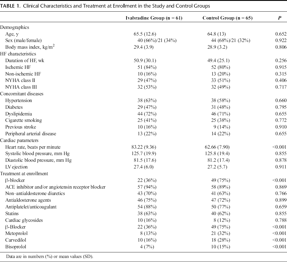
The heart rate was considerably higher in the ivabradine group [83.22 (9.36) beats per minute] compared to the control group [62.66 (7.90) beats per minute] (P < 0.001). However, once ivabradine was initiated in the study group, heart rate was found to be similar across the groups during the visits performed at 3 months and at the end of 12 months.
Left ventricular ejection was measured as 27.4 (6.0) in the ivabradine group and 27.2 (5.7) in the control group. There was no significant difference between the 2 groups in terms of EF. During the visit at 3 months, heart rate was decreased in the ivabradine group, but reflected no significant difference in terms of EF compared to the control group. However, at 12 months, EF was significantly different between the groups (P = 0.03).
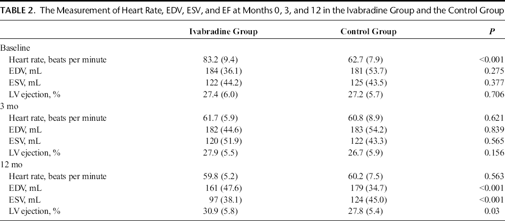
End-diastolic volume was measured as 184 (36.1) in the ivabradine group at baseline compared to 181 (53) in the control group, and ESV was measured as 122 (44.2) in the ivabradine group compared to 125 (43.5) in the control group. As was the case with EF, there was no significant difference regarding EDV and ESD at baseline and at 3 months, whereas EDV and ESV were considerably different in the ivabradine group compared to the control group during the visit at 12 months (P < 0.001). Heart rate, EDV, ESV, and EF of the groups are presented in Table 2.
The Measurement of Heart Rate, EDV, ESV, and EF at Months 0, 3, and 12 in the Ivabradine Group and the Control Group
Aortic Elasticity
Aortic strain (%) at the time of enrollment was 6.5 (2.9) in the ivabradine group, and 7.4 (3.4) in the control group. No statistically significant difference was found between the groups at baseline and 3 months. However, during the visit at 12 months, aortic strain was significantly increased in the ivabradine group compared to the control group (P < 0.001). Distensibility was measured as 2.9 ± 1.4 (cm2 [BULLET OPERATOR] dyne−1) in the ivabradine group at the time of enrollment compared to 3.4 ± 1.6 (cm2 [BULLET OPERATOR] dyne−1) in the control group. As was the case with aortic strain, distensibility was significantly increased in the ivabradine group compared to the control group during the visit at 12 months (Table 3).
The Measurement of Aortic Elastic Parameters at Months 0, 3, and 12 in the Ivabradine Group and the Control Group
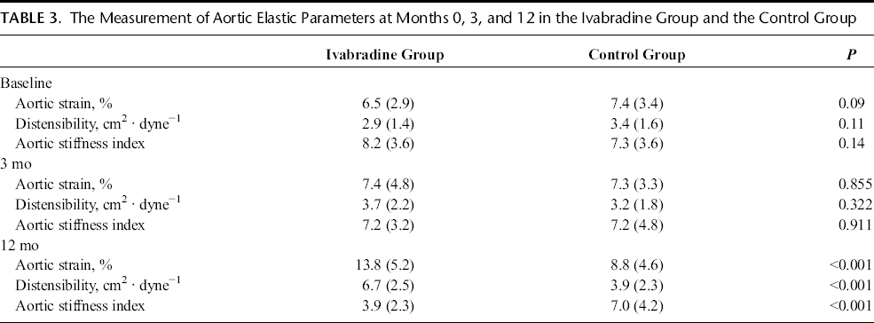
Aortic stiffness index was 8.2 (3.6) at the time of enrollment in the ivabradine group, and 7.3 (3.4) in the control group. There was no statistically significant difference between the groups at baseline and at 3 months. However, at 12 months, it was significantly decreased in the ivabradine group compared to the control group (P < 0.001). Figure 1A shows the levels of aortic elastic parameters at baseline; Figure 1B shows the levels of aortic elastic parameters at 3 months; and Figure 1C shows the levels of aortic elastic parameters at 12 months in the ivabradine group and the control group.
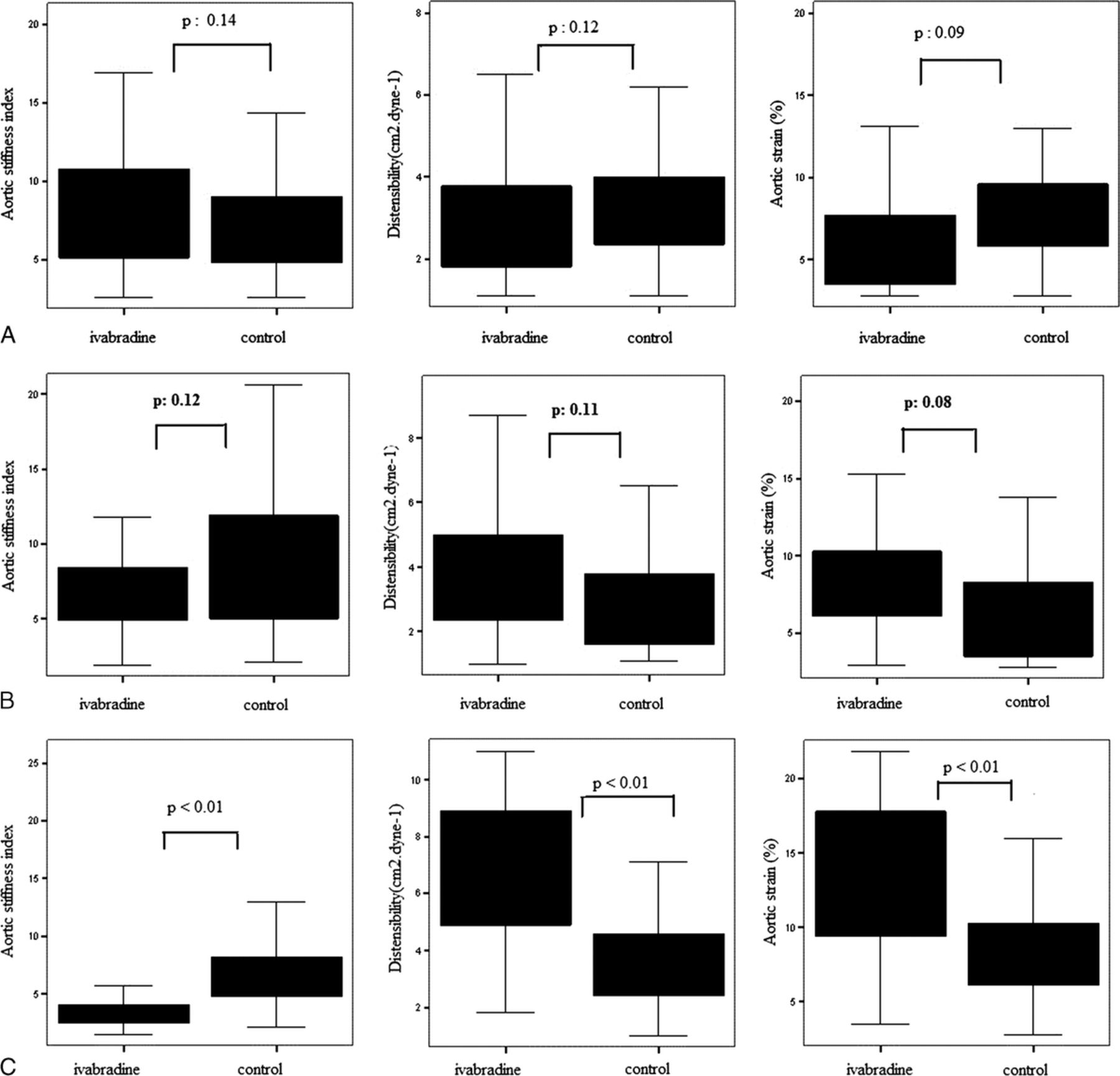
A, The levels of aortic elastic parameters at baseline in the ivabradine group and the control group. B, The levels of aortic elastic parameters at 3 months in the ivabradine group and the control group. C, The levels of aortic elastic parameters at 12 months in the ivabradine group and the control group.
Discussion
The present study is the first to investigate the effect of ivabradine treatment on aortic stiffness in patients with HF and that ivabradine treatment may reduce aortic stiffness after 1 year of follow-up. The reason for this improvement in aortic stiffness may be a result of the positive effects of ivabradine on aortic wall and the decrease in LVED volume and LVES volume as well as the increase in EF and cardiac output.
Mortality is considerably high in systolic HF, where arterial stiffness and pulsatile load contribute to the pathophysiology of HF. 20 Increased arterial stiffness increases PP, leading to cardiovascular adverse events and increased mortality.3,4 Studies have shown increased arterial stiffness in systolic HF.21–24
β-blockers have been shown to decrease morbidity and mortality in patients with systolic HF, and they also have favorable effects on arterial stiffness owing to their heart rate decreasing effects. Studies with new-generation β-blockers such as carvedilol and nebivolol have shown improved endothelial function and decreased arterial stiffness with these agents.25–28
Ivabradine is a new pharmacological agent which is a specific inhibitor of the If channel on sinoatrial node. 29 The only known effect of this agent is decreased heart rate. Moreover, ivabradine treatment has been shown to improve cardiac remodelling. 30 However, details of the favorable contribution of ivabradine in HF remain unknown.
In literature, there are no studies examining the effect of ivabradine treatment upon aortic stiffness. Our study is the first reporting the effect of ivabradine on aortic stiffness in patients with HF. Arterial stiffness is one of important signs of endothelial dysfunction. 31 Because of this, preclinic experimental studies of ivabradine on endothelial function may help us understand the relationship between aortic stiffness and ivabradine. Druoin et al. 14 have shown improved endothelial function with ivabradine in an animal model study, and associated the improvement with the rapid decrease in heart rate as well as the effect on nitric oxide and acetylcholine mechanism in vascular endothelium. Similarly, the decreased heart rate reduced vascular oxidative stress markers, improved endothelial function, and regressed atherosclerotic plaque formation in another animal model study by Custodis et al. 15 In light of the studies previously mentioned, the decreased heart rate obtained with ivabradine treatment may have affected endothelial function positively and contributed to the decrease of aortic stiffness.
As a matter of fact, studies have shown increased arterial stiffness with increased resting heart rate.32,33
In a study comparing the effects of ivabradine and β-blockers upon vascular functions, Nerla et al. 34 compared the effects of ivabradine versus atenolol on endothelial functions in patients with type 2 diabetes mellitus, and found no improvement regarding endothelial functions in ivabradine arm with a follow-up of 1 month. Also in our study, although aortic stiffness parameters were not significantly improved at 3 months, significant improvement was observed at the end of 1 year. We believe that the long study period of our study is beneficial in terms of demonstrating the net effect of ivabradine on aortic stiffness.
It is known that HF is characterized by the activation of the renin-angiotensin-aldosterone (RAAS) system. The RAAS activation is the leading cause of fibrosis and impaired cardiac functions. The RAAS activation is also known to contribute to development of arterial stiffness. Milliez et al. 16 investigated the effects of ivabradine on chronic and severe HF in adult Wister rat hearts, and concluded that ivabradine treatment led to a decrease in cardiac ACE proteins and mRNAs levels, which are the activation markers of cardiac RAAS and angiotensin 1 (AT1) receptors. In another study, ivabradine led to a decrease in AT2 levels and AT1 receptor activation in cases of endothelial dysfunction caused by hypercholesterolemia. 35 In light of the aforementioned studies, the favorable effects of ivabradine observed in our study may be associated with the effects on renin angiotensin aldosterone system.
The balance between elastin and collagen, components of extracellular matrix which comprises the structural skeleton of the vascular wall, is impaired during the development of aortic stiffness and this process leads to overproduction of collagen and impaired elastin.36,37 Dedkov et al. 38 demonstrated decreased amounts of interstitial and periarteriolar collagen with ivabradine treatment in an animal model study. The effect of ivabradine on arterial collagen deposition may be contributing to the regression of arterial stiffness.
The specifically decreased in heart rate reduces myocardial oxygen requirement, prolongs diastolic period, and increases myocardial perfusion. 39 Furthermore, decreased heart rate decreased the sympathetic denervation of the heart, aiding in improving myocardial pump performance and effectiveness. In our study, the decreased LVEDV and LVESV and the increased EF observed at the end of 12 months may be associated with the specific heart rate decreasing effects of ivabradine and are consistent with the subgroup analysis of BEAUTIFUL and SHIFT studies.30,40
Study Limitations
One of the limitations of this study is that it is a 3-center, nonrandomized study with a relatively small study population. And because other drug treatments given to the patients (Ace inh, statins, B-blockers, etc) and comorbid conditions may affect aortic stiffness, it is another limitation of our study. One of the reasons for the limitations of this study is that the observational nature is insufficient to definitely clarify the mechanism of the relation between ivabradine treatment and aortic stiffness in patients with HF. Moreover, there was no opportunity to use pulse wave rate instead of aortic stiffness to measure the arterial stiffness, as it is not among the routine methods used in our clinical practice.
Conclusions
In the current study in which 12-month follow-up of the patients with HF who received ivabradine treatment was investigated, we observed that ivabradine affected aortic elastic parameters positively and reduced LVEDV, LVESV, and increased EF; which pointed out that ivabradine treatment may decrease aortic stiffness among the patients with HF.