Abstract

2015 Combined Annual Meeting April 23 – 24, 2015 Chicago, IL
Combined Annual Meeting Abstracts
Cardiology/Cardiovascular Disease
Id: 2 Microrna Expression Pattern in Patients with Ventricular Arrhythmias and End Stage Heart Failure
T. Calway, T. Bak, K. Furlough, G. Kim. Medicine, University of Chicago, Chicago, IL.
Id: 3 Low Myocardial Infarction and Stroke Incidence in Hispanic Patients with Psoriasis
J. Nieves, C. Sulia, L. Figueroa, M. Crespo, N. Escobales. Medicine, Physiology and Dermatology, University of Puerto Rico, Medical Sciences Campus, San Juan, Puerto Rico. P.I. Altieri, H.L. Banchs. Cardiovascular Center of Puerto Rico and the Caribbean, San Juan, Puerto Rico.
Id: 4 Trpv1 Residues Vital to Protection against 4-Hne Modification, Prservation of Channel Activity and Microvascular Function
D.J. DelloStritto, P. Connel, I. Bratz. Integrative Medical Scineces, Northeast Ohio. Medical University, Rootstown, Ohio.
W. Geldenhuys. Pharmaceutical Sciences, Northeast Ohio. Medical University, Rootstown, Ohio.
We previously demonstrated enhanced 4-hydroxynonenal (4-HNE) post-translational modification (PTM) of TRPV1 decreases TRPV1 functional expression and contributes to microvascular dysfunction in diabetes. Accordingly, we hypothesized that manipulation of residues associated with 4-HNE PTM would preserve TRPV1 function and restore vascular integrity. 4-HNE decreased capsaicin mediated increases in myocardial blood flow and capsaicin-mediated relaxation in isolated coronary microvessels. TRPV1 functional analysis using electrophysiology revealed blunted capsaicin-mediated currents in the presence of 4-HNE which were reversed by the reactive carbonyl species scavenger aminoguanidine (AGD). Using computer modeling we identified three probable residues, previously identified to be important for oxidative modification, as potential sites for 4-HNE modification. The corresponding residues, C616, C621 and C634, were mutated to alanine (individually and in combination) and subsequently we examined the effects of 4-HNE on TRPV1 currents induced by capsaicin via electrophysiology. The mutation of the three pore cysteine (individually or in combination) abrogated the effects of 4-HNE on capsaicin-mediated currents. These data suggest that TRPV1 is targeted by redox-active substances that directly modulate channel activity at numerous sites in diabetes to decrease TRPV1 functional expression and contribute to microvascular dysfunction. The results obtained demonstrate an optimal redox state is critical for a properly functioning TRPV1 channel.
Id: 5 Amelioration of Nadph-Mediated Stress Reduces Cell Death following Blast-Induced Traumatic Brain Injury
B.P. Lucke-Wold, R. Turner, C. Rosen. West Virginia University School of Medicine, Morgantown, West Virginia.
Z. Naser. Drexel University, Philadelphia, Pennsylvania, UNITED STATES. A. Logsdon, K. Smith, J. Huber. West Virginia University School of Pharmacy, Morgantown, West Virginia. M. Robson. Vanderbilt University, Nashville, Tennessee. J. Bailes, J. Lee. Northshore University, Evanston, Illinois.
1.7 million traumatic brain injuries (TBIs) occur each year in the United States. Recent evidence suggests that repetitive TBIs can lead to chronic neurodegenerative changes over time. Currently, available pharmacologic options for the treatment of acute neurotrauma are limited. Oxidative stress is an important secondary mechanism of injury that can lead to cellular apoptosis and behavioral changes such as impulsivity. Utilizing a clinically relevant and validated rodent blast model, we investigated how NADPH oxidase expression and associated oxidative stress contributes to cellular apoptosis following single and repeat blast injuries. Nox4 forms a complex with p22phox following injury, both of which are important subunits of the NADPH oxidase system found within the brain. Using immunohistochemical-staining methods, we found a visible increase in Nox4 following single blast injury in Sprague Dawley rats. Interestingly, Nox4 was also increased in post-mortem human samples obtained from athletes diagnosed with chronic traumatic encephalopathy (CTE). Nox4 activity correlated with an increase in superoxide formation. Alpha lipoic acid, an oxidative stress inhibitor, prevented the development of superoxide acutely, and increased anti-apoptotic markers Bcl-2 (t = 3.079, p<0.05) and heme oxygenase 1 (t = 8.169, p<0.001) after single blast exposure. Subacutely, alpha lipoic acid treatment reduced pro-apoptotic markers Bax (t=4.483, p<0.05), caspase 12 (t=6.157, p<0.001), and caspase 3 (t=4.573, p<0.01) following repetitive blast, and reduced tau hyperphosphorylation indicated by decreased CP-13 and PHf staining. Alpha lipoic acid ameliorated impulsive-like behavior 7 days after repetitive blast injury (t=3.573, p<0.05) compared to blast exposed animals without treatment. TBI can cause debilitating symptoms, disability, and psychiatric disorders. Secondary mechanisms of injury, such as oxidative stress, are ideal targets for neuropharmacologic intervention. Alpha lipoic acid warrants further investigation as a therapeutic for the treatment of acute neurotrauma and prevention of chronic neurodegeneration.
Id: 6 Transgenic Or Pharmacological Inhibition of Sphk1 and S1Pr2 Prevents Hypoxiamediated Pulmonary Hypertension
J. Chen, J.R. Sysol, H. Tang, L. Moreno-Vinasco, K.M. Shioura, J.X. Yuan, J.G. Garcia, V. Natarajan, R.F. Machado. University of Illinois at Chicago, Chicago, Illinois. H. Tang, J.X. Yuan, J.G. Garcia. University of Arizona, Tucson, Arizona.
Previously we showed that Sphingosine kinase 1 (SphK1) and S1PR2 are up-regulated in patients with pulmonary hypertension (PH) and experimental PH models. We hypothesized that transgenic or pharmacological Inhibition of SphK1 and S1PR2 may prevent hypoxia-mediated PH (HPH). SphK1-deficient mice (SphK1KO) and C57Bl6 wild-type littermates (WT) were exposed to normoxia or 10% fiO2 for four weeks (n = 8 per group). In a separate experiment, C57Bl6 mice were injected with a S1PR2 inhibitor (JTE013, 8 mg/kg body weight every other day for 4 weeks). Additionally, male salt-sensitive rats (250–300 g, n= 6 per group) were exposed to normoxia or 10% fiO2 for 3.5 weeks and received SphK inhibitor (SKI2, 10 mg/kg body weight) every other day for 3.5 weeks. RVSP, right ventricle: left ventricle + septum (RV/LV+S) ratio and pulmonary vessel thickness were measured. After fourweeks of hypoxic exposure, SphK1KO mice developed significantly less severe PH (RVSP 30.09±0.68 vs. 36.77±1.07 mmHg, p<0.001) and right ventricular hypertrophy (RVH) (RV/LV+S 0.31±0.01 vs. 0.37±0.01, p<0.001), when compared to WT mice. After 3.5 weeks of hypoxic exposure, rats receiving SphKI2 developed significantly less severe PH (RVSP 45.30±1.20 vs. 30.10± 1.10 mmHg, p<0.01, figure B) and less RVH (RV/LV+S 0.34±0.04 vs. 0.26±0.01, p <0.05), when compared to vehicle treated rats. The similar data were observed in JTE013-treated mice after 4-week hypoxia exposure. All the vascular remodeling data are consistent with RVSP and RVH data. We concluded that transgenic or pharmacological Inhibition of SphK1 or S1PR2 blocker protects rodents against the development of HPH.
ID: 7 Exercise-Induced B-Natriuretic Peptide Elevation in Patients with and without Left Ventricular Hypertrophy
A. Abdul Jabbar, R. Markert. Internal Medicine / Cardiology Division, Wright State University Boonshoft School of Medicine, Dayton, Ohio. A. Abdul Jabbar, K. Makam, R. Mulamalla, C. Ahsan. Internal Medicine / Cardiology Division, University of Nevada School of Medicine, Las Vegas, Nevada.
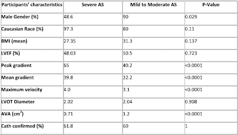
ID: 8 Right Ventricular Outflow to Aortic Valve Velocity Ratio: A Relative Dimensionless Index for Severe Aortic Stenosis
A. Abdul Jabbar, O. Ali, R. Markert, G. Broderick, B. White. Internal Medicine / Cardiology Division, Wright State University Boonshoft School of Medicine, Dayton, Ohio.
Id: 9 Hyperglycemia Induces Stress and Inflammatory Signaling that Leads to Hypertension in Pregnancy- a Translational Study
M.N. Uddin, C. Cawyer, N. Drever, S.R. Allen, T.J. Kuehl. Obstetrics and Gynecology, Baylor Scott & White Health/Texas A&M Health Science Center College of Medicine, Temple, Texas. M.N. Uddin, M.A. Co, M.M. Beeram, T.J. Kuehl. Pediatrics, Baylor Scott & White Healthcare/Texas A&M Health Science Center College of Medicine, Temple, Texas A.F. Pantho, S. Munir. University of Texas at Austin, Austin, Texas.
ID: 10 Simvastatin Regulates Human Lung Endothelial Sphingosine1-Phosphate Receptor 1 Via Kruppel-Like Factor 2
X. Sun, B. Mathew, S. Sammani, J.R. Jacobson. Medicine, University of Illinois at Chicago, Chicago, IL. J.G. Garcia. The University of Arizona, Tucson, AZ.
Id: 11 Cardiac Inflammation Increases with Contractile Dysfunction in Cardiomyopathies Caused by Sarcomere Protein Mutation
T.L. Lynch, S. Sadayappan. Cell and Molecular Physiology, Loyola University Chicago, Maywood, Illinois.
Id: 12 Urea and Thiourea-Based Tipodal Ligands Induce anti Angiogenic Profile on Human First Trimester Cytotrophoblast Cells
M.N. Uddin. Obstetrics and Gynecology, Baylor Scott & White Health/Texas A&M Health Science Center College of Medicine, Temple, Texas. B.M. Madhava. Pediatrics, Baylor Scott & White Health/Texas A&M Health Science Center College of Medicine, Temple, Texas. M.N. Uddin. Pediatrics, Baylor Scott & White Health/Texas A&M Health Science Center College of Medicine, Temple, TX. A.F. Pantho. University of Texas at Austin, Austin, TX. J. Castor II, A. Ashraf, D.C. Sprague. Biotechnology, Temple College, Temple, TX. M. Hossain. Chemistry and Biochemistry, Jackson State University, Jackson, Mississippi.
Id: 13 L-Name Attenuates the Cardiotonic Steroids-Induced Monolayer Permeability in Lymphatic Endothelial Cells
T. Kuehl, M.N. Uddin. Obstetrics and Gynecology, Baylor Scott & White Health/Texas A&M Health Science Center College of Medicine, Temple, Texas. J. Castor II, D.C. Sprague. Biotechnology, Temple College, Temple, Texas. W.E. Cromer, S.H. Afroze, D.C. Zawieja. Medical Physiology, Texas A&M Health Science Center College of Medicine, Temple, Texas. M.R. Beeram, T. Kuehl, M.N. Uddin. Pediatrics, Baylor Scott & White Health and Texas A&M Health Science Center College of Medicine, Temple, Texas.
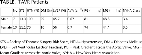
Id: 14 Feasibility of Transcutaneous Aortic Valve Replacement in a Non-Academic Community Hospital
D. Pappas, K. Hungate, R. Graf, T. Alexander, S. Damaraju. Cardiology, Spohn Shoreline, Corpus Christi, Texas. A.M. Damaraju. W B Ray High School, Corpus Christi, Texas.
TAVR Patients
Id: 15 the Role of Endothelial Microrna-130A in Angiogenesis
K. Furlough, T. Calway, S. McSharry, C. Harmon, D. Kim. University of Chicago-Molecular Cardiology, Chicago, IL.
Angiogenesis, the growth of new capillaries from preexisting blood vessels, is a complex process involving endothelial cell (EC) activation, disruption of vascular basement membranes, and migration and proliferation of ECs. Angiogenesis, which serves an important role in embryonic and postnatal development, is vital for the growth of new blood vessels from pre-existing vasculature and is subject to complex regulation in health and disease. MicroRNAs, short noncoding RNAs that downregulate gene expression at the post- transcriptional level, are important for proper organ function. Given their inherent pleiotropic actions to repress multiple gene targets simultaneously, microRNAs (miRNAs) are well poised to play a comprehensive and integrative role coordinating the interplay among the diverse pathways critical for angiogenesis. There has been emerging evidence that miR-130a plays a role in angiogenesis, though this role remains largely undefined. To study the effects of miR130a on angiogenesis, we created an inducible mouse model to express miR-130a, under the control of the VE-cadherein promoter, in ECs (VEcad- miR130a). Overexpression of miR-130a was initiated in adult ECs at weaning, approximately 4 weeks post-birth. We conclude that the overexpression of miR-130a in endothelial cells plays a role in the regulation of angiogenesis with likely anti-angiogenic effects. Loss of capillary density may be a mechanism for the induction of heart failure in this model.
Id: 16 Deficiency of Raptor in Smooth Muscle Cells Attenuates Hypoxia-Induced Pulmoanry Hypertension
H. Tang, Y. Gu, J.J. Yuan. Department of Medicine and Physiology, University of Arizona, Tucson, Arizona. D.R. Fraidenburg. Department of Medicine, University of Illinois, Chicago, IL. A. Makino. Department of Physiology, University of Arizona, Tucson, AZ.
Id: 17 Cha2Ds2Vasc Score in Predicting Future Development of Atrial Fibrillation after Successful Atrial Flutter Ablation
S.M. Hussain. Internal Medicine, UIC/AdvocateChrist Medical Center, Oak Lawn, Illinois. A. Bhan, M. Duggal, P. Dunskis. Cardiology, UIC/AdvocateChrist Medical Center, Oak Lawn, Illinois. A. Bhan, M. Duggal, P. Dunskis, H. Jibawi. Cardiology, Advocate South suburban hospital, Hazel crest, Illinois.
Id: 18 Interactions of Acidic Glycosaminoglycans with Basic Proteins and Peptides
S. Woodburn, A. Ellington. Chemistry, Bowling Green State University, Bowling Green, Ohio.
Heparin, the common sulfated glycosaminoglycan, has been in long-time clinical use as an anticoagulant prior to surgical procedures. Subsequent to surgery, it is neutralized by administration of the basic, low m.w. polypeptide, protamine, which is derived from salmon sperm. In past years, it has been reported that protamine itself exhibits a small anticoagulant effect when tested with a prothrombin time (PT) assay. In the current study, this laboratory reports that pharmacological quantities of protamine display considerable anticoagulant effects in PTassays and that the anticoagulant effect is readily neutralized by Dermatan Sulfate (DS). Whereas 2, 4, 8, 20, and 40 ug protamine generate PTs of 15.1, 17.3, 19.8, 24.0, and 31.1 seconds relative to a control of 10.3 seconds, addition of 90 ug DS to plasma/protamine mixtures reduces PT to 13.2, 13.0, 13.6, 24.0, and 14.5 seconds, respectively. The secondary control for 90 ug DS was only 13.6 seconds. Furthermore, the basic protein, calf thymus histone, displays a very slight anticoagulant effect in PT assays. However, it exhibits a significant inhibition of the anticoagulant effect of heparin when added sequentially to plasma. Relative to PTs of 12.3, 24.8, 47.3, and 53.3 seconds for addition of 2, 4, 8, and 10 ug heparin to plasma, the corresponding values are 10.4, 15.4, 42.8, and 48.3 seconds with the further sequential addition of 2 ug of calf thymus histone. Histone alone prolongs plasma by 2.5 seconds, from 10.3 to 12.8 seconds. More profound neutralizing results are obtained with 5 ug histone. Hence, the characteristic reaction of the basic proteins and peptides in their interaction with sulfated glycosaminoglycans such as heparin and DS may be a far more common phenomenon in nature than heretofore perceived. (The authors acknowledge, with profound appreciation, the contribution of the Medical Technology Program at BGSU for its donation of coagulation proteins in this study).
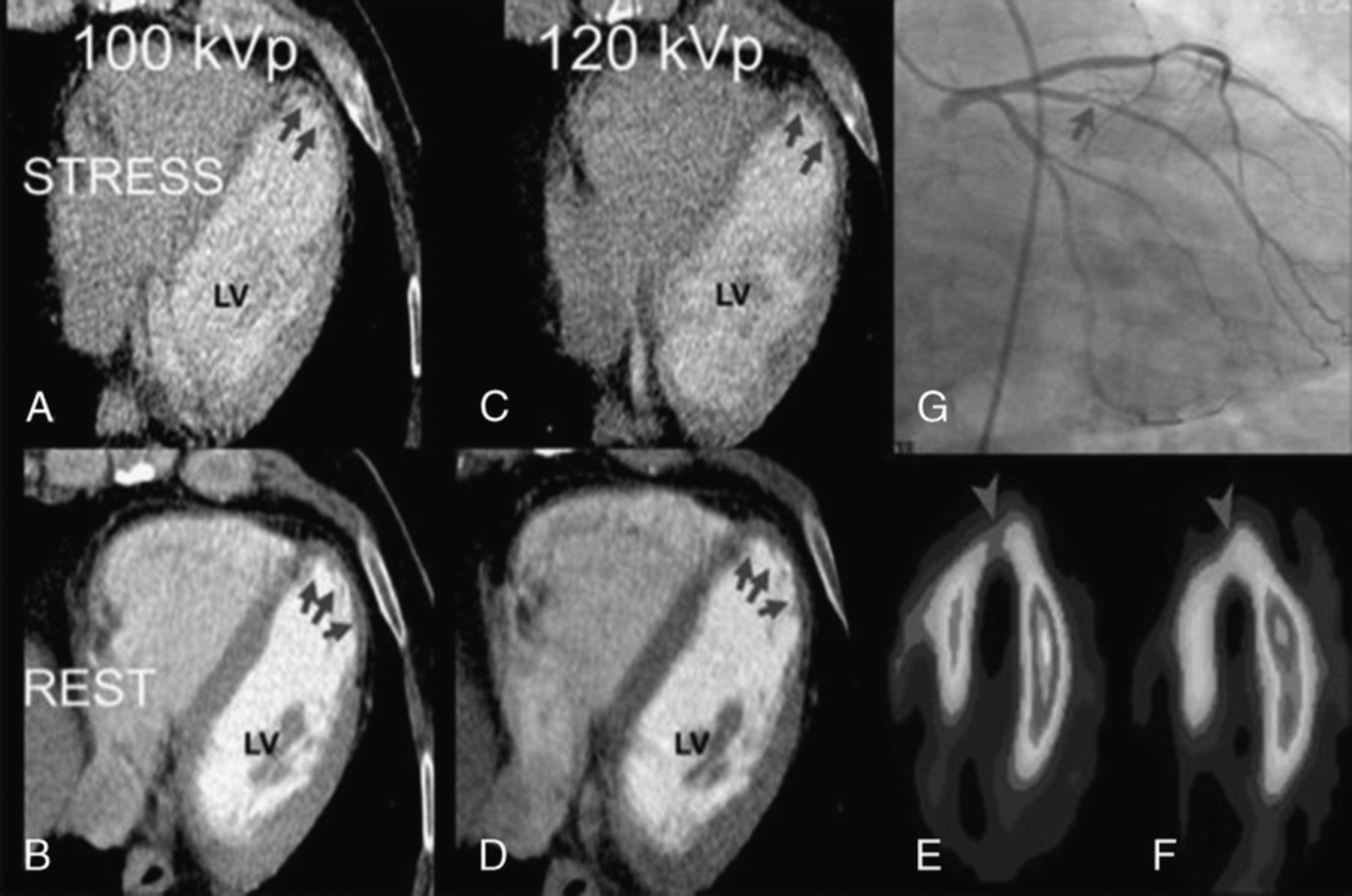
Id: 19 Dual - Energy Ct Myocardial Perfusion Imaging of Vasodialator Induced Myocardial Ischemia
M. Sidhu. Cardiovascular Medicine, University of Missouri, Columbia, Missouri. S. Uthamalingam, L. Engel, A.M. Lee, B. Ghoshhajra. Department of Radiology, Massachusetts General Hospital and Harvard Medical School, Boston, Massachusetts.
Comparative analysis of Stress and Rest images of Cardiac CT using different energy spectra (Panel AYD) with Stress (Panel E) and Rest (Panel F) Sestamibi Nuclear scan and a left coronary angiogram (Panel G). A thin perfusion defect (arrows) is more obvious on a 100 kVp scan (Panel A) when compared to 120 kvp energy scan (Panel C). Perfusion scan is absent on the rest scan (arrows Panel B and D). Panel E. Nuclear stress scan shows thin area of reversible perfusion defect when compared to the rest image (Panel F) that matches the area on stress CT scan Panel A. Panel G. Left coronary angiogram showing a significant hemodynamic lesion in the high diagonal branch supplying that area of reversible perfusion defect.
Id: 20 Isolated Aneurysms of the Ventricular Membranous Septum
A. Abdul Jabbar, O. Mufti, B.K. S. Internal Medicine / Cardiology Division, Wright State University Boonshoft School of Medicine, Dayton, Ohio. W. M. Cardiology, The Christ Hospital, Cincinnati, Ohio. B.M. Q, V. Tivakaran. Cardiology, Dayton VA Medical Center, Dayton, Ohio.
Id: 21 Severe Mrsa Endocarditis Treated with Veno-Venous Extracorporeal Membrance Oxygenation
J. Vargha, S. Mihalov, T. Yousuf. Department of Internal Medicine, Advocate Christ Medical Center, Oak Lawn, Illinois.
Id: 22 a Case of Polythemia Vera with Pulmonary Hypertension, Pulmonary Embolism, Hypertension and Renal Artery Stenosis
H. Tang. University of Arizona, Tucson, Arizona.
Q. gu, D. Yang, C. Xiong, Z. Zhao, Q. Luo, J. He, Z. Liu. fuwai Hospital National Center for Cardiovascular Diseases, Beijing, CHINA.
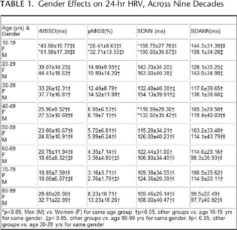
ID: 23 High Heart Rate Variability; a Marker of Healthy Longevity. Do Women have an Advantage over Men?
U. Zulfiqar. Internal Medicine, Peninsula Regional Medical Center, Salisbury, Maryland.
D.H. Singer. Dept of Medicine, Section of Cardiology, University of Illinois at Chicago, Chicago, IL.
Gender Effects on 24-hr HRV, Across Nine Decades
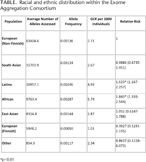
Endocrinology/Metabolism ID: 24 RELATIVE ETHNIC AND RACIAL fREQUENCY OF GCK-HYPERGLYCEMIA (GCK-MODY)
S.W. Greeley, R.N. Naylor. Pediatrics/Medicine, University of Chicago, Chicago, Illinois, UNITED STATES. D. Carmody. Medicine, University of Chicago, Chicago, Illinois. L.H. Philipson. Medicine/Pediatrics, University of Chicago, Chicago, Illinois.
Racial and ethnic distribution within the Exome Aggregation Consortium
ID: 25 Testosterone Therapy Interacts with Previously Undiagnosed Familial Thrombophilia, Facilitating Development of Osteonecrosis
R. Riaz, M. Prince. Internal Medicine Residency Training Program, Jewish Hospital of Cincinnati, Cincinnati, Ohio. C.J. Glueck, P. Wang. Cholesterol, Metabolism, and Thrombosis Center, Jewish Hospital of Cincinnati, Cincinnati, Ohio. R.A. Freiberg. Department of Orthopedics, VA Hospital, Cincinnati, Ohio.
Id: 26 Pancreatic Islet Glp-1 Secretion is Increased in Non-Diabetic Obesity and Glp-1 Directly Regulates Beta-Cell Cholecystokinin Production
A.K. Linnemann, T.J. Battiola, M.E. Kimple, D.B. Davis. Medicine/Endocrinology, University of Wisconsin-Madison, Madison, Wisconsin. J.C. Neuman. Nutritional Sciences, University of Wisconsin-Madison, Madison, Wisconsin. D.B. Davis. William S. Middleton Memorial Veterans Hospital, Madison, Wisconsin.
finding ways to protect pancreatic beta-cells from apoptosis is of particular interest in the prevention and treatment of diabetes. The use of glucagon-like peptide-1 (GLP-1) mimetics for the treatment of type 2 diabetes is increasing rapidly. However, despite in vitro and ex vivo evidence that GLP-1 protects beta-cells from apoptosis, the exact mechanism is not fully understood. Although GLP-1 is classically produced by intestinal L-cells after a meal, it was recently demonstrated that islet alpha-cells can also produce GLP-1 in response to stimuli such as cytokines and nutrients. Furthermore, islets isolated from obese mice express increased levels of prohormone convertase 1/3, the enzyme responsible for processing of GLP-1. This raises the question of whether islet production of GLP-1 is increased as a function of obesity in vivo. We now show that active GLP-1 is also secreted from human pancreatic islets as a function of BMI (R2=0.406, p=0.01). Similar to human, islets from obese C57BL/6J mice with a leptin mutation (ob/ob mice) secrete significantly more GLP-1 than islets from lean controls (p=0.0016). Considering the short half-life of GLP-1, it is likely that this islet-produced GLP-1 acts in a paracrine fashion to regulate a signaling network that is activated in obesity and may function to preserve beta-cell mass. Similarly, it was recently discovered that the classic gut hormone cholecystokinin (CCK) is also expressed in pancreatic beta-cells and is the most highly upregulated islet gene in response to obesity. Loss of CCK in obese mice results in decreased beta-cell mass and increased beta-cell death, leading to elevated fasting blood glucose. Additionally, we have recently found that beta-cell specific overexpression of CCK in lean mice confers protection from streptozotocin (STZ) induced apoptosis. This suggests that CCK intra-islet signaling is also important for apoptosis protection. As both GLP-1 and CCK are made in the obese islet, we hypothesized that GLP-1 regulates CCK in non-diabetic obesity. Indeed, GLP-1 treatment of betacells stimulates both CCK production (p=0.047) and secretion (p=0.004) by ∼2-fold. GLP-1 signals through cyclic AMP (cAMP) and treatment of beta-cells with a membrane permeable cAMP analogue stimulates Cck transcription by up to ∼9-fold (p=0.0005) and secretion by ∼8-fold (p=0.004). We used chromatin immunoprecipitation (ChIP) to show that both cAMP and GLP-1 treatment promote recruitment of RNA polymerase 2 and the cAMP-modulated transcription factor, CREB, to the promoter of the Cck gene in beta-cells (p<0.05 for all experiments). Inhibition of cAMP with sulprostone reduces Cck transcription by ∼50% indicating that cAMP-mediated signaling is required. However, reduction of glucose does not abolish the ability of cAMP to stimulate CCK production or secretion. Importantly, we find that CREB recruitment to the Cck promoter is present in the islets of ob/ob mice, but not in lean mice.
In summary, we provide a novel description of how islet production of incretins is regulated. We show that both human and mouse islets secrete active GLP-1 as a function of BMI/obesity. In turn, GLP-1 can promote beta-cell CCK production and secretion through direct transcriptional regulation of the gene in a cAMP-dependent manner. This mechanism of regulation likely explains the increase in islet CCK production seen in obesity. As both CCK and GLP-1 offer protection of beta-cells from apoptosis, we propose that these hormones may be co-regulated within the islet and exert a local effect of beta-cell protection.
Id: 27 Differential Effects of Leptin on Adiponectin Expression with Weight Gain versus Obesity
P. Singh, P. Sharma, K.R. Sahakyan, D.E. Davison, F.H. Sert-Kuniyoshi, A. Romero-Corral, F. Lopez-Jimenez, T. Kara, V.K. Somers. Cardiovascular Diseases, Mayo Clinic, Rochester, Minnesota. J.M. Swain. Department of Surgery, Mayo Clinic, Rocehster, Minnesota. M.D. Jensen. Endocrinology Research Unit, Mayo Clinic, Rochester, Minnesota.
Id: 28 Targeted Disruption of Atp Sensitive Potassium Channel Expression in Skeletal Muscle Promotes Energy Consumption during Physical Activity
S. Koganti, D. Zhu, D. Subbotina, D. Gao, D. Sierra, M. Proenza, L. Yang, D. Zingman. Internal Medicine, The University of Iowa, Iowa City, Iowa. D. Zingman, A. Alekseev. Mayo Clinic, Rochester, Minnesota.
Despite the medical, social and economic impact of obesity, only a few therapeutic options, focused largely on reducing caloric intake, are currently available and these have limited success rates. A major impediment is that any challenge by caloric restriction is counterbalanced by activation of systems that conserve energy to prevent body weight loss. Therefore, targeting energy-conserving mechanisms to promote energy expenditure is an attractive strategy for obesity treatment. Skeletal muscles account for about 40-50% of body mass and their function relies on the energy demanding processes of ion homeostasis and actin-myosin cycling. Even under sedentary conditions, spontaneous physical activities required to maintain muscle tone, body posture and “fidgeting” account for about 7- 10% of daily body energy usage. During exercise, energy consumption by skeletal muscles increases 20-100 times over the basal level. Considering this level of muscle metabolic activity, even small changes in energy efficiency could have a substantial effect on bodily energy consumption. Here, in order to suppress muscle energy efficiency, we target sarcolemmal ATP-sensitive potassium (KATP) channels which have previously been shown to be important in maintaining muscle energy economy. Specifically, we employ intramuscular injections of cell-penetrating oligonucleotides to prevent translation of the channel pore-forming subunit. This intervention results in significant reduction of KATP channel expression and function in treated areas, without affecting the channel expression in nontargeted tissues. Furthermore, suppression of KATP channel function in a group of muscles causes a substantial increase in activity-related energy consumption, with little effect on exercise tolerance. These findings establish a proof-of-principle that selective skeletal muscle targeting of sarcolemmal KATP channel function is possible and that this intervention can alter overall bodily energetics to treat or prevent obesity.
Id: 29 Ethanol Activates Midkine and Alk Signaling in Neuroblastoma Cells and in the Brain
D. He, H. Chen, H. Muramatsu, A. Lasek. Psychiatry, University of Illinois at Chicago, Chicago, Illinois.
Id: 30 Prevalence of Preeclampsia in Pre-Gestational Diabetic Pregnancy in Bangladeshi Patients
T.J. Kuehl, M.N. Uddin. Obstetrics and Gynecology/Pediatrics, Baylor Scott & White Health/Texas A&M Health Science Center College of Medicine, Temple, Texas. G.U. Ahsan, M. Jahan, M. Hasanuzzaman, S. Mahbuba, H. Jahan, S. Chowdhury, K. Leena, M.N. Uddin. Public Health, North South University, Dhaka, Dhaka, BANGLADESH.
Id: 31 the Extracellular Matrix Regulates Adipose Function and Expansion
M.K. Vaicik, E.M. Brey. Biomedical Engineering, Illinois Institute of Technology, Chicago, Illinois. M.K. Vaicik, E.M. Brey. Research Service, VA Hines, Hines, Illinois. M. Morse, A. Blagajcevic, R. Cohen. Section of Endocrinology, Department of Medicine, University of Chicago, Chicago, Illinois.
Obesity is a global epidemic that contributes to the increasing medical burdens related to type 2 diabetes, cardiovascular disease and cancer. The extracellular matrix (ECM) has been shown to regulate the development and function of numerous tissues and organs. An understanding of the role the ECM plays in adipose tissue function and expansion could lead to new therapeutics that eliminate or reduce obesity-associated morbidity and mortality. Laminin a4 is upregulated during adipogenesis and is present in the ECM surrounding fully differentiated adipocytes. However, there is little understanding of its function in adipose tissue. We have found that mice with a null mutation of the laminin α4 gene (Lama4−/−) exhibit reduced weight gain and fat mass accumulation in response to both aging and high-fat diet when compared to wild-type (Lama4+/+) animals. However, the underlying mechanisms by which Lama4 regulates fat mass have not yet been defined. We have now found that physical activity and food intake does not differ between Lama4+/+ and Lama4−/− mice. However, Lama4−/− mice have a significantly increased metabolic rate at 25C (room temperature) and 16C (cold) compared to Lama4+/+ mice. Interestingly, Lama4−/− mice exhibit significantly increased UCP-1 expression in subcutaneous adipose tissue [18.79 ± 4.97% UCP-1 positive compared to 2.62 ± 1.63% (n=5, p£0.01)]. In contrast, in thermoneutral conditions at 30C both Lama4+/+ and Lama4−/− mice exhibit equivalent metabolic rates. These results suggest that beiging of subcutaneous adipose tissue in Lama4−/− mice may lead to decreased adipose tissue accumulation and potentially improved metabolic function. Thus, alterations in laminin composition suggest that the ECM plays a role in modulating cellular behavior in adipose tissue expansion in a temperature- and depot-specific manner.
Id: 32 Significant Differences in Fecal Microbiota Are Associated with Various Stages of Glucose Tolerance in African-American Male Veterans
I. Ciubotaru, S. Kukreja, E. Barengolts. Endocrinology, University of Illinois at Chicago, Chicago, Illinois. I. Ciubotaru, A. Akbar, S. Kukreja, E. Barengolts. Endocrinology, Jesse Brown VA, Chicago, Illinois. S. Green. CRC, DNA Services, University of Illinois at Chicago, Chicago, Illinois.
There is emerging evidence that intestinal microbiota is a contributor to the metabolic/glycemic phenotype. While changes in microbiota have been described in obesity and diabetes, little is known about microbiota composition in various dysglycemic states. This study aimed to investigate the relationship between microbiota and changes in the glycemic control of prediabetic subjects. Stool was collected from African American men participating in a randomized controlled trial of vitamin D Intervention at VA. Four groups (Gr) were characterized based on changes in OGTT between baseline (T0) and the study completion at 12 months (T12): Gr 1- stable normal glucose tolerance (GT); Gr 2- stable impaired fasting glucose or stable impaired GT; Gr 3 - worsened GT; and Gr 4 - improved GT. Microbiota DNA was extracted from stool collected at T12, analyzed using high-throughput next-generation sequencing of microbial rRNA genes and data processed using established bioinformatics pipelines. Microbiota (composition, alpha diversity, abundance) was analyzed in 116 subjects: Gr 1= 35, Gr 2 = 27, Gr 3 = 24, and Gr 4 = 29. At Phylum level significant differences in bacterial composition were observed between Gr 1 and Gr 2 (p= 0.03) and a trend to significance for Gr1 vs Gr3 (p= 0.06), and Gr 1 vs Gr4 (p= 0.06). Bacteroidetes was higher, firmicutes lower, and hence the Bacteroidetes/firmicutes ratio (B/f) was lower with worsening glycemic control (B/f: Gr 1 vs Gr 2 = 1.9 vs 0.9, p= 0.01; and Gr 1 vs Gr 3 = 1.9 vs 1.1, p= 0.04). Proteobacteria decreased in Gr 2 and Gr 4 compared to Gr 1 (p=0.01 for both). Similarly, there were significant differences in microbiota at the family and Genus levels. In Gr 2 vs Gr1 there was less Prevotella (hence higher Bacteroides/Prevotella ratio, 5.6 vs 2.7, p= 0.05), less Enterobacteriaceae (p=0.03), and more Ruminococcaceae (p= 0.01) and Veillonellaceae (p= 0.02). Notably, Akkermansia was more abundant in Gr 4 vs Gr 1 (p=0.04). We speculate that lower abundance of Prevotella may be associated with worsening glycemia, and conversely higher abundance of Akkermansia might be associated with improving glycemia, thus corroborating suggestions from previous studies. Ruminococcaceae might be associated with higher insulin level (seen in previous research), and therefore conducive to maintenance of stable glycemia. Observed association of Veillonellaceae and glycemia was novel. In Conclusion, significant differences in microbiota (composition, alpha diversity, abundance) were observed in various GT states. Interesting most of the differences were seen between normoglycemic subjects and those who were remained prediabetics at the end of the study. These findings suggest that there may be a certain makeup of the gut microbiota associated with steady glycemic states. Further studies are needed to evaluate whether causative relationship exist between microbiota and changes in GT.
Id: 33 Roles of Growth Hormone/Insulin-Like Growth Factor-I Axis in Prostate Stem/Progenitor Cells Amplification
J.D. Rinaldi, D. Hu, W. Hu, G. Shi, L. Birch, G.S. Prins. Urology, UIC, Chicago, Illinois. J.D. Rinaldi, L. Justulin. Morphology, UNESP, Botucatu, Sao Paulo, BRAZIL.
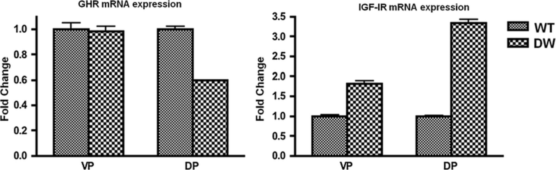
Prostate stem/early progenitor cells were isolated from ventral (VP) and dorsal (DP) prostate using 3-D Matrigel culture for 7 days. GHR and IGF-1R gene expressions were quantified by real-time qPCR (N=5). Of note, both genes were expressed in prostate stem/progenitor cells from WT and DW rats. Although rat prostate expressed lower levels of GHR, IGF-1R was increased in both VP and DP from DW rats when compared to WT rats.
ID: 34 the Impact of Prostaglandin E2 Levels on Glycemic Control and Therapeutic Response in Human Subjects with Type 2 Diabetes Mellitus
A.C. Weeks, D.B. Davis, M. Kimple. Endocrinology, Diabetes & Metabolism, University of Wisconsin, Madison, Wisconsin. M. Dart. Department of Surgery, University of Wisconsin, Madison, Wisconsin. X. Li. Department of Biostatistics and Medical Informatics, University of Wisconsin, Madison, Wisconsin.
The varied response of patients with type 2 diabetes mellitus (T2DM) to therapeutic agents is well recognized, but the underlying etiology remains unclear. Given the substantial morbidity and mortality associated with T2DM, as well as the cost of pharmacotherapy, elucidating causal factors is of great interest. As incretin mimetics function in part by directly stimulating the insulin-producing b-cells, factors down-regulating b-cell function are of particular concern. Using islets isolated from diabetic mice and humans, we showed prostaglandin E2 (PGE2) acts through a specific b-cell receptor to reduce insulin secretion, directly competing with incretin signaling[i]. We hypothesized (1) individuals with high PGE2 production have poorer glycemic control and a weaker response to incretin-based therapeutics than those with low PGE2 production and (2) plasma PGE2 level can be a biomarker of T2DM status and therapeutic response. In a preliminary study using 75 anonymized patient samples, individuals with T2DM (n=65) had significantly elevated plasma PGE2 levels (18.0 ± 22.4 pg/ml) as compared to non-diabetics (ND) (11.0 ± 7.3 pg/ml, n=10). Subjects with diabetic nephropathy, indicating poorer glycemic control, demonstrated the greatest elevation (19.9 ± 11.6 pg/ml, n=16, p=0.04 vs. ND). This data gave support to a new cohort study of T2DM and ND subjects recruited from the UW Endocrinology and Diabetes Clinic. Exclusion criteria included age <18 or>70, chronic or recent use of most COX inhibitors or oral steroids, active infection, anemia of any type confounding hemoglobin A1c measurement, and most autoimmune diseases. A power analysis with α=0.05 and b=0.08 indicated 35 ND and 137 T2DM subjects (with 20-30% on incretin therapy) needed. PGE2 levels are correlated with (1) donor demographics and measures of (2) diabetes/obesity status, (3) glycemic control, and (4) inflammation. Chart review is performed at 6 and 12 months to record diabetes status, glycemic control and medication failure. We performed an interim analysis of 62 subjects (14 ND and 47 T2DM, with 19 T2DM subjects on incretin–based therapy). Multivariable analysis of all subjects adjusted for covariates demonstrated no significant effects on PGE2 levels with respect to age, BMI, triglycerides, HgA1c, ESR, or aspirin use; however, age (p=0.0795) and TG (p=0.0588) demonstrated a strong trend. Within the T2DM group alone, triglycerides again showed a strong trend (p=0.0678), as did ESR (p=0.1363) and age (p=0.18). The correlation of ESR with PGE2 levels in the T2DM group appeared to be more relevant with incretin therapy (p=0.2398) than without (p=0.9732), whereas the opposite was true for triglyceride levels (p=0.3262 with incretin therapy vs. 0.0489 without). Although results did not show a significant correlation between PGEM levels and T2DM status, this may reflect additional unidentified covariates rather than a true rejection of our hypothesis, providing guidance for the full analysis.
Id: 35 Hypoglycemia in Sulfonylurea-Treated Kcnj11-Related Diabetes is Infrequent and Usually Mild
D. Carmody, M. Lanning, L. Szczerbinski, L.H. Philipson, S.W. Greeley. Section of Pediatric and Adult Endocrinology, Diabetes and Metabolism, University of Chicago, Chicago, Illinois.
ID: 36 Expression Of Receptors For Growth Hormone-Releasing Hormone (Ghrh-R) In Human Papillary Thyroid Cancer Cells: Effects Of Ghrh Antagonists On Matrix Metalloproteinase-2
P. Catanuto, J. Tashiro, M.K. Glassberg, N.L. Block. Medicine, University of Miami Miller School of Medicine, Miami, florida. P. Catanuto, J. Tashiro, J.I. Lew, S.J. Elliot. Surgery, University of Miami Miller School of Medicine, Miami, florida. F.G. Rick. Urology, florida International University, Miami, florida. A.V. Schally. Endocrine, Polypeptide and Cancer Institute, Veterans Affairs Medical Center and South florida Veterans Affairs foundation for Research and Education, Miami, florida.
Papillary thyroid cancer (PTC) is the most prevalent of all endocrine cancers. In recent studies, the presence of receptors for pituitary-type growth hormone-releasing hormone (pGHRH-R) has been demonstrated in various human cancers, including human prostate, brain, and other cancer lines. Thyroid malignancies however, have not yet been investigated in this regard. In this study, we found that pGHRH-R and its functional splice variant, SV1, are present in normal thyroid and PTC cells. We also treated seven normal and PTC tumor thyroid cells in vitro with a GHRH antagonist, MIA-602, to compare its anti-proliferation and anti-invasion potential against untreated cells. We found that treatment with GHRH antagonist increases the expression of SV1 and pGHRH-R in tumor cells compared to tumor cells exposed to vehicle only, a response which may alter the sensitivity of signaling kinases within the cells. GHRH antagonist treatment of tumor cells also reduced activity of the tumor invasion marker, matrix metalloproteinase (MMP)- 2, compared to tumor cells exposed to vehicle only. The expression of pGHRH-R and SV1, as well as MMP-2 activity, in normal thyroid cells remained unaffected by GHRH antagonist treatment. Similarly, cell proliferation rates for tumor or normal thyroid cells were not affected by GHRH antagonist treatment. Our findings have important implications for the therapeutic use of GHRH antagonist in cases of aggressive PTC refractory to conventional treatment modalities, and in which protein expression and MMP-2 activity in normal thyroid tissue is left unaltered.
Id: 37 Pituitary Tumor Apoplexy and Influenza: Case Report
T.M. Yousuf, N. Libo, A. Lara, A. Bacal, T. Yasmeen. Internal Medicine, Advocate Christ Medical Center, Oak Brook, Illinois.
Epidemiology/Health Care Outcomes/Quality Improvement/Bioinformatics
ID: 38 Design Of Pilot Study To Improve Quality-Of-Life In Chronic Diseases Using Custom Web-Based Educational And Monitoring Platform
O. Oni. Clinical Research, Kansas City VA Medical Center, Kansas, Missouri. A. Goel, V. Savin. Nephrology, Kansas City VA Medical Center, 4801 E Linwood Blvd, Kansas City, Missouri. K. Gandy. Play-it Health, LLC, 1800 Baltimore Avenue, Kansas City, Missouri. E. Witten. Witten and Associates, LLC, 8318 Connell Street, Overland Park, Kansas.
Id: 39 An Online Discussion Group for Rare Monogenic Diabetes Patients: Supporting Families and Fueling New Research
M.E. Perrone, D. Carmody, L.H. Philipson, S.W. Greeley. Section of Pediatric and Adult Endocrinology, Diabetes and Metabolism, University of Chicago, Chicago, Illinois.
Id: 40 Effect of Vitamin D Supplementation during Pregnancy on Maternal and Neonatal Outcomes: A Systematic Review and Meta-Analysis of Randomized Clinical Trials
P. Thota. Infectious Diseases, Case Western Reserve University, Cleveland, Ohio. FR. Perez-Lopez. University of Zaragoza, Zaragoza, SPAIN. V. Pasupuleti. Case Western Reserve University, Cleveland, Ohio. E. Mezones-Holguin, V. Benites-Zapata. Instituto Nacional de Salud, Lima, PERU. A. Deshpande. Medicine, Cleveland Clinic, Cleveland, Ohio. A.V. Hernandez. Cleveland Clinic, Cleveland, Ohio.
Gastroenterology/Clinical Nutrition
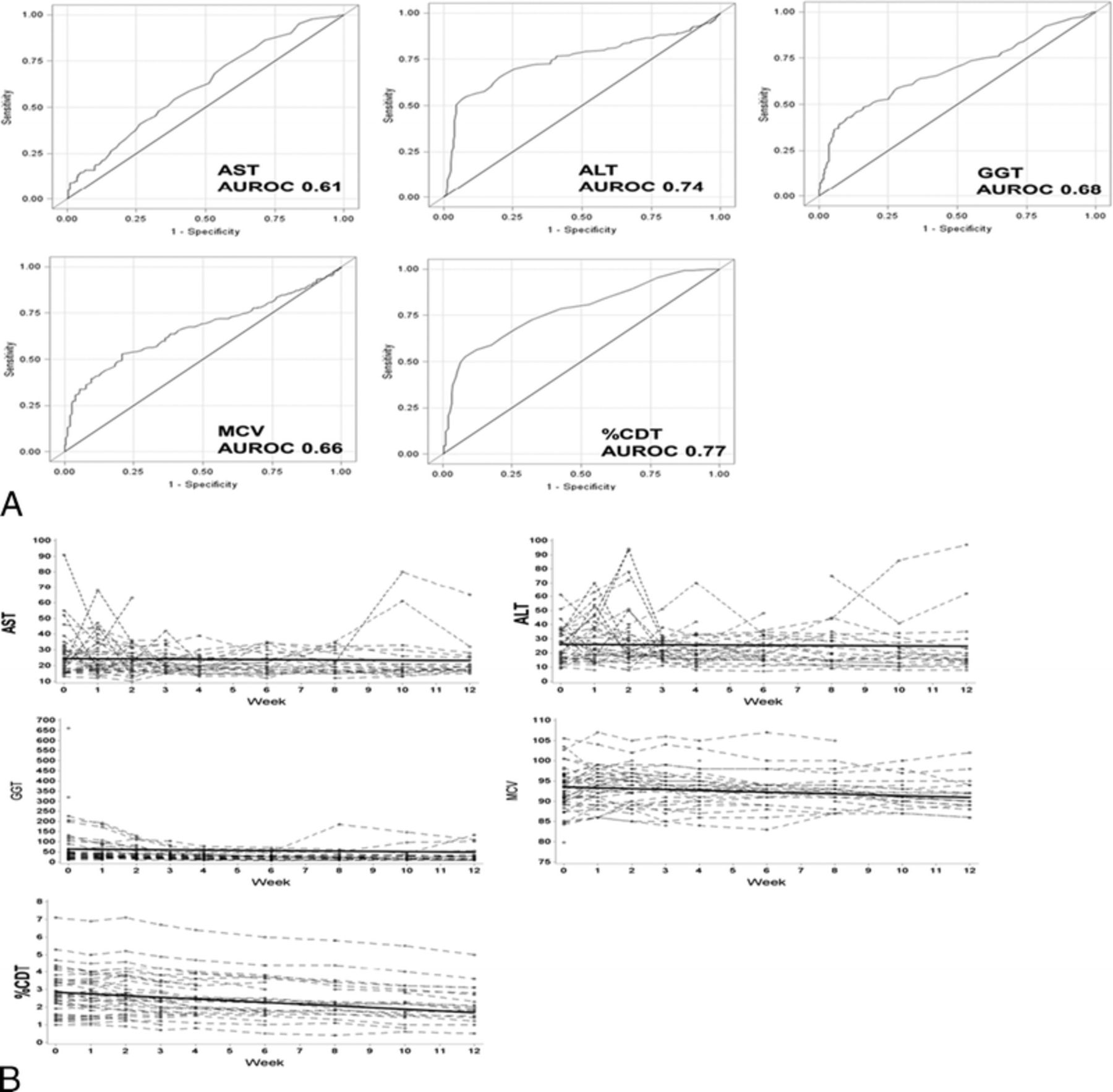
Id: 41 the Utility of Commonly Used Laboratory Tests to Screen for Excessive Alcohol Use in Clinical Practice
G. Gough, S. Liangpunsakul. Medicine, Indiana University, Indianapolis, Indiana.
Id: 42 Comparative Effects of Protamine and Poly-L-Lysine upon Prothrombin Time
A. Brecher, A. Riaz. Chemistry, Bowling Green State University, Bowling Green, Ohio.
Alcoholism is responsible for the observance of prolonged blood coagulation times in a significant fraction of the alcoholic population. This laboratory has reported earlier that acetaldehyde, the primary intermediate in ethanol metabolism, readily prolongs coagulation times upon covalent interaction with thrombin, prothrombin, factors Xa, IXa, XIa, XIII, and fibrinogen, among the proteins tested. In this communication, it is reported that protamine, the basic polypeptide isolated from salmon sperm and utilized to neutralize the anticoagulant effect of heparin, has anticoagulant properties which are enhanced upon exposure to acetaldehyde. It is further demonstrated that the basic polypeptide, poly-L-lysine, similarly prolongs prothrombin time (PT), and that mixtures of plasma, poly-L-lysine, and acetaldehyde have additional PT times at 4.47mM acetaldehyde, and synergistic anticoagulant effects at 44.7 mM acetaldehyde. The pattern for protamine and poly-L-lysine are quite analogous. Whereas 9mM protamine prolongs PT at 30 sec relative to 11.5 sec for the control, 4.4 mM acetaldehyde prolongs PT at 13.2 sec, and 44.7 mM acetaldehyde extends PT at 33.7 sec. Pre-mixtures of 9 mM protamine/4. 4 mM acetaldehyde produce an additive 33.1 sec PT, while a 9 mM protamine/44.7 mM acetaldehyde generates a synergistic 91.2 sec PT. Comparable numbers with poly-L-lysine generate values of 11.3, 15.1, 34.8, 12.4, 17.1, and 45.9 sec, respectively. Poly-L-lysine is a bacterial fermentation product. The commercial source was tested at concentrations from 0.035 to 0.64 mM/L, and mixtures ranging from 0-0.5, 1-5, and 4-15 kDa/mol. The higher molecular weights produced prolonged PTs for the same concentrations. These data may reflect the decreased solubility of the poly-L-lysine-fibrinogen complexes. (The authors appreciate the generous donation of reagents by the BGSU Medical Technology program to this study).
Id: 43 Correlation of Foxm1 and Oxidative Stress Induced Dna Damage in Human Hepatocellular Carcinoma
B.L. Sun, N. Ronquillo, P. Raychaudhuri, G. Guzman. University of Illinois Hospital & Health Sciences System, Chicago, Illinois.
Id: 44 Cholestatic Drug-Induced Liver Injury Treated with Corticosteroid in a Patient with Miliary Tuberculosis and Associated Adrenal Insufficiency
S. Lee, P. Khankhanian, A. Schneier, O.N. Machado. Medicine, Icahn School of Medicine at Mount Sinai, Elmhurst, New York. S. Liu. Pathology, Icahn School of Medicine at Mount Sinai, Elmhurst, New York.
Drug-induced liver injury (DILI) in a patient with multiple comorbidities is challenging to diagnose because liver injury can be attributed to multiple disease processes. Many medications have been reported to cause DILI, but the pathophysiology is multifactorial and difficult to pinpoint. However, delayed treatment of DILI could have fatal consequences, and therefore, understanding the features and risks of DILI is crucial. A 82 year-old woman a remote history of cholecystectomy presented with a pre-syncopal episode. Two days prior, she was discharged from another hospital after prescribed ciprofloxacin and metronidazole for colitis diagnosed by abdominal computational tomography (CT). She took ciprofloxacin and metronidazole for 3 days in total. At admission, she was found to have severe sepsis with elevated AST (111 IU/L) and ALT (450 IU/L) with normal alkaline phosphatase (AP) (111 IU/L) and hyponatremia to 125 mEq/L. She was treated with vancomycin and imipenem/cilastatin in addition to fluid resuscitation. She became hemodynamically stable after three days. At this time, vancomycin and imipenem/cilastatin were discontinued, and cefepime was started, given negative cultures. However, AP began to increase (figure A). After five doses of cefepime, it was discontinued as a possible cause of liver injury. She was restarted on imipenem/cilastatin for two more days for a total of 10 days of antibiotic treatment, but her AP continued to rise. A search for common non-drug causes of acute liver injury was all negative. Liver biopsy revealed cholestasis and noncaseating granulomatous disease, suggesting DILI or infectious etiology (figure B). An ACTH stimulation test for persistent hyponatremia and fatigue confirmed the diagnosis of adrenal insufficiency for which corticosteroids were started. The AP peaked at 1,004 IU/L and started to trend down two days after steroid was started. Chest CT revealed multiple subcentimeter central and peripherally located densities. A diagnosis of miliary tuberculosis with associated adrenal insufficiency was made, and the patient improved with anti-TB medications and corticosteroids. A few weeks after discharge, induced sputum cultures grew mycobacterium tuberculosis confirming the diagnosis. This patient received five different antibiotics including ciprofloxacin, metronidazole, vancomycin, imipenem/cilastatin, and cefepime. Danan et al. provide a consensus method for assessing the causal role of a drug in liver injury. Only ciprofloxacin and metronidazole are “suggestive” agents for cholestatic liver injury in terms of both the onset and cessation of medications. Metronidazole is known to cause hepatocellular liver injury, and pharmacology of each drug implies that ciprofloxacin was the most likely antibiotic causing DILI. This case is unique because miliary TB was complicated by adrenal insufficiency and drug-induced cholestatic liver injury, but acute liver injury was fully reversed after corticosteroid treatment. This implies an immune-mediated etiology of DILI, especially ciprofloxacin-induced cholestatic liver injury.
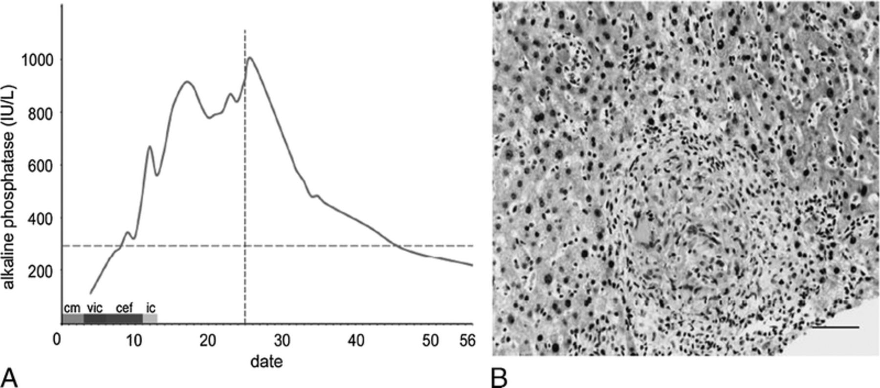
(A) Daily trend of alkaline phosphatase (AP). Day 1 is the first day of ciprofloxacin and metronidazole. Liver injury is cholestatic when AP is greater than two times the upper limit, represented as blue horizontal dotted line. The date on which corticosteroid was given is represented as the red vertical line. Liver injury started on day 8. “cm” stands for ciprofloxacin and metronidazole (for 3 days); “vic,” vancomycin and imipenem / cilastatin (for 3 days); “cef,” cefepime (for 5 days); “ic,” imipenem / cilastatin (for 2 days). (B) Liver biopsy of this patient, demonstrating a non-caseating epithelioid granuloma involving both portal tracts and lobules with evidence of cholestasis. Scale bar = 150 μm.
Id: 45 Spirochetes in the Gut! a Case Report of An Hiv Related Chronic Diarrhea
L. dakhoul, F. Abou Obeid, H. Raddawi. Internal Medicine, Advocate Christ Medical Center, Oak Lawn, Illinois.
Id: 46 Your Appendix Can Tell Your Story! An Unusual Cause of Appendicitis
L. dakhoul, H. Raddawi. Internal Medicine, Advocate Christ Medical Center, Oak Lawn, Illinois.
Id: 47 Primary Amyloidosis Diagnosed by Endoscopic-Ultrasound Guided Celiac Lymph Node Biopsy
N. AKBAR, V. Puri, A. Kubbara, U. Ahmad, A. Nawras. Internal Medicine, University of Toledo medical Center, Toledo, Ohio.
Id: 48 Herpes Esophagitis in An Immunocompetent Teenager
T.M. Yousuf, S. Wang, A. Khan, B. Blumenstein. Internal Medicine, Advocate Christ Medical Center, Oak Brook, Illinois.
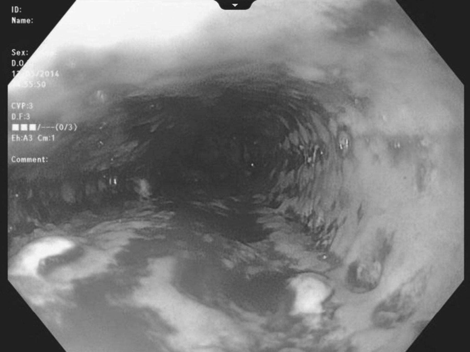
Herpes esophagitis is a common infection in the immunocompromised host. However, it is a rare condition in the immunocompetent population, and usually presents with the constellation of symptoms of odynophagia, fever and retrosternal chest pain. In immunocompetent patients, symptoms of odynophagia and chest pains are usually attributed more to pill induced esophagitis, toxic ingestion or reflux esophagitis. Rarely, if ever, is infectious esophagitis from HSV, CMV or candida considered. HSV-1 is more commonly the cause of esphoagitis, but typically as a reactivation. In very rare cases does it present as a primary infection in the esophagus. We describe a case of HSV esophagitis in an eighteen year old immunocompetent host with no significant past medical history. He initially presented to his primary care physician with complaints of odynophagia and was prescribed a course of amoxicillin and prednisone syrup. He presented four days later to the emergency room with worsening odynophagia, retrosternal chest pain and anorexia. He was evaluated by the gastroenterology team and was taken for esphsophagogastroduodenoscopy (EGD), which revealed diffuse bleeding superficial ulcerations along the entirety of his esophagus. Biopsies were taken and subsequently found to be HSV positive. The patient was treated with intravenous acyclovir, proton pump inhibitor, and sucralfate. HIV status on the patient was found to be negative, and no other causes of immunosuppression was found. We believe the patients’ initial presentation was in fact, HSV esophagitis, which was exacerbated by his prednisone use. The purpose of this report is to highlight the rare occurrence of infectious esophagitis in otherwise healthy, young individuals and to be able to promptly diagnose and begin treatment on such patients. EGD with biopsy is the gold standard diagnostic modality for HSV esophagitis and should always be considered in young patients who present with odynophagia with or without other alarm features. EGD findings often can point to the cause as HSV has a very typical appearance on EGD. Often we see superficial, well-demarcated ulcers, typically along the mid to distal esophagus. The treatment of choice for HSV esophagitis is intravenous acyclovir 5mg per kg every eight hours for seven to fourteen days. Treatment should be initiated promptly following the EGD to expedite resolution. Symptoms for most immunocompetent patients resolve spontaneously in about one to two weeks. Although rare, HSV esophagitis should be entertained especially given the correct constellation of history and symptoms. In confirmed cases of HSV esophagitis, it is important to reassess the patient for any underlying immunodeficiencies, especially HIV as a cause for the infection.
ID: 49 Elevated Liver Transaminases; One More Etiology To Look For
N. AKBAR, A. KUBBARA, R. QADIR. Internal Medicine, University of Toledo medical center, Toledo, Ohio.
AST and ALT progression in our patient
Id: 50 a Rare Cause of Heartburn and Abdominal Pain in a Teenager Female
N. AKBAR, A. KUBBARA. Internal Medicine, University of Toledo medical center, Toledo, Ohio. A. NAWRAS. Gastroenterology, University of Toledo medical center, Toledo, Ohio.

Id: 51 Irony of Transfusional Support for Refractory Anemia in Hemophagocytic Lymphohistiocytosis
S. Lee. Medicine, Icahn School of Medicine at Mount Sinai, Elmhurst, New York. M. Chary. Icahn School of Medicine at Mount Sinai, New York, New York.
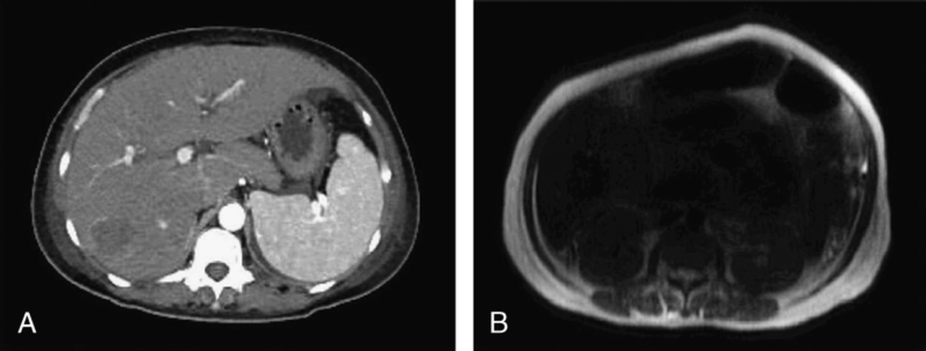
Hemophagocytic lymphohistiocytosis (HLH) is an aggressive disorder from immune system derangement with uncontrolled macrophages engulfing patients’ own blood cells. Patients with myelodysplastic syndrome, leukemias, sickle cell disease, or beta-thalassemia major depend on chronic transfusion, requiring 10 - 20 units of RBC transfusion a year. However, these patients do not develop iron deposition in the organ parenchyma until the late stage of diseases. Here we report a unique case of a patient with HLH and associated severe refractory anemia, who developed rapid iron deposition in the liver and pancreas parenchyma after 16 units of RBC transfusion. This case shows the irony of managing refractory anemia with transfusional support because patients with HLH require continuous transfusional support and are simultaneously more susceptible to developing iron overload. A 44 year-old woman with no significant past medical history presented with fatigue, fevers, and jaundice. She was found to have hepatosplenomegaly and severe jaundice at admission. Her blood tests showed hemoglobin of 8.4 g/dL, mean corpuscular volume 85.0 fL/cell, iron 138 μg/dL, ferritin 4,170 ng/mL, transferrin saturation 93%, aspartate transaminase/alanine transaminase 123/191 U/L, gamma-glutamyl transpeptidase 57 U/L, lactate dehydrogenase 1,310 U/L, and total/conjugated bilirubin 20.7/18.7 U/L. Computerized tomography (CT) of the abdomen and pelvis showed hepatosplenomegaly and a 4.7 cm hemangioma (figure A). Liver biopsy showed hemophagocytosis with negative iron staining. Bone marrow biopsy showed hypercellular marrow with a large number of CD4- and CD68- positive histiocytes and hemophagocytosis: she was diagnosed with HLH, for which steroid and chemotherapy were started. Sixteen units of RBC transfusion were provided for refractory anemia, and MRI of the abdomen was performed: severe iron deposit in the liver and pancreas (MRI T2* relaxation time of 8 msec and R2* (1/T2*) = 125 Hz) was revealed, which was not shown at admission on CT scan or liver biopsy (figure B). Per Wood et al., the liver iron concentration (LIC) can be estimated, based on the shortening of T1, T2, and T2* relaxation times on MRI: her LIC was estimated to be 13 mg/g dry weight. Drasar et al. demonstrated the followings: 1) patients with sickle cell disease who had serum ferritin > 1,000 ng/mL and received less than 20 units (range 12 - 19) of RBC transfusions showed a mean LIC of 2.1 - 2.3 mg/g dry weight; 2) the total unit of transfusion correlates more strongly with LIC than the rate of transfusion. This result demonstrates that patients with HLH could reach the iron overload state more rapidly than patients with other diseases requiring transfusional support. A large amount of iron from breakdown of phagocytosed RBCs by reticuloendothelial macrophages is stored as ferritin, resulting in a very high transferrin saturation in HLH. Therefore, the reticuloendothelial system in HLH has a low iron binding capacity to store extra iron generated by transfusional products, triggering more rapid iron deposition in the organ parenchyma.
Abdominal CT and MRI images. (A) is an abdominal CT image at the early stage of hospital admission, showing splenomegaly and hepatomegaly with a 4.7 cm irregular mass at the posterior right hepatic lobe. (B) is an abdominal MRI imaging after the patient received 16 units of RBC transfusional supprt with diffuse signal dropout due to iron desposition in the liver and pancreas (T2* = 8 msec).
Genetic & Molecular Medicine
Id: 88 a Gene Regulatory Network Shared between Neurulation and Orofacial Development
Y.A. Kousa, R.R. Roushangar. Biochemistry and Molecular Biology Department, Michigan State University, East Lansing, Michigan. H. Zhu, Y. Lei, R.H. Finnell. Dell Pediatric Research Institute, Department of Nutritional Sciences, University of Texas at Austin, Austin, Texas, UNITED STATES. W.D. Fakhouri. Department of Diagnostic and Biomedical Sciences, University of Texas at Houston, Houston, Texas. T.D. Busch, J.C. Murray, A. Bassuk. Department of Pediatrics, University of Iowa, Iowa City, Iowa. G.M. Shaw. Department of Pediatrics, Stanford University School of Medicine, Stanford, California. A. Ashley-Koch, S. Gregory. Duke Molecular Physiology Institute, Department of Medicine and Molecular Genetics and Microbiology, Duke University, Durham, California. N. Patel, B.C. Schutte. Department of Microbiology and Molecular Genetics, Michigan State University, East Lansing, Michigan. E.J. Leslie. Center for Craniofacial and Dental Genetics, University of Pittsburgh, Pittsburgh, Pennsylvania. T.J. Williams. Department of Craniofacial Biology, University of Colorado Denver at Anschutz Medical Campus, Aurora, Colorado. Y. Chai. Center for Craniofacial Molecular Biology, University of Southern California Ostrow School of Dentistry, Los Angeles, California.
IRf6, TfAP2A and GRHL3 encode transcription factors that are required for orofacial development in humans and mice. We showed that rs642961 is highly associated with orofacial clefting and perturbs a TfAP2A binding site in an enhancer element (MCS9.7) that recapitulates Irf6 expression. In keratinocyte culture, TfAP2A regulates IRf6 expression via MCS9.7. We also showed that Irf6 regulates Grhl3 expression in zebrafish and that mutations in IRf6 and GRHL3 cause nearly identical human phenotypes. These observations suggest that TfAP2A, IRf6 and GRHL3 form a network in craniofacial morphogenesis. However, mice that lack either Tfap2a or Grhl3 also have neural tube defects. These neurulation defects are located rostrally (exencephaly) and caudally (curly tail) in both mutant mice. In addition, Tfap2a knockout embryos have an exceptional ventral wall defect involving both the thoracic and abdominal cavities. Therapeutically, neurulation defects in Grhl3 knockout embryos are rescued by inositol supplementation, but not folate. In this study, we discover a critical role for Irf6 in neural tube and ventral wall morphogenesis by characterizing an allelic series, including both loss- and gain-of-function alleles. We find that reducing Irf6 expression leads to a curly tail and reductions in both Tfap2a and Grhl3 expression. Remarkably, reducing Irf6 expression in Tfap2a haploinsufficient embryos rescues exencephaly. Irf6 over-expression leads to a ventral wall defect that is highly analogous to Tfap2a knockout embryos. Molecularly, we find that Irf6 is expressed in neural ectoderm and early migrating neural crest cells and that MCS9.7 is active in these cells. Strikingly, we show that Tfap2a regulates MCS9.7 in multiple tissues, including tail and skin. Consistently, we find that Tfap2a and Grhl3 interact genetically in rostral and caudal neurulation and ventral wall development in a dose-dependent manner. Therefore, Irf6 homeostasis is required for at least three ectoderm derived tissues via positive and negative regulation of Tfap2a and Grhl3. Lastly, we sequence IRf6 in 96 individuals with spina bifida and find a rare variant previously shown to be disease causing in orofacial clefting. We further genotype common SNPs in 2,500 individuals with spina bifida to cover 28% of the genetic variation at the IRf6 locus but do not detect a common association. With roles in neural tube, ventral wall and craniofacial morphogenesis, Tfap2a-Irf6-Grhl3 appear to be a key gene regulatory network in ectoderm development. Considering that Grhl3 is a distal node in this pathway, inositol supplementation and inhibition may provide an environmental lever to alter multiple developmental programs of the ectoderm based on genetic risk.
Id: 89 An Epigenetic Map of Age-Associated Autosomal Loci in Northern European Families at High Risk for the Metabolic Syndrome
D. Cerjak, R. James, Y. Zhang. Medicine, Medical College of Wisconsin, Brookfield, Wisconsin. O. Ali Pediatrics, Medical College of Wisconsin, Milwaukee, Wisconsin. J.W. Kent, J. Blangero, M.A. Carless. Pediatrics, Medical College of Wisconsin, Milwaukee, Wisconsin. Genetics, Texas Biomedical Research Institute, San Antonio, Texas.
ID: 90 Liver Eqtls for Warfarin dose Response Genes Reveal Susceptibility to Venous Thromboembolism among African Americans
W. Hernandez, E.R. Gamazon, A. Konkashbaev, M.A. Perera. Deparment of Medicine, Section of Human Genetics, The University of Chicago, Chicago, Illinois. L.H. Cavallari. University of florida, Gainesville, florida. R.A. Kittles. University of Arizona Cancer Center, Tucson, Arizona.
Venous thromboembolism (VTE) is a chronic disease encompassing deep vein thrombosis (DVT), pulmonary embolism (PE), or both. In the US, African Americans (AAs) have the highest incidence and mortality rates of DVT/PE. Warfarin is used to treat and prevent DVT/PE and the dose requirement has been shown to be higher among AAs as well as for DVT/PE patients regardless of ethnicity. Because genetic variation within cis-regulatory elements or trans-acting regulators can affect gene expression in a cell type specific manner, we aimed to investigate the role between VKORC1, CYP2C9, and CALU eQTLs and DVT/PE susceptibility using liver eQTL data. We identified and genotyped 72 cis and trans eQTLs in a study population of 462 AAs on stable warfarin dose; of which 256 individuals were treated with warfarin due to DVT/PE (cases) and the remaining due to a variety of other conditions (controls). We found a significant decrease in risk of DVT/PE for carriers of the minor allele of two VKORC1 eQTLs and one CALU eQTL (rs9925964, OR=0.53, P=0.01; rs12597511, OR=0.48. P=0.02; and rs11054879, OR=0.61, P=0.03 respectively). We also found our DVT/PE patients had a higher percentage of West African ancestry and were younger compared to controls (t=-1.991, p=0.04 and t=2.720, p=0.007 respectively). The frequency of these protective alleles were much lower (10%) in our study population and among the HapMap Yourbans (YRI) but significantly higher in the HapMap European Americans (CEU) at approximately 40%. By investigating eQTLs for genes known to contribute to warfarin dose requirement we have uncovered novel disease loci involved in the risk of DVT/PE. These findings may help explain the increased risk of DVT/PE seen in the African American population.
Id: 91 Interstitial Triplication of 15Q11-Q13: An Assessment of Methylation Status and Gene Expression
A.M. Goetjen, N. Germain, S. Chamberlain. University of Connecticut Health Center, Farmington, Connecticut.
The majority of genes in the human genome are biallelically-expressed, but this is not true for a number of genes that are located at the 15q11-q13 locus, which are regulated by a process called genomic imprinting. Genomic imprinting is a phenomenon in which genes are expressed in a parent-of-origin specific manner. Imprinted genes are functionally haploid. The master regulator of gene expression from this imprinted region is a differentially methylated region known as the Prader-Willi imprinting center (PWS-IC). A number of genes are selectively expressed from the paternal allele, whereas one gene (UBE3A) is maternally-expressed in neuronal cells. There are three clinical syndromes that result from duplication or deletion of these mono-allelically expressed genes: Prader-Willi syndrome, Angelman syndrome, and 15q duplication syndrome. Here I focus on 15q duplication syndrome. Children with 15q duplication syndrome present with cognitive dysfunction, speech/language disorders, and autism. Duplication of the 15q11-q13 locus may occur as an interstitial duplication or as an isodicentric chromosome 15 (idic(15)) that contain two or three copies of the maternal locus, respectively. Those with paternal interstitial duplication are clinically unaffected. An increased number of maternal copies of the locus correlates with a greater severity of the clinical phenotype, however little is known about the effect that chromosome structure has on the phenotypic presentation and gene expression. Thus, the focus of this project was to characterize gene expression and methylation status of the 15q11-q13 locus in a patient with a maternal interstitial triplication. Induced pluripotent stem cells (iPSCs) and iPSC-derived neurons were used as the model system. Methylation at the PWS-IC, was shown to be 75%, consistent with the individual's cells having three maternal copies and one paternal copy of the 15q11-q13 locus. Based on observations from iPSCs generated from patients with maternal interstitial duplication or idic(15), it was hypothesized that there would be a positive correlation between the level of gene expression and copy number in iPSCs generated from the patient with the maternal interstitial triplication. Indeed, 15q gene expression from the maternal interstitial triplication clones is positively correlated with the number of maternal copies of the locus. We observe minor differences in gene expression between interstitial triplication and idic(15) iPSCs or neurons, suggesting that different structural presentations of the three maternal copies of 15q11-q13 may be regulated similarly.
Id: 92 Effects of Rpd3 Mutation on Mitochondrial Function and Metabolism in Aging Drosophila
J. Woods, B. Rogina. Genetics and Genome Sciences, University of Connecticut Health Center, West Hartford, Connecticut.
Previously our lab showed that mutations in rpd3 (Drosophila HDAC1 homologue) extend the lifespan of Drosophila through a mechanism that overlaps with caloric restriction (CR). CR is known to increase lifespan by altering many physiological processes, including mitochondrial function and nutrient metabolism. The objective of this project is to determine the mechanism of lifespan extension in heterozygous rpd3-mutant flies as it relates to mitochondrial function, stress response, and metabolic homeostasis. The mitochondrial-to-nuclear gene ratio, level of spargel (Drosophila PGC-1 homologue) mRNA, and quantification of electron micrographs were examined as indicators of mitochondrial biogenesis and quantity. Mitochondrial respiration was examined to analyze differences in mitochondrial function. qPCR and mRNA sequencing were used to examine changes in genes responsible for metabolism, as well as other cellular pathways that affect longevity. We have found significant differences in mitochondrial respiration in fruit flies with heterozygous mutations of rpd3. However, our results indicate no difference in mitochondrial biogenesis. Many genes of the insulin-signaling pathway are differentially expressed in rpd3-mutant flies. Based on these results, we conclude differences in mitochondrial biogenesis are not the reason for lifespan extension in rpd3-mutant flies as initially hypothesized. The insulin-signaling pathway remains a candidate pathway for the lifespan extending effects. Future work will be needed to determine how targeting this pathway could promote healthy aging and lifespan extension.
Id: 93 Heterozygous Mutations in Aggrecan Cause Short Stature, Accelerated Bone Maturation, and Early Growth Cessation
M. Guo, C. Jacobsen, J. Hirschhorn. Division of Endocrinology, Boston Children's Hospital, Boston, Massachusetts. O. Nilsson, J. Lui, J. Baron. Program in Developmental Endocrinology and Genetics, National Institutes of Health, Bethesda, Maryland. J. Quintos. Warren Alpert Medical School of Brown University, Providence, Rhode Island. J. Popovic, D. Flynn. University of Pittsburgh Medical Center, PIttsburgh, Pennsylvania. A. Dauber. Cincinnati Children's Hospital Medical Center, Cincinnati, Ohio. N. Dunbar. Connecticut Children's Medical Center, Hartford, Connecticut.
Short stature is a common presentation to pediatric endocrinology clinics and is frequently associated with delayed skeletal maturation. In contrast, short stature with advanced skeletal maturation is a rare presentation. We recruited four families with autosomal dominantly inherited short stature and advanced skeletal maturation. Affected family members presented with childhood short stature (height SDS -1.9 to -3.5), advanced bone age, early growth cessation, and adult short stature (height SDS -2.6 to -4.7). Additional features that were variably present include osteochondritis dissecans, early onset osteoarthritis, macrocephaly, midface hypoplasia, and exaggerated lumbar lordosis. To identify the genetic cause of this phenotype, we performed whole exome sequencing in selected individuals from each family. In all four families, we identified novel heterozygous variants in the aggrecan gene (ACAN), which encodes a proteoglycan that serves as a major structural component of the extracellular matrix in growth plate and other cartilaginous tissues. The sequence variants were present in all affected but in none of the unaffected family members. Each of the mutations identified was predicted to result in loss of protein function. Our study indicates that heterozygous mutations in ACAN cause a mild skeletal dysplasia that presents as short stature with advanced bone age and early growth cessation. This contrasts with previous reports of ACAN mutations identified in individuals with severe skeletal dysplasias. Our findings thus expand the spectrum of ACAN defects and provide a new molecular genetic etiology for the unusual child who presents with short stature and accelerated skeletal maturation.
Hematology and Oncology
Id: 94 Central Retinal Artery Occlusion Associated with Familial Thrombophilia in Young Healthy Females
P. Shah, C.J. Glueck, J. Patel, Schockman, D. Smith. Internal Medicine/Endocrinology, Jewish-Mercy Cholesterol and Metabolism Center, Cincinnati, Ohio.
Id: 95 Falls and Fractures in Patients with Breast Cancer on Aromatase Inhibitors Compared with Their Age-Matched Controls
M. Williams, P. Choksi, K. Kidwell, C. Van Poznak. Internal Medicine, University of Michigan, Ann Arbor, Michigan. J. Stella School of Medicine, University of Michigan, Ann Arbor, Michigan. D. Hanauer. Pediatrics, University of Michigan, Ann Arbor, Michigan.
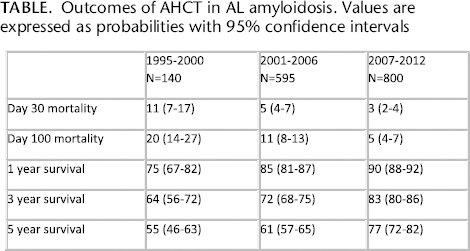
Id: 96 Improved Outcomes of Autologous Hematopoietic Cell Transplantation (Ahct) for Light Chain Amyloidosis: A Center for International Blood and Marrow Transplant Registry (Cibmtr) Study
A. D'Souza, P. Hari. Medicine, Medical College of Wisconsin, Milwaukee, Wisconsin, UNITED STATES. A. D'Souza, J. Huang, M. Zhang, P. Hari. Center for International Blood and Marrow Transplant Registry (CIBMTR), Milwaukee, Wisconsin.
Outcomes of AHCT in AL amyloidosis. Values are expressed as probabilities with 95% confidence intervals
Id: 97 Diagnostic Ramifications of Ocular Vascular Occlusion as a First Thrombotic Event Associated with Factor V Leiden and Prothrombin Gene Heterozygosity
S. Schockman, M. Prince, R. Riaz. Internal Medicine Residency Program at Jewish Hospital- Mercy Health, Cincinnati, Ohio. C.J. Glueck, P. Wang. Cholesterol, Metabolism, and Thrombosis Center of the Jewish Hospital- Mercy Health, Cincinnati, Ohio. C.J. Glueck. Mercy Medical Physicians, Cincinnati, Ohio. R. Hutchins. Cincinnati Eye Institute, Cincinnati, Ohio. R. Hutchins. Ophthalmology, University of Cincinnati College of Medicine, Cincinnati, Ohio.
Id: 98 Central Retinal Vein Thrombosis 3, 3, and 4 Months after Starting Testosterone Therapy in Men with Previously Unrecognized Familial Thrombophilia
S. Schockman, R. Riaz, M. Prince. Internal Medicine Residency Program at The Jewish Hospital – Mercy Health, Cincinnati, Ohio. C.J. Glueck, P. Wang. Cholesterol, Metabolism, and Thrombosis Center of the Jewish Hospital- Mercy Health, Cincinnati, Ohio. C.J. Glueck. Mercy Medical Physicians, Cincinnati, Ohio.
Id: 99 Identification of Signaling Lipid Lysophosphatidic Acid as a Critical Link between Diet-Induced Obesity, Angiogenesis and Breast Cancer Progression
L. Dong, Y. Chen, R. Yuan, R. Silverstein, B. Ren. Blood Research Institute, Blood Center of Wisconsin, Milwaukee, Wisconsin. Y. Yuan, S. Wu. Edison Biotechnology Institute and Department of Chemistry and Biochemistry, Ohio University, Athens, Ohio. I. Aguilera-Barrantes. Pathology, Medical College of Wisconsin, Milwaukee, Wisconsin. A. Sturich, R. Silverstein, B. Ren. Medicine, Medical College of Wisconsin, Milwaukee, Wisconsin.
Obesity increases cancer risk including breast cancer (BC) but the direct link and mechanisms by which obesity promotes BC progression remain largely unknown. Nutritional obesity is accompanied by autotaxin (ATX)-mediated synthesis of the bioactive signaling phospholipid, lysophosphatidic acid (LPA). We have shown that LPA stimulates angiogenesis by turning off CD36 antiangiogenic switch in microvascular endothelial cells (MVECs) via protein kinase (PKD-1) signaling pathway that may be associated with mitochondrial bioenergetic metabolism. We hypothesize that LPA-PKD-1 signaling is a key BC promoter by modifying mitochondrial bioenergetics in both tumor and endothelial compartments. Using the Seahorse Bioscience Extracellular flux Analyzer, we showed that LPA enhanced mitochondrial respiration in human breast adenocarcinoma MDA-MB231 cells transduced with wild type PKD-1 (PKD-WT) in the presence of high glucose concentration, resulting in elevations in both oxygen consumption rate (OCR) and ATP-linked OCR, but not in the extracellular acidification rate (ECAR). The conditional medium from human MVECs transduced with PKD-WT reduced levels of basal OCR, ATP-linked OCR, and ECAR in breast cancer cells when compared with the control medium. However, in tumor-associated ECs, exposure to LPA decreased not only CD36 expression but also basal OCR, ATP-linked mitochondrial respiration and basal ECAR. Furthermore, overexpressing PKD-WT or constitutively active PKD-1 decreased basal OCR and ATP-linked mitochondrial respiration. To determine in vivo mechanisms of obesity-derived LPA, we established a syngeneic breast adenocarcinoma model in diet induced obese female mice. BC growth was significantly larger in the diet-induced obese mice than in the lean controls after tumors were subcutaneously implanted for 21 days (3884.6±804.4 mm3 vs 843.4±392.1 mm3). The tumor endothelium demonstrated increased LPA receptor 1, along with reduced CD36 expression and increased periphery vessels when compared to the lean control group (81.5±12.75vs 34.5±7.25 mm2). Proteomic angiogenic profiling showed significant elevations in several angiogenic proteins, particularly leptin level in the serum of the obese mice. Our study indicates that LPA changes cellular bioenergetics in both endothelial and BC cells. This may be associated with the metabolic switch between mitochondrial oxidative phosphorylation and aerobic glycolysis. The switch could be regulated by PKD-1 signaling and associated with proangiogenic responses and BC initiation and progression. Targeting LPA/PKD-leptin-metabolic signaling axis in the tumor microenvironment could provide a novel therapeutic strategy against breast cancer.
Id: 100 Deregulated Cell Cycle Confers Resistance to Aromatase Inhibitors in Postmenopausal Breast Cancer Patients
I. Doostan, C. Karakas, K. Keyomarsi. Experimental Radiation Oncology, The University of Texas MD Anderson Cancer Center, Houston, Texas. I. Doostan, K. Keyomarsi. Graduate School of Biomedical Sciences, The University of Texas at Houston, Houston, Texas. S. Moulder. Department of Breast Medical Oncology, MD Anderson, Houston, Texas. K. Hunt. Department of Breast Surgical Oncology, Houston, Texas.
Doostan I1, Karakas C2, Moulder S3, Hunt KK4, Keyomarsi K5 1,5 University of Texas, Graduate School of Biomedical Sciences & 1,2,5 Department of Experimental Radiation Oncology, 3Department of Breast Medical Oncology and 4 Department of Breast Surgical Oncology, MD Anderson Cancer Center, Houston, Texas, 77030 Contact author:
Id: 101 Histone Deacteylase Inhibition Regulates Inflammation and Enhances Tregs after Allogeneic Hematopoietic Cell Transplantation in Humans
E. Gatza, G. Hou, Y. Song, S. Choi. Pediatrics-Hematology/Oncology, University of Michigan, Ann Arbor, Michigan. Y. Sun, K. Oravecz-Wilson, P. Reddy. Internal Medicine- Hematology/Oncology, University of Michigan, Ann Arbor, Michigan. J. Whitfield. Comprehensive Cancer Center Immunology Core, University of Michigan, Ann Arbor, Michigan. C. Dinarello. Internal Medicine-Infectious Diseases, University of Colorado, Aurora, Michigan.
Id: 102 Ceramide Mediated Lethal Mitophagy is a Novel Cell Death Mechanism in Flt3 Targeted Therapy of Acute Myeloid Leukemia
M. Dany. Medical Scientist Training Program, Medical University of South Carolina, Charleston, South Carolina. B. Ogretmen. Biochemistry and Molecular Biology, Medical University of South Carolina, Charleston, South Carolina. M. Dany, B. Ogretmen. Medical University of South Carolina, Hollings Cancer Center, Charleston, South Carolina.
Mutations in fLT3 receptor tyrosine kinase are common in Acute Myeloid Leukemia (AML) and confer a worse prognosis. FLT3 inhibitors are promising therapeutic agents; however, clinical trials show limited success due to development of resistance. A better understanding of the cell death mechanism in response to fLT3 inhibitors helps in identifying alternative therapeutic strategies. Ceramide, a bioactive sphingolipid, is synthesized de novo by Ceramide Synthases (CerS) and mediates cancer cell death in response to chemotherapeutic agents. This study investigates the biological role of ceramide in the response of AML to fLT3 targeted therapy. We show that AML patient samples and cell lines expressing fLT3 have suppressed CerS1 expression and lower levels of its product C18-ceramide. Silencing fLT3 expression or its pharmacological inhibition increased CerS1 and C18-ceramide levels while fLT3 overexpression suppressed them. Mechanistically, fLT3 signaling increases the activity of Histone Deacetylase 1 (HDAC1) that prevents the recruitment of Sp1 transcription factor to CerS1 promoter. However, upon fLT3 inhibition, CerS1 promoter becomes associated with acetylated histones leading to the recruitment of Sp1 and resulting in increased levels of CerS1 and C18-ceramide. The increase in C18-ceramide mediates cell death as silencing CerS1 expression or inhibiting its enzymatic activity protected from fLT3 inhibitors-induced cell death. Mass spectroscopy, confocal, and electron microscopy reveal that the increase in C18-ceramide occurs in mitochondria that form contact sites with autophagosomes. Indeed, fLT3 inhibitors resulted in the formation of LC3B-II containing autophagosomes that co-localize with ceramide and was accompanied by mitochondrial depolarization and decreased ATP generation. LC3B-II has a hydrophobic domain at the amino terminal that binds to ceramide. This ceramide binding domain is required for execution of cell death since disrupting it by overexpressing LC3B-I35A and LC3Bf52A mutants failed to sensitize the cells to cell death mediated by fLT3 inhibition. Interestingly, treatment with C18- pyridinium-ceramide, which accumulates selectively in mitochondria due to the conjugated pyridinium ring, is able to induce cell death in cells sensitive or resistant to fLT3 targeted therapy, through the same mechanism of LC3B-II dependent lethal mitophagy.
Id: 103 Fatty Acid Synthase, Cyclooxygenase-2 and Osteoprtegerin Expression in Invasive Breast Cancer: Implications in Carcinogenesis
N. Sharma-Walia, S. Goswami. RFUMS, North Chicago, Illinois.
Breast cancer, the leading cause of death among women with an onset frequency of one in eight, is the most common type of cancer among women. However, despite the advancement in therapy, the mortality rate in breast cancer patients still remains high. The role of various different genes, directly or indirectly, regulating these processes have tremendously added to our knowledge of the complex process of proliferation, which has remained the most important prognostic factor so far. In our previous study, we established Osteoprotegerin (OPG)'s role in aneuploidy, cell proliferation and angiogenesis in breast cancer. We demonstrated that OPG is expressed and secreted at very high levels from the highly invasive breast cancer cell lines SUM149PT and SUM1315MO2 as compared to human normal mammary epithelial cells (HMEC). Here, we demonstrated by mass spectrometry that OPG pulls down fatty acid synthase (fASN), a key enzyme of the fatty acid biosynthetic pathway in breast cancer cells. We observed large number of lipid droplets in SUM149PT and SUM1315MO2 cells in comparison to HMEC by electron microscopy. Human breast cancer tissue samples showed high expression of fASN. Treatment with fASN inhibitor C75 decreased the number of lipid droplets/cell, altered expression of genes involved in cell proliferation, cell signaling (Akt/PI3K, ERK, GSK3b) apoptosis (caspase-3 and caspapse-9) and cell cycle. Treatment with C75 lowered the inflammatory pathway enzyme cyclooxygenase-2 (COX-2) expression and reduced secretion of its inflammatory metabolite prosataglandin E2 (PGE2). We reasoned that there might be crosstalk between OPG, fASN, and COX-2, that sustains the inflammatory pathways, and drives the progression of breast cancer. Here, we identified cis-acting elements involved in the transcriptional regulation of COX-2 by recombinant human OPG. Collectively, our study indicates the crosstalk between fASN, COX-2 and OPG. Combinatorial drug treatment of these main players of carcinogenesis can be used as a potential therapeutic target to treat highly invasive breast cancer.
Id: 104 Sphingosine Kinases Play Important Role in Breast Cancer Cell Survival
L. Yogendran, N. Sharma-Walia. RfUMS, North Chicago, Illinois.
Breast cancer is the most common cancer and the second leading cause of death from malignancy in women in the United States. Highly metastatic Inflammatory Breast Cancer (IBC) is an extremely rare and lethal form of breast cancer, affecting roughly 1-5% of all breast cancer patients. The spread of IBC is, in part, regulated by a series of signaling lipids. We discovered high gene expression and protein levels of sphingosine kinase 1 (SPHK1) and sphingosine kinase 2 (SPHK2) in SUM149PT and SUM1315MO2 inflammatory and invasive breast cancer cell lines as compared to human mammary epithelial cells (HMEC). Sphingolipid pathway centers on three molecules: the cell growth inhibiting/arresting sphingosine and ceramide (N-acyl sphingosine), and the pro-cell survival bioactive sphingosine 1 phosphate (S1P). These sphingolipids with opposing functions are interconvertible inside cells, suggesting that a finely tuned balance between them can determine cell fate. Imbalances in sphingolipid levels, especially with an excessive production of S1P correspond to unhealthy cell proliferation and differentiation. We observed high secretion of lipid messenger SIP and its receptors S1PR2 and S1PR3 in SUM149PT and SUM1315MO2 cells when compared to HMEC cells. Targeting SPHK mediated signaling in SUM149PT and SUM1315MO2 cells using clinically relevant pharmacologic inhibitor ABC294640, induced dose dependent caspase-3 cleavage, G2-M cell cycle blockage and apoptosis in IBC cells. SUM149PT and SUM1315MO2 cells revealed activation of protective autophagy under nutrient starvation when compared to HMEC cells. Since S1P promotes cancer cell survival or protective autophagy under nutrient starvation via activation of antiapoptotic signal transduction, we evaluated the effect of SPHK inhibition on autophagic pathway. Treatment with SPHK inhibitor resulted in accumulation of proapoptotic ceramides and reduction of intracellular S1P. SPHK inhibition downregulated autophagic pathway suggesting that autophagic survival pathways during starvation are dependent upon the induction of SPHK mediated signal transduction events. Collectively, these results implicate interrelated mechanisms and SPHK inhibition in the induction of IBC cell death ABC294640 and rationalize evaluation of novel SPHK inhibitor in future studies clinical trials for IBC. Since SPHK1, SPHK2, and S1PRs play multiple roles in cancers, effective inhibition of these pathways via their targeted inhibitors could potentially be tested in breast cancer progression.
ID: 105 Red Blood Cells Store And Release Interleukin-33
J. Wei, J. Zhao, Y. Zhang, M. Gladwin, Y. Zhao. Medicine, University of Pittsburgh, Pittsburgh, Pennsylvania. V. Schrott, M. Gladwin, G. Bullock. Vascular Medicine, Pittsburgh, Pennsylvania. Y. Zhao, V. Schrott, G. Bullock. Department of Pathology, Pittsburgh, Pennsylvania.
Interleukine-33 (IL-33) is a member of the IL-1 cytokine superfamily that potently drives production of a variety of cytokines and contributes to the pathogenesis of inflammatory diseases. Serum IL-33 levels are increased in various diseases, such as atopic dermatitis, chronic hepatitis C infection, and asthma. IL-33 is a nuclear protein and is released from apoptotic or necrotic cells. Here, we show that red blood cells (RBCs) are one of the major sources of plasma IL-33. IL-33 levels are significantly increased in supernatants from lysed RBCs. Plasma IL-33 levels are increased in patients during hemolysis and plasma IL-33 levels show a positive correlation with degree of hemolysis. IL-33 protein and mRNA levels were detected in the late stages of differentiation in ex vivo primary human erythroid progenitor cell cultures, suggesting that IL-33 is expressed during maturation of RBCs. Furthermore, hemoglobin depleted red cell lysates induced IL-8 expression in human epithelial cells. These results suggest that erythroid progenitor cells produce IL-33 and circulating RBCs represent a major source of IL-33 that is released upon hemolysis.
Id: 106 the Role of Sirt2 in Regulating the Critical Basal-Like Breast Cancer Determinant Slug
W. Zhou, C. Kuperwasser. Developmental, Molecular, and Chemical Biology, Tufts University, Boston, Massachusetts.
Breast cancer is a strikingly heterogeneous disease comprised of a multitude of discrete molecular subtypes. Basal-like breast cancer (BLBC) represents the most deadly form of these subtypes, and affects 20% of breast cancer patients. Unlike other subtypes, few driver mutations have been identified and, thus, no targeted therapies have been established. Moreover, knowledge of the molecular processes underlying BLBC is limited. We previously reported that the transcriptional repressor Slug is frequently overexpressed in BLBC in patients harboring BRCA1 mutations, and may in fact represent an important determinant in BLBC genesis and progression. Notably, Slug is a short-lived protein that is strictly regulated by proteasomal degradation in normal breast tissue. The mechanism by which it becomes deregulated in cancer remains unknown. To identify potential homeostatic regulators of Slug, we performed a chemical inhibitor screen and found that sirtinol, a drug causing Sirt2 inhibition, leads to rapid turnover and depletion of Slug protein. Conversely, Sirt2 overexpression stabilizes Slug and increases its level and activity. Mechanistically, we showed that Sirt2 binds to and deacetylates Slug protein to protect it from ubiquitinationmediated proteasomal degradation, thereby stabilizing Slug protein and increasing its bioavailability. Analysis from The Cancer Genome Altas (TCGA) dataset revealed that basal-like breast tumors frequently exhibited Sirt2 copy number amplification compared to other breast cancer subtypes (23%, p<0.05). Furthermore, we found that knocking down of Sirt2 in BLBC cell lines abolishes Slug stability leading to decreased Slug abundance, as well as suppression of metastasis-associated markers of the epithelial-to-mesenchymal transition (EMT). Taken together, these results illuminate an important role of Sirt2-dependent deacetylation in controlling Slug abundance. Co-opting this molecular mechanism to aberrantly stabilize transcription repressors such as Slug could be a general feature of cancer cells for adopting malignant behaviors. As such, targeting Sirt2 may be a rational strategy to molecularly dampen the Slug hyperactivity and to halt BLBC progression.
Id: 107 Impact of Insurance Status on the Day of Admission and Clinical Outcome in Acute Myeloid Leukemia (Aml) Patients
B. Ahmad, N. Pierson, H. Maymani, M. Khawandanah, S. Srour, A. Asch, M. Cherry. Hematology/Oncology, University of Oklahoma health sciences center, Oklahoma city, Oklahoma, UNITED STATES. H. Saeed. Internal Medicine, University of oklahoma health science center, Oklahoma city, Oklahoma, UNITED STATES. M. Machiorlatti. Biostatistics and Epidemiology, Research Design and Analysis Center, University of Oklahoma health science center, Oklahoma city, Oklahoma.
Id: 108 Time from Hospital Admission to Induction Chemotherapy Adversely Affects Outcomes in Patients with Acute Myeloid Leukemia
H. Maymani. Internal Medicine, University of Oklahoma Health Science Center, Oklahoma City, Oklahoma. B. Ahmad, S. Srour, A. Asch, G. Selby, M. Cherry. Hematology Oncology, University of Oklahoma Health Science Center, Oklahoma City, Oklahoma. M. Machiorlatti, S. Vesely. Biostatistics, University of Oklahoma Health Science Center, Oklahoma City, Oklahoma.
Id: 109 Malignant Salivary Gland Tumors: A Single Institution Epidemiological Report
B. Ahmad, S. Nabeel, M. Khawandanah, W. Razaq, M. Razaq. Hematology/Oncology, University of Oklahoma health science center, Oklahoma city, Oklahoma. H. Saeed, M. Muqeet Adnan. Internal Medicine, University of oklahoma health science center, Oklahoma city, Oklahoma. M. Machiorlatti, S. Vesely. Department of Biostatistics and Epidemiology, University of oklahoma health science center, Oklahoma City, Oklahoma.
Demographic Statistics
Id: 110 Predicting Response to Histone Deacetylase Inhibitors Using High-Throughput Genomics
P. Geeleher, D. Lenkala, F. Wang, B. LaCroix, J. Wang, S. Karovic, M.L. Maitland, R. Huang. Univ of Chicago, Chicago, Illinois.
A. Loboda, M. Nebozhyn, M. Chisamore, J. Hardwick. Merck Research Laboratory, North Wales, Pennsylvania.
Id: 111 Healthcare Disparities According to Insurance Status among Patients with Salivary Gland Tumors
B. Ahmad, S. Nabeel, M. Khawandanah, W. Razaq. Hematology/Oncology, University of Oklahoma Health Sciences Center, Oklahoma City, Oklahoma. M. Razaq, B. Ahmad, H. Saeed, M. Muqeet Adnan. Internal Medicine, University of Oklahoma Health Sciences Center, Oklahoma City, Oklahoma. M. Machiorlatti, S. Vesely. Department of Biostatistics and Epidemiology, University of Oklahoma Health Sciences center, Oklahoma City, Oklahoma.
Id: 112 Cinobufotalin Inhibits Ovarian Cancer Cells Proliferation, Migration and Invasion
A.B. McDowell Jr, T.C. McCormick, T.J. Kuehl, M.N. Uddin Obstetrics and Gynecology, Baylor Scott & White Health/Texas A&M Health Science Center College of Medicine, Temple, Texas. M. Co, M.R. Beeram, M.N. Uddin. Pediatrics, Baylor Scott & White Health/Texas A&M Health Science Center College of Medicine, Temple, Texas. S.H. Afroze, D.C. Zawieja. Medical Physiology, Texas A&M Health Science Center College of Medicine, Temple, Texas.
Id: 113 Acute Myeloid Leukemia: Comparison of Epidemiology and Treatment Outcomes between Insured and Uninsured Patients
S.A. Srour, U. Bhutta. Medical Service, Department of Veterans Affairs, Oklahoma City, Oklahoma. M. Machiorlatti, D. Thompson. Biostatistics and Epidemiology, OUHSC, Oklahoma City, Oklahoma. U. Bhutta. Internal Medicine, OUHSC, Oklahoma City, Oklahoma. S.A. Srour, N. Pierson, M.A. Cherry, C. Kurkjian. Medicine, Hematology/Oncology Section, The University of Oklahoma Health Sciences Center (OUHSC), Oklahoma City, Oklahoma.
ID: 114 Arsenic Dysregulates Human Prostate Stem/Progenitor Cell Homeostasis, Perturbs Autophagy And Drives Transformation Through Accumulation Of SQSTM1/P62
L. Xie, D. Hu, W. Hu, J. Yang, G.S. Prins. Urology, Univ of Illinois at Chicago, Chicago, Illinois.
Inorganic arsenic (iAs) is an environmental toxin which increases cancer risk, including prostate cancer, for chronically exposed populations worldwide. Presently, the underlying biological mechanism for iAs-induced prostate carcinogenesis is poorly understood. Since recent studies indicate that iAs can target tissue stem cells, we sought to determine whether iAs can modify normal human prostate stem and progenitor cell homeostasis and drive their transformation. Primary prostate epithelial cells (PrEC) of young, disease-free donors were utilized and stemprogenitor cells isolated using fACS and 3D prostasphere (PS) culture. Treatment with 1 mM iAs significantly increased stem-like cell numbers in 2D PrEC cultures and PS formation in 7-day 3D cultures whereas 5 mM iAs markedly reduced these parameters indicating a biphasic effect. Furthermore, iAs at either dose hindered differentiation of PS and in vitro derived prostate organoids. Together these results show that short-term iAs exposure perturbs homeostasis and differentiation of normal human prostate stem-progenitor cells. Autophagy is a cell selfprotective mechanism that plays a critical role in self-survival of stem/progenitor cells and suppression of tumorigenesis. While iAs had no effect on autophagy in PrEC parental cells, it markedly increased LC3B-II and p62 protein levels (but not mRNA) in day 7 PS in a dose-dependent manner (0, 0.1, 1, 5 mM iAs), suggestive of autophagy flux blockade. Serial passage of PS through passage 4 showed continued elevation of SQSTM1/p62(hereafter referred to as p62), presumably due to p62 mRNA induction with 4-5 weeks exposure. p62 is known to be involved in tumorigenesis and p62 protein levels are positively associated with prostate cancer Gleason score; however, it is not known whether p62 can transform the normal prostate epithelial cells. To test this, we stably overexpressed p62 in the normal human prostate cell line RWPE-1 by lentiviral vector and grew the cells as subcutaneous xenografts in nude mice for 3 months. Overexpression of p62 transformed the RWPE1 cells, forming high Gleason grade tumors with faster growth and size vs empty vector cells. Immunohistology and qRT-PCR data revealed EMT (elevated vimentin, Twist, Snail, N-cadherin, reduced E-cadherin), nuclear localization of YAP, and induction of YAP and b-catenin downstream genes suggesting that these pathways are involved in p62-induced tumorigenesis.
Id: 115 Cell-Free Hemoglobin, Hmox1 and Apol1 in Sickle Cell Nephropathy
S.L. Saraf, X. Zhang, B. Shah, J. Arruda, R.F. Machado, V.R. Gordeuk. Medicine, University of Illinois at Chicago, Chicago, Illinois. K.P. Gudehithlu, A. Singh, John H Stroger Jr. Hospital of Cook County, Chicago, Illinois. R. Kittles. University of Arizona, Tucson, Arizona. M.T. Gladwin. University of Pittsburgh, Pittsburgh, Pennsylvania.
We recently reported that hemoglobinuria is associated with chronic kidney disease (CKD) stage and progression in sickle cell disease (SCD). To further investigate a potential role of cell-free hemoglobin in SCD nephropathy, we measured urine concentrations of kidney injury molecule-1 (KIM-1), a biomarker of tubular injury, and nephrin, a biomarker of glomerular injury, in 25 University of Illinois at Chicago (UIC) SCD patients. Urine KIM-1 concentration directly correlated with increasing urine cell-free hemoglobin concentration (P=0.002)(figure 1) but urine nephrin did not. We then added lyophilized hemoglobin to cultured human kidney-2 (HK2) tubular cells (n=5). Supernatant KIM-1 concentrations increased progressively with increasing cell-free hemoglobin exposure (P=0.01). To determine whether enzymes for metabolizing hemoglobin and protecting from reactive oxygen species are affected by exposure of HK2 cells to cell-free hemoglobin, we evaluated candidate gene expression using rt-PCR. The expression of HMOX1 (P=0.0001)(figure 2) progressively increased with increasing cell-free hemoglobin dose while expression of SOD1, SOD2, CAT, GSR, GSS, and NfE2L2 did not change. We then focused on variants of HMOX1 in a cohort of 247 UIC SCD patients. Genotyping was carried out using Affymetrix Axiom genome-wide Pan-African array in DNA isolated from peripheral blood mononuclear cells. Examination of 11 tag SNPs within +/-10 kb of HMOX1 identified a SNP in HMOX1 (rs743811, minor allele frequency 0.14) that had a significant association with CKD stage (OR=3.0, P=0.0001) and non-significant associations of the same direction with end stage renal disease (ESRD)(OR=2.5, P=0.2). Validation studies were conducted in 517 SCD patients from the Walk-Treatment of Pulmonary Hypertension and Sickle Cell Disease with Sildenafil Therapy (Walk-PHaSST) cohort. Genotyping was performed using the Illumina Human 610-Quad SNP array and imputed using the HapMap II reference panels for HMOX1 tag SNPs. HMOX1 rs743811 had a significant associated with ESRD (OR=9.8, P=0.0003) and non-significant associations of a similar direction with CKD stage (OR=1.4, P=0.2). Homozygosity or compound heterozygosity for the G1/G2 variants of APOL1 have been implicated in CKD in African Americans with and without SCD and we therefore examined their association with hemoglobinuria-associated CKD in the UIC cohort. S342G and I384M substitutions are in almost complete linkage disequilibrium and are termed G1; deletion of two amino acids, N388 and Y389, is termed G2. The G2 variant was further imputed using reference panels of the 1000 Genomes Project. Homozygosity or compound heterozygosity of the G1/G2 variant was associated with hemoglobinuria (OR=7.3, P=0.0002), CKD stage (OR=2.6, P=0.02), and ESRD (OR=6.5, P=0.04). For validation studies in Walk-PHaSST, the G1 and G2 variants of APOL1 were imputed using the 1000 Genomes Project. Homozygosity or compound heterozygosity of G1/G2 in the Walk-PHaSST cohort was also significantly associated with hemoglobinuria (OR=2.6, P=0.003). Our findings are consistent with the possibility that cell-free hemoglobin contributes to sickle cell nephropathy through tubular injury. A tag-SNP in HMOX1 is associated with CKD stage in the UIC cohort and ESRD in the Walk-PHaSST cohort, raising the possibility that altered HMOX1 expression or function can have a role in SCD-associated CKD. Our results also point to a novel association of the G1/G2 variants of APOL1 with cell-free hemoglobin-mediated CKD in SCD subjects. Future studies to explore the roles of HMOX1 and APOL1 in cell-free hemoglobin-associated sickle cell nephropathy are warranted.
Id: 116 Corticosteroid-Induced Morphological Changes in Cells of the Myeloid Lineage
S. Lee, P. Khankhanian. Medicine, Icahn School of Medicine at Mount Sinai, Elmhurst, New York. J. Mascarenhas. Tisch Cancer Institute, Hematology and Oncology, Icahn School of Medicine at Mount Sinai, New York, New York.
A 60 year-old previously healthy woman presented with generalized body swelling, found to have IgA nephropathy. She received intravenous pulse corticosteroids, followed by prednisone 60 mg daily. Her symptoms improved, and she was discharged on a monotherapy of prednisone 60 mg daily for a six month course. The onemonth blood smear showed morphological changes in myeloid cells as follows, which were not present on the initial blood smears and smears after steroid therapy was discontinued. In response to chronic steroid exposure, peripheral smears displayed a high degree of heterogeneity in cells of the myeloid lineage. (A) shows ring-shaped nucleus of a neutrophils. (B) demonstrates a nuclear club in the upper lobe of the nucleus previously reported in patients with androgen-secreting tumors. The neutrophil in (C) shows pseudo-Pelger-Huët anomaly (PHA) previously reported in myelodysplastic syndrome (MDS) or acute myeloid leukemia (AML) with MDS-related feature (AML-MDS). (D) is an example of hypersegmented neutrophils reported in B12 or folate deficiency. A neutrophil in (E) is mono-lobed with a highly clumped chromosome previously reported in MDS or AML-MDS. Nuclear lobes in (f) are interconnected via thin filaments, resembling myelokathexis in the WHIM syndrome. (G) shows dysmorphic a nucleus with clubs/hooks in eosinophils, previously not reported. The cytoplasm reveals cytoplasmic clearing and basophilic granules reportedly observed in myeloid leukemia or hypereosinophilic syndrome. The eosinophil in (H) demonstrates pseudo-PHA with an indented single-lobed nucleus as reported in MDS or AML-MDS. (I) shows a hyper-segmented eosinophil also reported in B12 or folate deficiency. This cell displays basophilic cytoplasmic granules. (J) is an example of a large number of eosinophils demonstrating a “satellite” nuclear lobe that is highly clumped, lightly stained, and smaller than other lobes. This finding was never reported in other pathologies. This cell also shows a small number of basophilic cytoplasmic granules. (K) is an eosinophil with a highly clumped chromosome and a large degree of cytoplasmic clearing as reported in MDS or AML-MDS. (L) is a bi-lobed eosinophil with cytoplasmic clearing, basophilic granules, and a nuclear hook in the lower lobe. A basophil in (M) reveals the cytoplasmic granules displaying a large degree of heterogeneity in size with empty granules near the plasma membrane, suggesting partial degranulation, reported in AML. A monocyte in (N) shows a highly dysmorphic nucleus, previously reported as monocytic pseudo-PHA observed in leukemoid reactions or severe inflammation. (O) displays a highly clumped nucleus with a nuclear club in a monocyte, which was never reported in other pathologies.
Id: 117 Primary Cardiac Prosthetic Valve-Associated Lymphoma: Case Report and Literature Review
S. George, S.A. Srour. Hematology and Oncology, University of Oklahoma Health Science Center, Oklahoma City, Oklahoma.
Patient characteristics and outcome of reported cases
ID: 118 Saved From Hit
S.S. Tirunagari, D. Frieri. Medicine, Nassau University Medical Center, Westbury, New York.
Id: 119 Retinal Artery Occlusion as a First Thrombotic Event Associated with High Factor Viii and Low Free Proteins in the First Trimester of Pregnancy
S. Schockman, M. Prince, R. Riaz. Internal Medicine Residency Program at The Jewish Hospital – Mercy Health, Cincinnati, Ohio. C.J. Glueck, P. Wang. Cholesterol, Metabolism, and Thrombosis Center of the Jewish Hospital- Mercy Health, Cincinnati, Ohio. C.J. Glueck. Mercy Medical Physicians, Cincinnati, Ohio.
Id: 120 More than a Headache: A Case of Cetuximab-Induced Aseptic Meningitis
D. Prasanna, E. Shum, T. Elrafei. Jacobi Medical Center, Bronx, New York.
Id: 121 Tretinoin-Induced Thrombocytosis, a Rare Finding with Potential Complications: Two Case Reports
S.A. Srour, M. Cherry. Medicine, Hematology/Oncology Section, The University of Oklahoma Health Sciences Center, Oklahoma City, Oklahoma. S.A. Srour, U. Bhutta. Medical Service, Department of Veterans Affairs, Oklahoma City, Oklahoma. U. Bhutta. Internal Medicine, OUHSC, Oklahoma City, Oklahoma.
Id: 122 Pseudo-Pelger-Huët Anomaly and Granulocytic Dysplasia Associated with Human Granulocytic Anaplasmosis
S. Lee, P. Khankhanian, C. Salama, M. Brown, J. Lieber. Medicine, Icahn School of Medicine at Mount Sinai, Elmhurst, New York.
Human granulocytic anaplasmosis (HGA) is a rare tick-borne disease caused by Anaplasma phagocytophilum, a Gram negative cocco-bacilli residing in the endoplasm of granulocytes. Hematological abnormalities associated with HGA including reactive leukopenia, leukocytosis, thrombocytopenia, thrombocytosis, or anemia occur during the early stage of infection and normalize by the end of second week. Pseudo-Pelger-Huët anomaly (PHA) designates mono- or bi-lobed granulocytes, reportedly shown in patients with severe infections and inflammation or hematological malignancies including myelodysplastic syndrome (MDS) or acute myeloid leukemia (AML). Dysplastic granulocytic changes are also typical manifestations in MDS and acute or chronic granulocytic leukemias. Here, we report a unique case of a male patient who presented with fever, maculopapular rash sparing face, palms and soles, lower extremity edema, headache, arthralgias, and unilateral vision loss for 10 days. He was found to have HGA confirmed by the peripheral smear finding (morulae in neutrophils, figure H), who also showed striking hematological manifestations including a large number of pseudo-PHA (figure A-C), a severe degree of a left shift (figure D and E), and dysplastic granulocytes (figure A-C, f and G). These hematological presentations on the peripheral smear all resolved with doxycycline treatment, implying that the changes are most likely reactive manifestations secondary to HGA, rather than underlying hematological malignancies including MDS or AML. Dysplastic eosinophils included hypodense eosinophils with both basophilic and eosinophilic granules (proeosinophilic granules) reported in myeloid leukemia, as well as cytoplasmic clearing with reduced cytoplasmic areas occupied by cytoplasmic granules as previously reported in hypereosinophilic syndrome (figure f). Karyorrhexis of neutrophils, a nucleic fragmentation in apoptotic cells, is a dysplastic feature when it is revealed on the peripheral smear, as reported in AML or MDS (figure G). In the natural disease course of HGA, reactive leukocytosis, leukopenia, thrombocytosis, or thrombocytopenia resolve by two weeks after the onset of symptoms. Therefore, this patient's clinical presentation at hospital admission is a unique feature of HGA: no reactive leukocytosis, leukopenia, thrombocytosis, or thrombocytopenia with “reactive” pseudo-PHA and dysplastic granulocytes.
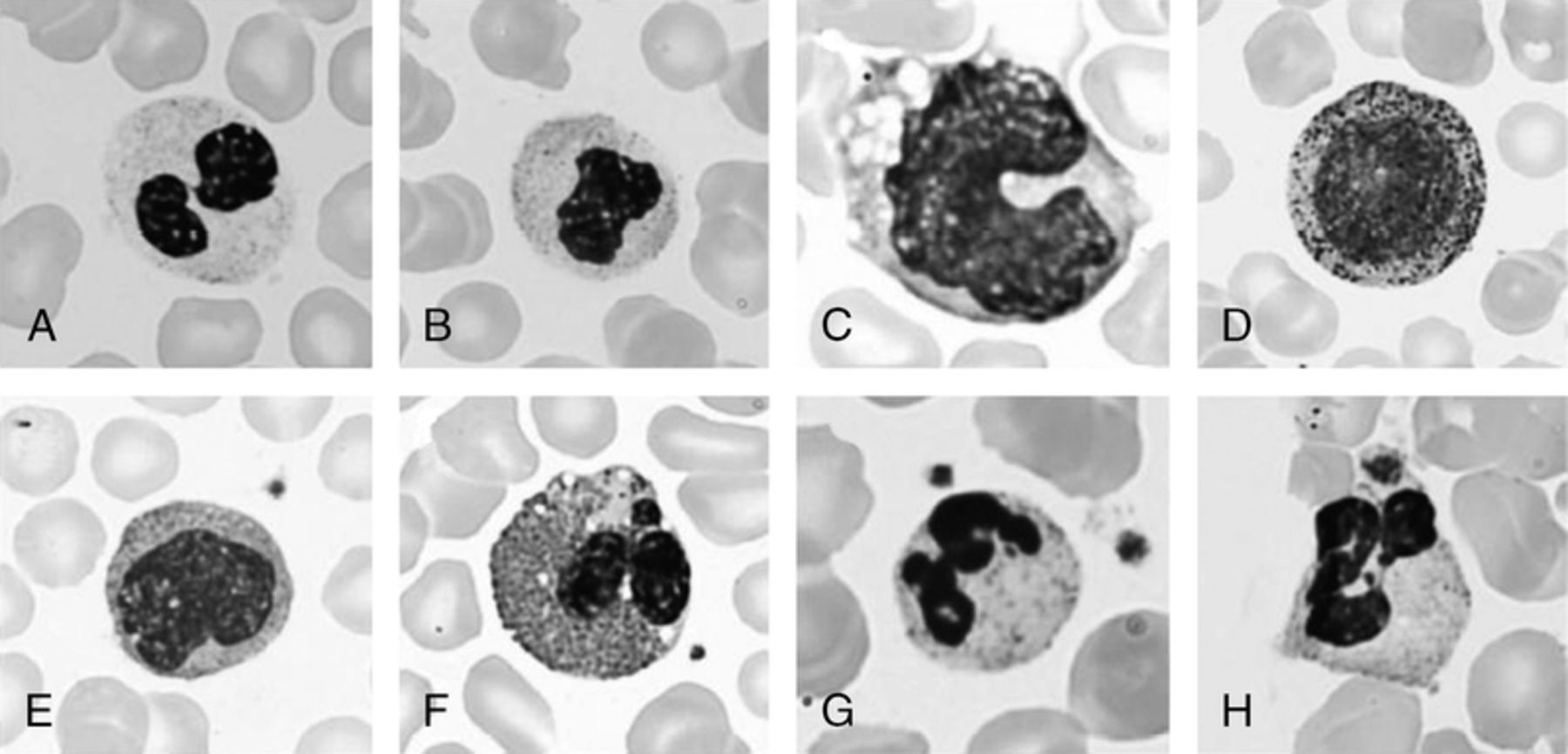
Pseudo-Pelger-Huët anomaly and a toxic left shift of granulocytes with dysplastic features, associated with human granulocytic anaplasmosis. (A) and (B) demonstrate Pseudo-Pelger-Huët anomaly in a bi-lobed neutrophil (A) and a mono-lobed neutrophil with toxic granules and Döhle bodies (B). (C) is a reactive monocyte with an irregular nuclear membrane contour typically shown in Pelger-Huët anomaly. (D) and (E) demonstrate a left shift showing immature granulocytes. (F) shows a dysplastic hypodense eosinophil having cytoplasmic clearing and basophilic granules in the cytoplasm. (G) is an example of karyorrhexis of neutrophils, a dysplastic feature of granulocytes when displayed on peripheral smears. (H) shows a morula, Anaplasma phagocytophilum in the endosome of granulocytes. Distinct cocco-bacilli are shown in the cytoplasm of the neutrophil.
ID: 123 Paraneoplastic Orthostatic Hypotension Associated With Acute Myeloid Leukemia With Trisomy 8
S. Lee, M. Chary. Medicine, Icahn School of Medicine at Mount Sinai, Elmhurst, New York. B. Petersen, J. Mascarenhas. Tisch Cancer Institute, Hematology and Oncology, Icahn School of Medicine at Mount Sinai, New York, New York.
Paraneoplastic neuropathies (PNP) associated with leukemia are rare, and consensus guidelines for the treatment of PNP are lacking. Since those associated with leukemia are not mediated by autoimmune antibodies, therapeutic approach is focused on treatment of the underlying leukemia. Most PNP cause irreversible neurologic damage, and early diagnosis and treatment are crucial to lessen the morbidity associated with PNP. This is the first report to describe severe paraneoplastic orthostatic hypotension which resolved after treatment of acute myeloid leukemia (AML). A 76 year-old woman presented with progressive dizziness and fatigue. She had severe orthostatic hypotension (supine blood pressure 186/82 mmHg; standing 79/53 mmHg). Flow cytometry demonstrated an aberrant myeloid phenotype: CD33+, CD13 dim+, HLA-DR+, CD56+, CD4+, CD38+, CD11b dim+, CD15 dim+, CD7 partial dim+, CD34-, CD117-, CD14-, CD3-, CD5-, CD10-, and CD19-. Bone marrow showed blasts comprising approximately 90% of overall cellularity (see figure); immunoperoxidase stains performed on the biopsy revealed that neoplastic cells were also positive for CD43 and CD68, and negative for CD123; labeling for myeloperoxidase was equivocal. Trisomy 8 was also detected. She was diagnosed with AML, for which azacitidine was initiated. Orthostatic hypotension resolved after initiation of treatment. The diagnosis of PNP in leukemia is usually based on clinical signs and symptoms due to the lack of detectable autoimmune antibodies. The resolution of symptoms after azacitidine is sufficient to diagnose this autonomic instability as a PNP: this patient's neurological disorder is categorized as a “definite” paraneoplastic neurological syndrome, based on the criterion established by Graus et al.: “A non-classical syndrome that resolves or significantly improves after cancer treatment without concomitant immunotherapy, provided that the syndrome is not susceptible to spontaneous remission.” This case demonstrates a unique example of paraneoplastic sequelae remitting with treatment of the underlying hematologic neoplasm. Paraneoplastic autonomic dysfunction has been reported concurrently with other irreversible neurological disorders, which attributes to increased mortality complicating the disease course of a primary neoplasm. This patient presented with severe orthostatic hypotension, which resolved after treatment of the leukemia. This report demonstrates that neurological deficits associated with leukemia require immediate attention, and treatment of the hematologic malignancy can reduce the morbidity associated with PNP. Physicians should be aware of this unusual occurrence of autonomic neuropathy with AML as delay in treatment of the hematologic malignancy can lead to irreversible neurologic deficit and increased morbidity and mortality.
Peripheral blood and bone marrow aspirate smears. (A) Peripheral blood smear showing a blast cell with a nucleolus and multiple cytoplasmic vacuoles (Wright Giemsa stain, ×1,000). (B) Bone marrow aspirate smear with sheets of blasts similar to those in the peripheral blood (Wright Giemsa stain, ×1,000).
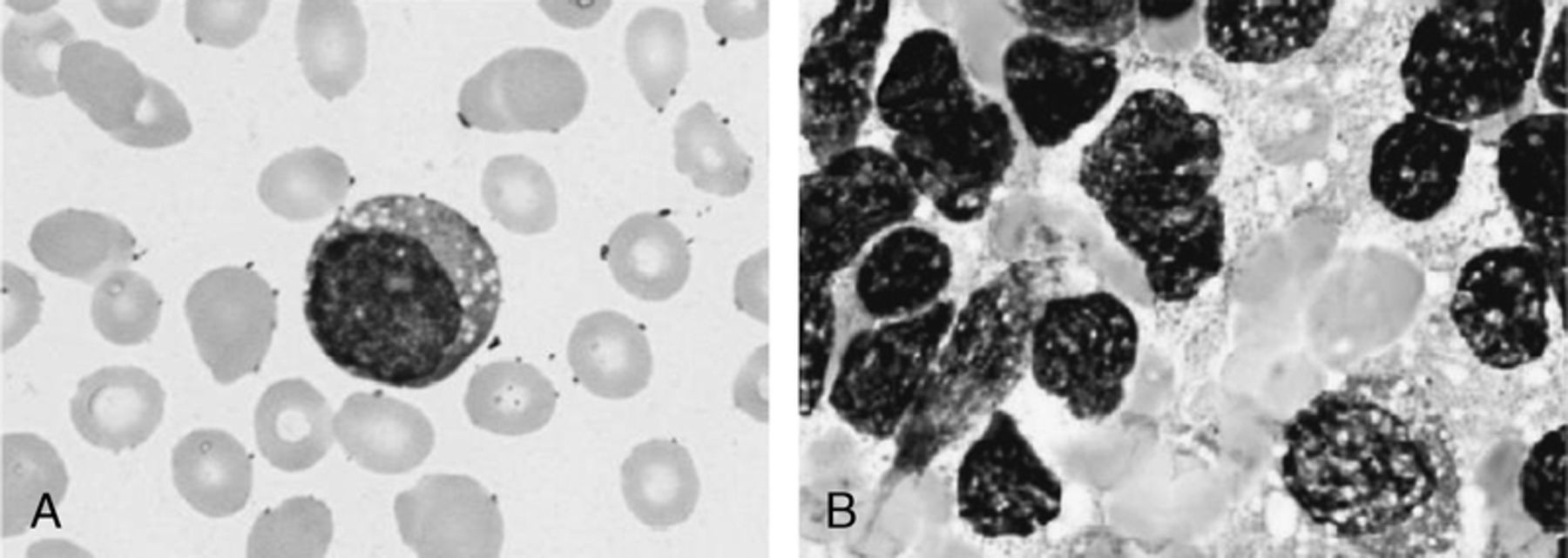
Id: 124 Fulminant Hepatic Failure from Hemophagocytic Lymphohistiocytosis Secondary to Hepatosplenic T-Cell Lymphoma, Mimicking Chinese Herb-Induced Liver Failure
S. Lee, A. Schneier. Medicine, Icahn School of Medicine at Mount Sinai, Elmhurst, New York. B. Petersen. Pathology, Icahn School of Medicine at Mount Sinai, New York, New York. J.L. Gabrilove, J. Lin. Tisch Cancer Institute, Hematology and Oncology, Icahn School of Medicine at Mount Sinai, New York, New York.
A 40 year-old previously healthy women presented for jaundice and fatigue. Two weeks prior, she had rashes on lower extremities, spreading to the entire body, for which she received Chinese herbs. Her rashes resolved, but she developed progressive jaundice and fatigure. At admission, she was found to be febrile with hepatosplenomegaly and severe jaundice on physical exam. Initial laboratory results are as follows: white blood cell 3,800 /μL with neutrophil 78.0% and lymphocytes 12.0%, hemoglobin 8.4 g/dL, platelets 35,000 /mm3, MCV 85.0 fL/cell, transferring saturation 93%, TIBC 189 μg/dL, iron 138 μg/dL, ferritin 4,170 ng/mL, haptoglobin < 7 mg/dL, negative direct Coomb's test, folic acid 12.35 ng/mL, vitamin B12 592 pg/mL, INR 1.4, AST/ALT 123/191 U/L, GGT 57 U/L, total bilirubin/conjugated 20.7/18.7 mg/dL, LDH 1,310 U/L, triglyceride 384 mg/dL, and creatinine 0.4 mg/dL. Liver biopsy showed lymphocyte infiltration and mild hemophagocytosis. A search for common causes of liver failure including infectious etiologies was all negative. Bone marrow biopsy (figure) was performed for severe pancytopenia requiring continuous transfusional support and showed a hypercellular marrow although she was profoundly pancytopenic in peripheral blood count. It showed hemophagocytosis (figure A) with 73% histiocytes positive for CD4, CD68, and CD163 (figure B) and 27% T-lymphocytes positive for CD3 and CD8, localized interstitially and in sinusoids (figure C). Staining and flow cytometry came back positive for CD3, CD5 (dim to moderate), CD7, CD8, CD38, and CD52. The T-cell receptor gene rearrangement study sent when she was severely neutropenic (WBC 100 /μL) came back indeterminate. Based on bone marrow biopsy and flow cytometry result, a diagnosis of hemophagocytic lymphohistiocytosis (HLH) secondary to hepatosplenic T-cell lymphoma (HSTL) was made. Because of elevated total bilirubin (48.7 mg/dL), etoposide was not started; instead methylprednisolone, pentostatin, and alemtuzumab were started. She deteriorated with worsening pancytopenia up to as low as WBC of 0.1 /μL and platelets of 15 /mm3, ferritin 56,856 ng/mL, LDH 2,334 U/L, and total/direct bilirubin 71.3/66.6 mg/dL. On the fifth day after these medications were started, she developed cardiopulmonary arrest and died. This case demonstrates fulminant hepatic failure from HLH and HSTL, mimicking acute hepatotoxicity attributed to Chinese herbal ingestion. She was pancytopenic with very high bilirubin, all of which seemed to be consistent with acute hepatotoxicity from Chinese herbs per the patient's history. However, given constant fever, very high ferritin, and pancytopenia, bone marrow biopsy was performed and confirmed HLH. This case suggests that diagnostic tests, especially bone marrow or liver biopsy, should not be delayed if a patient has unexplained fever, pancytopenia, and evidence of transaminitis or very high MELD scores, even though the patient's history suggests other toxic or infectious etiologies.
Bone marrow biopsy of this patient with HLH secondary to HSTL. (A) is a bone marrow smear displaying a histocyte with red blood cells in the cytoplasm. Wright-Giemsa stain, ×1,000. (B) show a large proportion of CD163-positive histiocytes. (C) shows CD3-positive T-cells localized in sinusoids of the bone marrow. (B) - (C) ×1,000.
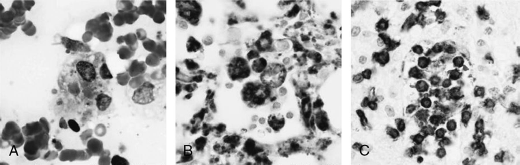
ID: 125 Navigating Care in a Challenging Case of T(6;9) and fLT3 Positive Aml
Y. Song, D. Bixby, D. Roulston, J. Magenau, S.W. Choi. University of Michigan, Ann Arbor, Michigan.
Translocation t(6;9)(p23;q34) is a rare cytogenetic abnormality found in fewer than 5% of pediatric and adult cases of acute myelogenous leukemia (AML). This translocation results in the formation of chimeric fusion gene DEK-NUP214 on the der(6) chromosome, which is translated to nucleoporin fusion proteins and leads to altered nuclear transport and upregulated myeloid protein synthesis. The outcomes of t(6;9) AML are generally poor, with lower five-year overall survival (OS) (£40%) and higher relapse risk (RR) (>50%). Patients with t(6;9) AML demonstrate lower response rates to induction chemotherapy and higher relapse rates despite achieving remission. Additionally, t(6;9) is often seen in AML with the fMS-like tyrosine kinase 3 gene (fLT3) internal tandem duplication (ITD) mutation, one of the most common abnormalities in AML associated with increased risk of treatment failure and mortality, detected in 15 to 25% of patients. FLT3-ITD with a high ITD allelic ratio has been associated with five-year OS rates <35% and RR >60%. We report a challenging case of t(6;9) and fLT3-positive AML in a young adult male. The patient was a 23-year-old Caucasian male who initially presented to his primary care physician with fever, night sweats, and bilateral lower extremity rash. Laboratory testing revealed white blood cells 8.5 x10³/μL, hemoglobin 10.4g/dL, and platelets 41.0 x10³/μL, with a manual differential count showing 66% blasts, 10% neutrophils, 1% myelocytes, 1% bands, and 18% lymphocytes. Bone marrow biopsy showed 74% blasts with Auer rods. Flow cytometry detected an immunophenotype of CD13, CD33, CD38, CD117, and HLA-DR. These cells also expressed low levels of CD4 and CD45; partially expressed CD7, CD25, and CD64; and variably expressed CD34. Cytogenetic and molecular testing confirmed a diagnosis of AML with karyotype 46, XY, t(6;9)(p23;q34), DEK/NUP214 and the presence of the fLT3-ITD mutation, respectively. The patient responded poorly to multiple standard induction and intensification regimens, demonstrating persistent disease. Morphologic remission was eventually achieved with a fLT3 inhibitor (sorafenib) and a hypomethylating agent (azacytidine), a novel investigative combination therapy with a reported response rate of >40%. The patient proceeded with a 9 of 10 HLA matched (HLA-B mismatched) unrelated donor peripheral blood hematopoietic cell transplantation (HCT) (10 months from initial diagnosis). His post-HCT course was complicated by grade I acute graft-versus-host disease of the skin (palms and soles, non-biopsied) requiring high-dose steroids. Bone marrow testing on days 30 and 100 showed no morphologic, flow cytometric, cytogenetic, or molecular evidence of AML. However, despite re-initiation of sorafenib in the post-HCT setting, he experienced early relapse on day 139 with the original [fLT3-ITD and t(6;9)] and new (fLT3-D835 and +8) molecular and cytogenetic markers, respectively, perhaps reflecting selection of these sub-clones within the preexisting disease or clonal evolution. This case highlights the need for improved strategies in the post-HCT setting for high-risk AML. While the incorporation of non-cytotoxic targeted post-HCT maintenance agents is currently being explored, the type of drug, disease-specific characteristics, timing of initiation, dosing/schedule, and hold/re-start parameters for myelosuppression need to be better defined and studied in appropriate clinical trials to ensure uniform practices across institutions.
Id: 126 Endothelial Cell Cd36 Transcription is Downregulated by Lpa/Pkd-1-Hdac7-Foxo1 Signaling Axis and is Associated with Proangiogenic Reprogramming
B. Ren, R. Silverstein. Blood Research Institute/Medicine, BloodCenter of Wisconsin/Medical College of Wisconsin, Milwaukee, Wisconsin. B. Best, D. Ramakrishnan. Blood Research Institute, Blood Center of Wisconsin, Milwaukee, Wisconsin. B. Walcott. Cardiovascular Research Center, Massachusetts General Hospital, Charlestown, Massachusetts. P. Storz. Cancer Biology, Mayo Clinic, Jacksonville, florida.
CD36 is a scavenger receptor that plays an important role in athero-thrombotic diseases, diabetes and cancer. Lysophosphatidic acid (LPA), a bioactive phospholipid signaling mediator, abolishes endothelial cell responses to antiangiogenic proteins containing thrombospondin type 1 homology domains by down-regulating CD36 transcription. The mechanism as to how angiogenic signaling is integrated to regulate endothelial specific CD36 transcription remain unknown. We here describe that in microvascular endothelial cells (MVECs) LPA represses CD36 transcription by activating a PKD-1 signaling pathway that induces the formation of a HDAC7/NCoR1-foxO1 complex in the nucleus. Intriguingly, we show that turning off CD36 transcription is associated with reprogramming MVECs to express ephrin B2, a critical “molecular signature” involved in angiogenesis and arteriogenesis, and phosphorylation of Erk2, an important MAP kinase in arterial differentiation. We also demonstrate that activation of this signaling pathway in the vasculature of mice with transplanted Lewis lung carcinomas is associated with low CD36 expression or with CD36 deficiency, as shown by tumor vessel staining for phospho-PKD-1, arterial markers ephrinB2 and smooth muscle cell actin in the tumor vasculature of both wild type and CD36 deficient mice. Functionally, turning off CD36 transcription promotes angiogenesis in a PKD-1-dependent manner as an in vivo Matrigel assay showed that pharmacologic inhibition of PKD signaling significantly reduced the inhibitory effect of LPA on TSR/CD36-mediated anti-angiogenic effects in response to VEGf in vivo (p<0.01). These studies suggest that a LPA/PKD1-HDAC7-foxO1 signaling axis regulates endothelial CD36 transcription and mediates silencing of the antiangiogenic switch, resulting in proangiogenic reprogramming and arteriogenic responses, and a HDAC7-mediated epigenetic mechanism may be involved in angiogenesis regulated by LPA. Targeting this signaling cascade could be a novel approach for cancer, cardiovascular complications of diabetes and athero-thrombotic diseases as well as obesity.
Infectious Disease
Id: 127 Antibiogram of Vre Causing Urinary Tract Infections as a Restrospective Study from Mmc, Springfield Illinois
M. das, V. Sundareshan. Internal Medicine, Southern Illinois University, Springfield, Illinois.
The prevalence of Vancomycin-resistant Enterococci (VRE) has steadily been increasing in the hospital setting for nosocomial infections. Data from the Centers for Disease Control and Prevention reported that during 2006 and 2007, 30% of all enterococci infections within hospitals in the United States are VRE. Of the total percentage of VRE, approximately 80% are species E. Faecium. These organisms can be extremely difficult to treat due to their high resistance levels to various antimicrobial agents. E. Faecium in particular usually have higher levels of resistance to beta lactams and aminoglycosides compared to E. Faecalis. The objective of our study was to determine the prevalence of VRE at our institution and determine the best therapy for treatment based on the susceptibilities to a panel of antimicrobials. We performed a retrospective analysis of any enterococci isolate found in the urine from August 2012 to August 2014 using the VITEK system. The isolates were then broken down into those susceptible and those resistant to a select group of antimicrobials, thus generating an antibiogram for each isolate. The antimicrobials which were tested included: gentamycin, streptomycin, nitrofurantoin, daptomycin, linezolid, ciprofloxacin, levofloxacin, erythromycin, tigecycline, tetracycyline, and doxycycline. The results showed a total of 180 enterococci isolates collected from in urine cultures. Of these 180, nearly 60% were E. Faecium and 40% were E. Faecalis. Of the E. Faecium group, only 94% were resistant to ampicillin and 100% produced beta lactamase. From the E. Faecalis group 100% were susceptible to ampicillin and 100% were beta lactamase producers. As noted in from our results, E. Faecium was the predominant organism isolated which was also found to be more resistant to ampicillin compared to E. Faecalis. The higher resistance pattern is thought to be due in part to the low affinity of Penicillin Binding Protein and beta lactamase production. Ampicillin resistant strains, which produce beta lactamase, can be treated with ampicillin with sulbactam, and some case reports have noted treatment with high doses of ampicillin. Some of these resistant strains are treated with daptomycin. However, there are many downsides to the use of it such as, it is not fDA approved for E. Faecium infections, and its inability to concentrate in the genitourinary system. Similarly, the side effects and drug interactions limit the use of linezolid in treatment of VRE, especially with prolonged use.
Id: 128 Kaposi—s Sarcoma-Associated Herpesvirus (Kshv) Latently Infected Cells Derived Nanovesicles Promote Endothelial Mesenchymal Transitions (Endmt) and Vascular Permeability
N. Sharma-Walia, K. Patel. RfUMS, North Chicago, Illinois.
Nanovesicles (NVs) are nano-sized extracellular vesicles that are released from a wide range of mammalian cell types such as fibroblast, epithelial, and endothelial cells. NVs are abundant in proteins, mRNAs, DNA fragments and microRNAs, which help NVs act as intercellular communicasomes. NVs production, transport and their significance in tumor microenvironment has been demonstrated in several cancer models but their prevalence and role(s) in Kaposi's sarcoma herpes virus (KSHV) associated human malignancies have not yet been studied extensively. KSHV is associated with a neoplastic angioproliferative endothelial malignancy called Kaposi sarcoma (KS) and two other AIDS-related lymphoid cell malignancies called primary effusion lymphoma and multicentric Castleman's disease. KS progression and pathogenesis has been linked to a number of critical events such as endothelial mesenchymal transition (EndMT), cell migration and invasion. The role(s) of NVs derived from KSHVinfected cell conditioned medium was investigated in this study. NVs from KSHV latently infected primary effusion lymphoma (PEL) cells (KSHV+/EBV-; BCBL-1, BC-3), non-infected (KSHV-/EBV-; BJAB) human Burkitt's lymphoma (BL) cells, KSHV latently infected endothelial cells (TIVE-LTC), and their control counterpart TIVE cells were isolated and purified. Our study revealed that KSHV+/EBV- PEL and KSHV latently infected endothelial cells secrete NVs. Fluorescently labeled NVs derived from either KSHV+ or KSHV- cell lines were efficiently/rapidly internalized by primary human microvascular dermal endothelial cells (HMVEC-d). KSHV+ cell line derived NVs were enriched in inflammatory pathway proteins such as cyclooxygenase-2 (COX-2), 5 lipoxygenase (5LO), and leukotriene A4 hydrolase (LTA4H). We hypothesized that infected cell derived NVs are taken up by surrounding uninfected endothelial cells and induce oncogenesis. The effect of infected cell derived NVs on phenotypic changes and endothelial cell biology controlling factors such as angiogenesis, vasoconstriction, vasodilation, inflammatory response, apoptosis, cell adhesion, and EndMT was studied. Uptake of KSHV+ cells derived NVs by recipient uninfected endothelial cells reduced the expression of proteins such as VE-cadherin, Tie-1/2, VEGfR-1/2, PECAM/CD31, upregulated smooth muscle actin, type I and III collagens, N-cadherin, and vimentin, and also upregulated the expression of EndMT regulating transcriptions factors, including Snail, Slug, Twist, ZEB1 and ZEB2. Furthermore, this uptake of NVs by endothelial cells induced Rac1-GTPase, and enhanced the migration and invasive potential of uninfected endothelial cells. KSHV+ cell line derived NVs, when compared to non-infected BJAB derived NVs induced vascular permeability leading to the disassembly of cell junctions. Together, this study for the first time reports that NVs secreted from KSHV+/EBV- cells have a unique inflammatory signature and these NVs induce EndMT in primary endothelial cells. The crucial role of NVs in KSHV pathogenesis via manipulation of extracellular vesicles in the infected cell microenvironment is highlighted in this study. Thus, KSHV infection induced NVs represent a novel strategy employed by KSHV to promote inflammation, migration, invasion, and vascular permeability.
Id: 129 Dissection of Usher-Chaperone-Subunit Interactions during Pilus Biogenesis in Escherichia Coli
G.T. Werneburg, D.G. Thanassi. Molecular Genetics & Microbiology, Stony Brook University, Stony Brook, New York. G.T. Werneburg. Medical Scientist Training Program, Stony Brook University, Stony Brook, New York.
The chaperone/usher (CU) pathway is a conserved secretion system employed by many species of Gram-negative bacteria to construct virulence-associated surface structures known as pili or fimbriae. Our model system, type 1 pili, facilitates colonization of the bladder by uropathogenic Escherichia coli, and subsequent urinary tract infection. The CU pilus biogenesis pathway requires both a periplasmic chaperone protein and an integral outer membrane protein, the usher. The chaperone facilitates proper folding of pilus subunits in the periplasm and prevents premature interactions. The usher catalyzes the exchange of chaperone-subunit for subunit-subunit interactions, and promotes ordered polymerization and secretion of the pilus fiber. The usher, a putative multimer, has 5 domains: a periplasmic N-terminal domain (N), a transmembrane beta-barrel domain that is gated by an internal plug domain, and two periplasmic C-terminal domains (C1 and C2). Chaperone-subunit complexes first bind to the N domain of the usher and then move to the C domains as the plug is expelled and the pilus subunits are inserted into the barrel channel. To understand the mechanism of the usher (fimD) in catalyzing the assembly and secretion of pili and to identify mechanistic targets for pharmacological inhibition, we monitored interactions of chaperone-subunit (fimCfimH) complexes with usher domains during pilus assembly using fluorescence-based approaches. We calculated an overall affinity for fimC-fimH binding to the full length fimD usher that is in agreement with previous surface plasmon resonance measurements. We then combined our approach together with domain deletion mutants of fimD to reveal the contributions of individual usher domains to chaperone-subunit binding and gain insight into the sequential order of chaperone-subunit-usher interactions. Our data suggest that the C domains are fimD's high-affinity binding site for fimC-fimH complex. However, in the absence of the usher's N domain, no binding was detected. These findings support a model wherein the high affinity C domains are only accessible once the chaperone-subunit complex binds the usher's N domain, and the N to C domain handoff is then driven by differential affinity. We show that in the context of the full-length usher, the plug domain does not contribute directly to fimD's affinity for chaperone-subunit complexes, but likely “masks” fimD's C domains prior to usher activation. Further, in vivo we show that fimD is functional in trans, suggesting that the non-translocating fimD protomers of the multimer do not remain idle during pilus biogenesis, but rather actively recruit and contribute to incorporation of chaperone-subunit complexes. Based on these findings, we are currently developing and implementing additional fluorescence and biochemical assays to further our understanding of the mechanism of the usher in the assembly and secretion of pili, and to identify targets and test inhibitors of our newly elucidated mechanistic steps of pilus assembly.
Id: 130 Lipoxins: Implications in Kaposi—s Sarcoma Associated Herpesvirus (Kshv) Biology
J.A. Chandrasekharan, N. Sharma-Walia. RfUMS, North Chicago, Illinois.
Kaposi's sarcoma-associated herpesvirus (KSHV), a γ-2 herpesvirus, is etiologically associated with angioproliferative endothelial Kaposi's sarcoma (KS), primary effusion body cavity B-cell lymphoma (PEL or BCBL) and B cell-lymphoproliferative multicentric Castleman's disease (MCD). Our exciting paradigm shifting previous studies established that KSHV utilizes host pro-inflammatory pathways such as cyclooxygenase-2 (COX-2), prostaglandin E2 (PGE2), and 5-lipoxygenase (5LO) to promote its latent gene expression and consequent pathogenesis. COX-2 and 5-LO inhibitors are associated with gastrointestinal side effects and adverse cardiovascular effects at high doses; additionally, these inhibitors do not completely eradicate the viral load. Therefore, there is a need for targeted, endogenous, and safe therapeutics with high antiviral potential. Lipoxins are host cell endogenous anti-inflammatory, pro-resolving and short-lived bioactive lipids. Here, we discovered that KSHV infection of human dermal microvascular endothelial cells (HMVEC-d) significantly reduces the secretion of anti-inflammatory lipoxin (LXA4). We demonstrate that KSHV infected HMVEC-d cells and human KS lesion cells express lipoxin receptor ALXR, and more importantly incubation of KSHV infected cells with stable small molecule analogs of lipoxins and aspirin triggered lipoxins significantly downregulated KSHV's latent promoter activity and latent gene expression. Thus, the anti-inflammatory lipoxin pathways offer possibilities for regulating KSHV biology. Further studies on therapeutic potential of lipoxin in KS would offer novel and effective treatment method.
Id: 131 Leukotriene A4 Hydrolase Inhibitors: Potential Chemotherapeutic Modality for Primary Effusion Lymphoma
P. Sowa, N. Sharma-Walia. RfUMS, North Chicago, Illinois.
Kaposi's sarcoma-associated herpesvirus (KSHV), a γ-2 herpesvirus, is etiologically associated with angioproliferative endothelial Kaposi's sarcoma (KS), primary effusion lymphoma (PEL) and B cell-lymphoproliferative multicentric Castleman's disease (MCD). The significance of leukotriene A4 hydrolase (LTA4H) in KSHV biology prompted us to examine the role of LTA4H inhibitors in PEL, an aggressive AIDS-linked KSHV-associated non- Hodgkin's lymphoma using Bestatin, well-known LTA4H inhibitor. We demonstrate that (1) Bestatin is efficacious in specifically inducing proliferation arrest in PEL cells (KSHV+ BCBL-1 and BC-3) when compared to non-infected (KSHV-; BJAB) Burkitt's lymphoma cells; (2) Bestatin treatment reduced the number of KSHV + PEL cells in S phase; (3) Bestatin treatment induced ER stress proteins such as calnexin, BiP, CHOP. Overall, our study provides a comprehensive molecular framework that links LTA4H with ER stress induced PEL cell death and identify the chemotherapeutic potential of Bestatin in treating PEL.
Id: 132 Accuracy of Loop-Mediated Isothermal Amplification for the Diagnosis of Clostridium Difficile Infection: A Systematic Review
P. Thota, C.J. Donskey. Infectious Diseases, Case Western Reserve University, Cleveland, Ohio. A. Lloyd, V. Pasupuleti. Medicine, Case Western Reserve University, Cleveland, Ohio. C. Pant. University of Kansas Medical Center, Kansas City, Kansas. D.D. Rolston. Geisinger Medical Center, Danville, Pennsylvania. A.V. Hernandez, T.G. Fraser, A. Deshpande. Cleveland Clinic, Cleveland, Ohio. V. Benites-Zapata. Universidad Peruana de Ciencias Aplicadas (UPC), Lima, PERU.
Id: 133 Evolution of Peripherial Blood Smear and Lymph Node Histology in Miliary Tuberculosis
S. Lee, D. Chang. Medicine, Icahn School of Medicine at Mount Sinai, Elmhurst, New York. M. Chary. Icahn School of Medicine at Mount Sinai, New York, New York, UNITED STATES. F. Salem. Pathology, Icahn School of Medicine at Mount Sinai, New York, New York. J. Lin. Tisch Cancer Institute, Hematology and Oncology, Icahn School of Medicine at Mount Sinai, New York, New York.
A 40 year-old previously healthy man presented with an enlarging left neck mass. He denied difficulty breathing, fever, chills, night sweats, or weight loss. On admission, he was febrile and tachycardic. Physical exam was notable for a firm globular mass on his left neck and hepatomegaly with no oropharyngeal exudate or uvular deviation. Computerized tomography of the neck and chest demonstrated diffuse lymphadenopathy of the left neck and multiple lung nodules all less than 1 cm. Initial laboratory studies were remarkable for leukocytosis with white blood cell (WBC) 22,200 /μL, alkaline phosphatase 217 U/L, and gamma glutamyl transpeptidase 521 U/L. An initial peripheral blood smear (figure A) revealed many pseudo-Pelger-Huët neutrophils with toxic granules and vacuoles, previously reported in acute myeloid leukemia (AML) or severe infections. Hypersegmented neutrophils were noted, an extremely rare finding in a patient with no evidence of underlying B12 or folate deficiency. Nuclear projections in neutrophils were noted, previously reported in patients with androgen-producing tumors. Eosinophils had cytoplasmic clearing with reduced cytoplasmic areas occupied by cytoplasmic granules, as reported in hypereosinophilic syndrome. Both basophilic and eosinophilic granules were observed in eosinophils, previously reported in myeloid leukemia. Basophils revealed a reduced number of granules with vacuoles, signifying partial degranulation as reported in AML. Two days later, a fine needle aspiration and core biopsy of the left neck mass demonstrated well-formed granulomas with negative fungal and acid-fast bacilli (AfB) staining (figure C). Empirical itraconazole, vancomycin and piperacillintazobactam were initiated. Blood and urine cultures remained without growth. Ten days after admission, a peripheral smear (figure B) showed more pseudo-Pelger-Huët anomaly and thrombocytosis (1,200,000 /μL). Twenty five days after admission, he developed pancytopenia (WBC 3,200 /μL and platelets 80,000 /μL). Subsequently, he developed blisters and ulcers on the tongue, and its biopsy showed ulcerated squamous mucosa with acute inflammatory exudate and reactive histiocytosis (figure D). Suspicion for military tuberculosis (MTB) grew given his clinical manifestations, for which rifampin, isoniazid, moxifloxacin and ethambutol were started. He became afebrile. A peripheral smear at discharge demonstrated no Pelger-Huët anomaly or dysplastic granulocytes. The culture of neck lymph node biopsy came back with Mycobacterium tuberculosis (figure E). This case report shows a unique presentation of MTB and associated hematological manifestations including dysplastic granulocytes and alternating leukocytosis, leukopenia, thrombocytopenia, and extreme thrombocytosis. These hematological presentations seem to be reactive responses to MTB. This report suggests that severe infections can trigger striking hematological manifestations previously reported in myeloid leukemias.
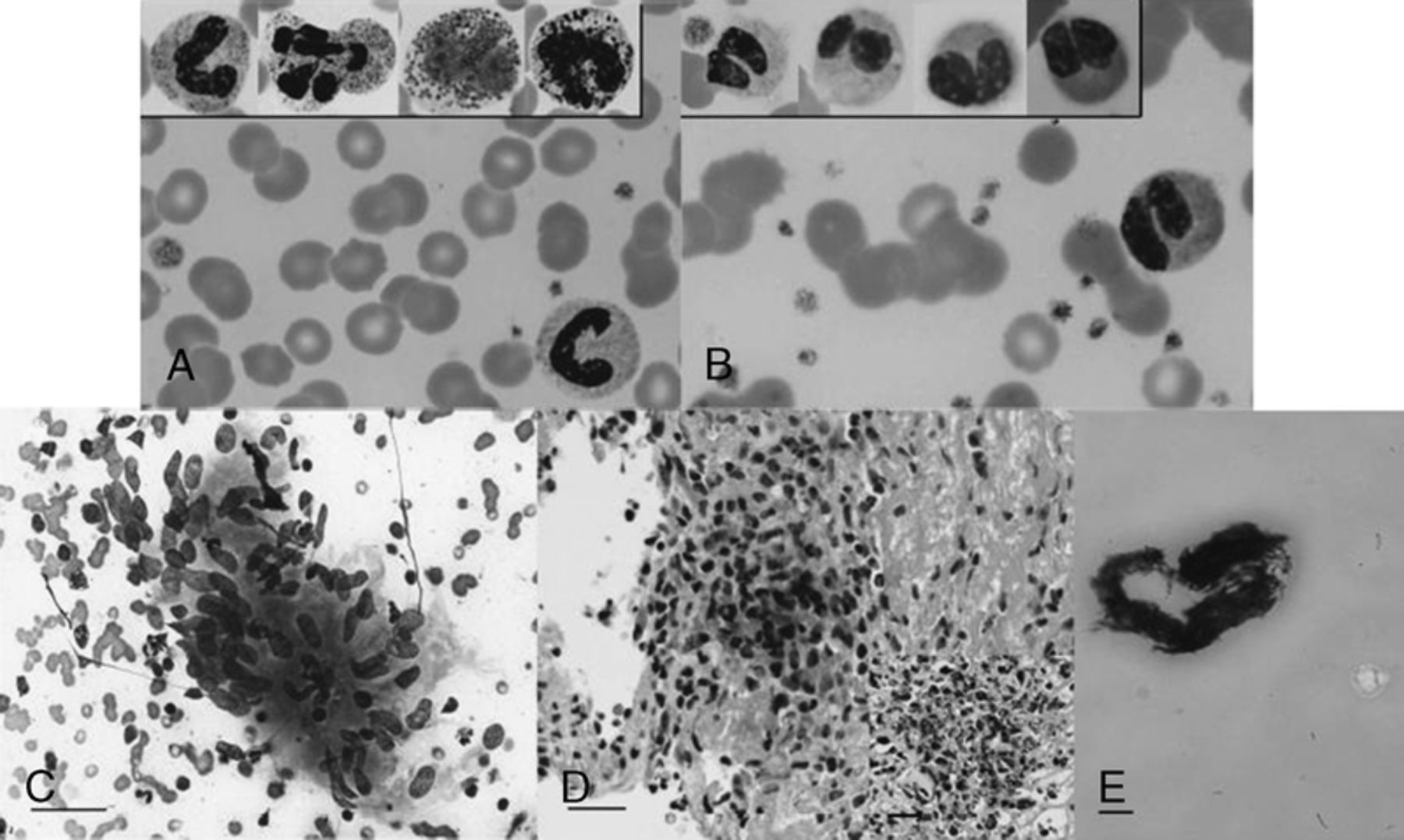
(A) Peripheral blood smear at admission; (B), day 10 after admission. Insets in (A) and (B) showfurther examples of neutrophils, eosinophil, basophil on the indicated day. (C) Fine-needle aspiration of a left neck lymph node (Diff-Quik stain) showing a well-formed granuloma consisting of epithelioid cells characterized with ovoid to elongated nuclei. (D) Tongue biopsy showing a granuloma (H&E stain). The inset in (D) shows CD68+ histiocytes in the granuloma. (E) Lymph node culture growing Mycobacterium tuberculosis six weeks after the lymph node biopsy was performed (Ziehl-Niessen stain). Scale bar in (A) and (B) = 100 μm; (C) = 20 μm.
Id: 134 Cyp2C19*17 Genetic Polymorphism and Therapeutic Challenges in An Allogeneic Stem Cell Transplant Recepient with Invasive Pulmonary Aspergillosis
M. Abidi, A. D‘Souza, P. Hari. of Medicine, Medical College of Wisconsin, Brookfield, Wisconsin. K. Kuppalli. Department of Medicine, Loyola University, Chicago, Illinois.
A 48-year-old Caucasian male was seen in consultation for pulmonary Invasive Aspergillosis. He had undergone allogeneic peripheral blood hematopoietic stem cell transplant for myelodysplastic syndrome 6 months. Post-transplant course was complicated by extensive graft versus host disease. He was treated with prednisone, tacrolimus and tocilizumab and photophoresis. Upon presentation, he was intubated secondary to multifocal pneumonia and respiratory failure. A chest computerized tomography (CT) scan showed bilateral upper lobe lung opacities. Empiric antimicrobial coverage included intravenous cefepime, azithromycin and voriconazole 300mg Q12h. Bronchoalveolar lavage (BAL) fluid cultures grew 10,000 cfu/ml Pseudomonas aeruginosa and 1,000 cfu/ml Aspergillus fumigatus. Nucleic acid amplification testing was positive for Mycoplasma pneumoniae. He was switched to doxycycline and he completed a 14-day course of doxycycline and cefepime. He improved clinically, was extubated and switched to oral voriconazole 300 mg q12h a week. However, his respiratory status worsened a week after this transition. Chest CT showed worsening opacities bilaterally in the upper and lower lobes. Voriconazole serum level was undetectable, as was a repeat voriconazole level a week later. We suspected that he might be an ultra metabolizer of voriconazole and a cytochrome assay was requested from an outside laboratory (LabCorp, Burlington NC). The methodology employed was DNA analysis of the cytochrome P450 2C19 gene including the alleles for poor metabolizers *1, *2, *3 *4, *5, *6, *7 and *8 as well as the ultra-metabolizer alleles *17. This was performed on the Tm Bioscience /Luminex Universal Array Platform using primer extension chemistry. Multiplex PCR amplifies DNA fragments containing mutations associated with the alleles mentioned above. Primer extension then generates a biotinlabeled product that hybridizes to complementary, bead-immbolized probes to permit flow-sorted detection of both normal and mutation sequences. We increased the dose of intravenous voriconazole to 400 mg every 12 hours and added intravenous micafungin. Repeat imaging one week after the addition of micafungin showed improvement in the bilateral ground glass opacities. CYP2C19 genotype testing was positive for the *17 translocation indicating he was an ultra metabolizer for voriconazole. Voriconazole was discontinued and he was discharged home on intravenous micafungin 150 mg daily. A month later, chest CT demonstrated slow improvement. He was continued on micafungin with plans to switch him to liposomal amphotericin B if there was any worsening. He was readmitted with recurrent pulmonary infections with evidence of aspergillus infection by BAL culture. He was treated with antibiotics and continuation of intravenous micafungin, and each time he improved clinically. He had multiple bronchoscopies with BAL including lung biopsies in order to differentiate between a new bacterial superinfection, pulmonary aspergillus, drug toxicity and pulmonary GVHD. We considered oral posaconazole as his gut GVHD improved but given cost he remained on intravenous micafungin. Nine months after his original presentation, he developed respiratory failure with new areas of lung consolidation and evidence of acute eosinophilic reaction. Aspergillus fumigatus was detected on BAL cultures and he was switched fto liposomal amphotericin B and posaconazole. sirolimus was discontinued given concern that lung pathology could be due to sirolimus toxicity. While work up was underway the patient suffered a cardiac arrest and died.
Nephrology
Id: 52 Discovery of Mesencephalic Astrocyte-Derived Neurotrophic Factor as a Urine Biomarker for Endoplasmic Reticulum Stress-Related Kidney Diseases
Y. Kim, H. Lee, J. Miner, Y.M. Chen. Renal Division, Department of Internal Medicine, Washington University in St. Louis, St. Louis, Missouri. F. Urano. Division of Endocrinology, Metabolism and Lipid Research, Department of Internal Medicine, Washington University School of Medicine, St. Louis, Missouri.
Accumulating evidence has highlighted the important role of endoplasmic reticulum (ER) stress and disrupted proteostasis in the pathogenesis of a variety of glomerular and tubular diseases. Thus, it is imperative to develop non-invasive biomarkers for detecting ER stress in podocytes or tubular cells in the incipient stage of disease, when clinically a kidney biopsy is not yet indicated. Moreover, restoration of ER homeostasis before irreversible kidney cell injury may hold significant promise as an attractive therapeutic strategy. However, there are no known biomarkers for detecting ER stress in associated kidney disease to date. Here, using mouse models of human nephrotic syndrome caused by mutant laminin b2 protein-induced podocyte ER stress and of acute kidney injury triggered by the ER stress inducer tunicamycin in renal tubular cells, we identified mesencephalic astrocyte-derived neurotrophic factor (MANf) as a potential urine biomarker for detecting ER stress in podocytes or renal tubular cells respectively. We found that MANf was upregulated by ER stress in the podocytes and tubular cells. Most importantly, urinary MANf excretion concurrent with podocyte or tubular cell ER stress preceded clinical or histological manifestations of the corresponding disease. Thus, MANf can potentially serve as a urine diagnostic or prognostic biomarker to help stratify disease risk, predict disease progression, and monitor treatment response in the ER stress-related kidney diseases.
ID: 135 Focal Segmental Glomerulosclerosis And Papillary Renal Cell Carcinoma: Causal Link?
M. Haroon Al Rasheed, S. Setty. Department of Pathology, University of Illinois Hospital and Health Sciences System, Chicago, Illinois. A. Dudek. Department of Medicine, University of Illinois Hospital and Health Sciences System, Chicago, Illinois. M. Abern. Department of Urology, University of Illinois Hospital and Health Sciences System, Chicago, Illinois.
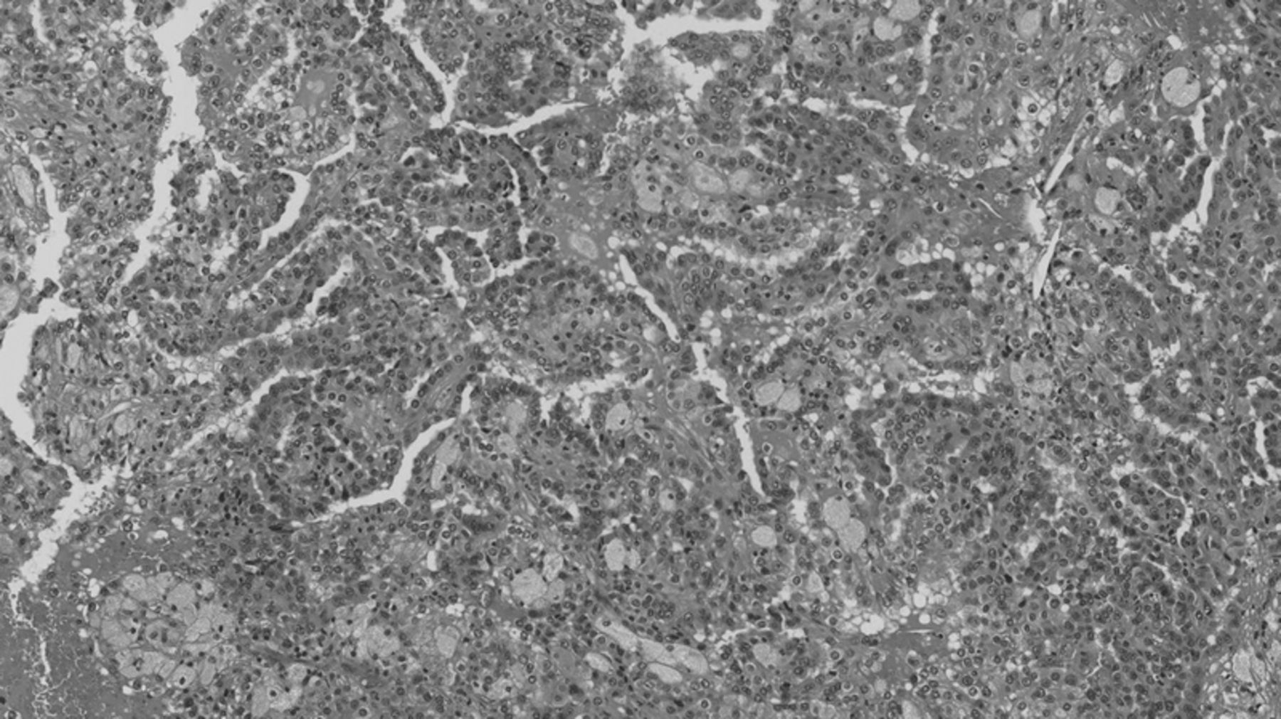
Original magnification 100x view of papillary renal cell carcinoma forming papillae lined by malignant epithelial cells with hemorrhage and necrosis.
ID: 136 Medullary Tophi in Chronic Kidney Disease: A Causal Link between URIC ACID and Progressive Renal Failure?
I. Ayoub, L. Hebert, B. Rovin. Medicine/Nephrology, The Ohio State University Wexner Medical Center, Columbus, Ohio. S. Brodsky, T. Nadasdy. Pathology, The Ohio State University Wexner Medical Center, Columbus, Ohio.
Clinical characteristics of patients with medullary/papillae tophi
Id: 137 Acute Renal Infarct Case Series
T.M. Yousuf, H. Iqbal, A. Said, A. Chadaga, J. Oyama, E. Lerma. Internal Medicine, Advocate Christ Medical Center, Oak Brook, Illinois.
Acute Renal Infarction is a commonly under-diagnosed condition. Acute renal infarction usually presents with the constellation of signs and symptoms of abdominal or flank pain, elevation of LDH, elevation of white blood count, hematuria, oliguria or anuria and fever. Differential diagnosis includes acute appendicitis, nephrolithiasis, cholecystitis, urinary tract infection and biliary pathology. This report will hopefully highlight the rarity of acute renal infarction and the importance of considering the diagnosis of acute renal infarction in patients presenting with acute kidney injury. The myriad of findings of 1) acute abdominal pain or flank pain, 2) elevation of LDH and 3) a high risk of thromboembolism in a patient presenting with acute kidney injury should make the astute clinician consider the possibility of acute renal infarction in the differential diagnosis. Case 1 is a 55 year old male with a past history of coronary artery disease, AAA and atrial fibrillation who presented to the emergency room with a 2 day history of intractable abdominal discomfort localized to the left flank and right lower quadrant (RLQ) with radiation to the right femoral area. On physical exam, the patient was in moderate distress and had significant tenderness over the RLQ. In addition, he also had notable left sided costovertebral angle tenderness. Pertinent laboratory studies included: WBC of 8.0, creatinine of 1.41mgs/dL and INR of 1.2. He was known to be non-compliant with warfarin anticoagulation. CT of abdomen revealed areas of decreased perfusion involving the lower pole of the right kidney with perinephric stranding, new occlusion of the left internal iliac artery and aneurysms involving the right common iliac artery as well as both common femoral arteries. Duplex ultrasonography revealed the presence of aneurysms in both common femoral arteries and increased velocity in the right proximal third of the renal artery. Patient was afforded supportive measures and an adequate analgesic regimen and subsequently showed significant clinical improvement as well as resolution of laboratory abnormalities. Case 2 is that of a 53 year old male who has a past history of chronic kidney disease and atrial fibrillation who presented to the emergency room with an acute onset of abdominal discomfort localized to the left flank with radiation to the back. Physical exam revealed stable vitals and an irregular pulse. On examination, he had left CVA tenderness. Initial labs revealed creatinine of 3.5mgs/dL, INR of 1.7 and significantly elevated LDH (1323). CT scan of the abdomen demonstrated the absence of the right kidney. A MAG3 nuclear medicine scan revealed diminished flow to the left kidney. A CT angiogram revealed evidence of thrombus in the left renal artery. He was then started on anticoagulation with heparin and a wire guided thrombolysis with thrombectomy was performed. Although there was brisk urine output production post-procedure, there was no evidence of renal recovery in the ensuing days; therefore, he was subsequently initiated on hemodialysis. Acute renal infarction is a commonly missed phenomenon and it should be high on the list of differential diagnosis if a patient presents with abdominal pain, elevated LDH and predispositions to increased risk of thromboembolism. The mainstay of treatment remains to be appropriate anticoagulation. Our cases demonstrate acute renal infarction likely secondary to atrial fibrillation with and without therapeutic anticoagulation. Atrial fibrillation, as the most common arrhythmia identified in hospitalized patients, warrants increased awareness of its association with acute renal infarction so that it can be promptly recognized and managed appropriately.
Pediatrics
Id: 53 Preeclampsia Has Detrimental Impact on Health of Offspring
S. Munir, N. Drever, S.R. Allen, T. Kuehl, M.N. Uddin. Obstetrics and Gynecology, Baylor Scott & White Health/Texas A&M Health Science Center College of Medicine, Temple, Texas, M.A. Co, M.R. Beeram, T. Kuehl, M.N. Uddin. Pediatrics, Baylor Scott & White Health/Texas A&M Health Science Center College of Medicine, Temple, Texas. S.H. Afroze. Medical Physiology, Texas A&M Health Science Center College of Medicine, Temple, Texas.
Pulmonary/Critical Care
Id: 54 Cigarette Smoking Inhibits Rig-I and Interferon Responses to Influenza in Human Small Airway Epithelial Cells
W. Wu, W. Zhang, J. Booth, J. Metcalf. Internal Medicine, University of Oklahoma Health Sciences Center, Edmond, Oklahoma.
Id: 55 Refractile Foreign Material Deposits and Alveolar Hemorrhage in Crack Cocaine Smoker
R. Sogomonian, V. Gandhi, T. McGarry, M. Moezzi. Internal Medicine, Icahn School of Medicine at Mount Sinai Elmhurst Hospital Center, Elmhurst, New York.
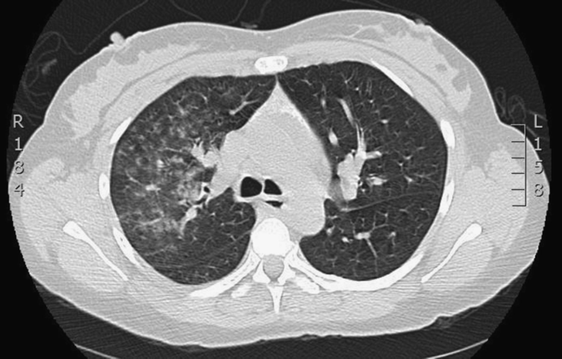
Id: 56 Stability of Protein Inhibitor of Activated Sata1 (Pias1) is Regualted in the Ubiquitinproteasome System
J. Zhao, B. Chen. Medicine, University of Pittsburgh, Pittsburgh, Pennsylvania.
The ubiquitin-proteasome system regulates intracellular protein stability, thus modulating cellular responses including cytokine release. PIAS1 (protein inhibitor of activated STAT1) exhibits an anti-inflammatory property via negatively regulating pro-inflammatory pathways such as JAK-STAT and Nf-kB. However, the stability of PIAS1 has not been revealed. Here we demonstrate that a novel ubiquitin E3 ligase, HECTD2, targets PIAS1 for its ubiquitination and degradation, thus increasing inflammatory responses. Glycogen synthase kinase (GSK3b) binds and phosphorylates PIAS1 serving as a phosphodegron motif for HECTD2 interacting with PIAS1. We identified a naturally occurring HECTD2 polymorphism (HECTD2A19P) in 8.5% of the population that functions to blunt inflammatory responses by exclusively causing the mislocalization of HECTD2A19P in the cytosol preventing nuclear interaction with PIAS1. We also developed a small molecule inhibitor, BC-1382, to target HECTD2 and attenuates proinflammatory stimuli-induced Nf-κB activation. These studies provide a new innate immunity pathway suggesting that mutation or antagonism of E3 ligase HECTD2 results in reduced severity of inflammation by selectively modulating the abundance of anti-inflammatory protein PIAS1.
Id: 57 Glycogen Synthase Kinase 3Beta Regulates Il-33 Receptor Internalization and Il-33 Signaling
J. Zhao, J. Wei, R.K. Bowser, R.S. Traister, M. Fan, Y. Zhao. Medicine, University of Pittsburgh, Pittsburgh, Pennsylvania.
IL-33, a member of the IL-1 cytokine family, contributes to the pathogenesis of lung inflammatory diseases including asthma, fibrosis, and lung injury. ST2L, the membrane bound receptor for IL-33, is expressed on immune effector cells and lung epithelia, and plays a critical role in triggering inflammation. We have previously shown that ST2L is an ubiquitinated protein and its protein level is regulated by the ubiquitin-proteasome system. Here, we investigate the molecular mechanisms of ST2L internalization and demonstrate that glycogen synthase kinase 3b (GSK3b) regulates ST2L internalization and IL-33 signaling. Immunofluorescence staining, flow cytometry analysis, and cell surface protein isolation and detection kit were used to determine cell surface ST2L expression. Plasmids containing GSK3b wild type and site-directed mutants were generated and transfected to mouse lung epithelial cells. IL-33 treatment reduces ST2L cell surface expression, an effect was attenuated by inhibition or down-regulation of GSK3b. GSK3b was phosphorylated and interacted to ST2L in response to IL-33 treatment. Serine residue 446 in ST2L was identified as docking stie for GSK3b. GSK3b binding site mutant (ST2LS446A) and phosphorylation site mutant (ST2LS442A) are resistant to IL-33-induced ST2L internalization. IL-33 also activated focal adhesion kinase (FAK), a protein tyrosine kinase. Inhibition of FAK impaired IL-33-induced GSK3b activation and ST2L internalization. Further, inhibition of ST2L internalization enhanced IL-33-induced cytokine release in lung epithelial cells. These results suggest that FAK/GSK3b modulates ST2L internalization and IL-33-induced inflammatory responses. Activation of FAK/GSK3b might serve as a unique strategy to lessen pulmonary inflammation.
Id: 58 Lysocardiolipin Acyltransferase (Lycat) Regulates Tgf-B-Induced Differentiation of Human Lung Fibroblasts
L. Huang, S. Reddy, V. Natarajan. UIC, Chicago, Illinois. C. Feghali-Bostwick. Medical University of South Carolina, Charleston, South Carolina. J. Garcia. University of Arizona, Tuscon, Arizona.
Id: 59 Genetic and Epigenetic Regulation of Group V Phospholipase A2 in Pulmonary Inflammation
X. Sun, D. Adyshev, W. Zhang, M. Wade, S.M. Dudek. Medicine, University of Illinois at Chicago, Chicago, Illinois. A.R. Leff. The University of Chicago, Chicago, Illinois.
Id: 60 the Lysophosphatidic Acid Receptor 1 Antagonist Ki16425 Blunts Abdominal and Systemic Inflammaton in a Mouse Model of Peritoneal Sepsis
A.M. Jacko, J. Zhao. Medicine, University Of Pittsburgh School of Medicine, Pittsburgh, Pennsylvania. J. Wei, N. Weathington, Y. Zhao, H. Huang, A. Tsung. Surgery, University Of Pittsburgh School of Medicine, Pittsburgh, Pennsylvania.
Lysophosphatidic acid (LPA) is a bioactive lipid mediator of inflammation via the LPA receptors 1-7. We and others have previously described pro-inflammatory and pro-fibrotic activity of LPA signaling in pulmonary systems in bleomycin- or lipopolysaccharide (LPS)-induced lung injury models. In this study, we investigated if LPA signaling is active in the pathogenesis of systemic sepsis from an abdominal source. We report here that antagonism of the LPA receptor LPA1 with the small molecule ki16425 reduces the severity of abdominal inflammation and organ damage in the setting of peritoneal endotoxin exposure. Pretreatment of mice with intraperitoneal ki16425 reduces the induction of peritoneal neutrophil chemokine production, liver oxidative stress, liver injury, and cellular apoptosis in visceral organs in the setting of LPS exposure. Mice pretreated with ki16425 are also protected from LPS-induced mortality. Tissue myeloperoxidase activity is not affected by LPA1 antagonism. We have shown that LPA1 is associated with LPS co-receptor CD14, the association is suppressed by ki16425. LPS-induced phosphorylation of PKCδ and p38 MAPK in liver cells and IL-6 production in Raw264 cells are likewise blunted by LPA1 antagonism. These studies indicate that the small molecule inhibitor of LPA1, ki16425, suppresses cytokine responses and inflammation in a peritoneal sepsis model by blunting downstream signaling through the LPA1-CD14-TLR4 receptor complex. This anti-inflammatory effect may represent a therapeutic strategy for the treatment of systemic inflammatory responses to infection of the abdominal cavity.
Id: 61 Effect of Altitude and Cigarette Smoke Exposure on Bronchoalveolar Lavage Fluid Cellularity
M.K. Ishaq, C. Cross, H. Youness, L. Booth, E. Duggan, L. Howard, B.A. Petrone- Holtslander, A. Awab. Pulmonary and Critical Care Medicine, Oklahoma University Health Sciences Center, Oklahoma City, Oklahoma. D. Hutchings. Cherokee Nation Distributors, Catoosa, OK, Catoosa, Oklahoma. V. White, D. Self, D.M. Kupfer, D. Burian. Civil Aerospace Medical Institute, federal Aviation Administration, Oklahoma City, OK, Oklahoma City, Oklahoma. J.P. Metcalf. Oklahoma Medical Research foundation, Oklahoma City, OK., Oklahoma City, Oklahoma.
Id: 62 Fty720 S-Phosphonate Does Not Activate Grk2-Mediated S1Pr1 Phosphorylation and Ubiquitiation
L. Wang, S. Dudek. Univ of Illinois at Chicago, Chicago, Illinois. R. Bittman. The City University of New York, New York, New York.
Id: 63 Inhibition of the Actin Related Protein 2/3 Complex Decreases Pulmonary Endothelial Barrier Function and Recovery
B.N. Brinley. Pulmonary and Critical Care, University of Illinois Hospital and Health Science System, Chicago, Illinois. B.N. Brinley. Internal Medicine, Mercy Hospital and Medical Center, Chicago, Illinois.
Id: 64 the Central Role of Nuclear Factor (Erythroid-Derived 2)-Like 2 in Ventilatorinduced Lung Injury
T. Wang, T. Jiang, T. Zhou, H. Quijada, J.B. Mascarenhas, V. Ramamoorthi Elangovan, L. Hecker, D.D. Zhang, J.G. Garcia. University of Arizona, Tucson, Arizona.
Id: 65 Mice with Sickle Cell Disease Are Less Susceptible to Ventilator-Induced Lung Injury but Not Lps-Induced Lung Injury
J.R. Sysol. Pharmacology, University of Illinois College of Medicine at Chicago, Chicago, Illinois. J.R. Sysol, S. Sammani, E. Letsiou, S. Singla, S.M. Dudek, R.F. Machado. Medicine, University of Illinois College of Medicine at Chicago, Chicago, Illinois.
ID: 66 Anti-Inflammatory Effects Of C-Abl Inhibition In Lps-Induced ALI
A.N. Rizzo. Pharmacology, UIC College of Medicine, Chicago, Illinois. A.N. Rizzo, E. Letsiou, S. Sammani, M.E. Brown, S.M. Dudek. Medicine, University of Illinois College of Medicine, Chicago, Illinois. J.G. Garcia. University of Arizon, Tuscon, Arizona.
Id: 67 Parkin Deficiency Protects against Lps-Induced Acute Lung Injury
E. Letsiou, S. Sammani, S.M. Dudek. University of Illinois at Chicago, Chicago, Illinois.
Id: 68 Maladaptive Apoptosis Responses to Cigarette Smoke in Human Bronchial Epithelial Cells from Individuals with Copd
G.E. Holt, L. Kuenzi, M. Salathe. Medicine/Pulmonary, Allergy, Critical Care and Sleep Medicine, University of Miami, Miami, florida.
Id: 69 Role of Voltage-Gated Na+ Channels in Hypoxia-Induced Pulmonary Vasoconstriciton
Y. Gu, Q. Gu, H. Tang. Department of Medicine, University of Arizona, Tucson, Arizona. D.R. Fraidenburg. Department of Medicine, University of Illinois, Chicago, Illinois. A. Makino. Department of Physiology, University of Arizona, Tucson, Arizona. J.J. Yuan. Department of Medicine and Physiology, University of Arizona, Tucson, Arizona.
Id: 70 Hemin-Induced Acute Lung Injury is Mediated by Gvpla2 in Mice
S. Sammani, E. Letsiou, A. Rizzo, J. Sysol, L. Meliton, R. Machado, S. Dudek. UIC, Chicago, Illinois.
Id: 71 Therapeutic Benefits of Young, but Not Old, Adipose-Derived Mesenchymal Stem Cells in a Chronic Mouse Model of Bleomycin-Induced Pulmonary Fibrosis
J. Tashiro, D.J. Gerth, S.J. Elliot, M.K. Glassberg. Medicine, University of Miami Miller School of Medicine, Miami, florida.
Id: 72 Aspergillus Tracheobronchitis: A Successful Treatment of An Unusual Infection
M.U. Khawar. Internal Medicine, Oklahoma University Health Sciences Center, Oklahoma City, Oklahoma. M.K. Ishaq, K. Jones. Pulmonary & Critical Care Medicine, Oklahoma University Health Sciences Center, Oklahoma City, Oklahoma.
Id: 73 Pulmonary Embolism in Neurofibromatosis
W. Wong. Medicine, Hennepin County Medical Center, Shoreview, Minnesota.
A 58-year-old man with a history of neurofibromatosis type 1 and seizure disorder was brought to the emergency room after being found down at his apartment for an unknown period of time. The patient was unresponsive but hemodynamically stable. External survey was significant for a scalp laceration on the left parietal skull. The patient was immediately intubated for airway protection. CT of the head revealed a large left subdural hematoma. He was taken to the operating room for an emergent craniotomy. He was subsequently admitted to the surgical intensive care unit for further management. CT of the chest showed that patient has a right lower lobe obstructing pulmonary embolism. Ultrasound of bilateral lower extremities was negative for a deep vein thrombosis. PT and INR were within normal limits. Hypercoagulability panel was sent and was negative. Due to bleeding risk from subdural hematoma, patient was not a candidate for anticoagulation. He gradually improved and was discharged following sessions of physical and occupational therapy. The etiology of his presentation remained unclear. However, it is likely that patient lost consciousness secondary to a hypoxic event from the obstructing pulmonary embolism. Here we present a unique case of pulmonary embolism of unknown etiology in a patient with neurofibromatosis. Upon literature review, pulmonary neurofibromatosis is a common finding in patients who have mucosal neurofibromatosis. This is indeed the case for our patient, who not only has >70% of body surface area affected by neurofibromatosis, but also has mucosal neurofibromata. Pulmonary neurofibromatosis could erupt later on in life, or increase in number with age, thus predisposing patients to thromboembolism in the pulmonary vasculature. Our presentation aims at increasing clinical awareness of this possible link, so that timely therapeutic interventions would be provided. In particular, anticoagulation should be initiated for an optimal outcome. Mucosal neurofibromata.
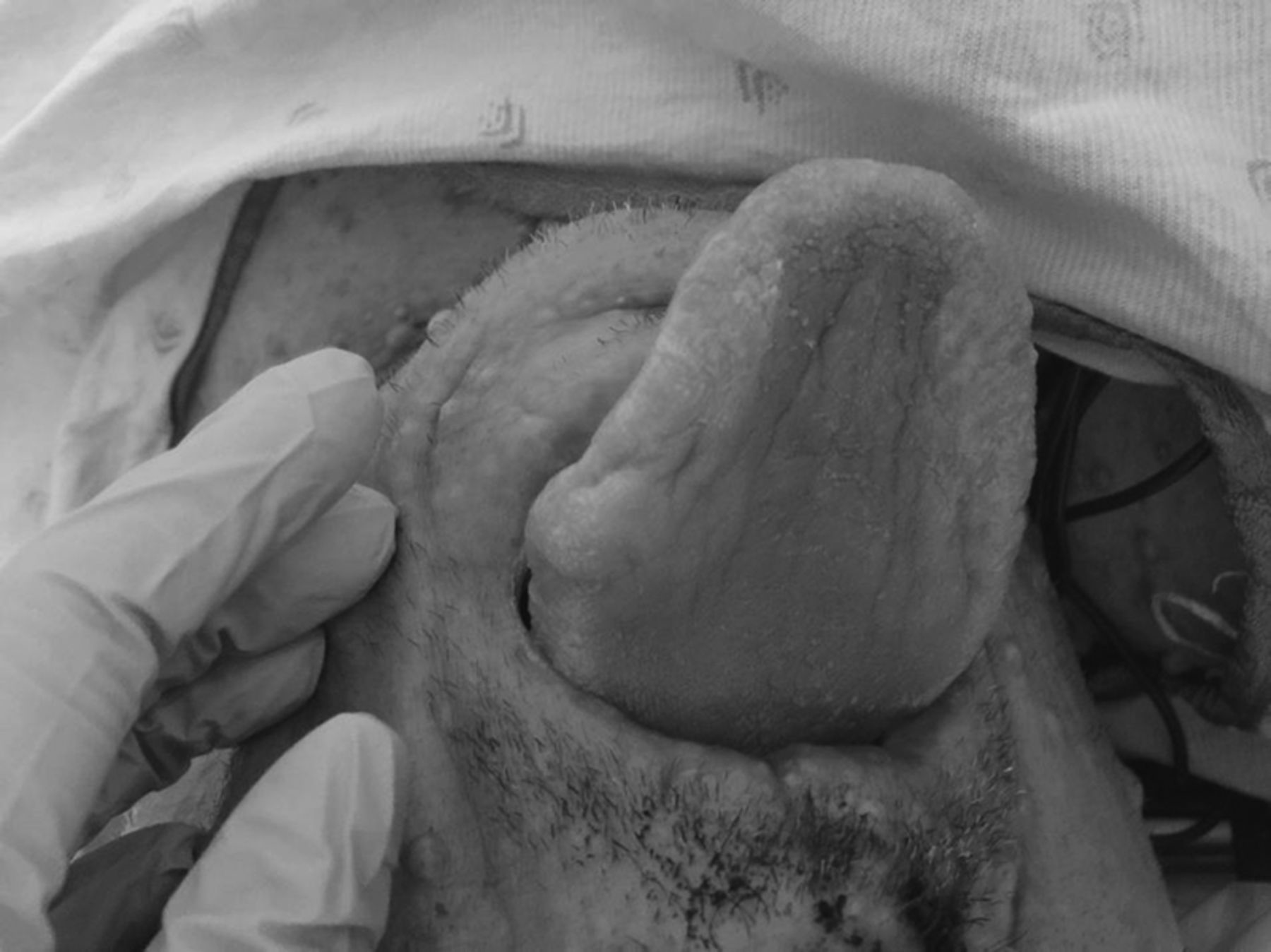
Mucosal neurofibromata
Mucosal neurofibromata
Id: 74 Death Due to Dysautonomia in a Patient with Fulminant Guillain-Barre Syndrome Mimicking Brain Death
D. Prasanna, N. Madan, S. Kapoor. Jacobi Medical Center, Bronx, New York.
Id: 75 a Unique Presentation and Management of Hypercapnic Respiratory Failure in Overlap Syndrome
D. Lin, A.R. Abreu. Division of Pulmonary, Allergy, Critical Care, Sleep Medicine, UHealth Sleep Medicine Program, University of Miami - Miller School of Medicine, Miami, florida.
Id: 76 Metastatic Glioblastoma – a Deceptive Killer
S. Naqvi, K. Allen. Pulmonary & Critical Care Medicine, University of Oklahoma Health Sciences Center, Edmond, Oklahoma. M. Khawar. Internal Medicine, University of Oklahoma Health Sciences Center, Oklahoma City, Oklahoma.
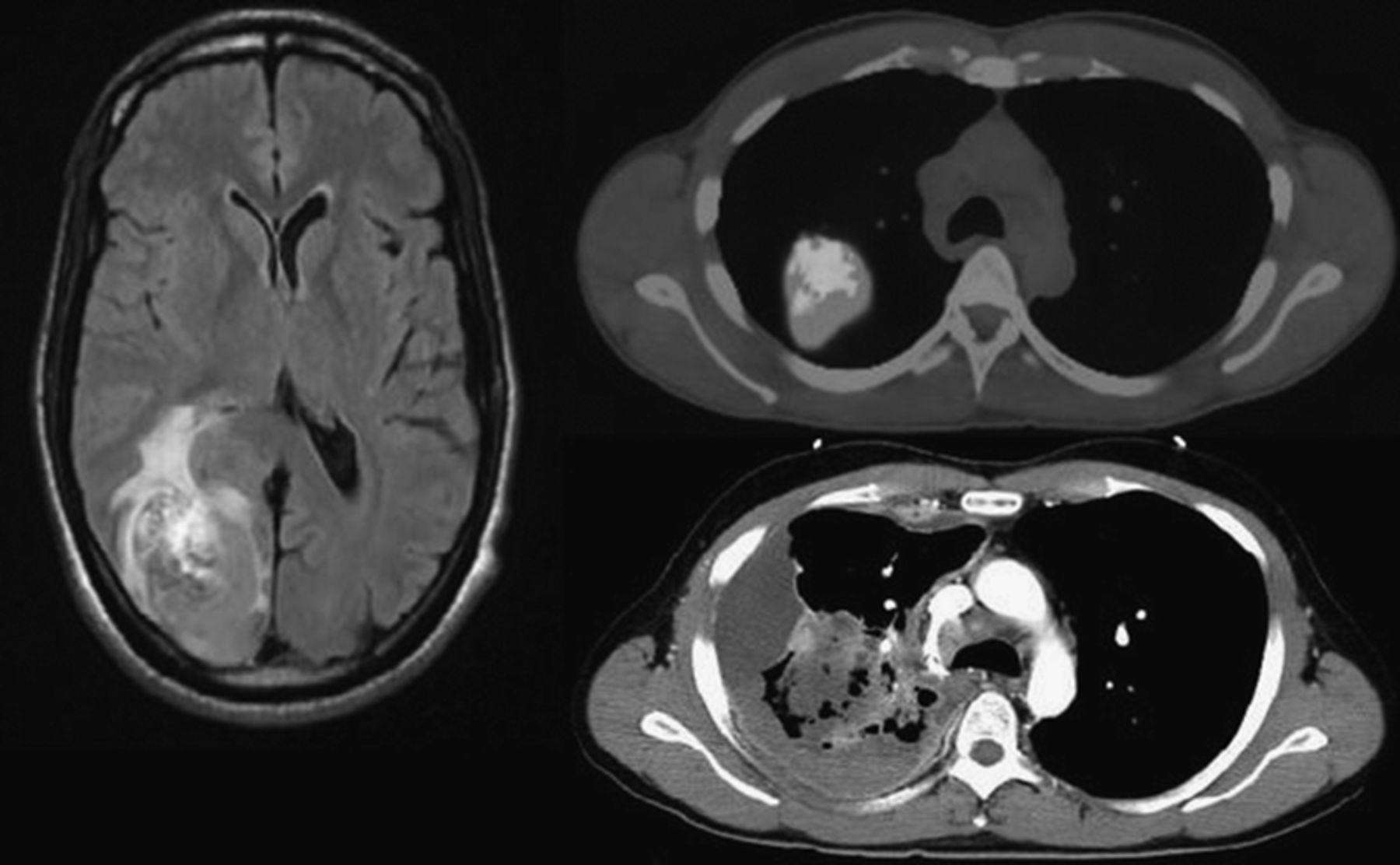
Id: 77 a Critical Central Airway Obstruction Managed with Adjuvent Cardio-Pulmonary Bypass
H. Bhardwaj, H. Youness, B. Brown. Pulmonary & Critical Care Medicine, University of Oklahoma Health Sciences Center, Oklahoma City, Oklahoma.
Id: 78 An Unusual Cause of Recurrent Pleural Effusion
M.U. Khawar, M.A. Farooq. Internal Medicine, Oklahoma University Health Sciences Center, Oklahoma City, Oklahoma. H. Youness. Pulmonary & Critical Care Medicine, Oklahoma University Health Sciences Center, Oklahoma City, Oklahoma.
ID: 79 Myofibroblast Differentiation Requires Signaling Via Mtor Complexes 1 And 2
K.T. Ferguson. Medicine, University of Wisconsin, Madison, Wisconsin, Sherris, RestorGenex, Buffalo Grove, Illinois. E.E. Torr, K. Bernau, N.K. Sandbo. Allergy, Pulmonary, & Critical Care Medicine, University of Wisconsin, Madison, Wisconsin.
Id: 80 the Crucial Role of Pecam1 Induced by Simvastatin for Endothelial Barrier Function
W. Chen, M. Habeck, X. Ni, J.R. Jacobson. Medicine, Univ of Illinois at Chicago, Chicago, Illinois.
Rheumatology/Immunology/Allergy
Id: 81 Baseline Serum Interferon Beta/Alpha Ratio Predicts Response to Tumor Necrosis Factor Alpha Inhibitoin in Rheumatoid Arthritis
T. Wampler Muskardin, J.M. Dorschner, M.A. Jensen, T.B. Niewold. Mayo Clinic, Rochester, Minnesota P. Vashisht, University of Nebraska Medical Center, Omaha, Nebraska. R. Aggrawal, B.S. Chrabot. Gwen Knapp Center for Lupus and Immunology Research, University of Chicago, Chicago, Illinois. M. Kern, P.K. Gregersen. Center for Genomics and Human Genetics, feinstein Institute Medical Research, North Shore LIJ Health System, Manhasset, New York. S. Bridges. University of Alabama at Birmingham, Birmingham, Alabama.
Id: 82 Single Cell Gene Expression in Human Lupus Monocytes Reveals a Differential Impact of Interferon Signaling between Monocyte Subsets
Z. Jin, M.A. Jensen, J.M. Dorschner, D.M. Vsetecka, S. Amin, A. Makol, F.C. Ernste, T. Osborn, K.G. Moder, V. Chowdhary, T.B. Niewold. Division of Rheumatology and Department of Immunology, Mayo Clinic, Rochester, Minnesota.
Id: 83 a Novel Model of Allergen-Specific Immunotherapy for House Dust Mite-Induced Asthma
S.J. Bracken, A.J. Adami, A. Mills, L.A. Guernsey, K. Parham, R.S. Thrall. Uconn Health Center, Unionville, Connecticut. E. Rafti, A.P. Matson, C.M. Schramm. Connecticut Children's Medical Center, Hartford, Connecticut.
Id: 84 Exposure to House Dust Mite is Associated with the Appearance of Pro-Regulatory Gut Microorganisms and Increases in Pulmonary Regulatory T Cells
A.J. Adami, S.J. Bracken, L.A. Guernsey, R.S. Thrall. Immunology, University of Connecticut Health Center, West Hartford, Connecticut. J. Graf. Molecular and Cell Biology, University of Connecticut, Storrs, Connecticut. C.M. Schramm. Pediatrics, Connecticut Childrens Medical Center, Hartford, Connecticut.
Id: 85 T Cell-Mediated Systemic Recruitment of Neutrophils and Monocytes after Intranasal Staphylococcal Enterotoxin a Exposure
J. Svedova, A.T. Vella. Immunology, University of Connecticut Health Center, Farmington, Connecticut.
Staphylococcus aureus (S. aureus), a common pathogen colonizing the human nose and skin, is one of the most important causes of life-threatening infections. The high morbidity and mortality of S. aureus infections are ascribed to various virulence factors, including superantigens. Superantigens, such as staphylococcal enterotoxin A (SEA), mediate food poisoning and toxic shock syndrome and are also implicated in a number of pulmonary diseases and even sepsis. Their potency is embedded in their ability to directly bind to MHC II on antigen presenting cells and then crosslink with specific TCR Vb chains on T cells (e.g. Vb3 for SEA). The subsequent oligoclonal expansion and activation of T cells unleashes a massive immune response. Although SEA has been studied for decades, the exact mechanism of its action is not completely understood. We previously showed that intranasal administration of SEA in mice leads to a T-cell mediated recruitment of neutrophils and monocytes to the lungs. We hypothesized that the local migration of these innate cells to the lungs would be coincident with a systemic immune response. The aim of this study was to understand the early systemic responses to intranasal SEA exposure, and in particular, to illuminate the mechanism of how SEA-activated T cells orchestrate migration and activation of neutrophils and monocytes. Intranasal instillation of SEA in mice caused a rapid influx of neutrophils and monocytes to peripheral blood, followed by their recruitment to spleen and draining and non-draining lymph nodes. This migration was dependent on T cells since TCR bδ-/- mice were unable to recruit these cells unless they received an adoptive T cell transfer. Confocal microscopy imaging showed that the recruited neutrophils localized to the T cell zone within the lymph nodes. Utilizing qPCR and multiplex assays, an increased expression of several known neutrophil and monocyte chemotactic factors, including CXCL1, CXCL2, CCL2, CCL3, CCL4 and CCL7 was detected in the lymph node tissue as early as 1 h after SEA exposure. By sorting specific cell populations we found that these chemokines were differentially expressed. Interestingly, CXCL1 and CXCL2 were mostly produced by dendritic cells while CCL3 and CCL4 were made by SEAactivated Vb3+ T cells. Our future studies will focus on illuminating the mechanism of chemokine release by T cells and dendritic cells and on describing the consequences of neutrophil and monocyte recruitment to lymph nodes. Understanding how SEA-activated T cells affect the migration and function of these innate cells is not only important for designing appropriate countermeasures against SEA-impacted diseases but it will also enhance our knowledge of inflammatory mechanisms.
Id: 86 a Commensal Bacteria-Derived Lipid, Deficient in the Serum of Multiple Sclerosis Patients, Causes Attenuation of Experimental Autoimmune Encephalomyelitis and An Increase in Cd39+ Tregs
E.J. Anstadt, M. Fujiwara, R.B. Clark. Immunology, University of Connecticut Health Center, Farmington, Connecticut. F. Nichols. Oral and Health Diagnostic Sciences, University of Connecticut Health Center, Farmington, Connecticut. R. Nemati. Chemistry, University of Connecticut, Storrs, Connecticut.
The role of the microbiome in multiple sclerosis (MS) remains unknown. We have identified a Toll-like receptor 2 agonist produced by bacteria of the phylum Bacteroidetes (composing 30% of the normal gastrointestinal flora), called Lipid 654 (L654). We found that L654 can be recovered in healthy human serum and that levels are significantly lower in patients with MS. To determine the relevance of this finding, we administered L654 to mice that were actively immunized to induce experimental autoimmune encephalomyelitis (EAE) and found significant attenuation of disease. The mechanism underlying the attenuation induced by L654 is not yet known. CD39, the ratelimiting ectonucleotidase responsible for the conversion of adenosine triphosphate to adenosine, has been implicated as a critical suppressive mechanism of foxP3+ T regulatory cells (Tregs) in both EAE and MS. In MS, circulating CD39+ Tregs are decreased and also impaired in their ability to control Th17 proliferation. Intriguingly, CD39+ Tregs are increased during periods of remission in MS patients. The purpose of this study was to 1) determine if L654 could attenuate clinical disease severity in a transfer model of EAE to better test the effect of L654 on activated, encephalitogenic T cells and 2) determine the role of CD39+ Tregs in the disease inhibition caused by L654 administration. An adoptive transfer model of EAE in SJL/J mice was utilized. Mice received transfer of PLP-specific activated T cells and EAE developed 8-11 days later. A 1 ug dose of L654 was administered to mice at day 0 or at day 0 and 7. Mice were observed for disease severity or spinal cords were harvested for cellular analysis. L654 administration significantly attenuated clinical disease severity in the transfer model of EAE (p < 0.0001). Cellular analysis of mononuclear cells in the spinal cord at day 18 showed a significant decrease in total cell number (p = 0.02) as well as a titrating decrease in percentage of IL-17 producing, IFNγ producing and IL-17/IFNγ double-producing CD4+ T cells in mice administered L654. At day 10, cellular analysis showed a decrease in total mononuclear cell number and an increase in percentage of CD39+ Tregs in mice administered L654. Therefore, L654 is of significant interest as a potential treatment for MS, as it may increase CD39+ Tregs, known to be deficient in MS patients, and potentially suppress clinical disease. Future directions will determine if the suppression induced by L654 is dependent on CD39+ Tregs, and what effect L654 administration has on spontaneous relapses in EAE.
Id: 87 Inhibition of Ezh2 Simultaneously Decreases Pro-Inflammatory and Enhances Antiinflammatory Microglial Phenotypes
E. Koellhoffer, R. Ritzel, L. McCullough. Neuroscience, University of Connecticut Health Center, Farmington, Connecticut. J. Grenier. Immunology, University of Connecticut Health Center, Farmington, Connecticut.
The aging central nervous system (CNS) is characterized by a chronic baseline elevation in inflammation which contributes to the pathogenicity of many neurological diseases. Understanding the source of this inflammation is critical as manipulations may allow for the reversal of these age-related changes, thereby reducing the risk of CNS disease. This has led to a focus on the role of microglia, the resident innate immune cell of the brain, in CNS inflammation. Microglia possess the ability to adapt to the environment by altering their phenotype between a resting (M0) state to either an activated pro-inflammatory (M1) or anti-inflammatory (M2) phenotype. In the setting of aging, microglia have a more predominant M1 phenotype at baseline. However, the molecular mechanisms underlying these changes with age remain largely unknown. Recent work has shown that Jumonji Domain Containing 3 (Jmjd3), a histone demethylase, is essential for M2 polarization. By demethylating histone H3 lysine 27 trimethylation (H3K27me3) to a monomethylation mark (H3K27me1), Jmjd3 alters the epigenetic landscape of target promoters from a transcriptionally repressed state to a transcriptionally active state. This in turn allows for expression of target genes to occur. In contrast, Enhancer of Zeste Homologue 2 (Ezh2) functions in an opposite manner by selectively writing the H3K27me3 mark and repressing transcription of target genes. We hypothesized that inhibition of Ezh2 may tilt the balance between Jmjd3 and Ezh2 respective activities in favor of higher Jmjd3 activity, thereby enhancing polarization toward an anti-inflammatory and neuroprotective M2 phenotype. Mixed glial cultures were isolated from P0.5-P2 C57Bl/6 mice and cultured for 10-14 days before microglia were isolated. Primary microglia were treated with GSK343 (an inhibitor of Ezh2) for 1hr before being stimulated with LPS+IFNγ or IL-4. After 4hr or 24hr RNA was isolated for qRT-PCR analysis. In our model of primary microglia, pre-treatment with GSK343 significantly diminished LPS+IFNγ- induced IL6 and IL1B expression at both 4hr and 24hr (p<0.05). Additionally, Ezh2 inhibition rescued the expression of M2-associated genes ARG1, CD206, and IRf4 at 24hr (p<0.05) which were otherwise significantly down-regulated in the presence of LPS+IFNγ. In line with our hypothesis, inhibition of Ezh2 also resulted in an augmented IL-4- induced upregulation of ARG1, CD206, and IRf4 at 4hr (p<0.05). Our data implies that there is epigenetic regulation of M1/M2 microglia polarization and that Ezh2 has a role in this regulation. Pre-treatment with an inhibitor of Ezh2 limits LPS+IFNγ-induced microglial M1 polarization and restores M2-associated gene expression to baseline. This suggests that altering the balance between Ezh2 and Jmjd3 allows for reprograming of both M1- and M2-associated genes.