
Abstract
There is no single methodology that can fully explain the nature of human development and learning. Yet, headway is being made on how cognitive milestones are achieved during development with the use of magnetic resonance imaging (MRI) technology. With this methodology, it is possible to assess changes in brain structure, function, and connectivity. Recent findings suggest that both progressive and regressive processes—as opposed to simple linear patterns of change—underlie changes in cognitive abilities. Functional MRI studies suggest that both biological maturation and learning correspond to a fine-tuning of neural systems with enhanced recruitment of task-relevant regions. This fine-tuning of cortical systems corresponds with their enhanced connectivity with cortical and subcortical circuitry. In sum, imaging has helped to move the field of cognitive development beyond questions of what develops and when, to how these changes may occur.
Developmental cognitive psychology is the study of how individuals' cognitive abilities change over time and of the emergent processes that support those changes. Behavioral measures used to assess cognitive development typically vary by the age of the population of interest. For example, investigations in infancy are often dependent on looking-time measures, while studies in children use verbal-report or manual-response (button-press, joystick) measures. In either case, the measure obtained is an indirect estimate of some underlying process or body of knowledge. Broadly speaking, such measures have been most successful at addressing what develops and when.
Although traditional research methods have been informative in understanding cognitive change over time, recent advances in brain-imaging methods promise to be useful at addressing the biological mechanisms underlying those changes. The methods that will be emphasized in this article are all forms of magnetic resonance imaging (MRI) that provide information about structure, function, and brain connectivity.
One could argue that neuroimaging provides no unique information to the study of cognitive development. That is, identifying a structure with which a function is associated (i.e., brain mapping) is interesting, but it provides no more information about cognitive development than can be obtained from simple behavioral measures. When, between populations, performance differs along some dimension of interest, behavioral data can be informative about developmental processes. However, infants and children may arrive at the same behavioral outcomes adults do by using very different neural pathways and associated cognitive strategies. In such cases, having only behavioral information may be misleading, resulting in the formulation of models and theories that devalue the contribution of other factors to development.
Understanding the development of pathways underlying cognition and the experiences that alter those pathways is imperative to the study of cognitive development. As noted by Karmiloff-Smith (1994), “the mind does not begin with pre specified modules; rather, development involves a gradual process of modularization” (p. 693). Perhaps the single greatest contribution that neuroimaging can make to the study of cognitive development involves unmasking the biological mechanisms that support developmental behavioral change. Before the advent of sophisticated imaging techniques, assumptions about relationships between the brain and behavior had no empirical grounding. We suggest that imaging techniques provide a unique opportunity to assess biological and behavioral changes simultaneously, allowing for quantitative evidence of brain–behavior associations. We provide evidence from developmental studies of cognitive control during learning to illustrate these contributions. Learning to predict environmental information is a key element of cognitive development. Knowing what to expect and in which context to expect it is critical to planning and maintaining appropriate thoughts and actions. Although learning to predict certain events in the world (e.g., anticipatory eye movements in response to regularly presented stimuli) may be intact early in development, the ability to adjust behavior when these predictions are violated develops more gradually as the underlying neural circuitry is organized (Casey, Amso, & Davidson, in press).
IMAGING METHODOLOGIES: WHAT THEY DO AND DO NOT REVEAL ABOUT DEVELOPMENT
MRI technologies have introduced a new set of tools for capturing features of brain development in living, developing humans. This method permits repeated scanning of the same individual over time, thus providing precise measurements of neuroanatomical changes during learning and development. MRI became especially important to cognitive and developmental scientists when its functional capabilities were discovered and developed. Whereas MRI is used to produce structural images of the brain, the functional component of MRI (fMRI) provides an index of brain activity by measuring changes in localized blood-oxygen levels in the living brain (e.g., Kwong et al., 1992). The assumption that these localized increases in blood oxygenation reflect increases in neuronal activity has found empirical grounding in combined fMRI and electrophysiology studies of nonhuman primates (Logothetis, Pauls, Augath, Trinath, & Oeltermann, 2001).
While fMRI provides a measure of brain function and can help identify regional changes with development, diffusion tensor imaging (DTI) provides an index of brain connectivity. DTI measures change in the microstructure of white matter (tissue containing nerve fibers with fatty insulating material called myelin), based on the properties of water diffusion (e.g., Pierpaoli, Jezzard, Basser, Barnett, & Di Chiro, 1996). Diffusion of water in white-matter tracts is constrained by myelin and the orientation and regularity of fibers. Water diffuses more readily in parallel to a tract than perpendicular to it—a phenomenon called anisotropic diffusion—and thus can provide information about directionality of connectivity. MRI can be sensitized to water diffusion to quantify myelination and white-matter microstructure in the living brain in order to provide information about changes in connectivity with development (Klingberg, Vaidya, Gabrieli, Moseley, & Hedehus, 1999; Liston et al., in press). This technique can be informative as to how regional connectivity relates to the development of behavior and changes in cortical activity underlying that behavior.
Conventional imaging methods have advanced the field of developmental neuroscience by providing evidence of changes in structural architecture and functional organization in the living brain as it develops. However, magnetic resonance (MR) methods (e.g., MRI, fMRI, DTI) only provide indirect measures of brain structure, function, and connectivity. Differences in the volume of a structure or amount of activity, as measured by MR methods, lack the resolution to definitively characterize the mechanisms of change. Evidence from histology, the microscopic study of tissue, suggests that brain development is a dynamic process of regressive and progressive processes. As such, developmental changes observed using MR techniques may reflect a combination of these processes.
HOW DOES THE STRUCTURAL AND FUNCTIONAL ORGANIZATION OF THE HUMAN BRAIN CHANGE WITH COGNITIVE DEVELOPMENT?
Several imaging studies have mapped the neuroanatomical course of human brain development. Longitudinal MRI studies (Gogtay et al., 2004; Sowell et al., 2004) have shown that cortical maturation parallels developmental cognitive milestones. Regions supporting primary functions, such as motor and sensory systems, mature earliest (see Fig. 1). Next come temporal and parietal association cortices involved in basic language skills and spatial attention. Evidence suggests that higher-order association areas, such as the prefrontal and lateral temporal cortices, which integrate primary sensorimotor processes and modulate basic attention and language processes, mature last (see Fig. 1; Gogtay et al., 2004; Sowell et al., 2004). This progression has been determined via MRI-based studies showing that loss of cortical gray-matter (which contains mostly cell bodies) volume occurs earliest in the primary sensorimotor areas and latest in the dorsolateral prefrontal cortex (e.g., Gogtay et al., 2004; Sowell et al., 2004). The prevalent hypothesis that changes in gray-matter volume detected by MR reflect synapse formation and elimination cannot be supported by imaging studies because of the limited resolution of this methodology.
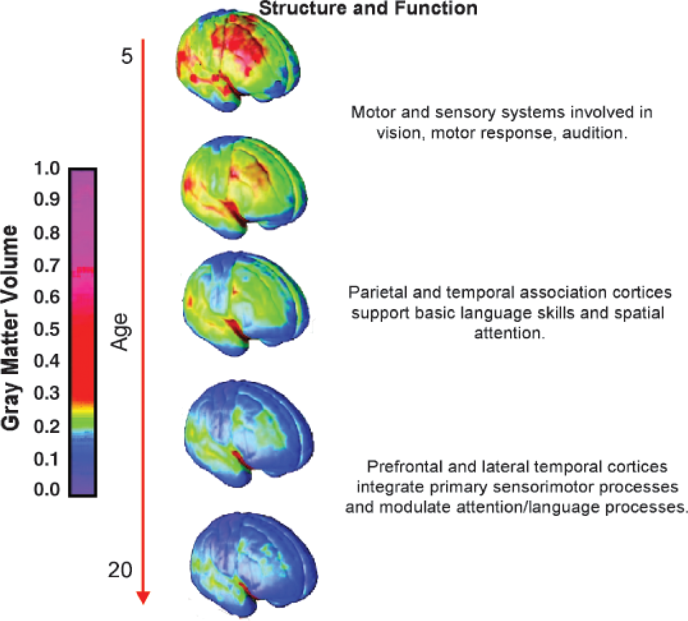
The sequence of gray-matter maturation (indicated by loss) with age. Areas in blue correspond to the specific cortices undergoing gray-matter loss. These structures and their functional significance are described to the right. Adapted from Gogtay et al., 2004.
Cross-sectional studies of normative brain maturation during childhood and adolescence have shown similar patterns, leading to the conclusion that gray-matter loss during this period reflects a sculpting process of the immature brain (e.g., Caviness, Kennedy, Richelme, Rademacher, & Filipek, 1996). While gray-matter volume has an inverted-U-shaped pattern of development, increases in white-matter volume and density with age are roughly linear (Gogtay et al., 2004). These changes presumably reflect ongoing myelination of axons, which enhances neuronal conduction and may play a role in the speed of cognitive processing.
How do these changes in brain structure relate to cognitive development? With development, a child's capacity to filter competing information and suppress inappropriate actions, termed cognitive control, improves dramatically; thus susceptibility to interfering and competing actions diminishes with maturity (Casey, Amso, & Davidson, in press). Durston, Thomas, Worden, Yang, and Casey (2002) used a go/no-go task in combination with fMRI to examine the neural basis of cognitive control and its development. In a go/no-go task, subjects are presented with stimuli to which they are instructed either to respond (go trials) or withhold a response (no-go trials). In an attempt to isolate cognitive and neural processes underlying susceptibility to interference, they parametrically manipulated the number of responses a subject made before having to withhold a response. The ability to accurately withhold a response decreased as the number of preceding responses increased. Simultaneously, the imaging data showed that inhibiting a response to a less-frequent nontarget was associated with increased activity in the ventral prefrontal cortex and the striatum, one of the nuclei that make up the basal ganglia, located below the cerebral cortex. Activity in the ventral prefrontal cortex correlated with performance across age, with children making more errors overall and maximally recruiting the ventral prefrontal cortex, even when a single response preceded them withholding a response.
These findings suggest that susceptibility to interference from competing sources is paralleled by maturational differences in recruitment of underlying frontostriatal circuitry, especially in the prefrontal cortex. Collectively, imaging studies of cognitive control show that children recruit larger, more diffuse prefrontal regions when performing these cognitive tasks than adults do. Activity within brain regions that correlates with task performance becomes more focal or fine-tuned with age, whereas brain regions not correlated with such task performance diminish in activity with age, as shown by cross-sectional (Brown, Lugar, Coalson, Miezin, Petersen, & Schlaggar, 2005) and longitudinal studies (Durston et al., 2006; see Fig. 2).
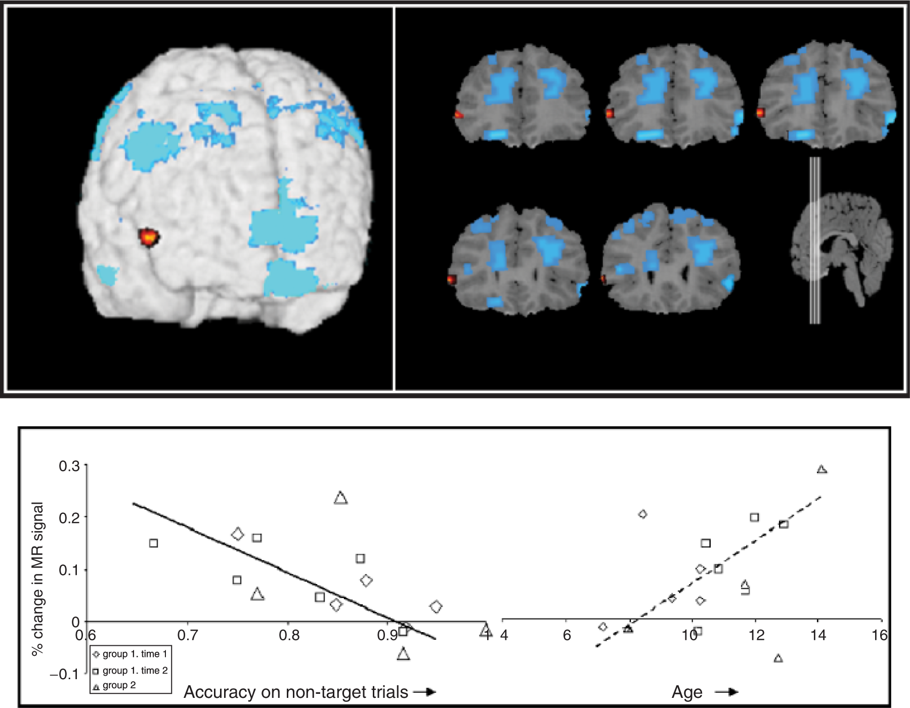
Regressive and progressive changes with development. Red areas indicate increases in activity in regions associated with cognitive control with age. Blue areas are those that do not correlate with cognitive-control task performance and show attenuated activity with age. The graphs show correlations between activity of key brain areas (VENTrAL PREFRonTal CORTEX) during performance of a go/no-go task with behavioral performance and age. These data reflect the increased impulsivity observed as children move into the early stages of adolescence and that later diminishes in adulthood. Adapted from “A Shift From Diffuse to Focal Cortical Activity With Development,” by S. Durston, M.C. Davidson, N. Tottenham, A. Galvan, J. Spicer, J.A. Fossella, & B.J. Casey, 2006, Developmental Science, 9, p. 5. Copyright 2006 by Blackwell Publishers. Adapted with permission.
There are only a few examples of the use of DTI to study cognitive development. One such study (Liston et al., in press) used activation maps from the Durston et al. (2002) developmental-fMRI study discussed earlier to identify fiber tracts that play a role in control over behavior. Specifically, they examined the degree of connectivity between the prefrontal cortex and the striatum, two brain regions in which activity has been shown to correlate with task performance. Levels of connectivity in the frontostriatal and a comparison fiber tract (the corticospinal fiber tract) both correlated with age, but only frontostriatal connectivity correlated with performance on the cognitive-control task (go/no-go paradigm). These findings suggest that the development of prefrontal connectivity and function contributes to a developing capacity for cognitive control.
The results from this select review of studies indicate that changes in prefrontal cortical volume, function, and connectivity as measured by MRI, fMRI, and DTI correspond with a developing capacity for cognitive control. The MRI data showed protracted development of lateral prefrontal cortical thickness. The fMRI data showed fine-tuning of prefrontal activity with development as the pattern of activity shifted from a diffuse to more focal pattern. The DTI results suggest that enhanced prefrontal connectivity may contribute to changes in cognitive abilities with development.
HOW DOES THE STRUCTURAL AND FUNCTIONAL ORGANIZATION OF THE HUMAN BRAIN CHANGE WITH LEARNING?
As development is an interactive process between biological maturation and experience, it is important to examine neural changes with learning. One of the first studies to examine learning with fMRI showed cortical changes as subjects learned sequential finger movements (Karni, Meyer, Jezzard, Adams, Turner, & Ungerleider, 1995). Activity in the primary motor cortex during motor-sequence learning was apparent within a single imaging session and increased over weeks of training. Specifically, activity in task-relevant regions became increasingly enhanced with training, whereas task-irrelevant regions become less active over time. This pattern, in adults, mimics the observed changes in cross-sectional (Brown et al., 2005) and longitudinal (Durston et al., 2006) developmental data described previously.
The previously described studies (Durston et al., 2002; Liston et al., in press) examined the neural mechanisms supporting changes in how behavior is altered when learned expectations are violated (i.e., inhibiting a response to a less frequent nontarget). Recently, Amso, Davidson, Johnson, Glover, and Casey (2005) used fMRI to examine neural mechanisms underlying simple learning in adults. A common measure of learning from developmental psychology is preference for novelty as measured by looking times. In novelty-preference paradigms, a stimulus becomes familiar or learned through repeated exposures. Adjusting behavior when these learned or expected events are violated is an essential element of cognitive control (Casey et al., in press), aspects of which are present early in life. Understanding the neural bases for these preferences may constrain developmental theories for how these abilities emerge. In this study, subjects were presented with cue and target stimuli in alternation. The frequency manipulation was designed so that frequent and novel target stimuli were preceded by and had an equal probability of co-occurrence or association with the same cue. The association manipulation was such that the target stimulus was identical in the novel and frequent-association condition. Here the manipulation rested solely on the probability of its co-occurrence with the preceding cue stimulus.
The behavioral results from this study showed longer response times to novel target stimuli—i.e., stimuli with a lower frequency of occurrence across the experiment—as well as learning-related behavioral changes (shorter response times) for both frequently occurring and frequently associated stimuli. The imaging results showed increased activity in the striatum to novel targets and increased left hippocampal activity to novel associations. The hippocampus was preferentially active to the infrequent association, suggesting its involvement in learning of new associations or linking a cue with a novel target (see Fig. 3). These behavioral and imaging findings are interesting in light of the developmental work using novelty preferences and looking-time measures to show learning in infants. The findings suggest that novelty preference is not based in a single system, but is rather a manifestation of various learning mechanisms that are interacting with the environment. When learned associations are violated is when cognitive control is needed to adjust behavior appropriately, and the previously described studies suggest that this ability is what changes most with development.
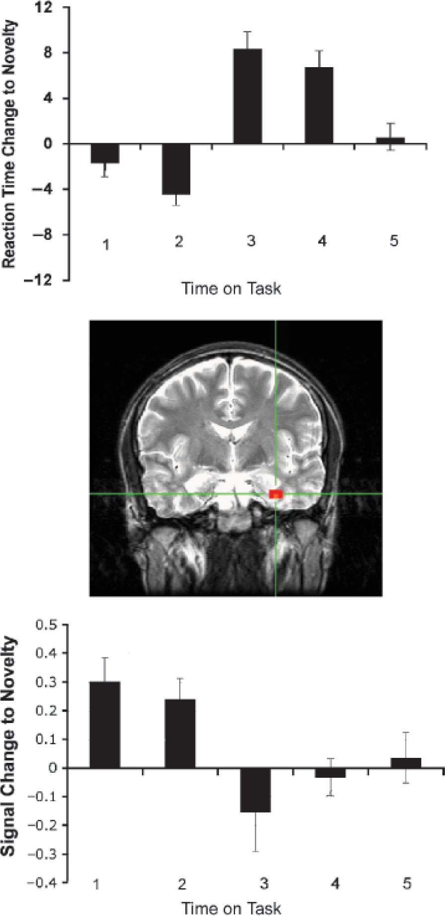
Infrequent (novel) relative to frequent association comparison. Top graph illustrates changes in reaction time to the novel relative to the frequent association. A greater response to the novel relative to the frequent association indicates learning; results show that learning is not evident at sampled time points early in the task, but becomes increasingly evident later, as participants are more exposed to the task structure. Image (center) shows hippocampal activity to novelty. Bottom graph shows pattern of activation (as measured by signal change) in the hippocampus to novel relative to frequent association as a function of time on task. Adapted from Amso, Davidson, Johnson, Glover, & Casey (2005).
WHAT HAS BEEN LEARNED AND FUTURE DIRECTIONS
How has imaging informed the understanding of cognitive development? First, the imaging studies of development and learning suggest that both progressive and regressive processes, as opposed to simple linear patterns of change, underlie changes in cognitive abilities and that these changes may differ regionally across the brain. In development, cortical-thickness changes occur last in higher cortical regions of the lateral prefrontal and temporal cortex and occur in an inverted U-shaped progression, with an increase and subsequent decrease. Second, development and learning correspond to a fine-tuning of neural systems with enhanced recruitment of task-relevant regions and suppression of less task-relevant regions. Finally, these changes correspond with enhanced connectivity of cortical circuitry as measured by DTI. Each of these imaging methods has begun to provide insight on how changes in cognitive processing occur with development. Neuroimaging and other sophisticated techniques such as computational modeling (Munakata & McClelland, 2003) permit the mechanisms supporting the behaviors under investigation to be more precisely characterized. This approach allows for formulation of testable theories and models of development that are consistent with neurobiology.
Footnotes
Acknowledgements
This work was supported in part by R01 MH63255, R01 DA018879, R21 DA15882, and P50 MH062196 awarded to B.J. Casey.