
Abstract
Primary idiopathic frozen shoulder can be mis-diagnosed in patients presenting with a painful stiff shoulder if radiographs of the shoulder are not undertaken. We report a patient who was referred to an orthopaedic upper limb clinic with a presumed diagnosis of a frozen shoulder who turned out to have the rather rare condition, melorheostosis. This was successfully treated by arthroscopic debridement.
Introduction
Melorheostosis is a rare, benign, non-familial bone dysplasia characterized by a classic radiographic feature of flowing hyperostosis resembling dripping candle wax, generally on one side of a long bone. The condition was originally described by Leri & Joanny [1]. Its aetiology remains unclear, and treatment in most instances is symptomatic. Melorheostosis usually affects one limb, more often the lower extremity, and rarely the axial skeleton. We report a case of melorheostosis affecting both the glenoid and proximal humerus in a 38 year old professional golfer.
Case Report
The patient was referred from physiotherapy triage with a diagnosis of a frozen shoulder but without a radiograph of the shoulder having been performed. He presented with a short but typical history of potential frozen shoulder including symptoms of a painful, stiff shoulder with no apparent precipitating cause. On examination, he had complete loss of active and passive external and internal rotation at the glenohumeral joint but with relative preservation of forward flexion to 160° and abduction to 100°. His Oxford shoulder score was 32/60. His golf handicap had dropped from 0 to 3! Initial X-rays revealed a typical dripping candle sign on the humeral shaft with hyperostotic lesions affecting both the glenoid rim and humeral head (Figs 1 and 2. A subsequent MRI revealed kissing type lesions affecting the posterior glenoid rim and posterior humeral head at the level of the bare area (Fig. 3).
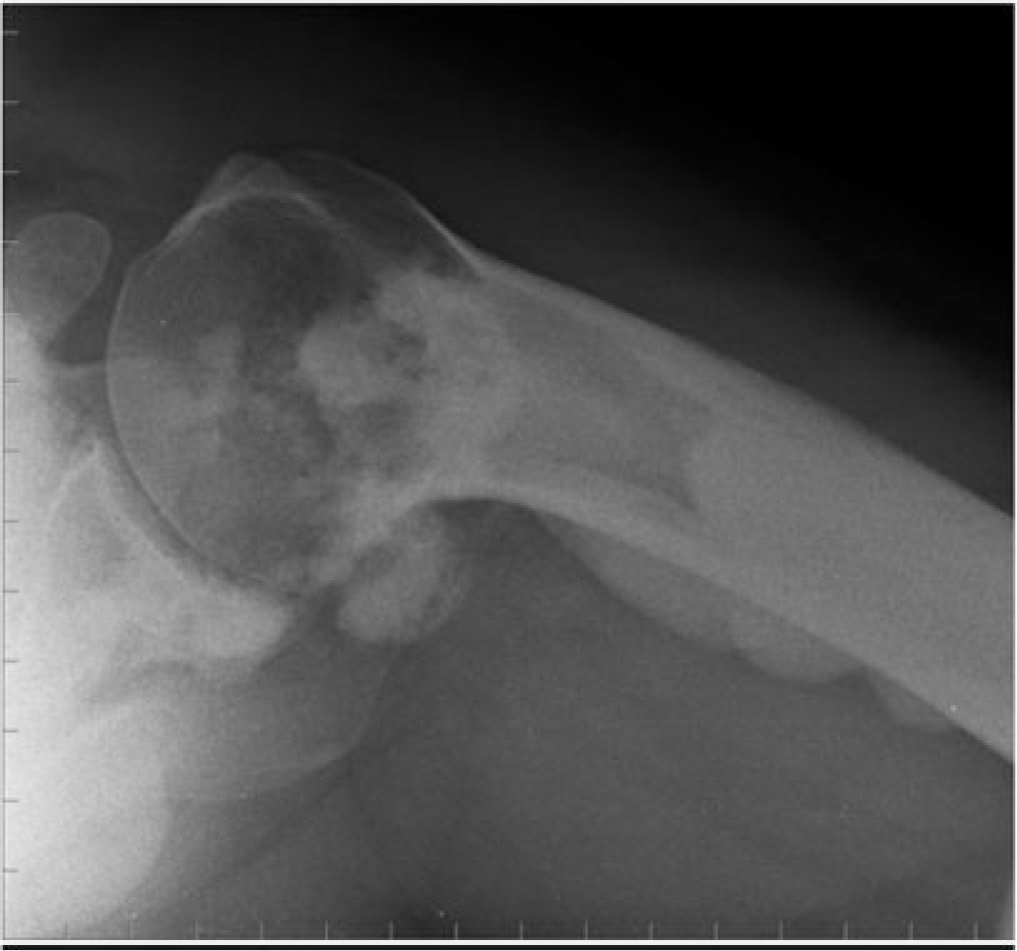
Shoulder radiograph—axillary view showing hyperostotic lesions on posterior glenoid and humeral head.
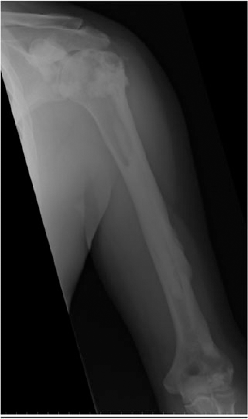
AP radiograph of shoulder and humerus showing typical dripping candle wax sign.
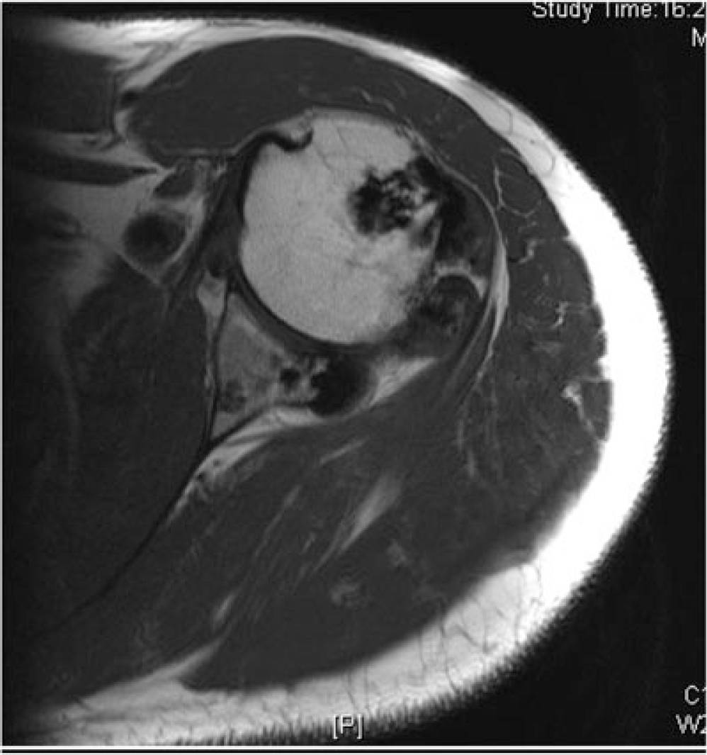
T2 MRI image—axial view of shoulder showing lesions on posterior glenoid and posterior humeral head.
He subsequently underwent arthroscopic excision of his glenohumeral overgrowth using an arthroscopic shaver with burr attachment. At follow-up 2 years later, the patient was very happy with the results of surgery and was back playing golf uninhibited. His ROM at the shoulder was 60° ER, 80° IR, 160° forward flexion, 160° abduction. His Oxford shoulder score had returned to normal and his golf handicap had improved again to 0.
Discussion
Both Codman [2] and Bunker [3] reiterate that a diagnosis of frozen shoulder can only be made on the back of a normal radiographic examination of the shoulder. If an X-ray is not undertaken, a variety of conditions may be missed such as osteoarthritis, chronic shoulder dislocations, fractures, chronic infection, heterotrophic ossification from head injury and any evidence of previous shoulder surgery. Our case was an unusual presentation of a rare condition. It was successfully treated with arthroscopic debridement. Owing to its rarity, there is a dearth of evidence as to whether the growths recur if resected. At 2° years follow-up, there were no signs of recurrence in our patient.
Footnotes
Conflicts of Interest
None declared