






Abstract
Several studies were performed to evaluate the degree of liver fibrosis by non-invasive markers. We aimed to assess the diagnostic value of both biglycan (BGN) and osteopontin (OPN) as non-invasive markers of hepatic fibrosis in patients with chronic hepatitis B (CHB) and chronic hepatitis C (CHC). This study was performed on 100 patients with CHB virus, 100 patients with CHC virus and 100 normal controls. All participants were subjected to the following laboratory tests: hemoglobin, platelet, alanine aminotransferase, aspartate aminotransferase, albumin, international normalized ratio, HBs Ag, hepatitis C virus (HCV) antibody, hepatitis B virus DNA, HCV RNA, liver biopsy, BGN and OPN. We found that BGN level was significantly increased in the CHB group compared with the controls (p<0.001), but the level was not different between the CHC group and the controls (p<0.96). OPN was increased in both the CHB and CHC groups compared with the controls (p<0.001). Positive correlation was found between fibrosis stages and BGN level of the CHB group (r=0.64; p<0.001) and between fibrosis stages and OPN level of the CHB (r=0.63; p<0.001) and CHC (r=0.59; p<0.03) groups. The area under the curve (AUC), sensitivity and specificity of BGN were 1.0, 100% and 100% in predicting fibrosis in patients with CHB, and 0.50, 26% and 78% in predicting fibrosis in patients with CHC. OPN had an AUC of 0.997, sensitivity of 96% and specificity of 100% in predicting fibrosis in patients with CHB, and 0.974, 96.5% and 100% in predicting fibrosis in patients with CHC. In conclusion, BGN and OPN could be considered non-invasive markers for liver fibrosis assessment.
Keywords
Significance of this study
What is already known about this subject?
Biglycan (BGN), a small leucine-rich proteoglycan, has many roles in the fibrogenesis processes.
Osteopontin (OPN) acts as a chemoattractant to inflammatory cells.
BGN may be considered a non-invasive marker for liver fibrosis assessment in hepatitis B infection.
What are the new findings?
BGN has higher sensitivity in detecting hepatic fibrosis in patients with chronic hepatitis B (CHB) than in patients with chronic hepatitis C (CHC).
OPN had higher sensitivity and specificity than BGN in detecting liver fibrosis in hepatitis C virus infection.
OPN is a good predictor of liver fibrosis in patients with CHB and CHC.
How might these results change the focus of research or clinical practice?
These results may consider BGN and OPN as non-invasive markers for liver fibrosis assessment.
Introduction
Chronic hepatitis C is considered the most common cause of chronic liver disease and cirrhosis,1 and Egypt has the highest hepatitis C virus (HCV) prevalence in the world.2 Hepatitis B virus (HBV) infection is a major health problem. It is known that there are about 350 million HBV carriers, and nearly one million patients die from HBV-related liver diseases annually.3 Both hepatitis C and B can progress to different pathological stages, which vary from mild hepatic inflammation without fibrosis to advanced hepatic fibrosis and cirrhosis.4
Liver biopsy is the gold standard and accurate method for the diagnosis of hepatic fibrosis, but it has many limitations: the biopsy size reflects only a small part of a liver condition, and it is an invasive maneuver, which may be complicated by pain, injury to liver parenchyma and hemorrhage. Also, liver biopsy cannot differentiate between early and advanced fibrosis. Therefore the need for non-invasive markers to assess the degree of liver fibrosis is essential.5
Biglycan (BGN), a small leucine-rich proteoglycan, has many roles in the fibrogenesis processes such as liver fibrosis.6 BGN acts as an activating signal to innate and adaptive immunity as in cases of tissue stress or inflammation; it is proteolytically released and turns into a host-derived non-microbial danger signal (damage-associated molecular patterns), which is recognized by innate immune cell receptors. Consequently cells are activated and produce cytokines that activate adaptive immunity cells, which in turn produce cytokines that produce inflammatory process.7
Osteopontin (OPN) is a multifunctional protein produced by many cells, such as T cells, B cells, dendritic cells and macrophages.8 OPN, also known as bone sialoprotein I, early T-lymphocyte activation protein, urinary stone protein, nephropontin, uropontin and secreted phosphoprotein 1, has a major role in bone remodeling. OPN has two forms: soluble (extracellular form) and insoluble (intracellular).9 Secreted OPN exerts many functions; it stimulates the innate immune cells and acts as a chemoattractant to inflammatory cells such as macrophages and neutrophils, then in the subsequent activation and cytokine production.10 OPN also modulates cell-mediated immune response through stimulation and activation of T cells to produce their cytokines. OPN is considered an extracellular matrix protein as it contains an arginine-glycine-aspartic acid (RGD) domain that is attached to type I collagen, fibronectin and osteocalcin, thereby increasing tissue fibrosis.11
Objective
In this study, we aimed to assess the diagnostic value of both serum BGN and OPN as non-invasive markers of hepatic fibrosis in patients with chronic hepatitis B (CHB) and chronic hepatitis C (CHC).
Subjects and methods
In this case study, 100 patients with CHB and 100 patients with CHC were recruited. All cases were enrolled from outpatients clinic and from the gastroenterology department in Al-Azhar University Hospitals. One hundred age-matched and sex-matched healthy controls were also enrolled in the study. Exclusion criteria included hepatitis D virus, non-alcoholic steatohepatitis (NASH), hepatocellular carcinoma, autoimmune hepatitis, drug-induced liver injury, hereditary liver diseases and alcoholic liver diseases. All patients were subjected to liver biopsy to assess the degree of fibrosis. Abdominal ultrasonography was done to detect any change of liver parenchyma.
Blood samples (10 mL) were collected from patients and controls, divided into (1) 2 mL EDTA blood for hemoglobin and platelet count measurements on automated cell counter (ABX Micros 60 hematology analyzer, France); (2) 2 mL sodium citrate blood for prothrombin time estimation using the semiautomated coagulometer (Coadata 504, Germany); and (3) 6 mL serum sample, which lifted for clotting then centrifuged for 15 min at 4000× g, then serum samples were subdivided into small aliquots to be stored at −80°C until tested for alanine aminotransferase (ALT), aspartate aminotransferase (AST), albumin, hepatitis B surface antigen (HBs Ag), HCV antibody (HCV Ab), HBV DNA, HCV RNA, BGN and OPN measurements.
ALT, AST, and albumin were measured using Indiko clinical and specialty chemistry kits and analyzer (USA).
Detection of HCV Ab and HBs Ag was done using Biokit ELISA kits (Spain) (lot B26851 and B26859, respectively) according to the manufacturer's instructions. The absorbance of each sample well was read at 450 nm on ELISA reader (Stat Fax 2100, USA).
Detection of HBV DNA and HCV RNA was done using real-time PCR (StepOne Real-Time PCR, Applied Biosystems, USA). HBV DNA and HCV RNA were extracted from all cases (Qiagen, Germany; lot 57704 and 52905, respectively). Pathogen detection by PCR is based on the amplification of specific regions of the pathogen genome. In real-time PCR the amplified product is detected via fluorescent dyes. These are usually linked to oligonucleotide probes which bind specifically to the amplified product.
Bgn Measurement
Serum BGN was measured by Human Biglycan PicoKine ELISA Kit (USA; lot 90111119810310) according to the manufacturer's instructions. A monoclonal antibody from mouse specific for BGN has been precoated onto 96-well plates. Standards and test samples were added to the wells, and a biotinylated detection polyclonal antibody from goat specific for BGN was added, subsequently followed by washing with phosphate-buffered saline (PBS). Avidin-biotin-peroxidase complex was added and unbound conjugates were washed away with PBS or Tris-buffered saline. Horseradish peroxidase (HRP) was added to each microplate well successively and incubated. A substrate TMB was used to visualize HRP enzymatic reaction. TMB was catalyzed by HRP to produce a blue color product that changed into yellow after adding acidic stop solution. The optical density was measured spectrophotometrically at a wavelength of 450 nm±2 nm on Stat Fax system (Awareness Technology, USA). The density of the yellow color is proportional to the human BGN amount of the sample captured in the plate.
Opn Measurement
Serum OPN was measured by Human Osteopontin PicoKine ELISA Kit (USA; lot 90111119810310) according to the manufacturer's instructions. Briefly, serum was added to ELISA plates, which precoated with monoclonal antibodies directed against OPN. After incubation and washing of the wells, an HRP was added and the plate was incubated, followed by washing and addition of the TMB substrate. The enzymatic reaction was stopped by adding acid solution and read at 450 nm on Stat Fax system (Awareness Technology).
Liver biopsy and histological assessment
Using 16 F true-cut biopsy needles and under ultrasonic direction, liver biopsies were performed in all patients. All samples were at least 20 mm in length containing more than 11 portal tracts. Tissue specimens were fixed in 10 formalin before paraffin-embedding and stained with Masson trichrome and hematoxylin-eosin-saffron stains. All samples were scored according to the Ishak's scoring12: F0, no fibrosis; F1, fibrous expansion of some portal areas, with or without short fibrous septa; F2, fibrous expansion of most portal areas, with or without short fibrous septa; F3, fibrous expansion of most portal areas, with occasional portal to portal bridging; F4, fibrous expansion of portal areas with marked bridging (portal to portal), as well as portal to central; F5, marked bridging (portal to portal and/or portal to central) with occasional nodules (incomplete cirrhosis); and F6, cirrhosis, probable or definite.
Results
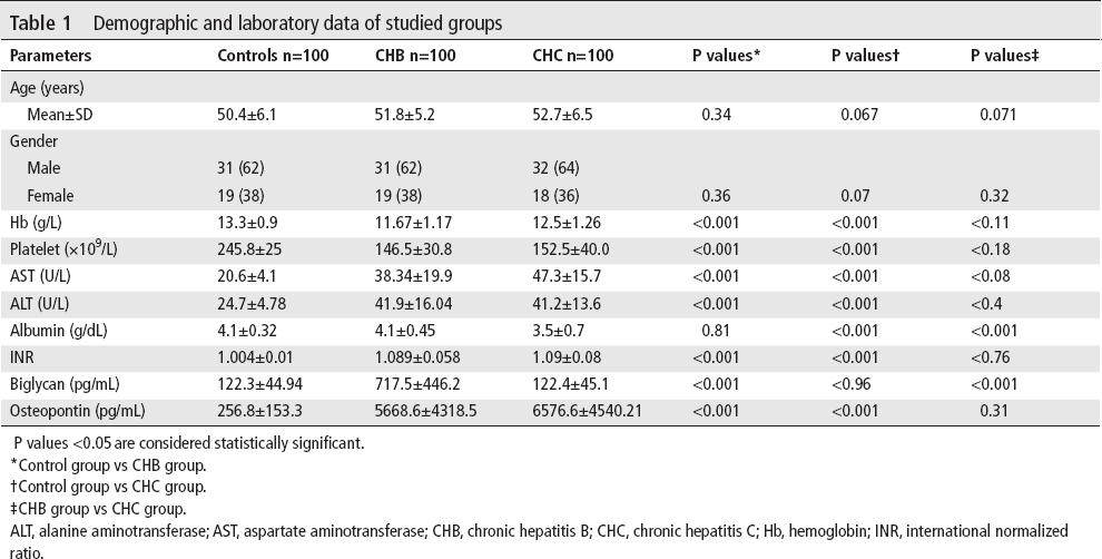
This study involves 100 patients with CHB, 100 patients with CHC and 100 healthy controls. With regard to age and sex, there were no statistical differences between all groups. The mean level of hemoglobin and platelet count was significantly lower in the CHB and CHC groups in comparison with the healthy control group (p<0.001), but there was no significant difference between the CHB and CHC groups (p<0.11 and p<0.18, respectively). The mean levels of ALT, AST and international normalized ratio (INR) were statistically higher in the CHB and CHC groups than in the control group (p<0.001); on the other hand, there was no significant difference between the CHB and CHC groups (p<0.4, p<0.08 and p<0.76 respectively) (table 1).
Demographic and laboratory data of studied groups
P values <0.05 are considered statistically significant.
Control group vs CHB group.
Control group vs CHC group.
CHB group vs CHC group.
ALT, alanine aminotransferase; AST, aspartate aminotransferase; CHB, chronic hepatitis B; CHC, chronic hepatitis C; Hb, hemoglobin; INR, international normalized ratio.
As regards albumin concentration, there was no statistical difference between the CHB group when compared with the healthy controls (p=0.81 and p=0.71), but it was decreased significantly in the CHC group in comparison with the control group and CHB group (p<0.001).
The mean level of BGN concentration in the CHB group (717.5±446.2) was significantly higher than that of the healthy controls (122.3±44.94) (p<0.001) and the CHC group (122.4±45.1) (p<0.001), but there was no statistical difference between the CHC group (122.4±45.1) when compared with the control group (p<0.96).
The mean level of OPN in the CHB group (5668.6±4318.5) and CHC group (6576.6±4540.21) was significantly higher than the healthy group (256.8±153.3) (p<0.001); on the other hand, there was no significant difference between the CHB and CHC groups (p<0.31) (table 1).
Fibrosis stages were determined by liver biopsy. The mean levels of BGN in patients with CHB with F0, F1–F3 and F4–F6 were 294.25±33.58, 624.71±222.59 and 1381.7±380.65, respectively, with a statistical difference (p<0.001). In patients with CHB with F0, F1–F3 and F4–F6, the OPN levels were 5012.29±4066.73, 5749.71±4718.5 and 5938.85±4229.34, respectively, with a significant increase (p<0.001). Also the OPN levels in patients with CHC with F0, F1–F3 and F4–F6 were 5800.64±4864.11, 6177.32±4546.68 and 7341±4458, respectively, with a statistical difference (p<0.001). BGN level was not analyzed in various fibrosis stages in patients with CHC, as no significant difference was noted in its level between CHC cases and healthy controls (table 2).
Biglycan and osteopontin levels in liver fibrosis stages in patients with CHB and CHC

CHB, chronic hepatitis B; CHC, chronic hepatitis C.
As regards correlation between laboratory studies and stages of fibrosis, among 100 patients with CHB, there was a significant negative correlation between fibrosis stages and platelet (r=−0.58; p<0.001), ALT (r=−0.57; p<0.001) and albumin (r=−0.6; p<0.001), but there was a significant positive correlation between fibrosis stages and AST (r=0.37; p=0.008), INR (r=0.58; p<0.001), BGN (r=0.64; p<0.001) and OPN (r=0.63; p<0.001). There was no significant correlation between fibrosis stages and hemoglobin (r=−0.15; p=0.28) and HBV DNA (r=−0.064; p=0.67), as seen in table 3.
Correlation between laboratory studies and stages of fibrosis, among patients with CHB and CHC

P values <0.05 are considered statistically significant.
ALT, alanine aminotransferase; AST, aspartate aminotransferase; CHB, chronic hepatitis B; CHC, chronic hepatitis C; Hb, hemoglobin; HBV, hepatitis B virus; HCV, hepatitis C virus; INR, international normalized ratio; r, correlation coefficient.
In the CHC group, there was a significant negative correlation between fibrosis stages and hemoglobin (r=−0.36; p=0.016), platelet (r=−0.87; p<0.001) and albumin (r=−0.92; p<0.001). On the other hand, there was a significant positive correlation between fibrosis stages and ALT (r=0.77; p<0.001), INR (r=0.90; p<0.001), and OPN (r=0.59; p<0.03), but there was no significant correlation between fibrosis stages and HCV load (r=−0.19; p=0.16) (table 3).
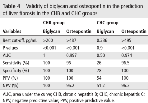
Relative operating characteristics (ROC) were performed to assess the validity of serum BGN and OPN in the assessment of the degree of liver fibrosis in the CHB and CHC groups. Based on the ROC curve, the optimal cut-off point of serum BGN was >200 pg/ mL, differentiating CHB with fibrosis from controls with statistical significance (p<0.001), area under the curve (AUC) of 1, sensitivity of 100%, specificity of 100%, positive predictive value (PPV) of 100% and negative predictive value (NPV) of 100%, while in the CHC group BGN had an AUC of 0.50, lower sensitivity of 26%, specificity of 78%, PPV of 54%, NPV of 51.2% and cut-off of 0.336 pg/mL (table 4).
Validity of biglycan and osteopontin in the prediction of liver fibrosis in the CHB and CHC groups
AUC, area under the curve; CHB, chronic hepatitis B; CHC, chronic hepatitis C; NPV, negative predictive value; PPV, positive predictive value.
As regards OPN as a predictor of liver fibrosis in the CHB group, it had an AUC of 0.997, lower sensitivity (96%) than BGN, specificity of 100%, PPV of 100%, NPV of 96.2% and a cut-off of >487 pg/mL. In contrast, in the CHC group, OPN had the highest sensitivity at 96.5%, specificity of 100%, PPV of 100%, NPV of 96.2%, AUC of 0.974 and a cut-off of >495 pg/mL (table 4).
Discussion
Liver fibrosis is a risky medical condition that occurs on top of many diseases as chronic viral hepatitis B, chronic viral hepatitis C, alcoholism, autoimmune hepatitis and NASH.13 14
Liver biopsy is the gold standard method for fibrosis assessment, but it has many disadvantages: it is invasive, there is a possibility of bleeding, and there is variability in the results.15–17 Therefore, the need for non-invasive markers that evaluate the degree of fibrosis and monitoring treatment is very essential.
The aim of this study is to evaluate both the serum BGN and OPN as non-invasive markers to predict liver fibrosis in CHB and CHC infections. BGN belongs to the proteoglycan family which is involved in the fibrosis process.18 To the best of our knowledge, there is only one study that evaluates BGN as a non-invasive marker for liver fibrosis assessment in human CHB infection,6 but no study about its role in human CHC infection. The BGN level increased significantly in the CHB group when compared with the control group (p<0.001), in agreement with the result of Ciftciler et al,6 but there is no significant difference in the BGN level between the CHC group when compared with the control group (p<0.96). Liver biopsy was performed to assess the degree of liver fibrosis according to the Ishak scale and classify fibrosis into six stages.19 In accordance with the result of Ciftciler et al,6 our study revealed that the BGN level increased in CHB as an increase in the degree of liver fibrosis (p<0.001). A positive correlation was found between fibrosis stages and serum BGN level in patients with CHB (r=0.64; p<0.001) as the BGN level increased with an increase in fibrosis stages.
OPN is an extracellular matrix protein that has many roles in physiological and pathological conditions such as cirrhosis, inflammation and tumor progression.20 OPN acts as a chemoattractant for inflammatory cells such as macrophages and neutrophils during inflammatory processes.21
In our study, OPN level increased markedly in the CHB and CHC groups when compared with the healthy control group (p<0.001). Our results matched the results of Zhao et al,22 Matsue et al,23 and Fouad et al.24 OPN level increased markedly as the degree of fibrosis increased either in the CHB group or in the CHC group (p<0.001). Our results are consistent with the results of Zhao et al,22 Matsue et al,23 and Fouad et al.24 A positive correlation was found between fibrosis stages and serum OPN level in patients with CHB (r=0.63; p<0.001) and its level in the CHC group (r=0.59; p<0.03).
In agreement with Ciftciler et al,6 the BGN level in the CHB group had a cut-off of >200, sensitivity of 100%, specificity of 100%, AUC of 1, PPV of 100% and NPV of 100%. These findings may prove BGN as a non-invasive marker for liver fibrosis assessment in CHB. In contrast, BGN level in patients with CHC had a sensitivity of 26% and a specificity of 78% with no statistical significance (p=0.9), which needs further studies to evaluate its role in CHC fibrosis.
On the other hand, in our study, the sensitivity, specificity, PPV, and NPV of OPN in the prediction of CHC fibrosis were 96.5%, 100%, 100%, and 96.2%, respectively, at a cut-off level of <495 pg/mL, and our results matched with Huang et al.25 Also, the OPN level in patients with CHB had a sensitivity of 96%, specificity of 100%, AUC of 0.997, PPV of 100% and NPV of 96.2%, with a cut-off level of <487 pg/mL, and our results matched the results of Zhao et al.22
Conclusion
Our study demonstrated that serum BGN and OPN concentrations have a strong positive correlation with liver fibrosis in patients with CHB, with a note that BGN has higher sensitivity in the detection of hepatic fibrosis in patients with CHB than in patients with hepatitis C. However, in patients with CHC, OPN had higher sensitivity and specificity than BGN in the detection of liver fibrosis. Collectively, BGN could be considered a non-invasive marker for the detection of liver fibrosis in patients with CHB, but in patients with CHC further studies are needed to assess its role. On the other hand, OPN is a good predictor of liver fibrosis in patients with CHB and CHC.